Embed Size (px)
Citation preview
EJD, vol. 18, n° 1, January-February 200878
Correspondence Eur J Dermatol 2008; 18 (1): 78-104
CORRESPONDENCE
Scleromyxedema diagnosis following unexplained encephalopathy
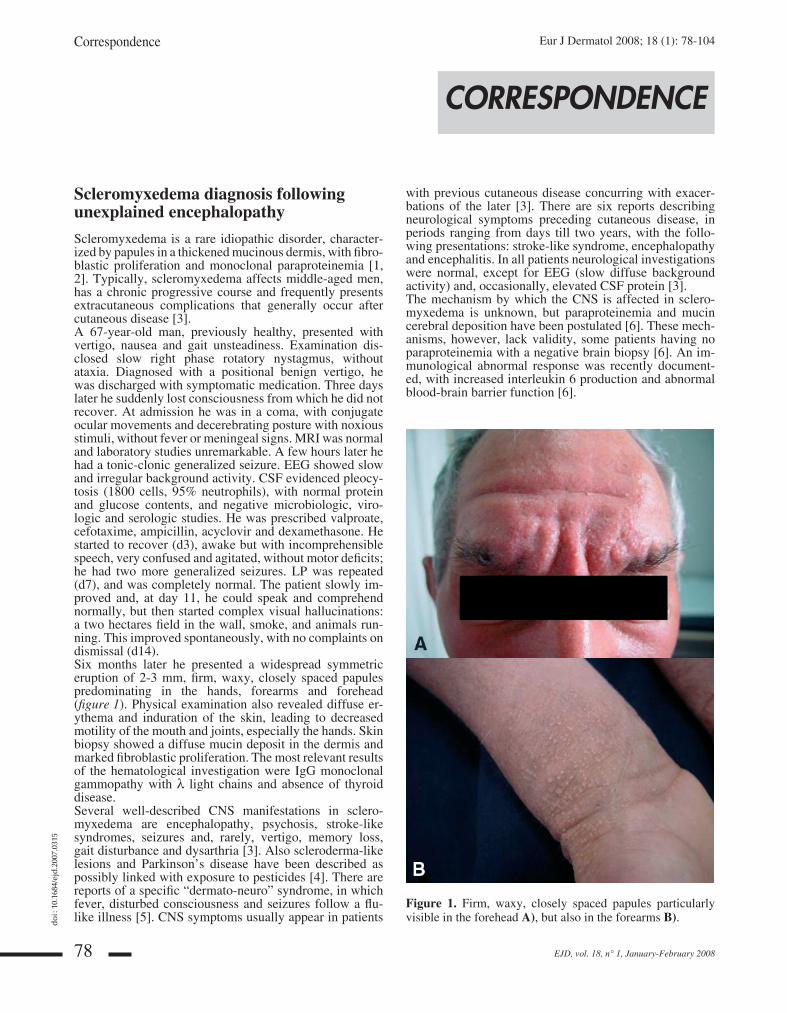
Scleromyxedema is a rare idiopathic disorder, character-ized by papules in a thickened mucinous dermis, with fi bro-blastic proliferation and monoclonal paraproteinemia [1, 2]. Typically, scleromyxedema affects middle-aged men, has a chronic progressive course and frequently pre sents extracutaneous complications that generally occur after cutaneous disease [3].A 67-year-old man, previously healthy, presented with vertigo, nausea and gait unsteadiness. Examination dis-closed slow right phase rotatory nystagmus, without ataxia. Diagnosed with a positional benign vertigo, he was discharged with symptomatic medication. Three days later he suddenly lost consciousness from which he did not recover. At admission he was in a coma, with conjugate ocular movements and decerebrating posture with noxious stimuli, without fever or meningeal signs. MRI was normal and laboratory studies unremarkable. A few hours later he had a tonic-clonic generalized seizure. EEG showed slow and irregular background activity. CSF evidenced pleocy-tosis (1800 cells, 95% neutrophils), with normal protein and glucose contents, and negative microbiologic, viro-logic and serologic studies. He was prescribed valproate, cefotaxime, ampicillin, acyclovir and dexamethasone. He started to recover (d3), awake but with incomprehensible speech, very confused and agitated, without motor defi cits; he had two more generalized seizures. LP was repeated (d7), and was completely normal. The patient slowly im-proved and, at day 11, he could speak and comprehend normally, but then started complex visual hallucinations: a two hectares fi eld in the wall, smoke, and animals run-ning. This improved spontaneously, with no complaints on dismissal (d14).Six months later he presented a widespread symmetric eruption of 2-3 mm, fi rm, waxy, closely spaced pa pules predominating in the hands, forearms and forehead (fi gure 1). Physical examination also revealed diffuse er-ythema and induration of the skin, leading to decreased motility of the mouth and joints, especially the hands. Skin biopsy showed a diffuse mucin deposit in the dermis and marked fi broblastic proliferation. The most relevant results of the hematological investigation were IgG monoclonal gammopathy with λ light chains and absence of thyroid disease.Several well-described CNS manifestations in sclero-myxedema are encephalopathy, psychosis, stroke-like syndromes, seizures and, rarely, vertigo, memory loss, gait disturbance and dysarthria [3]. Also scleroderma-like lesions and Parkinson’s disease have been described as possibly linked with exposure to pesticides [4]. There are reports of a specifi c “dermato-neuro” syndrome, in which fever, disturbed consciousness and seizures follow a fl u-like illness [5]. CNS symptoms usually appear in patients
with previous cutaneous disease concurring with exacer-bations of the later [3]. There are six reports describing neurological symptoms preceding cutaneous disease, in periods ranging from days till two years, with the follo-wing presentations: stroke-like syndrome, encephalopathy and encephalitis. In all patients neurological investigations were normal, except for EEG (slow diffuse background activity) and, occasionally, elevated CSF protein [3].The mechanism by which the CNS is affected in sclero-myxedema is unknown, but paraproteinemia and mucin cerebral deposition have been postulated [6]. These mech-anisms, however, lack validity, some patients having no paraproteinemia with a negative brain biopsy [6]. An im-munological abnormal response was recently document-ed, with increased interleukin 6 production and abnormal blood-brain barrier function [6].
Figure 1. Firm, waxy, closely spaced papules particularly visible in the forehead A), but also in the forearms B).
jleejd00492_cor1.indd 78jleejd00492_cor1.indd 78 12/10/2007 3:58:32 PM12/10/2007 3:58:32 PM
do
i : 1
0.16
84/e
jd.2
007.
0315

EJD, vol. 18, n° 1, January-February 2008 79
Our patient had a vertigo syndrome followed shortly by comatose state, seizures, CSF pleocytosis and, in the re-covery phase, hallucinations. This case, on neurological grounds, remained a puzzle until the detection of cutane-ous lesions. Although it is not possible to defi nitely link these two fi ndings, previous reports and the absence of any other known neurological disorder which could account for such a presentation, after exhaustive investigation and proper follow-up, strongly suggest that they are caused by the same systemic condition, that is, scleromyxedema. Af-ter this, proper treatment and anticipation of other extra-cutaneous complications was undertaken. Disclosure. The authors have reported no confl icts of in-terest. Funding There was no funding to this paper.
1 Neurology Department, Hospital de São Marcos, Braga, Portugal2 Dermatology Department, Hospital de São Marcos, Braga, [email protected]
Margarida RODRIGUES1
Álvaro MACHADO1
Filipa VENTURA2
Maria Luz DUARTE2
Carla FERREIRA1
1. Rudner EJ, Pomann J. Scleromyxedema revisited. Int J Dermatol. 2003; 42(1):31-35.2. Jablonska S, Blaszczyk M. Scleromyxedema is a scleroderma-like disorder and not a coexistance of scleroderma with papular mucinosis. Eur J Dermatol 1999; 9(7):551-4.3. Berger JR, Dobbs MR, Tehrune MH, Maragos WF. The Neurologic complications of Scleromyxedema. Medicine (Baltimore) 2001; 80(5):313-19.4. Stinco G, Piccirillo F, DE Francesco V, Patrone P. Scleroderma-like lesions and ParkinsonÊs disease: possible links with exposure to pesticides. Eur J Dermatol 2007; 17(3):256-7. 5. Gonzalez J, Palangio M, Schwartz J, Klainer A, Bisaccia E. Scleromyxedema with dermato- neuro syndrome. J Am Acad Dermatol 2000; 42(5 Pt 2): 927-8.6. Johkura K, Susuki K, Hasegawa O, Kuroiwa Y, Komatsumoto S. Encephalopathy in scleromyxedema. Neurology 1999; 53(5):1138-40.
Linear atrophoderma of Moulin
A 42-year-old Caucasian woman presented a 5-year his-tory of atrophic brown maculae on the left arm and trunk with an asymmetrical linear distribution forming an in-verted U-shape from the breast area up the upper arm, an S-shape on the anterior abdomen, a V-shape on the back toward the median line (fi gure 1). The patient reported that the lesions developed few days after working under sun exposure in contact with parasiticide substances and tomato plants. There was no history of atopic dermati-tis, psoriasis, or previous dermatitis. Past medical history was unremarkable. She denied any past treatment of the lesions. A biopsy revealed hyperpigmentation of epider-mal basal cells, slight thickening of the collagen fi bers in the mid-deep dermis with a rare perivascular lymphocytic infi ltrate (fi gure 1). Laboratory fi ndings were unremark-able except for an increase of hepatic enzymes compatible with hepatic steatosis. Antinuclear (ANA) and anti-DNA antibodies were negative and immunoglobulins were sug-gestive of previous common viral exanthematic diseases. The clinical picture, the histology, along with the lack of autoantibodies, were compatible with Linear Atropho-derma of Moulin (LAM) following Blaschko’s lines (BL). Cytogenetic analysis found a normal karyotype without genetic mosaicisms. Following treatment with a high dose of vitamin E (400 UI/d) and topical clobetasol propionate, a slight improvement of the lesions was observed.
Firstly described by Moulin et al. in 1992 in 5 patients [1], LAM is a distinct clinical entity characterized by acquired atrophic bandlike skin lesions that often show hyperpigmentation and always follow the lines of Blasch-ko. No preceding infl ammation is noted, but a transient infl ammatory stage is perhaps often unrecognized, and there is no induration or scleroderma. Usually the con-dition begins in childhood or adolescence, and there is no evidence of any long-term progression. Histopatho-logically, an irregular moderate hyperpigmentation of the lower part of the epidermis is found, along with a few perivascular lymphocytes in the dermis and slight thick-ening of the collagen bundles, as in our case [2]. The existence of LAM is controversial in its possible clini-cal overlap with linear scleroderma or morphea. Never-theless, this latter is characterized by one or more linear streaks of progressive induration that can extend through the dermis, subcutaneous tissue, and muscle to the un-derlying bone, causing signifi cant deformities [3]. The lack of autoantibodies, such as ANA, found in 73% of adult patients with linear scleroderma and the chronic and unvaried course make this diagnosis unlikely in our pa-tient, leading to the more compatible diagnosis of LAM [4]. The cause and pathogenesis of this disorder remain
Figure 1. Asymmetrical S-shaped linear distribution on the left arm and trunk of the atrophic brown maculae. Histology of the skin biopsy characterized by the hyperpigmentation of epidermal basal cells and the slight thickening of the collagen fi bers of the mid-deep dermis (H & E, × 150).
jleejd00492_cor1.indd 79jleejd00492_cor1.indd 79 12/10/2007 3:58:37 PM12/10/2007 3:58:37 PM
do
i : 1
0.16
84/e
jd.2
007.
0316