Embed Size (px)
Citation preview
JOURNAL OF EQUINE VETERINARY SCIENCE284
SUMMARY
Following reports of an increase in the number of Corynebacterium pseudotuberculosis infections in horses in the western counties of Colorado in 1997, an epidemiological assessment of the problem was conducted using a survey of premises with affected horses. No gender, age or breed predilections were found. Most diagnoses of disease were made in the summer or fall of 1997. Abscesses were reported to occur in various locations with a mean healing time of 77 days. A majority of premises owners reported potential contact of horses with sheep or goats. The days of lost use and treatment costs per case had a mean of 13 days and $139, respectively.
INTRODUCTION
Pigeon Fever (PF) is an infectious disease of horses caused by Corynebacterium pseudotuberculosis which results in mild to severe abscess formation.1 The disease is a self-limiting, suppurative inflammatory process most commonly affecting the pectoral, axillary,ventral abdomen, and inguinal regions.1 The muscles of these regions are affected, where disease is characterized by abscess then thickened outer walls of fibrous tissue. Systemic clinical signs are absent in the majority of horses. When present, they may include anorexia, depression, fever, and abortion.
Gait abnormalities, such as lameness or altered range of motion, are observed in cases with inguinal abscessation. Internal abscesses can occur resulting from lymphatic and/or hematogenous spread of the organism.
A similar disease occurs in sheep and goats. Corynebac-terium pseudotuberculosis serovar ovis is the etiologic agent of caseous lymphadenitis (CL), a chronic contagious disease of adult sheep and goats.2 Caseous lymphadenitis results in substantial economic losses to sheep producers due to unthriftiness and death as well as carcass condemnations at slaughter. Infection occurs following entry of the bacteria through skin wounds from shearing, grass awns, splinters, tail docking or castration. Abscess formation occurs in lymph nodes with the potential for rupture and subsequent discharge of purulent material and bacteria into the environment.
Diagnosis of PF in horses is based on the presence of the characteristic clinical signs and culture of the causal bacterium from abscess sites. Isolation and identification of C. psuedotuberculosis is the definitive test used for diagnosis of the infection. Serological testing is a screening procedure determining exposure to the bacterium. It does not indicate, however, whether an animal is currently infected or whether it will develop clinical signs of the disease.
In a retrospective study of PF in California, the median age of horses with external abscesses was similar to that of horses with internal abscesses.3 While cases were detected throughout the year, the disease was most frequently recognized in the fall and early winter. Breed and gender did not appear to be associated with infection and most of the horses (492/538 or 91.4%) had a single episode of clinical disease. Serologic testing was an unreliable method for diagnosis in horses with external abscesses. In another California study, a statistically significant spatial and temporal clustering of horses with PF was detected.4
Refereed
CORYNEBACTERIUM PSEUDOTUBERCULOSIS INFECTIONS (PIGEON FEVER) IN HORSES IN WESTERN COLORADO:
AN EPIDEMIOLOGICAL INVESTIGATIONKelly Hall, DVM1; Brian J. McCluskey, DVM, MS2 ; Wayne Cunningham, DVM, MS3
Authors’s addresses: 110562 Cedar Run Court, Las Vegas, NV 89135; 2United States Department of Agriculture, Centers for Epidemiology and Animal Health, 555 South Howes St, Fort Collins, CO 80521; 3Colorado Department of Agriculture, Animal Health Industries Division, 701 Kipling Ave, Lakewood, CO 80215.
Volume 21, Number 6, 2001 285
During 1997, cases of PF in the western counties of Colorado were reported by private veterinary practitioners to the State Veterinarian’s office. A concurrent outbreak of vesicular stomatitis was reported in these counties in the summer of 1997. The objectives of this study were to examine potential relationships between PF and specific demographic and environmental factors.
MATERIALS AND METHODS
An initial screening questionnaire was sent to all large animal veterinarians in five western counties in Colorado to obtain the names and addresses of horse owners with previous experience with PF and to determine the extent of the PF problem. A second questionnaire was developed with the intent to determine the epidemiological characteristics of the disease. Individual owners were contacted between February and March of 1999. Personal interviews were conducted on-site with owners by one of the authors (KH). The questionnaire focused on three aspects of PF infections. Clinical signs and disease, environmental influences, and economic impacts were assessed. Individual animal information was collected on horses that were exhibiting clinical signs consistent with PF or were culture positive for C. pseudotuberculsosis. Cultures of suspected PF cases were collected by aseptic techniques and sent immediately to the Colorado State University Diagnostic Laboratory. Abscesses were excised in their entirety to eliminate possible contamination. Second cultures were not performed. Cultures were not taken in all cases; therefore, diagnoses of PF were made by veterinarians based solely on clinical signs. Environmental factors such as proximity to sheep or goats, shelter access, and estimation of local insect populations were investigated. Frequency distributions and other descriptive statistics were then calculated.a
RESULTS
Overall, 14 horse owners from Montrose County and three from Delta County, Colorado completed the questionnaire. A total of 35 horses were included in the analysis.
The prevalence of PF infection on farms ranged from 3 to 100% with a mean and median of 26% and 20%, respectively. Sixty percent of positive horses (21/35) were mares, 34% were geldings (12/35) and 6% were stallions (2/35) . The disease was identified in 3 of 35 (9%) horses <2 years of age, 21 of 35 (60%) horses two to ten years of age and 11 of 35 (31%) horses >10 years of age. Approximately 48% of positive horses (17/35) were Quarter Horses. Overall, 5 of 35 (14.3%) of positive horses were Paints and Thoroughbreds, 3 of 35 (8.6%) Foxtrotters 14 of 35 (11.4%) Non- registered breeds and 1 of 35 (2.9%) were Warmbloods. Pigeon Fever occurred in 5/35 (14.3%), 15/35 (42%), 14/35
(40%), and 1/35 (2%) horses in spring, summer, fall, and winter, respectively.
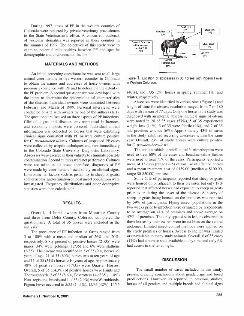
Abscesses were identified in various sites (Figure 1) and length of time for abscess resolution ranged from 7 to 180 days with a mean of 77 days. Only one horse in the study was diagnosed with an internal abscess. Clinical signs of edema were noted in 20 of 35 cases (57%), 5 of 35 experienced weight loss (14%), 3 of 35 were febrile (9%), and 2 of 35 had previous wounds (6%). Approximately 43% of cases in the study exhibited recurring abscesses within the same year. Overall, 23% of study horses were culture positive for C. pseudotuberculosis.
The antimicrobials, penicillin, sulfa-trimethoprim were used to treat 60% of the cases and betadine-saline flushes were used to treat 71% of the cases. Participants reported a mean of 13 days (range 0-75) of lost use of affected horses and a mean treatment cost of $139.00 (median = $100.00, range $0-850.00) per case.
Some 65% of participants reported that sheep or goats were housed on or adjacent to their premises but only 19% reported that affected horses had exposure to sheep or goats prior to or during the onset of the disease. A history of sheep or goats being housed on the premises was reported by 59% of participants. Flying insect populations in the two weeks prior to infection were estimated by respondents to be average on 41% of premises and above average on 47% of premises. The only type of skin lesions observed in these horses by their owners were insect bites on the ventral abdomen. Limited insect-control methods were applied on the study premises or horses. Access to shelter was limited or unavailable to many study animals. Overall, 6 of 35 cases (17%) had a barn or shed available at any time and only 6% had access to shelter at night.
DISCUSSION
The small number of cases included in this study, prevent drawing conclusions about gender, age and breed predilections. However, as reported in previous studies, horses of all genders and multiple breeds had clinical signs
Figure 1. Location of abscesses in 35 horses with Pigeon Fever in Western Colorado.
JOURNAL OF EQUINE VETERINARY SCIENCE286
consistent with PF or were culture positive in this study. 3,5,6 Even though Quarter Horses were reported to be positive for PF more often than other breeds, they are the most common breed found in this region.
Other reports have indicated that horses between one and two years of age are at higher risk due to the absence of maternal antibodies at the end of their first year or because they avoided exposure until that time.5 In the 35 cases in this study, this age range represented the lowest percentage of cases (9%) compared to the population two to ten years of age (60%). This may stem from regional management practices. Many owners maintained their animals in large pastures for the first two years with little handling or observation. Yearlings may have had lesions which went undetected and had resolved prior to observation. Limited access to shelter in these settings may have increased their exposure to insect populations.
Pigeon Fever was diagnosed predominantly in the summer and fall with 82% of the cases seen at this time. This trend has been reported in past studies in California.3 This may be due to more frequent use and observation of horses during this time period or to increased populations of arthropods. Corynebacterium pseudotuberculosis can be transmitted mechanically by a number of flying insects.7,8 Nearly half of the respondents reported that flying insect populations were above average two weeks prior to the occurrence of clinical signs of PF in their horses. The incidence of PF also varies according to rainfall and temperatures in the preceding winter. Milder winters are believed to result in optimal conditions for insect reproduction and survival. During the summer of 1997, an outbreak of vesicular stomatitis (VS) , a viral disease of horses and cattle, was occurring throughout Colorado, including Montrose and Delta counties.9 Arthropods are believed to be involved in the transmission of VS and similar to PF, VS outbreaks generally occur in the summer and fall following mild winters and wet springs. The potential importance of insects in the epidemiology to PF is given further credence by owners reporting that 86% of horses had no identifiable abrasions or wounds prior to or at the time they first observed clinical signs. Defects in the skin are thought to be the primary entry point of C. pseudotuburculosis in sheep and goats with CL.10 Insect bite lesions may create entry points for the organism in the environment or the insects themselves may mechanically transmit the organism.
A majority of participants reported both a history of sheep or goats on their premises or the presence of these species on or adjacent to their premises. Corynebacterium pseudotuburculosis can survive in the environment for extended time periods. Thus, direct exposure to sheep or goats may not be necessary for the organism to be transmitted.
The location of abscesses and other clinical signs reported in this study are similar to those previously reported.3 However, 13 of 35 (37%) horses in this study experienced
recurring abscesses which is much higher than previously reported.3 The mean abscess healing time of 77 days was also longer than previously reported.3 Extended abscess healing periods in individual horses may be the result of the inappropriate choice of anti-microbial treatment or improper wound management following abscess drainage. Participants commented that the length of time for wound healing was longer after horses were given penicillin as compared to sulfa-trimethoprim. The location of abscesses also influenced the length of healing time in individual horses. External genitalia (sheath, scrotum, and udder) abscesses were often the slowest to heal. This difference may be due to the lymphatic drainage of the site or difficulties experienced in access to these sites for flushing wounds.
There are no previous reports of the financial impact PF may have on horse owners. Treatment costs in this study included the cost of examination by a veterinarian, minor surgical procedures to lance abscesses, abscess flushing and antimicrobial use. Costs were minimized when owners administered the treatments. The cost of PF per premises would have been significantly higher if all cases were examined and treated by veterinarians.
FOOTNOTES
aEpi-Info ver. 6.04b, Centers for Disease Control and Prevention, Atlanta, Georgia, 30333.
REFERENCES
1. Davis WE: Corynebacterium pseudotuberculosis infections in animals. In: Smith, PB ed. Large Animal Internal Medicine. St. Louis: CV Mosby Co., 1990;1120-1126. 2. Jensen R, Swift BL: Diseases of Sheep 2nd ed. Phila-delphia: Lea& Febiger, 1982; p.314. 3. Aleman M, Spier SJ, Wilson WD, Doherr MG: Corynebacterium pseudotuberculosis infection in horses: 538 cases (1982-1993). J Am Vet Med Assoc 1996;209;804-809. 4. Doherr MG, Carpenter TE, Wilson WD, Gardner IA: Evalua-tion of temporal and spatial clustering of horses with Corynebacterium pseudotuberculosis infection. Am J Vet Res 1999;60:284-291. 5. Doherr MG, Carpenter TE, Wilson WD, Gardner IA: Application and evaluation of a mailed questionnaire for an epidemiological study of Corynebacterium pseudotuber-culosis infection in horses. Prev Vet Med 1998;35;241-253. 6. Songer JG, Beckenback K, Marshall MM, Olson GB, Kelly L: Biochemical and genetic characterization of Corynebacterium pseudotuberculosis. Am J Vet Res 1988;49:223-226. 7. Reid CH: Habronemiasis and Corynebacterium– chest abscess in the California horses. Vet Med Small Animal Clinician (equine issue). 1965;60:233-242. 8. Addo PB: Role of the common house fly (Musca domestica) in thespread of ulcerative lymphangitis. Vet Rec 1983:113:496-497. 9. McCluskey BJ, Hurd HS, Mumford EL: Review of the 1997 vesicular stomatitis outbreak in the southwestern United States. J Am Vet Med Assn 1999;215:1259-1262. 10. Serikawa S, Ito S, Hatta T, Kusaraki N, Senna K, et al.: Seroepidemiological evidence that shearing wounds are mainly responsible for Corynebacterium pseudotuberculosis infection in sheep. J Vet Med Sci 1993;55:691-692.