Embed Size (px)
Citation preview
Page 1 sur 12 Ronéo 12 UE1 Cours 8
UE1 Anglais
Pr Nagy
13/12/18
Marie VRAUX
Cours 8: The nervous system (part II)
Page 2 sur 12 Ronéo 12 UE1 Cours 8
I/ The Cranial Nerves
1) Vocabulary
2) Fill in the gap
3) True or False
4) Questions
II) The Spinal Nerves
1) Vocabulary
2) True or False
3) Questions
III) The Brain and Histology
1) Vocabulary
2) Fill in the gap
3) True or False
4) Questions
IV/ Brain Anatomy
1) Vocabulary
2) Fill in the gaps
3) Questions
4) True or False
V/ The Geography of Thought
1) Vocabulary
2) Fill in the gap
3) True or False
4) Questions
Page 3 sur 12 Ronéo 12 UE1 Cours 8
I/ The Cranial Nerves
1) Vocabulary
The thick, whitish cord of nerve tissue that extends
from the medulla oblongata down through the spinal
column and from which the spinal nerves branch off
to various parts of the body
Spinal cord (Spine) La moelle épinière
The part of the nervous system which consists of the
brain and spinal cord, to which sensory impulses are
transmitted and from which motor impulses pass out
and which supervises and coordinates the activity of
the entire nervous system
Central nervous
system (CNS)
Le system nerveux
central
1.To move upward
2.To come into being; originate. To result, issue or
proceed
Arise from Provenir de/ Procéder
de
To furnish or provide (something). To serve as a
conduit (medium of transmission)
Supply Alimenter, Innerver,
Desservir
The nerves outside the CNS including the cranial
nerves (excepting the optic nerve), the spinal nerves
and the sympathetic and parasympathetic nervous
systems
Peripheral nerves Les nerfs
périphériques
Called or known by a different name Referred to…as Connu sous le nom
de
A long flexible structure made up of strands or
fibers, such as a nerve or tendon
Cord Un cordon
A cluster (group) or strand (complex of filaments) of
closely bound muscle or nerve fibers
Bundle Un faisceau
Transmitting impulses from sense organs to nerve
centers; afferent
Sensory nerve Nerf sensoriel
Carrying impulses from the nerve centers to the
muscles
Motor nerve Nerf moteur
Two corresponding persons or items, similar in form
or function and matched or associated
Pair Une paire
Several nerves that arise in pairs from the brainstem
and reach the periphery through openings in the
skull. There are 12 such pairs in humans
Cranial nerves Les nerfs crâniens
The nerves that arise in pairs from the spinal cord.
There are 31 pairs in the human body
Spinal nerves Les nerfs rachidiens
The tenth and longest of the cranial nerves, passing
through the neck and thorax into the abdomen and
supplying sensation to part of the ear, the tongue, the
larynx and the pharynx, motor impulses to the vocal
cords and motor and secretory impulses to the
abdominal and thoracic viscera
Vagus nerve
= pneumogastric
nerve
Le nerf vague, le nerf
pneumogastrique
The section of the alimentary canal that extends from
the mouth and nasal cavities to the larynx, where it
becomes continuous with the oesophagus
Pharynx Le pharynx
The part of the respiratory tract between the pharynx
and the trachea, having walls of cartilage and muscle
and containing the vocal cords enveloped in folds of
mucous membrane
Larynx Le larynx
Page 4 sur 12 Ronéo 12 UE1 Cours 8
1. Continuing
2. Temporarily excluded. Not yet dealt with
Remaining Restant
To operate or regulate; direct, command Control Diriger, régler
The globe-shaped portion of the eye formed by the
sclera and cornea, surrounded by the socket and
covered externally by the eyelids
Eyeball Le globe occulaire
2) Fill in the gap
Of the twelve cranial nerves, four contain both sensory and motor fibres (V, VII, IX, X).
3) True or False
Most of the cranial nerves contain motor fibers.
Five cranial nerves contain motor fibers, four contain sensory and motor fibers. Then 9 out of 12
contain motor fibers.
→True
Most of the cranial nerves contain sensory fibers.
Three cranial nerves contain sensory fibers and four contain sensory and motor fibers. Then 7 out of
12 contain sensory fibers.
→True
Most of the cranial nerves contain both motor and sensory fibers.
Only 4 out of 12 contain both motor and sensory fibers.
→False
The 12 cranial nerves are responsible for conducting signals from the brain to the rest of the body and
vice versa.
Most of these nerves innervate the cranium and therefore don’t leave the skull. There are only 2
cranial nerves that leave the skull (vagus nerve (X) and accessory nerve (XI)).
→False
Most of the cranial nerves have a parasympathetic function.
The vagus nerve (X) is responsible for 80% of the body’s parasympathetic functions. It’s the most
important parasympathetic nerve in the body.
The oculomotor nerve (III) is responsible for miosis (= constriction of the pupil).
Glossopharyngeal nerve (IX) and facial nerve (VII) innervate the salivary glands. They both increase
the secretion of saliva.
The facial nerve (VII) also increases the production of tears by the lacrimal gland.
These 3 nerves (oculomotor, glossopharyngeal and facial nerves) reach their respective tissues through
the trigeminal nerve (V).
So 5 out of the 12 cranial nerves have a parasympathetic function.
→False
Page 5 sur 12 Ronéo 12 UE1 Cours 8
4) Questions
Which is the most important cranial nerve?
What sorts of fibers does it contain?
What parts of the body does it supply?
The most important cranial nerve is the vagus nerve (X).
It contains sensory and motor fibers.
It supplies thoracic viscera, abdomen, pharynx and larynx.
It innervates the abdomen and the thoracic viscera with parasympathetic fibers.
In the gastro intestinal tract, it increases peristaltic movement, secretion in glandular organs and
blood flow due to vasodilation.
Concerning the heart, it lowers the heart rate, the contractility of heart muscle and conduction
velocity.
II/ The Spinal Nerves
1) Vocabulary
The spinal nerves of the cervical region of which
there are eight on each side in humans
Cervical nerves Les nerfs cervicaux
The spinal nerves of the thoracic region that consist
of 12 pairs of which one pair emerges just below
each thoracic vertebra
Thoracic nerves Les nerfs dorsaux, nerfs
thoraciques
The five pairs of spinal nerves of the lumbar region
of which one on each side passes out below each
lumbar vertebra and the upper four unite by
connecting branches into a lumbar plexus
Lumbar nerves Les nerfs lombaires
The spinal nerves of the sacral region of which there
are five pairs and which have anterior and posterior
branches passing out through the sacral foramina
Sacral nerves Les nerfs sacrés
The 31st or lowest pair of spinal nerves Coccygeal Nerfs coccygiens
To become separated into parts Divide into Se diviser en, se répartir
Located behind or toward the rear. Situated toward
the hind part of the body (dorsal)
Posterior Postérieur
The membranous tissue forming the external
covering or integument of an animal and consisting
of the epidermis and dermis
Skin La peau
The posterior portion of the trunk of the human body
between the neck and the pelvis; the dorsum
Back Le dos
Located on or near the front of the body. Located on
or near the front of an organ or the ventral surface of
the body in human beings
Anterior Antérieur
To make or form a circle around; enclose Circle Entourer, ceindre
The part of the human body between the neck and
the diaphragm, partially encased by the ribs and
containing the heart and lungs; the chest
Thorax Le thorax
The short muscles that extend between the ribs
filling in most of the intervals between them and
serving to move the ribs in respiration
Intercostal
muscles
Les muscles intercostaux
Page 6 sur 12 Ronéo 12 UE1 Cours 8
A structure in the form of a network, especially of
nerves, blood vessels, or lymphatics
Plexus Un plexus
A system of lines or channels that cross or
interconnect. A complex, interconnected group or
system
Network Un réseau
To move or extend away from the origin and toward
a different point
Pass out
(from…to)
Sortir (pour aller à…)
Relating to the neck (part of the body joining the
head to the shoulders or trunk)
Cervical Cervical
Relating to the structure of the skeleton, composed of
the innominate bones on the sides, the pubis in front,
and the sacrum and coccyx behind, that rests on the
lower limbs and supports the spinal column
Pelvic Pelvien
Higher in place; superior Upper Supérieur
Physically situated below; inferior Lower Inférieur
One of the jointed appendages, such as an arm or a
leg, used for locomotion
Limb Un membre
A sensory and motor nerve originating in the sacral
plexus and running through the pelvis and upper leg
Sciatic nerve Le nerf sciatique
To come into existence; grow out of, originate Emerge from Émerger, sortir
A nerve plexus that lies against the posterior and
lateral walls of the pelvis, is formed by the union of
the lumbosacral trunk and the first, second and third
sacral nerves and continues into the thigh as the
sciatic nerve
Sacral plexus Le plexus sacré
The portion of the human leg between the hip and
knee and supported by a single large bone (the
femur)
Thigh La cuisse
The two lower human limbs that extend from the top
of the foot (especially the part between the knee and
the ankle)
Leg La jambe
2) True or False
Most spinal nerves contain both motor and sensory fibers.
All spinal nerves contain both motor and sensory fibers.
→False
Limb nerves contain nerve fibers from different regions of the spinal column.
For example, the arm is innervated by the brachial plexus which originates from the cervical and
thoracic regions.
→True
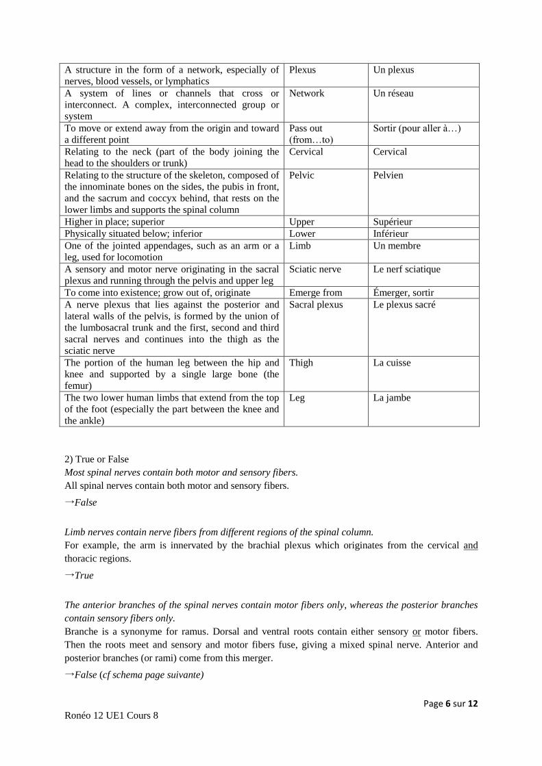
The anterior branches of the spinal nerves contain motor fibers only, whereas the posterior branches
contain sensory fibers only.
Branche is a synonyme for ramus. Dorsal and ventral roots contain either sensory or motor fibers.
Then the roots meet and sensory and motor fibers fuse, giving a mixed spinal nerve. Anterior and
posterior branches (or rami) come from this merger.
→False (cf schema page suivante)
Page 7 sur 12 Ronéo 12 UE1 Cours 8
The nerves of the sympathetic nervous system originate in the spinal column.
Nerves that belong to the parasympathetic nervous system leave the central nervous system (CNS) at
the level either of the cranium or the sacral region. They have a craniosacral outflow.
Nerves belonging to the sympathetic nervous system leave the CNS at the level of the thorax and the
lumbar region. They have a thoracolumbar outflow (from T1 to L2).
→True
3) Questions
What are limb nerves made up of?
Why is this the case?
Limb nerves are made up of several spinal nerves because spinal nerves meet to form a plexus and
then come limb nerves, so one limb nerve comes out of several spinal nerves.
For example, in the leg, the sciatic nerve comes from the sacral plexus that is made up of several
spinal nerves.
III/ The brain and histology
1) Vocabulary
The portion of the CNS that is enclosed within the cranium,
continuous with the spinal cord, and composed of gray
matter and white matter. It is the primary center for the
regulation and control of bodily activities, receiving and
interpreting sensory impulses and transmitting information to
the muscles and body organs. It is also the seat of
consciousness, thought, memory and emotion
Brain Le cerveau
The faculties by which stimuli from outside or inside the
body are received and felt, as the faculties of hearing, sight,
smell, touch, taste and equilibrium
Sense Un sens, une
sensation
The delicate network of branched cells and fibers that
supports the tissue of the CNS
Glia, neuroglia La glie, la
névroglie
The three membranes that enclose the vertebrate brain and
spinal cord: the pia mater, arachnoid and dura mater
Meninges (sing.
Meninx)
Les méninges
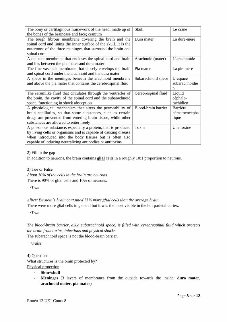
Page 8 sur 12 Ronéo 12 UE1 Cours 8
The bony or cartilaginous framework of the head, made up of
the bones of the braincase and face; cranium
Skull Le crâne
The tough fibrous membrane covering the brain and the
spinal cord and lining the inner surface of the skull. It is the
outermost of the three meninges that surround the brain and
spinal cord
Dura mater La dure-mère
A delicate membrane that encloses the spinal cord and brain
and lies between the pia mater and dura mater
Arachnoid (mater) L’arachnoïde
The fine vascular membrane that closely envelops the brain
and spinal cord under the arachnoid and the dura mater
Pia mater La pie-mère
A space in the meninges beneath the arachnoid membrane
and above the pia mater that contains the cerebrospinal fluid
Subarachnoid space L’espace
subarachnoïdie
n
The serumlike fluid that circulates through the ventricles of
the brain, the cavity of the spinal cord and the subarachnoid
space, functioning in shock absorption
Cerebrospinal fluid Liquid
céphalo-
rachidien
A physiological mechanism that alters the permeability of
brain capillaries, so that some substances, such as certain
drugs are prevented from entering brain tissue, while other
substances are allowed to enter freely
Blood-brain barrier Barrière
hématoencépha
lique
A poisonous substance, especially a protein, that is produced
by living cells or organisms and is capable of causing disease
when introduced into the body tissues but is often also
capable of inducing neutralizing antibodies or antitoxins
Toxin Une toxine
2) Fill in the gap
In addition to neurons, the brain contains glial cells in a roughly 10:1 proportion to neurons.
3) Tue or False
About 10% of the cells in the brain are neurons.
There is 90% of glial cells and 10% of neurons.
→True
Albert Einstein’s brain contained 73% more glial cells than the average brain.
There were more glial cells in general but it was the most visible in the left parietal cortex.
→True
The blood-brain barrier, a.k.a subarachnoid space, is filled with cerebrospinal fluid which protects
the brain from toxins, infections and physical shocks.
The subarachnoid space is not the blood-brain barrier.
→False
4) Questions
What structures is the brain protected by?
Physical protection:
- Skin+skull
- Meninges (3 layers of membranes from the outside towards the inside: dura mater,
arachnoid mater, pia mater)
Page 9 sur 12 Ronéo 12 UE1 Cours 8
- Between the arachnoid mater and the pia mater, there is the subarachnoid space, filled with
cerebrospinal fluid which provides the most important protection against physical shocks.
Chemical protection:
- Blood-brain barrier (contains microglia, astrocytes that filter substances, tightly packed
endothelial cells in blood vessels. These endothelial cells don’t have fenestrations.)
IV/ Brain anatomy
1) Vocabulary
The portion of the brain, consisting of the
medulla oblongata, pons Varolii and midbrain
that connects the spinal cord to the forebrain
and cerebrum
Brainstem Le tronc cerebral
The trilobed structure of the brain, lying
posterior to the pons and medulla oblongata and
inferior to the occipital lobes of the cerebral
hemispheres that is responsible for the
regulation and coordination of complex
voluntary muscular movement as well as the
maintenance of posture and balance
Cerebellum Le cervelet
The large rounded structure of the brain
occupying most of the cranial cavity, divided
into two cerebral hemispheres that are joined at
the bottom by the corpus callosum. It controls
and integrates motor, sensory and higher mental
functions, such as thought, reason, emotion and
memory
Cerebrum Le cerveau, le
télencéphale
1. The outer layer of an organ or body part such
as the cerebrum or the adrenal glands.
2. The convoluted layer of gray substance
covering each cerebral hemisphere
Cortex Le cortex (cerebral)
Gray areas of brain and spinal cord made up of
cell bodies and dendrites of nerve cells rather
than myelinated axons
Gray (grey) matter La matière grise
1. A half of a sphere
2. Either of the lateral halves of the cerebrum
Hemisphere Un hémisphère
The faculty or act of speaking. The faculty or
act of expressing or describing thoughts,
feelings, or perceptions by the articulation of
words
Speech La parole, le langage
A sudden loss of brain function caused by a
blockage or rupture of a blood vessel to the
brain, resulting in necrosis of brain tissue and
characterized by loss of muscular control,
diminution or loss of sensation or
consciousness, dizziness, slurred speech or other
symptoms that vary with the extent and severity
of brain damage. Also called cerebral accident,
cerebral infarction, cerebrovascular accident
Stroke Un accident vasculaire
cérébral
The arched bridge of nervous tissue that
connects the two cerebral hemispheres, allowing
communication between the right and left sides
of the brain
Corpus callosum Le corps calleux
Page 10 sur 12 Ronéo 12 UE1 Cours 8
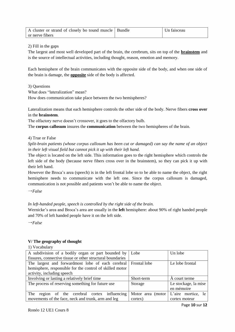
A cluster or strand of closely bo tound muscle
or nerve fibers
Bundle Un faisceau
2) Fill in the gaps
The largest and most well developed part of the brain, the cerebrum, sits on top of the brainstem and
is the source of intellectual activities, including thought, reason, emotion and memory.
Each hemisphere of the brain communicates with the opposite side of the body, and when one side of
the brain is damage, the opposite side of the body is affected.
3) Questions
What does “lateralization” mean?
How does communication take place between the two hemispheres?
Lateralization means that each hemisphere controls the other side of the body. Nerve fibers cross over
in the brainstem.
The olfactory nerve doesn’t crossover, it goes to the olfactory bulb.
The corpus callosum insures the communication between the two hemispheres of the brain.
4) True or False
Split-brain patients (whose corpus callosum has been cut or damaged) can say the name of an object
in their left visual field but cannot pick it up with their left hand.
The object is located on the left side. This information goes to the right hemisphere which controls the
left side of the body (because nerve fibers cross over in the brainstem), so they can pick it up with
their left hand.
However the Broca’s area (speech) is in the left frontal lobe so to be able to name the object, the right
hemisphere needs to communicate with the left one. Since the corpus callosum is damaged,
communication is not possible and patients won’t be able to name the object.
→False
In left-handed people, speech is controlled by the right side of the brain.
Wernicke’s area and Broca’s area are usually in the left hemisphere: about 90% of right handed people
and 70% of left handed people have it on the left side.
→False
V/ The geography of thought
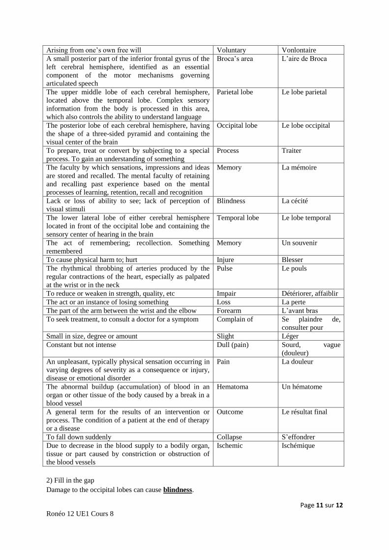
1) Vocabulary
A subdivision of a bodily organ or part bounded by
fissures, connective tissue or other structural boundaries
Lobe Un lobe
The largest and forwardmost lobe of each cerebral
hemisphere, responsible for the control of skilled motor
activity, including speech
Frontal lobe Le lobe frontal
Involving or lasting a relatively brief time Short-term À court terme
The process of reserving something for future use Storage Le stockage, la mise
en mémoire
The region of the cerebral cortex influencing
movements of the face, neck and trunk, arm and leg
Motor area (motor
cortex)
L’aire mortice, le
cortex moteur
Page 11 sur 12 Ronéo 12 UE1 Cours 8
Arising from one’s own free will Voluntary Vonlontaire
A small posterior part of the inferior frontal gyrus of the
left cerebral hemisphere, identified as an essential
component of the motor mechanisms governing
articulated speech
Broca’s area L’aire de Broca
The upper middle lobe of each cerebral hemisphere,
located above the temporal lobe. Complex sensory
information from the body is processed in this area,
which also controls the ability to understand language
Parietal lobe Le lobe parietal
The posterior lobe of each cerebral hemisphere, having
the shape of a three-sided pyramid and containing the
visual center of the brain
Occipital lobe Le lobe occipital
To prepare, treat or convert by subjecting to a special
process. To gain an understanding of something
Process Traiter
The faculty by which sensations, impressions and ideas
are stored and recalled. The mental faculty of retaining
and recalling past experience based on the mental
processes of learning, retention, recall and recognition
Memory La mémoire
Lack or loss of ability to see; lack of perception of
visual stimuli
Blindness La cécité
The lower lateral lobe of either cerebral hemisphere
located in front of the occipital lobe and containing the
sensory center of hearing in the brain
Temporal lobe Le lobe temporal
The act of remembering; recollection. Something
remembered
Memory Un souvenir
To cause physical harm to; hurt Injure Blesser
The rhythmical throbbing of arteries produced by the
regular contractions of the heart, especially as palpated
at the wrist or in the neck
Pulse Le pouls
To reduce or weaken in strength, quality, etc Impair Détériorer, affaiblir
The act or an instance of losing something Loss La perte
The part of the arm between the wrist and the elbow Forearm L’avant bras
To seek treatment, to consult a doctor for a symptom Complain of Se plaindre de,
consulter pour
Small in size, degree or amount Slight Léger
Constant but not intense Dull (pain) Sourd, vague
(douleur)
An unpleasant, typically physical sensation occurring in
varying degrees of severity as a consequence or injury,
disease or emotional disorder
Pain La douleur
The abnormal buildup (accumulation) of blood in an
organ or other tissue of the body caused by a break in a
blood vessel
Hematoma Un hématome
A general term for the results of an intervention or
process. The condition of a patient at the end of therapy
or a disease
Outcome Le résultat final
To fall down suddenly Collapse S’effondrer
Due to decrease in the blood supply to a bodily organ,
tissue or part caused by constriction or obstruction of
the blood vessels
Ischemic Ischémique
2) Fill in the gap
Damage to the occipital lobes can cause blindness.
Page 12 sur 12 Ronéo 12 UE1 Cours 8
3) True or False
The cerebral cortex can be divided into 5 lobes: the frontal, parietal, occipital, temporal and
cerebellum.
Cerebellum is not a lobe.
→False
The motor area is located on the right side in left-handed people, and on the left side in right handed-
people.
There are motor areas on both sides, in the rearmost portion of each frontal lobe.
→False
4) Questions
Which areas of the brain are specialized in abstract thought and/or sensory perception?
Frontal lobe is specialized in abstract thought. It is also responsible for emotion, inhibition and short
term memory.
Parietal lobe is specialized in sensory perception.
Temporal lobe → hearing, long term memory
Occipital lobe → vision