Embed Size (px)
Citation preview


Course: VPM 201 Fall, 2010Lecturer: C. Anne Muckle
All sorts of bacteria

Small non-spore-forming, capsulated gram-negative coccobacilli
Previously was in the genus Pasteurella, but reclassified as a new genus after its discoverer Dr. Edward Francis
Four subspecies; the subspecies tularensis is most virulent
Fastidious growth requirements in lab, requires cysteine-supplemented media
Primary isolation from clinical samples can be done in Level 2 diagnostic lab, but identification requires Biosafety Level 3
“Many laboratories actively avoid opportunities to cultivate it” (Songer & Post text pg 212)
Bioterrorism Category A agent
High potential for lab-acquired infections by aerosol inhalation
Has been used as a biological warfare agent in WW II, and it has received renewed attention recently owing to concerns about bioterrorism (high infection risk by inhalation, low infection dose, high mortality rate, no licensed vaccine)
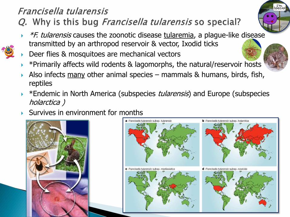
*F. tularensis causes the zoonotic disease tularemia, a plague-like disease transmitted by an arthropod reservoir & vector, Ixodid ticks
Deer flies & mosquitoes are mechanical vectors
*Primarily affects wild rodents & lagomorphs, the natural/reservoir hosts
Also infects many other animal species – mammals & humans, birds, fish, reptiles
*Endemic in North America (subspecies tularensis) and Europe (subspecies holarctica )
Survives in environment for months

Francisella tularensis is a facultative intracellular parasite (FIP)
Invades & multiplies inside macrophages, but its virulence factors are ??
F. tularensis invades macrophages

Infectious dose is very low (10 bacteria);
Can penetrate unbroken skin ( Doesn’t need any help like P. aeruginosa does !)
Clinical syndromes depend on infection route → plague–like septicemia
Contact or infected insect bite → ulceroglandular form, with skin ulcer at point of
entry
Ingestion → oropharyngeal and typhoidal forms
Inhalation → primary pneumonic form (this form has the highest mortality)
Eye infection → oculoglandular form
Lymph node infection without obvious skin ulcer → glandular form
Cat, lung. Numerous small pale foci disseminated throughout all lung lobes. Credit: Dr. J. Niefteld, Kansas State University,College of Veterinary Medicine
Cat, spleen and liver. Numerous small pale foci disseminated throughout the spleen; fewer pale foci in the liver lobe.Credit: Dr. J. Niefteld, Kansas State University,College of Veterinary Medicine
Beaver liver - disseminated small pale foci of necrotizing hepatitis. Credit: Dr. G. Wobeser
Canadian Cooperative Wildlife Health Centre

Cats - clinical illness can be severe, differential diagnosis is Y. pestis/plague
Dogs – can be infected by eating wild rabbits, & bites by infected ticks illness is milder than cats
Humans – highest risk is from arthropod vector bites and handling infected tissues, examples – vets, farmers, hunters & trappers skinning rabbits and other wild species – ex. squirrels, beavers, muskrats, pheasants
Humans also can be infected by contact with cats and dogs, mowing lawns, cutting brush in endemic areas
Treatment – first choice is gentamicin; tetracycline, chloramphenicol
PHAC – Canada Fact Sheet http://www.phac-aspc.gc.ca/tularemia/tul-qa-eng.php

Include tularemia as a differential diagnosis of febrile illness in endemic areas
Cats and dogs – flea and tick control, limiting hunting & other outdoor adventuring in endemic areas
Use of disinfectants to clean equipment
Hunters - wear latex gloves when handling carcasses and cook meat thoroughly before eating.
Black-tailed prairie dogs are highly sociable,a trait that facilitates the spread of infectious
diseases like plague and tularemia. NPS photo.

Taxonomy of the Genus Actinobacillus has been reviewed *→ definition of
the nine “true” actinobacilli species associated with animals
Habitat is mucosal membranes of upper respiratory tract ,GIT, & genital tract; carrier animals are needed for disease transmission
Non-motile gram-negative bacilli
Are pleomorphic, a mix of rod & coccoid shapes, giving a “Morse code” (dot & dash) appearance on Gram stain
Most have capsules
Urease positive Some species can grow on Mac agar as tiny LF colonies
(*Reference :Christensen H, Bisgaard, M. 2004. Vet. Microbiol. 99:13-30)

“Actino” means “rays” referring to the radiating structures (sulfur granules)
formed by A. lignieresii in tissue
Actinobacillus species of veterinary importance:
1. A. pleuropneumoniae – pigs
2. A. suis (pigs only)
3. A. equuli subspecies equuli - horses & pigs
4. A. equuli subspecies haemolyticus - horses only
5. A. lignieresii - ruminants and others

The only Actinobacillus species considered a primary pathogen Causes contagious porcine pleuropneumonia *Necrotizing haemorrhagic pneumonia with pleurisy Highly infectious Worldwide, 15 serotypes (1,5,7 in N.A); & 2 biotypes Usually young pigs affected, high morbidity & mortality (30-50%)
Acute signs – sudden death, pyrexia (shivering), coughing, expiratory dyspnea in young pigs, bloody froth from nose or mouth
Necrotizing hemorrhagic pneumonia of the caudodorsal aspect of caudal lung lobe + fibrinous pleuritis
Survivors – chronic lung lesions (lung scarring, abscesses, pleural adhesions, necrosis, sequestra) poor doers
Outbreaks (fall/winter) preceded by introduction of carrier into “clean herd”; or stresses in a low carrier herd (ventilation/temp problems, viral or mycoplasma infections)
A. pleuropneumoniae -acute respiratory distress

A. pleuropneumoniae virulence is associated with having *RTX toxins (pore-forming cytolytic toxins; RTX = “repeats in toxins”)
There are several types of RTX genes found in A. pleuropneumoniae, A. suis, A. lignieresii, and A. equuli subsp. haemolytica
A. pleuropneumoniae has RTX toxins Apxl, Apx ll, Apx lll & Apx lVA
Certain biotypes are more virulent
*urease (important for acquiring ammonia as a nitrogen source)
*capsule, LPS, transferrin binding proteins, hemoglobin- binding OMPs, iron binding proteins, proteases
Sustained inflammatory response tissue necrosis
Actinobacillus pneumoniaepneumonia - necrosis, hemorrhage, infiltration of neutrophils.
Laboratory for Genomics & BioinformaticsUniversity of Oklahoma Health Sciences Center

Disease control by vaccination:
Vaccines available passive protection from sow Bacterins reduce mortality but have variable efficacy PLEUROSTAR APP™ Novartis - a subunit vaccine (against RTX toxins & OM
proteins)
• Disease control by management procedures to prevent direct contact or aerosol exposure in intensive pig production environment:
quarantine segregated early weaning all-in/ all-out production cleaning & disinfection serological monitoring for and culling of carriers

How do we identify A. pleuropneumoniae in the lab?
Culture on BA, also on BA with Staph streak, or on chocolate agar because Biotype 1 requires NAD = Factor V
Look for tiny *hemolytic colonies that “satellite” around Staph streak
Urease-positive
*CAMP- positive (synergistic hemolytic action of RTX with Staph. aureus beta toxin)
We have to distinguish A. pleuropneumoniae from H. parasuis, and also from nonpathogenic commensal Haemophilus and Actinobacillus species in respiratory tract
Actinobacillus pleuropneumoniae cross streaked with a feeder colony of Beta-hemolytic staphylococcus demonstrating a CAMP reaction, hemolytic activity, & dependence on NAD (V-factor) for growth.

Genuine Actinobacillus suis has only been isolated from pigs
Resides in nostrils, tonsils, vaginal mucosal membranes of healthy pigs
Note: Because it is hard to distinguish from A. equuli subsp. haemolyticus using only biochemical tests (phenotypically) it has previously been reported from horses (Lab Identification errors).
*Hemolytic and urease-positive Virulence: Has *RTX toxins, capsule, urease, transferrin-binding proteins
Disease = septicaemia and localised infections
Three Syndromes:
Piglets (< 1 month) - septicaemia (50% mortality)
Grow-Finish pigs – septicaemia with bronchopneumonia
similar to hemorrhagic pneumoniae caused by A. pleuropneumoniae
Adult pigs - metritis, abortion, meningitis, red skin lesions
similar to those caused by Erysipelothrix rhusiopathiae
No commercial vaccines

A. lignieresii causes pyogranulomatous lesions in soft tissues of ruminants (tongue, head and neck, lungs, mammary glands, lymph nodes)
Called “Wooden tongue” also called “actinobacillosis” in cattle
Can have sporadic individual cases or can have outbreaks in cattle herds
Cutaneous form in ruminants: skin lesions and related lymphatics only (ex-outbreak in beef herd near Moncton, NB in 2008)
Granulomatous abscesses in sheep and cattle, humans, horses, dogs, rats, udder of cows and sows
* Must distinguish “actinobacillosis” from actimycosis = lumpy jaw, caused by Actinomyces bovis, which affects bone, usually the jaw bone of cattle
Lumpy jaw is caused by Actinomyces bovis.

A. lignieresii is a commensal in oropharynx and rumen of cattle and sheep
TRAUMA is the predisposing factor → penetrates mucosal or skin barrier →underlying submucosal soft tissues →pyogranulomatous infections (hard, tumorous masses) in tongue and soft tissues of neck and around jaw, can spread by lymphatics to lungs, stomachs & other organs
Examples of predisposing trauma – coarse feeds, hay, straw
Actinobacillus lignieresii – wooden tongue lesionsin tongue and retropharyngeal lymph nodes
Cows eating straw & wooden tongue

Clinical Signs: drooling, salivating, protruding tongue, dysphagia, weight loss Abscesses contain odourless purulent material
Lab Diagnosis: “Sulphur granules” in wooden tongue lesions, seen either on direct Gram-stain
or histology Central masses of gram-negative bacteria surrounded by spicules of calcium
phosphate, inflammatory debris (described as radiating, club-like filaments, pathologist’s term = Splendore-Hoeppli reaction)
In contrast, Actinomyces bovis granules in lumpy jaw lesions contain gram-positive bacteria, are yellow, and are larger than Actinobacillus lignieresii granules
Treatment requires surgical drainage + antibiotics (tetracyclines), potassium iodide (oral or i.v.)


Two subspecies now recognised:
A. equuli subsp. equuli, normal mucous membrane flora of horses and pigs
Can cause disease in horses and pigs Is nonhemolytic & CAMP-negative
A. equuli subsp. haemolyticus , normal mucous membrane flora of horses only
Is hemolytic & CAMP-positive

A. equuli ssp. equuli causes septicemia of neonatal foals
Disease is called “sleepy foal disease”, also called “joint ill” Sporadic infections largely affecting neonatal & young foals (causes1/3
of neonatal mortalities) Mare is source of infection, with transmission either in utero, at birth,
or via umbilicus, ingestion, inhalation Failure of foal to ingest adequate colostrum is important predisposing
cause = failure of passive transfer (FPT) Possibly carried by migrating larvae of Strongylus vulgaris from GIT to
bloodstream Septic emboli →microabscesses, particularly in kidney and joints Acute clinical signs due to neonatal septicaemia, frequently fatal Survivors show signs of chronic infections: purulent nephritis,
pneumonia and septic polyarthritis
Diagnosis: Blood culture of septic foal, kidneys, joints, & other organs at PM,
A. equuli ssp. equuli can also cause septicemia in pigs

Actinobacillus equuli septicemia - embolic nephritis .

Found in horses only
Sporadic cases of wound and joint infections, metritis, abortion, endocarditis and meningitis in adult horses
Not as common opportunistic pathogen as Streptococcus zooepidemicus, but an important pathogen of horses

Possible zoonotic risks of horse and pig bites:
Human wounds and bite wound infections caused by A. suisfrom pigs and A. equuli from horses

Genus Moraxella are short, plump, gram-negative bacilli/ coccobacilli, frequently in pairs “diplobacilli” or chains, can stain “gram-variable”
Nonmotile, fastidious, aerobic, do not grow on MacConkey agar
Several commensal strains of Moraxella and Neisseria on skin, mucous membranes and conjunctivae of animals
The important Moraxella species in animals are:
Moraxella bovis , hemolytic→ conjunctivitis in cattle
Moraxella ovis, hemolytic/ nonhemolytic → conjunctivitis in sheep & goats,
cattle
Moraxella equi, a non-hemolytic variant of M. bovis →conjunctivitis in horses

M. bovis causes infectious bovine keratoconjunctivitis (IBK or IBKC)
Also called “pinkeye”, “New Forest disease”, or “New Forest eye” (UK)
A highly contagious and painful eye disease common in cattle worldwide with significant economic losses ~ $ 200 million annually
Most common eye condition seen in cattle in UK, important in Australia & USA
Outbreaks in grazing cattle in summer, high morbidity, young cattle most susceptible
Herefords and cattle without pigmented ocular area more prone
Clinical Signs:
Early conjunctivitis, chemosis ( = conjunctival edema), lacrimation/serous discharge, photophobia, blepharospasm
Late purulent discharge, corneal edema, opacity, ulceration, scarring or rupture of cornea, panophthalmitis, blindness
PAINFUL!; cattle become temporarily or permanently blind; go off-feed

M. bovis is transmitted from asymptomatic adult cattle carriers by ocular/ nasal exudates, fomites, cows licking calves
*Face fly transmission is also important (*look at Figure 21-2, page 170, S& P text – what are those flies doing?)
Predisposing factors: ocular irritants - UV light, dust, wind, grasses, ammonia, other eye infections (mycoplasma, viral), also relationship to vaccination with modified live IBR vaccine

Moraxella bovis has these important virulence factors:
Type IV pili (Q and I) → attachment and maintenance of attachment
Hemolysin/cytolysin (pore-forming RTX toxin)
→damages conjunctival, corneal epithelial cells and PMNs; leakage of lysosomal enzymes from PMNs into cornea → liquefaction and ulceration (a very clever tactic, get the enemy to do the damage!)
*Only piliated and hemolytic M. bovis bacteria can cause IBK*
Capsule

IBK Management/Control: Provide shade (dark stall), third-eyelid flaps, eye patches,
tarsorrhaphy (sew eyelids partially together) to protect eye and reduce pain
topical corticosteroids, topical &/or systemic oxytetracycline
Vaccines available, but aren’t effective, do not increase ocular IgA, do not give protection against the different pilus serotypes
Fly control, pasture management (mowing weeds), vitamin A supplements, shade

Moraxella ovis causes infectious keratoconjunctivitis (IBK) in sheep and goats, and likely has a role in IBK of cattle
One report of M. ovis IBK in deer and moose in Wyoming, USA, 2000
Has pili and a hemolysin/cytotoxin similar to M. bovis hemolysin

Q - What is important for us to know about animal Neisseria species?
Are normal flora on mucous membranes
Can be significant in animal bite infections – ex. Neisseria canis & N. weaveri, also Moraxella canis