Embed Size (px)
Citation preview

Credit line: Breast. In: Greene, F.L., Compton, C.C., Fritz, A.G., et al., editors. AJCC Cancer Staging Atlas. New York: Springer, 2006: 219-233.
©American Joint Committee on Cancer.
Breast
Anatomic sites and subsites of the breast.

Credit line: Breast. In: Greene, F.L., Compton, C.C., Fritz, A.G., et al., editors. AJCC Cancer Staging Atlas. New York: Springer, 2006: 219-233.
©American Joint Committee on Cancer.
Breast
Schematic diagram of the breast and regional lymph nodes.
Credit line: Breast. In: Greene, F.L., Compton, C.C., Fritz, A.G., et al., editors. AJCC Cancer Staging Atlas. New York: Springer, 2006: 219-233.
©American Joint Committee on Cancer.
Breast
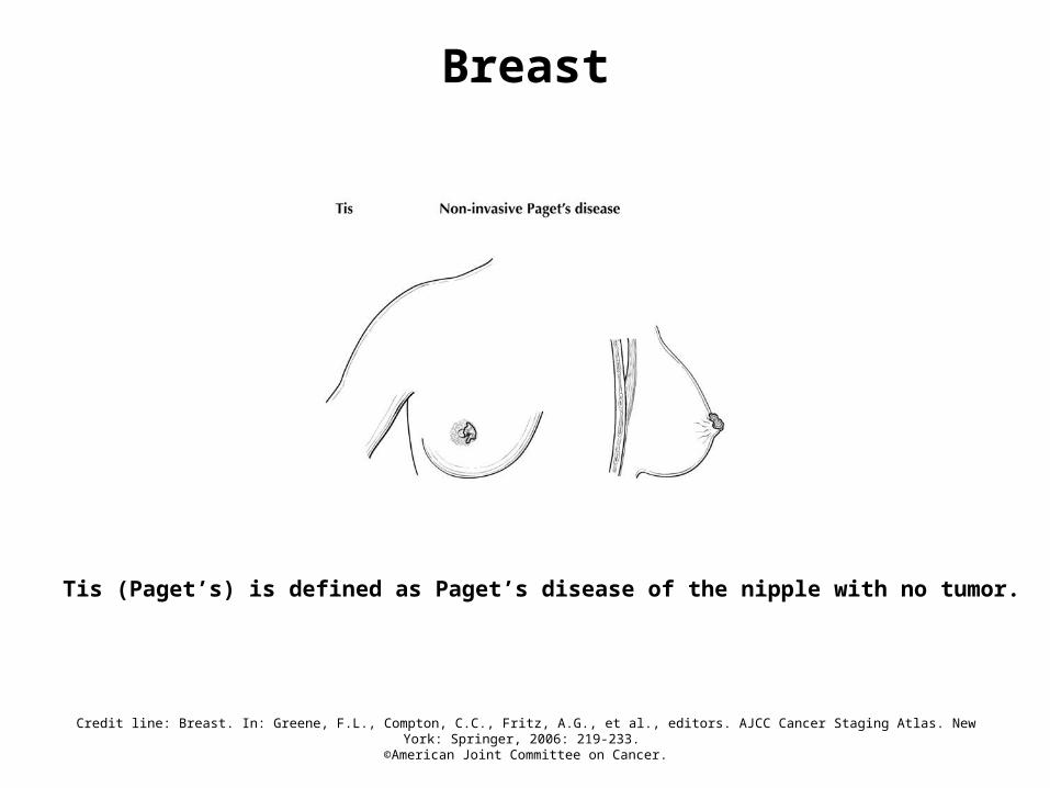
Tis (Paget’s) is defined as Paget’s disease of the nipple with no tumor.

Credit line: Breast. In: Greene, F.L., Compton, C.C., Fritz, A.G., et al., editors. AJCC Cancer Staging Atlas. New York: Springer, 2006: 219-233.
©American Joint Committee on Cancer.
Breast
T1mic is defined as microinvasion 0.1 cm or less in greatest dimension. The presence of multiple tumor foci of microinvasion (top of diagram) should be noted in parentheses.

Credit line: Breast. In: Greene, F.L., Compton, C.C., Fritz, A.G., et al., editors. AJCC Cancer Staging Atlas. New York: Springer, 2006: 219-233.
©American Joint Committee on Cancer.
Breast
T1 is defined as a tumor 2 cm or less in greatest dimension. T1a is defined as tumor more than 0.1 cm but not more than 0.5 cm in greatest dimension; T1b is defined as tumor more than 0.5 cm but not more than 1 cm in greatest dimension; T1c is defined as tumor more than 1 cm but not more than 2 cm in greatest dimension.

Credit line: Breast. In: Greene, F.L., Compton, C.C., Fritz, A.G., et al., editors. AJCC Cancer Staging Atlas. New York: Springer, 2006: 219-233.
©American Joint Committee on Cancer.
Breast
T2 (above dotted line) is defined as tumor more than 2 cm but not more than 5 cm in greatest dimension and T3 (below dotted line) is defined as tumor more than 5 cm in greatest dimension.

Credit line: Breast. In: Greene, F.L., Compton, C.C., Fritz, A.G., et al., editors. AJCC Cancer Staging Atlas. New York: Springer, 2006: 219-233.
©American Joint Committee on Cancer.
Breast
T4 is defined as a tumor of any size with direct extension to chest wall, not including pectoralis muscle.

Credit line: Breast. In: Greene, F.L., Compton, C.C., Fritz, A.G., et al., editors. AJCC Cancer Staging Atlas. New York: Springer, 2006: 219-233.
©American Joint Committee on Cancer.
Breast
T4b, illustrated here as satellite skin nodules, is defined as edema (including peau d’orange) or ulceration of the skin of the breast, or satellite skin nodules confined to the same breast. T4b illustrated here as edema (including peau d’orange).

Credit line: Breast. In: Greene, F.L., Compton, C.C., Fritz, A.G., et al., editors. AJCC Cancer Staging Atlas. New York: Springer, 2006: 219-233.
©American Joint Committee on Cancer.
Breast
T4c is defined as both T4a and T4b.
Credit line: Breast. In: Greene, F.L., Compton, C.C., Fritz, A.G., et al., editors. AJCC Cancer Staging Atlas. New York: Springer, 2006: 219-233.
©American Joint Committee on Cancer.
Breast
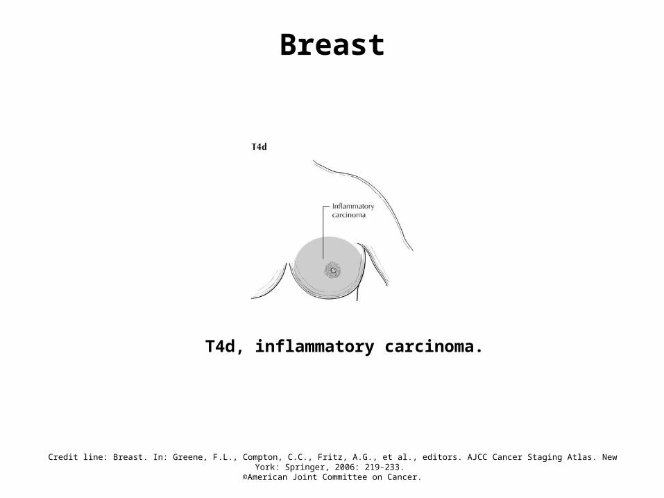
T4d, inflammatory carcinoma.

Credit line: Breast. In: Greene, F.L., Compton, C.C., Fritz, A.G., et al., editors. AJCC Cancer Staging Atlas. New York: Springer, 2006: 219-233.
©American Joint Committee on Cancer.
Breast
N1 is defined as metastasis in movable ipsilateral axillary lymph node(s).

Credit line: Breast. In: Greene, F.L., Compton, C.C., Fritz, A.G., et al., editors. AJCC Cancer Staging Atlas. New York: Springer, 2006: 219-233.
©American Joint Committee on Cancer.
Breast
N2a is defined as metastasis in ipsilateral axillary lymph nodes fixed to one another (matted) or to other structures.

Credit line: Breast. In: Greene, F.L., Compton, C.C., Fritz, A.G., et al., editors. AJCC Cancer Staging Atlas. New York: Springer, 2006: 219-233.
©American Joint Committee on Cancer.
Breast
N2b is defined as metastasis only in clinically apparent ipsilateral internal mammary nodes and in the absence of clinically evident axillary lymph node metastasis.

Credit line: Breast. In: Greene, F.L., Compton, C.C., Fritz, A.G., et al., editors. AJCC Cancer Staging Atlas. New York: Springer, 2006: 219-233.
©American Joint Committee on Cancer.
Breast
N3a metastasis in ipsilateral infraclavicular lymph node(s) without axillary or internal mammary lymph node involvement.

Credit line: Breast. In: Greene, F.L., Compton, C.C., Fritz, A.G., et al., editors. AJCC Cancer Staging Atlas. New York: Springer, 2006: 219-233.
©American Joint Committee on Cancer.
Breast
N3b metastasis in ipsilateral internal mammary lymph node(s) and axillary lymph node(s).
Credit line: Breast. In: Greene, F.L., Compton, C.C., Fritz, A.G., et al., editors. AJCC Cancer Staging Atlas. New York: Springer, 2006: 219-233.
©American Joint Committee on Cancer.
Breast
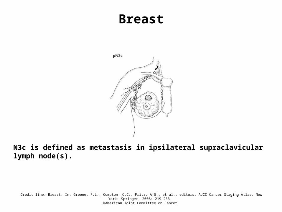
N3c is defined as metastasis in ipsilateral supraclavicular lymph node(s).

Credit line: Breast. In: Greene, F.L., Compton, C.C., Fritz, A.G., et al., editors. AJCC Cancer Staging Atlas. New York: Springer, 2006: 219-233.
©American Joint Committee on Cancer.
Breast
pN0(i+) is defined as no regional lymph node metastasis histologically, positive IHC, no IHC cluster greater than 0.2 mm.
Credit line: Breast. In: Greene, F.L., Compton, C.C., Fritz, A.G., et al., editors. AJCC Cancer Staging Atlas. New York: Springer, 2006: 219-233.
©American Joint Committee on Cancer.
Breast
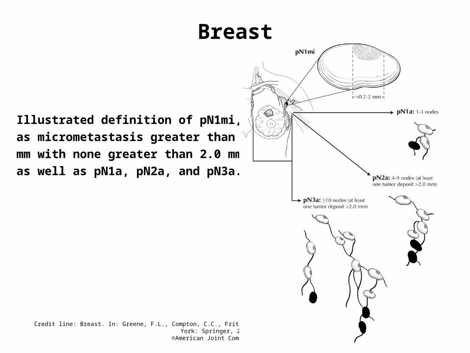
Illustrated definition of pN1mi, defined
as micrometastasis greater than 0.2
mm with none greater than 2.0 mm,
as well as pN1a, pN2a, and pN3a.

Credit line: Breast. In: Greene, F.L., Compton, C.C., Fritz, A.G., et al., editors. AJCC Cancer Staging Atlas. New York: Springer, 2006: 219-233.
©American Joint Committee on Cancer.
Breast
pN1b with isolated tumor cells in a single internal mammary node, sentinel lymph node positive.

Credit line: Breast. In: Greene, F.L., Compton, C.C., Fritz, A.G., et al., editors. AJCC Cancer Staging Atlas. New York: Springer, 2006: 219-233.
©American Joint Committee on Cancer.
Breast
pN1c illustrating 3 positive axillary lymph nodes with isolated tumor cells in a single internal mammary lymph node, sentinel lymph node positive.

Credit line: Breast. In: Greene, F.L., Compton, C.C., Fritz, A.G., et al., editors. AJCC Cancer Staging Atlas. New York: Springer, 2006: 219-233.
©American Joint Committee on Cancer.
Breast
pN2b illustrating clinically apparent metastasis in 2 positive internal mammary nodes with no axillary lymph node involvement.
Credit line: Breast. In: Greene, F.L., Compton, C.C., Fritz, A.G., et al., editors. AJCC Cancer Staging Atlas. New York: Springer, 2006: 219-233.
©American Joint Committee on Cancer.
Breast
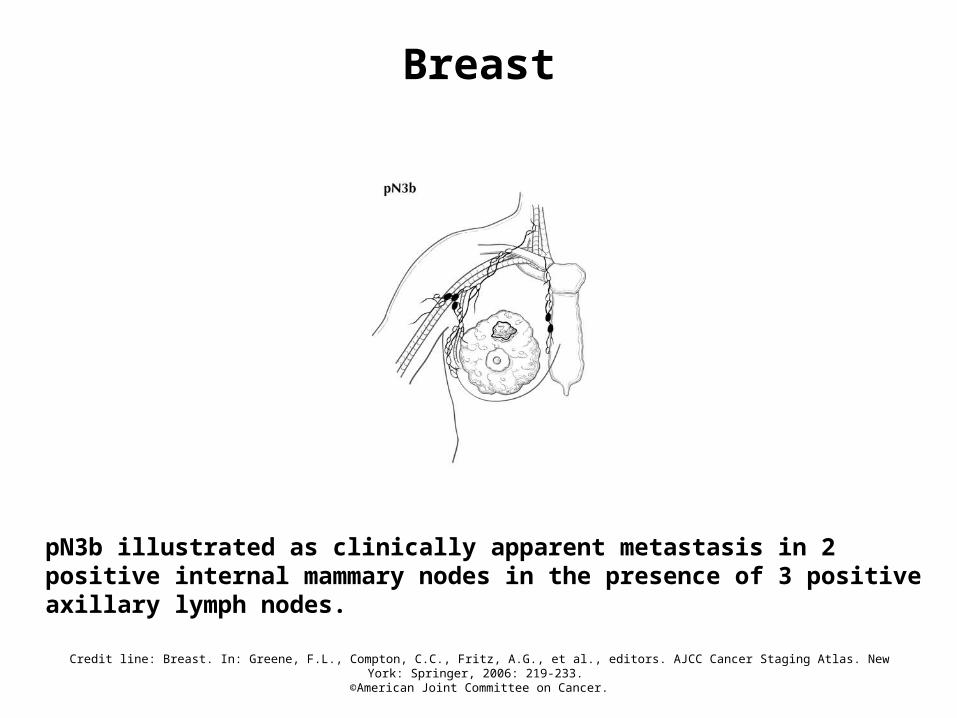
pN3b illustrated as clinically apparent metastasis in 2 positive internal mammary nodes in the presence of 3 positive axillary lymph nodes.
Credit line: Breast. In: Greene, F.L., Compton, C.C., Fritz, A.G., et al., editors. AJCC Cancer Staging Atlas. New York: Springer, 2006: 219-233.
©American Joint Committee on Cancer.
Breast
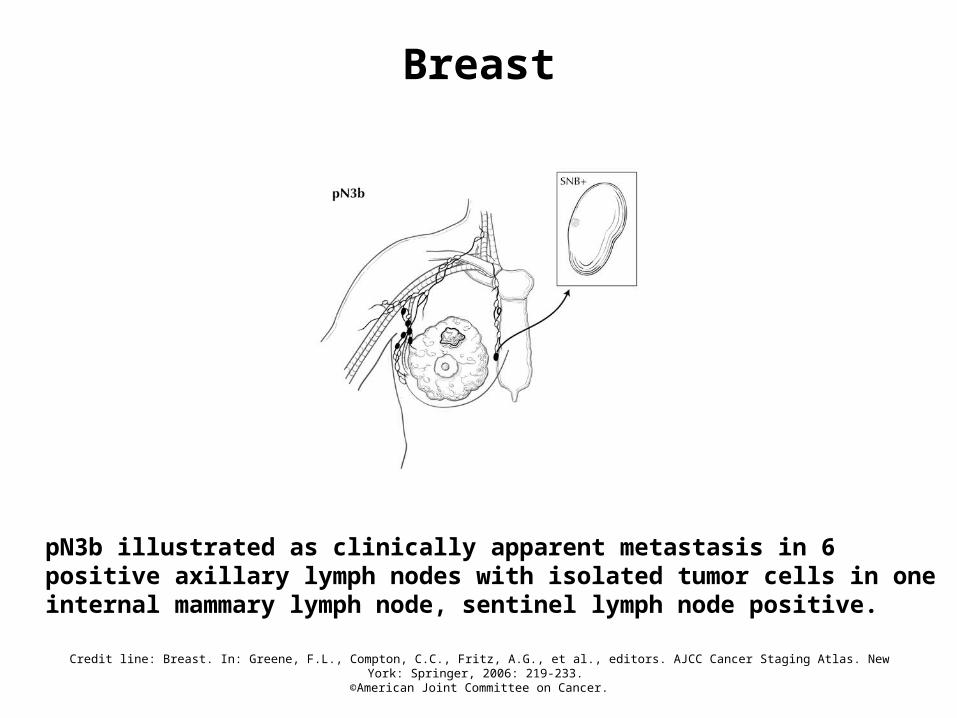
pN3b illustrated as clinically apparent metastasis in 6 positive axillary lymph nodes with isolated tumor cells in one internal mammary lymph node, sentinel lymph node positive.


![[Boschke F.L.] Medicinal Chemistry](https://img.pdfslide.net/doc/110x75/56d6bebf1a28ab3016936add/boschke-fl-medicinal-chemistry.jpg)