Embed Size (px)
Citation preview
CroniconO P E N A C C E S S EC DENTAL SCIENCE
Research Article
Effect of Air Abrasion Techniques on Marginal Adaptation of Class V Cavity
Ahmed Mohamed Hassan1*, Ahmed Mohammed Elmarakby1 and Samah Samir Bedir2
1Assistant Professor, Department of Restorative Dental Sciences, Alfarabi Dental College, Riyadh, KSA and Department of Operative Dentistry, Faculty of Dentistry, Al-Azhar University, Egypt2Instructor, Department of Restorative Dental Sciences, Alfarabi Dental College, Riyadh, KSA
*Corresponding Author: Ahmed Mohammed Hassan, assistant professor, department of restorative dental science, Alfarabi dental college, Riyadh, KSA and Department of Operative Dentistry, Faculty of Dentistry, Al-Azhar University, Egypt.
Citation: Ahmed Mohammed Hassan., et al. “Effect of Air Abrasion Techniques on Marginal Adaptation of Class V Cavity”. EC Dental Science 6.2 (2016): 1288-1296.
Received: November 28, 2016; Published: December 02, 2016
Abstract
Aims: The aim of this study was to evaluate the effect of air abrasion techniques with different alumina particle sizes and different pressures on marginal integrity of class V cavity restored with low shrinkable composite and resin modified glass nano ionomer.
Material and Methods: A total number of 150 sound human posterior teeth were used. They were divided into two subgroups (n = 75) according to the used restorative material [A1: low shrinkable composite resin (P90) and A2: Ketac nano ionomer (N100)]. Each group was divided into five groups (n = 15) according to the cavity preparation technique (B1: conventional bur, B2: small size alumina particles with low pressure, B3: small size alumina particles with high pressure, B4: large size alumina particles, with low pressure, and B5: large size alumina particles with high pressure). Each subgroup was further divided into three subgroups (n = 5) according to the storage time (C1: immediately, C2: three months, and C3 six months). Marginal integrity was measured using scan-ning electron microscope. Two-way analysis of variance was used to compare the mean values of marginal gaps for both enamel and dentin margins. Student’s t-test was used to compare the mean values of marginal gaps for each group.
Results: It was found that; largest gape was detected for group A2B1C3, while the smallest gap was detected for group A1B5C1.
Conclusion: Using of air abrasion technique with large size alumina particles under high pressure resulted in better marginal adap-tation.
Keywords: Air Abrasion; Low Shrinkable Composite; Marginal Adaptation
Introduction
Dental caries is one of the most common infectious diseases of mankind. With minimally invasive dentistry, dental caries is treated as an infectious condition rather than an end product of it. Now no longer radical “extension for prevention” is practiced but has changed to “constriction with conviction” [1]. The concept of conservative healthy tooth cavity preparation has gained popularity with the advent of adhesive resin bonding systems in which the retention and resistance form for cavity preparation has also been minimized [2]. Nowadays, common caries removal procedure was done by carbide and diamond bur which induces some disadvantages like producing pain, vibra-tion, noise and thermal changes resulting in pulp irritation [3]. New techniques and materials have yielded new concepts for tooth surface preparation and caries removal in conservative dentistry [4]. Air abrasion offers an alternative to conventional dental hand pieces as it minimizes the heat, pressure, noise and vibration [5].
1289
Effect of Air Abrasion Techniques on Marginal Adaptation of Class V Cavity
Citation: Ahmed Mohammed Hassan., et al. “Effect of Air Abrasion Techniques on Marginal Adaptation of Class V Cavity”. EC Dental Science 6.2 (2016): 1288-1296.
Air abrasion can be best described as a pseudo-mechanical, non-rotary method of cutting and removing dental hard tissue. Air abra-sion for restoration preparation removes tooth structure using a stream of aluminum oxide particles generated from compressed air, bottled carbon dioxide or nitrogen gas. The abrasive particles strike the tooth with high velocity and remove small amounts of tooth structure [4,6]. Air abrasion was originally developed by Robert Black in 1945 as an alternative pseudo-mechanical method for dental tissue removal and the first air abrasion unit marketed was called the Airdent by SS White [1].
Restoring cervical lesions is a challenge for the clinicians. Advances in material science have made it possible to restore these lesions with minimal tooth preparation using bonded restorations such as glass ionomer cement (GIC), resin-modified GIC (RM-GIC) or compos-ite resin [7]. With the introduction of nano-filled and low shrink types of esthetic restorative materials, it will be easier to restore these cavities with more bond strength and less tooth structure loss and minimal specification of GV Black’s concepts [8]. The patency of the tooth-restoration interface is critical to the restored tooth’s vitality, function, and appearance. Failure can occur from weak links between the tooth enamel or dentin and adhesive, the adhesive itself, the adhesive/restoration boundary and finally, the restoration itself [9]. Mi-croleakage is one of the major reasons for recurrent decays, pulp inflammation and necrosis [10].
Scanning electron microscope and confocal microscope have been used to examine the marginal adaptation of class V cavity restora-tions [11,12]. In this study, we will use scanning electron microscope to examine the marginal adaptation of class V cavities prepared by five techniques and restored by either low shrinkable composite or resin modified glass ionomer. The null hypothesis that will be investi-gated is that; there is no effect of preparation techniques on the marginal adaptation of class V restorations.
Materials and Methods
Sample selection and grouping
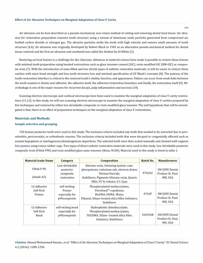
150 human posterior teeth were used in this study. The inclusion criteria included any teeth that needed to be extracted due to peri-odontitis, pericoronitis, or orthodontic reasons. The exclusion criteria included teeth that were decayed or congenitally affected such as enamel hypoplasia or amelogenesis/dentinogenesis imperfecta. The selected teeth were then scaled manually and cleaned with eugenol free pumice using rotary rubber cups. Two types of direct esthetic restorative materials were used in this study: low shrinkable posterior composite resin (Filtek P90) and resin modified glass nano-ionomer (Ketac N100). Material used in this study is listed in table 1.
Material trade Name Category Composition Batch No. Manufacturer
Filtek P 90
(shade A2)
Low shrinkable posterior
composite restorative
Silorane resin, Initiating system: cam-phorquinone, iodonium salt, electron donor,
Yttrium fluoride, Stabilizers, Pigments Silorane resin, Quartz
filler, 55 % volume, 0.1-2µm
4762A2
3M ESPE Dental Product St. Paul,
MN, USA
LS Adhesive Self-Etch Primer
self-etching Primer
especially for p90composite
Phosphorylated methacrylates, Vitrebond™ copolymer, BisGMA, HEMA, Water,
Ethanol, Silane-treated silica filler, Initiators, Stabilizers
4763P 3M ESPE Dental Product St. Paul,
MN, USA
LS Adhesive Self-Etch
Bond
self-etching bond especially for p90composite
Hydrophobic dimethacrylate, Phosphorylated methacrylates,
TEGDMA, Silane- treated silica filler, Initiators, Stabilizers
166926B 3M ESPE Dental Product St. Paul,
MN, USA

1290
Effect of Air Abrasion Techniques on Marginal Adaptation of Class V Cavity
Citation: Ahmed Mohammed Hassan., et al. “Effect of Air Abrasion Techniques on Marginal Adaptation of Class V Cavity”. EC Dental Science 6.2 (2016): 1288-1296.
Ketac N100
Resin modified nanoparticles glass ionomer
Paste A: Silane treated glass, silane treated zirconia, PEGDMA, silane treated silica,
HEMA, BisGMA, TEGDMA. Paste B: Silane treated ceramic, copolymer
acrylic and Iataconic acid, water, HEMA.
K04407 3M ESPE Dental Product St. Paul,
MN, USA
Ketac nano Primer
Primer for nano ionomer
HEMA (35-45%), Water (40-50%), copoly-mer acrylic and Iataconic acid (10-15%).
N265388 3M ESPE Dental Product St. Paul,
MN, USA
Table 1: The materials used in the study.
The teeth were divided into two main groups (n = 75) according to type of restorative material (A1: restored with P90 and A2: re-stored with N 100). Each main group was divided into five groups (n = 15) according to method of cavity preparation (B1: control group using the conventional bur, B2: using (10 µm) alumina particles and (15 psi) pressure, B3: using (10 µm) alumina particles and (40 psi) pressure, B4: using (50 µm) alumina particles and (15 psi) pressure, and B5: using (50 µm) alumina particles and (40 psi) pressure. Then each group was further subdivided into three subgroups (n = 5) according to storage time (C1: immediate, C2: three months and C3: six months).
Cavity preparation
For the control group, a round carbide bur (Midwest No.¼ Dentsply) was used. A standardized class V cavity (3mm height and 4mm width) was prepared in the buccal and lingual surfaces of each tooth. All cavity preparations procedures were carried out at speed of (100000 rpm) with air -water coolant. The bur used for cavity preparation was discarded after every three preparations. For the groups prepared with air abrasion, the air abrasion unit (Sandstorm Air Abrasion Sandblasting Unit, Kreativ, Inc., San Diego, CA) was used accord-ing to the manufacturer’s directions. Air abrasion was used to prepare the cavity in a micro-pulse mode at beam intensity of 2 gm/min for 15 seconds at distance of 5 mm between the nozzle and tooth surface. The application of the aluminum oxide jet was accomplished inside a closed transparent acrylic box, to avoid particle aspiration by the operator.
Restoration placement
For resin, composite Filtek P90, the P90 System Adhesive Self-Etch Primer was rubbed to the prepared cavity for 15 seconds with black micro brush, followed by gentle air dispersion and then light cured for 10 seconds with halogen device (QHL75 visible curing light, Dentsply). Then, the P90 System Adhesive Bond applied for 15 seconds with green micro brush, followed by gentle air dispersion and 10 seconds of light curing. After that, the resin composite was applied in three increments: the first against the gingival wall and the second against the occlusal wall, while the final increment was placed flush with the contour of the tooth and covered with a transparent matrix strip (Ruwa Matrix Strips). Each increment was light cured for 40 sec. Immediately after filling, excess materials were removed using finishing burs (Shofu SF 201 Ra) and polished with the Sof-lex disk system (3M ESPE).
For Ketac (N100) Light-Curing Nano-Ionomer, Ketac N100 Primer was applied to both enamel and dentinal surfaces of prepared cavity for 15 seconds with a micro brush and were kept wet with the primer for the full application time. Two clicks were dispensed from the clicker dispenser to provide an adequate amount of material. Both pastes were mixed together for 20 seconds. Ketac N100 restorative was placed in 2 mm increment, and light cured for 40 seconds. Finishing procedures were carried out as the same for resin composite.
Storage
After completion of samples preparation, the samples were stored in distilled water at 37°C and humidity 100% in an incubator (Kum-tex incubator device. GC. Japan). The distilled water was changed daily until the time of examination (immediate, three months, and six months).
1291
Effect of Air Abrasion Techniques on Marginal Adaptation of Class V Cavity
Citation: Ahmed Mohammed Hassan., et al. “Effect of Air Abrasion Techniques on Marginal Adaptation of Class V Cavity”. EC Dental Science 6.2 (2016): 1288-1296.
Scanning Electron Microscopy
Impressions of the restored teeth were taken using a silicone impression material (aquasil ultra XLV fast set smart wetting impression material, Dentsply, International Inc. Canada) and were then used for the fabrication of epoxy replicas (Devcon 5-minute epoxy, ITW Dev-con, UK). The replicas were sputter-coated with gold (Polaron E-5200 Energy Beam Sciences, Agawan, MA, USA) and were investigated in the SEM (JSM, 6360LV, JEOL, Tokyo, JAPAN) for detection of marginal gaps width. Measurement of marginal gaps was made at x500 magnification. All teeth were tested at both occlusal and gingival margins of the prepared class V cavity.
Statistical Analysis
Two-way analysis of variance was used to compare the mean values of marginal gaps for both enamel and dentin margins across groups (B1 – B5). Student’s t-test for two independent samples was used to compare between the two restorative materials. All statistical analysis was conducted at the significance level of 0.05.
Results and Discussion
Results
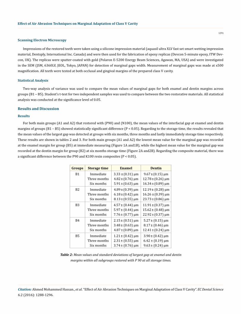
For both main groups (A1 and A2) that restored with (P90) and (N100), the mean values of the interfacial gap at enamel and dentin margins of groups (B1 – B5) showed statistically significant difference (P < 0.05). Regarding to the storage time, the results revealed that the mean values of the largest gap was detected at groups with six months, three months and lastly immediately storage time respectively. These results are shown in tables 2 and 3. For both main groups (A1 and A2) the lowest mean value for the marginal gap was recorded at the enamel margin for group (B5) at immediate measuring (Figure 1A and1B), while the highest mean value for the marginal gap was recorded at the dentin margin for group (B2) at six months storage time (Figure 2A and2B). Regarding the composite material, there was a significant difference between the P90 and K100 resin composites (P < 0.05).
Groups Storage time Enamel DentinB1 Immediate
Three months Six months
3.33 ± (0.31) µm 4.82 ± (0.76) µm 5.91 ± (0.63) µm
9.67 ± (0.15) µm 12.78 ± (0.26) µm 16.34 ± (0.89) µm
B2 Immediate Three months
Six months
4.89 ± (0.39) µm 6.18 ± (0.42) µm 8.13 ± (0.55) µm
12.19 ± (0.28) µm 16.26 ± (0.39) µm 23.73 ± (0.86) µm
B3 Immediate Three months
Six months
4.57 ± (0.44) µm 5.97 ± (0.44) µm 7.76 ± (0.77) µm
11.91 ± (0.37) µm 15.62 ± (0.48) µm 22.92 ± (0.37) µm
B4 Immediate Three months
Six months
2.15 ± (0.51) µm 3.48 ± (0.63) µm 4.87 ± (0.89) µm
5.27 ± (0.15) µm 8.17 ± (0.46) µm
12.41 ± (0.24) µmB5 Immediate
Three months Six months
1.21 ± (0.42) µm 2.31 ± (0.55) µm 3.74 ± (0.76) µm
3.90 ± (0.42) µm 6.42 ± (0.19) µm 9.63 ± (0.24) µm
Table 2: Mean values and standard deviations of largest gap at enamel and dentin margins within all subgroups restored with P 90 at all storage times.

1292
Effect of Air Abrasion Techniques on Marginal Adaptation of Class V Cavity
Citation: Ahmed Mohammed Hassan., et al. “Effect of Air Abrasion Techniques on Marginal Adaptation of Class V Cavity”. EC Dental Science 6.2 (2016): 1288-1296.
Groups Storage time Enamel DentinB1 Immediate
Three months Six months
4.25 ± (0.19) µm 7.53 ± (0.24) µm
10.16 ± (0.65) µm
10.34± (0.42) µm 12.57 ± (0.84) µm 9.66 ± (0.34) µm
B2 Immediate Three months
Six months
6.89 ± (0.52) µm 9.08 ± (0.53) µm
14.54 ± (0.87) µm
13.95 ± (0.77) µm 17.19 ± (0.31) µm 25.33 ± (0.17) µm
B3 Immediate Three months
Six months
6.36 ± (0.29) µm 8.96 ± (0.17) µm
13.93 ± (0.44) µm
13.18 ± (0.65) µm 16.74 ± (0.27) µm 24.89 ± (0.21) µm
B4 Immediate Three months
Six months
3.18 ± (0.64) µm 5.42 ± (0.13) µm 8.51± (0.64) µm
7.87 ± (0.32) µm 9.86 ± (0.64) µm
15.64 ± (0.56) µmB5 Immediate
Three months Six months
2.11 ± (0.35) µm 4.06 ± (0.34) µm 6.29 ± (0.47) µm
5.10 ± (0.89) µm 7.27 ± (0.87) µm
12.74 ± (0.66) µm
Table 3: Mean values and standard deviations of largest gap at enamel and dentin margins within all subgroups restored with K100 at all storage times.
Figure 1 A: SEM micrograph showing no gap between P 90 and enamel margin in group B5 at immediate storage time (at x500 magnification).
Figure 1 B: SEM micrograph showing no gap between N100 and enamel margin in group B5 at immediate storage time (at x500 magnification).

1293
Effect of Air Abrasion Techniques on Marginal Adaptation of Class V Cavity
Citation: Ahmed Mohammed Hassan., et al. “Effect of Air Abrasion Techniques on Marginal Adaptation of Class V Cavity”. EC Dental Science 6.2 (2016): 1288-1296.
Figure 2 A: SEM micrograph that shows a gap (22.7 um) between P 90 and gingival dentin margin in group B2 at six months storage time (at x500 magnification).
Figure 2 B: SEM micrograph that shows a gap (23.5 um) between N100 and gingival dentin margin in group B2 at six months storage time (at x500 magnification).
Discussion
Presently, several air abrasion devices are available for the purposes of tooth surface cleaning or caries removal whereby their pres-ence and emergence in restorative dentistry may signal an end to the use of mechanical instruments for caries removal. However, the air abrasion devices currently on the market are not without flaws; they do not afford good control to the amount of powder to be expelled [13]. Also, the depth of penetration during cavity cannot be controlled, so it has to be accompanied with visual inspection in regular in-tervals [6].
Cavity preparation characteristics using the air abrasion system are directly related to the tips and the operation parameters [14]. Air abrasion systems available today have either mechanical or digital operator controls. Mechanical control is standard in most devices, and its control of powder flow rate is more tenuous compared to that of digital control, which provides a consistent and minimal amount of powder while maintaining high efficiency. In selected devices, digital control also allows for a pulsed mode of operation, providing an interrupted air abrasive stream at settings from 0.5 to 2.0 seconds [15].
This method of cutting is relatively painless however, the total loss of tactile sensation, and the ability of alumina particles to remove sound tooth structure rather than the carious substrate in addition to the potential risk of inhalation problem should also be considered at the time of selection [1]. On the other hand, high speed burs yielded preparations 1.5 times quicker than air abrasion techniques [16].

1294
Effect of Air Abrasion Techniques on Marginal Adaptation of Class V Cavity
Citation: Ahmed Mohammed Hassan., et al. “Effect of Air Abrasion Techniques on Marginal Adaptation of Class V Cavity”. EC Dental Science 6.2 (2016): 1288-1296.
In the present study, the best marginal adaptation was observed for air abraded groups using large particle size with high pressure and restored with (P90) resin composite. This is in agreement with Knobloch., et al. 2005 which can be explained by the increased surface area of the tooth structure which would then improve the effectiveness of etching by increasing the wettability of tooth structure [17,18]. Another study also suggested the use of an acidic conditioner prior to application of resin is necessary to remove the smear layer created by the air abrasion to obtain good bonding, because the smear layer can prevent the diffusion of monomers into the superficial dental structure [11].
On the other hand, this result is in disagreement with Kumar., et al. 2014 who explained his result by the limited duration of air abrasion procedure (5 sec) to minimize the obstruction of dentinal tubules from residual dust layer [7]. While other study used sodium bicarbonate as the powder for air abrasion and we used aluminum oxide [10]. Another study showed the same results of microleakage with either bur preparation or air abrasion [19]. Freeman., et al. 2012 explain the marginal gap after air abrasion by the polymerization shrinkage that may result in tearing of the dentin surface damaged by air abrasion, compromising adhesion [12]. The reasons for this disagreement with other results may be attributed to different materials and techniques in each study.
The result of this study showed that the marginal adaptation was poorer at the dentin margins when compared to the enamel margins in all groups. This is may be explained by the superficial maceration of collagen fibers and tearing of damaged dentin surface by polymer-ization shrinkage [7]. Regarding the alumina particle size, the result of this study is augmented by the result of another study who found that; air-abrasion treatment with small particles is less effective in preventing microleakage compared to large particles [21]. This is can be explained as a larger particle size would remove a larger amount of tooth structure leading to more irregular surface with greater surface area available for bonding. Also, the higher presser of air abrasion resulted in a better the marginal adaptation. This is may be attributed to the greater force of the alumina particles that strikes the tooth surface making it more irregular with a greater surface area. The longer the storage time, the poorer the marginal adaptation was observed in the results of the present study. This is in agreement with another study that found 24 hours micro-tensile bond strengths were greater than the 6 and 12 months bond strengths [22].
The results of the present study showed superior marginal adaptation for main group restored with low shrinkable siloran based com-posite (P90) when comparing to the main group that restored with ketac nano ionomer (N100). This may be due to less polymerization shrinkage of low shrinkable siloran based composite (P90) that improved bonding of resin material to cavity walls especially to enamel walls. Results of the present study are in agreement with other studies which found that silorane restorative generated the lowest polym-erization stress and yielded the lowest dye penetration [23]. On the other-hand, results of the study of Boaro., et al. 2010 revealed that; not all low-shrinkage composites demonstrated reduced polymerization shrinkage. Among the materials considered as “low-shrinkage” by the respective manufacturers, although (P90) presented low post-gel shrinkage, Polymerization stress showed a strong correlation with post-gel shrinkage except for LS, which presented high stress [24]. These differences in results may be due to variation in testing technique or chemistry of materials used or type of adhesives.
Conclusion
Air abrasion cavity preparation resulted in better marginal adaptation than conventional bur preparation when used large particle size and high pressure. None of the restorative materials used in this study was capable to seal the cavity margins completely. The Filtek P90 low shrinkage silorane based composite resin yielded better results of marginal integrity along the occlusal enamel and gingival dentin walls when compared to ketac N100 nano ionomer system. The shorter the storage time, the better results of marginal integrity of tooth \ restoration interface.
Conflict of Interest
There are no conflicts of interest.
1295
Effect of Air Abrasion Techniques on Marginal Adaptation of Class V Cavity
Citation: Ahmed Mohammed Hassan., et al. “Effect of Air Abrasion Techniques on Marginal Adaptation of Class V Cavity”. EC Dental Science 6.2 (2016): 1288-1296.
Bibliography
1. Mm J., et al. “Minimal intervention dentistry – a new frontier in clinical dentistry”. Journal of Clinical and Diagnostic Research 8.7 (2014): 4-8.
2. Boob AR., et al. “Evaluation of the efficiency and effectiveness of three minimally invasive methods of caries removal: an in vitro study”. International Journal of Clinical Pediatric Dentistry 7.1 (2014): 11-18.
3. Pahlavan A., et al. “Effect of air abrasion and erbium-doped yttrium aluminum garnet (Er: YAG) laser preparation on shear bond strength of composite to dentin”. Journal of Lasers in Medical Science 4.3 (2013): 127-130.
4. Eshghi A., et al. “Effect of bioactive glass air abrasion on shear bond strength of two adhesive resins to decalcified enamel”. Journal of Dentistry (Tehran) 11.6 (2014): 644-654.
5. Oliveira MT., et al. “Influence of diamond sono-airabrasion, air-abrasion and Er:YAG laser irradiation on bonding of different adhesive systems to dentin”. European Journal of Dentistry 1.3 (2007): 158-166.
6. Hegde VS and Khatavkar RA. “A new dimension to conservative dentistry: air abrasion”. Journal of Conservative Dentistry 13.1 (2010): 4-8.
7. kumar U., et al. “Effect of air abrasion preconditioning on microleakage in class V restorations under cyclic loading: an in-vitro study”. Journal of Clinical and Diagnostic Research 8.5 (2014): 29-32.
8. Rodrigues SA Jr., et al. “Micro-structural characterization and fracture behavior of a micro-hybrid and a nano-fill composite”. Dental Materials 24.9 (2008): 1281-1288.
9. Thompson VP., et al. “Outside the (cavity-prep) box thinking”. Advances in Dental Research 25.1 (2013): 24-32.
10. Soleymani A., et al. “Evaluation of the effect of enameloplasty and air abrasion on sealant microleakage”. Journal of Dentistry (Tehran) 11.6 (2014): 639-643.
11. Sengun A., et al. “Adhesion of two bonding systems to air- abraded or bur-abraded human enamel surfaces”. European Journal of Dentistry 2.3 (2008): 167-175.
12. Freeman R., et al. “Effect of air abrasion and thermocycling on resin adaptation and shear bond strength to dentin for an etch-and-rinse and self-etch resin adhesive”. Dental Materials Journal 31.2 (2012): 180-188.
13. Honda K., et al. “Efficacy of a new jet nozzle for removal of carious dentin with an air abrasion system”. Dental Materials Journal 27.6 (2008): 835-841.
14. Peruchi C., et al. “Influence of air abrasion tips and operation modes on enamel cutting characteristics”. European Journal of Dentistry 7.1 (2013): 1-5.
15. Pelka M., et al. “Influence of air-polishing devices and abrasives on root dentin – An in vitro confocal laser scanning microscope study”. Quintessence International 41.7 (2010): 141-148.
16. Antunes LA., et al. “Effectiveness of high speed instrument and air abrasion on different dental substrates”. Brazilian Oral Research 22.3 (2008): 235-241.
17. Knobloch LA., et al. “Microleakage and bond strength of sealant to primary enamel comparing air abrasion and acid etch techniques”. Pediatric Dentistry 27.6 (2005): 463-469.
18. Asl Aminabadi N., et al. “Class III Restoration of Anterior Primary Teeth: In Vitro Retention Comparison of Conventional, Modified and Air-abrasionTreated Preparations”. Journal of Dental Research, Dental Clinics, Dental Prospects 8.2 (2014): 89-94.

1296
Effect of Air Abrasion Techniques on Marginal Adaptation of Class V Cavity
Citation: Ahmed Mohammed Hassan., et al. “Effect of Air Abrasion Techniques on Marginal Adaptation of Class V Cavity”. EC Dental Science 6.2 (2016): 1288-1296.
19. Zyskind D., et al. “Effect of etching on leakage of sealants placed after air abrasion”. Pediatric Dentistry 20.1 (1998): 25-27.
20. Arora A., et al. “A comparative evaluation of dentinal hypersensitivity and microleakage associated with composite restorations in cavities preconditioned with air abrasion - An ex vivo study”. Contemporary Clinical Dentistry 3.3 (2012): 306-313.
21. Fu B and Hannig M. “Effects of air abrasion and acid etching on the microleakage of preventive Class I resin restorations: an in vitro study”. Journal of Esthetic Dentistry 1.3 (1999): 143-148.
22. Giacobbi MF and Vandewalle KS. “Micro-tensile bond strength of a new silorane-based composite resin adhesive”. General Dentistry 60.3 (2012): 148-152.
23. Krifka S., et al. “Microleakage of silorane- and methacrylate-based class V composite restorations”. Clinical Oral Investigation 16.4 (2012): 1117-1124.
24. Boaro LC., et al. “Polymerization stress, shrinkage and elastic modulus of current low-shrinkage restorative composites”. Dental Mate-rial 26.12 (2010): 1144-1155.
Volume 6 Issue 2 December 2016© All rights reserved by Ahmed Mohammed Hassan., et al.