Embed Size (px)
Citation preview

doi:10.1016/j.jmb.2012.03.020 J. Mol. Biol. (2012) 419, 347–358
Contents lists available at www.sciencedirect.com
Journal of Molecular Biologyj ourna l homepage: ht tp : / /ees .e lsev ie r.com. jmb
Crystal Structure of Bacillus subtilis Signal PeptidePeptidase A
Sung-Eun Nam, Apollos C. Kim and Mark Paetzel⁎Department of Molecular Biology and Biochemistry, Simon Fraser University,South Science Building 8888 University Drive, Burnaby, British Columbia, Canada V5A 1S6
Received 12 January 2012;received in revised form16 March 2012;accepted 26 March 2012Available online1 April 2012
Edited by R. Huber
Keywords:signal peptide peptidase;self-compartmentalizedprotease;serine/lysine dyad;lysine general base;serine protease
*Corresponding author. E-mail addrAbbreviations used: SppA, signal
SEC, size-exclusion chromatographylight scattering; MPD, 2-methyl-2,4-Canadian Light Source.
0022-2836/$ - see front matter © 2012 E
Signal peptide peptidase A (SppA) is a membrane-bound self-compart-mentalized serine protease that functions to cleave the remnant signalpeptides left behind after protein secretion and cleavage by signalpeptidases. SppA is found in plants, archaea and bacteria. Here, we reportthe first crystal structure of a Gram-positive bacterial SppA. The 2.4-Å-resolution structure of Bacillus subtilis SppA (SppABS) catalytic domainreveals eight SppABS molecules in the asymmetric unit, forming a dome-shaped octameric complex. The octameric state of SppABS is supported byanalytical size-exclusion chromatography and multi-angle light scatteringanalysis. Our sequence analysis, mutagenesis and activity assays areconsistent with Ser147 serving as the nucleophile and Lys199 serving as thegeneral base; however, they are located in different region of the protein,more than 29 Å apart. Only upon assembling the octamer do the serine andlysine come within close proximity, with neighboring protomers eachproviding one-half of the catalytic dyad, thus producing eight separateactive sites within the complex, twice the number seen within Escherichia coliSppA (SppAEC). The SppABS S1 substrate specificity pocket is deep, narrowand hydrophobic, but with a polar bottom. The S3 pocket, which isconstructed from two neighboring proteins, is shallower, wider and morepolar than the S1 pocket. A comparison of these pockets to those seen inSppAEC reveals a significant difference in the size and shape of the S1pocket, which we show is reflected in the repertoire of peptides the enzymesare capable of cleaving.
© 2012 Elsevier Ltd. All rights reserved.
Introduction
Secretory proteins contain a signal peptide at theiramino-terminus that facilitates transport to andtranslocation across the cytoplasmic membrane.1,2
Once translocated, the signal peptide is cleaved off
ess: [email protected] peptidase A;; MALS, multi-anglepentanediol; CLS,
lsevier Ltd. All rights reserve
by signal peptidase. The remnant membrane-em-bedded signal peptide is subsequently degraded bysignal peptide peptidase (Spp). Spp is a membrane-bound peptidase that is found in all cells. Althoughall Spp share the same function—digesting signalpeptides—their size, membrane topology and cata-lytic mechanisms can be quite different. In archaea,plant chloroplasts and bacteria, these enzymes[signal peptide peptidase A (SppA)] utilize a serinenucleophile and a lysine general base.3–7 However,in eukaryotic cells, these enzymes are asparticproteases.8 This report focuses on the Gram-positivebacterial serine protease Bacillus subtilis SppA(SppABS).
d.

348 Octameric Signal Peptide Peptidase A Structure
Hussain et al. were the first to identify SppA as anenzyme involved in signal peptide digestion whenthey observed, in an in vitro experiment, thatEscherichia coli lipoprotein signal peptides weredigested upon the addition of a membrane extractcontaining SppA.9 Bolhuis et al. were the first toreport an SppA from B. subtilis. They showed thatSppABS can cleave the signal peptide of secretoryproteins in an in vivo assay.3
Crystallographic analysis has revealed that the E.coli SppA (SppAEC) contains two domains that arestructurally a tandem repeat, although the sequenceidentity between the N- and C-terminal domains isonly 18%.7 The structure has also revealed that theenzyme forms a tetramer, with four separate activesites. Each active site being located at the interfacebetween the N- and C-terminal domains, thecatalytic serine arrives from the C-terminal domain,and the catalytic lysine arrives from the N-terminaldomain of the same molecule. Mutagenesis exper-iments are consistent with SppAEC utilizing Ser409as the nucleophile and Lys209 as the general base.10
Based on topology predictions and analogy withSppAEC, the catalytic domain of SppABS is expectedto be located on the trans-side of the cytoplasmicmembrane with a single amino-terminal transmem-brane segment anchoring it to the membrane.10
Here, we report the first X-ray crystal structure ofa Gram-positive bacterial SppA. SppABS assemblesinto an octameric dome-shaped structure, whichcreates eight separate active sites, as opposed tothe tetrameric complex with four active sitesobserved in SppAEC. The SppABS active sites areconstructed from the interface of neighboringprotomers, suggesting that a proteolytically activeenzyme is only produced upon assembly of anoligomeric SppABS. The S1 and S3 substrate speci-ficity pockets (Schechter and Berger nomenclature11)are identified, described and compared to those inSppAEC. We find that the dimensions and polarityof the S1 pocket differ significantly betweenSppABS and SppAEC and that these differencesare consistent with the range of residues theenzymes will accommodate at the P1 position ofsubstrates.
Results and Discussion
SppABS purification, crystallization andstructure solution
We have observed that expressing the solubledomain of SppABS (SppABSΔ1–25) in the cytoplasmof E. coli results in slow cell growth and a limitedprotein expression level. Sequence alignments (Sup-plementary Fig. 1) suggest that Ser147 serves as thenucleophile and that Lys199 served as the generalbase in the SppABS catalytic mechanism; therefore,
we designed the mutants K199A and S147A to testthis hypothesis and to observe their effect on SppABSexpression level. Consistent with their proposed roleas active-site residues, our activity assays reveal thatthese mutant enzymes are inactive. In addition, theK199A mutant was highly overexpressed in thecytoplasm of E. coli and was easily purified inmilligram quantities. This protein was subjected tolimited proteolysis using thermolysin in order toimprove crystallization. The resulting proteolyticallyresistant fragment has a molecular mass of approxi-mately 28 kDa, approximately 8 kDa smaller than theoriginally purified protein. Amino-terminal sequen-cing analysis has revealed that the proteolyticallyresistant fragment starts at Leu51 (Fig. 1a). Thealcohols t-butanol and 2-methyl-2,4-pentanediol(MPD) and the detergent n-dodecyl-β-maltosidewere used to produce conditions that led to highlyordered plate-shaped crystals. The 2.4-Å-resolutionstructure reveals clear electron density for residues57–295, with the exception of a loop region betweenresidues 72 and 82.
The SppABS protein fold
The protomeric unit of the SppABS catalyticdomain (residues 57–295) has an α/β protein foldconsisting of seven β-strands, eight α-helices andone 310 helix (Fig. 1b). The SppABS protomer has tworegions: a globular region and an extension region.The globular region includes β-strands 1–4 and 7that are arranged in a parallel β-sheet, surroundedby α-helices 1–3 and 6–8. The extension regionconsists of α-helices 4 and 5 and β-strands 5 and 6.Each protomer contains a nucleophilic residue,Ser147, and a general base residue, Lys199 (Fig.1c). Ser147 is located at the turn before α-helix 3 ofthe globular region while Lys199 is located on theloop between β-strand 6 and α-helix 5 of theextension region (Fig. 1b and c). The distance fromthe Ser147 Oγ to the Lys199 Cβ is approximately29 Å, suggesting that a monomeric SppABS wouldnot be capable of catalysis. Eight molecules are in theasymmetric unit of this crystal (Fig. 2a). Analysis ofthe asymmetric unit reveals that the active-sitecatalytic dyad is created by Ser147 from oneprotomer and Lys199 from the neighboring proto-mer (Fig. 2b). Eight SppABS protomers cometogether to form a dome-shaped structure witheight separate active sites.
SppABS is octameric in solution
To confirm the existence of the octameric state ofSppABS in solution, we analyzed its size byanalytical size-exclusion chromatography (SEC)and multi-angle light scattering (MALS). We ob-serve that SppABS has an average molecular mass of225,400±4,508 g/mol, consistent with the SppABS

Fig. 1. The SppABS protein folds.(a) A schematic diagram showingthe full-length SppABS with itspredicted transmembrane segment(pink) and the confirmed amino-terminal thermolysin cleavage site.The light-green region is what isobserved in the electron density. (b)A topology diagram of SppABSshowing the full-length proteinwith β-strand as arrows and α-helices as cylinders. The protein iscolored as a gradient from N-terminus (blue) to C-terminus(red). The regions not seen in theelectron density are shown as bro-ken lines. (c) A cartoon diagramshowing the tertiary structure of theSppABS protomer. The β-strandsare shown as arrows, and the α-helices are shown as cylinders. Thecolor scheme is the same as in (b).The nucleophile Ser147 (red) andthe general base Lys199Ala (blue)are shown as spheres.
349Octameric Signal Peptide Peptidase A Structure
complex (proteolytically resistant fragment) being inan octameric arrangement in solution (Fig. 2c).
The SppABS dimensions and surface properties
The octameric SppABS catalytic domain is domeshaped with a wide opening created by the globularregions and a narrower opening made by theextension regions (Fig. 3). Based on membranetopology predictions and analogy to SppAEC,whose membrane topology has been experimentallydetermined, the wider opening of SppABS ispredicted to face the outer leaflet of the cytoplasmicmembrane10 (Fig. 3a). The outside of the dome has arelatively polar surface made up of both positivelyand negatively charged patches (Fig. 3b–d). Theinterior of the dome is predominantly hydrophobicwith some patches of negative charge (Fig. 3c and e).The positively charged rim at the narrower opening
of the dome is created by eight lysines, Lys185 fromeach of the SppABS protomers (Fig. 3b). The narrowopening is ∼22 Å in diameter. A cross-section of thedome shows a concave groove where the active sitesand substrate specificity binding pockets reside. Atthis point, the diameter is approximately ∼73 Å,while the narrowest region of the SppABS domeinterior is approximately 50 Å in diameter. Theheight of the dome is approximately 50 Å. The baseof the dome, 86 Å in diameter at the opening, isprimarily positively charged (Fig. 3e). The innercavity has a solvent-accessible surface area of17,220 Å2 and a volume of 56,456 Å3.
The SppABS Ser/Lys catalytic dyad and otheractive-site residues
The catalytic dyad of SppABS is made up of thenucleophilic Ser147 Oγ from one protomer and the

Fig. 2. Octameric arrangementof SppABS. (a) The SppABS octa-mer as shown in a cartoon dia-gram, viewed from the top (closestto the extension region that formsthe smaller opening). Each proto-mer is represented in a differentcolor. The serine nucleophile (red)and lysine general base mutated toalanine (blue) are shown asspheres, revealing eight active-sitecatalytic dyads. (b) A Cα trace oftwo neighboring protomers com-ing together to form one of theeight active sites: the serine (redsphere) comes from one protomer,and the lysine (blue sphere) ar-rives from the adjacent protomer.(c) A MALS analysis of purifiedSppABS proteolytic resistant frag-ment is represented by a molarmass (g/mol) versus time (min)plot overlaid with an analyticalsize-exclusion elution profile. Amolecular mass of 225,000 Da isapproximately equivalent to eightSppABS protomers. The SDS-PAGEgel stained with PageBlue showsthe fractions collected from theSEC column. The molecular massmarkers are shown on the right.
Next to the peak is a top view of the SppABS octamer as shown in molecular surface representation, each protomer iscolored alternating black and white.
350 Octameric Signal Peptide Peptidase A Structure
general base Lys199 Nζ from the adjacent protomer.Having eight protomers in the octamer thereforecreates eight separate active sites, each formed at theprotein–protein interface (Fig. 2). In order forSppABS to have one completely functional unit,three neighboring SppABS protomers are required toassemble (Fig. 4a). The nucleophile (Ser147), oxya-nion hole residues (Gly114 and Gly148), the generalbase coordinating residue (Ser169) and S1 specificitypocket residues arrive from the central molecule(salmon in Fig. 4); the lysine general base arrivedfrom one neighboring protomer (green in Fig. 4),and the S3 specificity pocket is partially formed fromthe other neighboring protomer (light blue in Fig. 4).Modeling the Lys199 side chain within this K199A
mutant structure shows that the Ser147 Oγ would beexpected to be within hydrogen bonding distance tothe Lys199 Nζ. The oxyanion hole is created by thehydrogen bond donor NH groups of Gly114 andGly148 from the same molecule that contributes thenucleophilic Ser147 to the catalytic dyad. Theamino-terminal end of the α3 helix dipole, whereSer147 is located, is also a possible contributor tooxyanion stabilization. This molecule also contrib-utes the side-chain hydroxyl group of Ser169, to theactive site. Similarly positioned hydroxyl groups inother Ser/Lys proteases have been proposed to help
coordinate and orient the lysine general baseepsilon-amino group12,13 (Fig. 4).
TheSppABS substrate specificity binding pockets
The eight active sites and substrate bindinggrooves of SppABS are located approximatelymidway up the interior of the octameric domecreating a continuous concave surface that encirclesthe entire inner bowl (as seen in surface cross-sectionin Fig. 3e). Analysis of the grooves reveals only twoclear specificity pockets for each of the eight activesites. Modeling an extended conformation for thesubstrate puts the P1 and P3 residue side chainsfacing the S1 and S3 specificity pockets and the P2and P4 residue side chains facing toward the solventaway from the binding groove. The S1 substratespecificity pocket of SppABS is created by residuesGly114, Val116, Ser119, Gly148, Tyr151, Gly171,Val172, Ser223, Gly226 and Phe227 from oneprotomer and Glu164 from a neighboring protomerwhile the S3 pocket is created by Val116, Met219 andSer223 from one protomer and Pro163, Glu164,Thr165, Leu166, Asp252 and Arg254 from a thirdadjacent protomer (Fig. 4). The S1 pocket is narrowand deep with hydrophobic walls (Phe227, Tyr151and Val116) and a more polar bottom (Glu164 Oɛ1,

Fig. 3. Surface properties anddimensions of SppABS. (a) A semi-transparent surface representationof the SppABS octamer, with oneprotomer shown in cartoon dia-gram. The widest opening in SppA-BS, formed from the globular region(“bottom”), is facing the cytoplas-mic membrane, and the transmem-brane segment is schematicallydrawn. The electrostatic propertiesof SppABS are shown from the top(b), bottom (c) and side (d) (blue,positive; red, negative). (e) A cross-section view of the SppABS octamer,with dimensions given.
351Octameric Signal Peptide Peptidase A Structure
Oɛ2 and Tyr151 Oη) (Fig. 4). The S3 pocket is not asdeep as the S1 pocket but is significantly wider. TheS3 entrance is hydrophobic however deeper into thepocket it has polar characteristics formed by themain-chain carbonyl oxygens of Pro163, Glu164 andMet219 and side chains of Ser223, Asp252 andArg254. Structural alignment of the residues in-volved in creating the S1 and S3 pockets in the eightbinding sites of SppABS showed that they superim-pose quite well except for residues Ser223, Glu164and Arg254 (Fig. 4c). The Ser223 side-chain rotamer,which is located at the S1 and S3 pocket boundary,varies among the eight chains. Alternate conforma-tions are observed for the side chains of residuesGlu164 and Arg254, which form part of the S3pocket, this suggests that these residues maybedynamic and may possibly be involved in aninduced fit process upon substrate binding.
Octameric SppABS versus tetrameric SppAEC
Both SppABS and SppAEC form oligomers thatassemble into dome-shaped structures.7 The SppA-BS protomer is half the size of the SppAEC protomer.Therefore, the SppABS dome complex is created byeight protomers while only four protomers arerequired for SppAEC (Fig. 5a and b). SppAEC's N-and C-terminal domains are structurally tandemrepeats (Fig. 5c). The SppABS protomer superim-
poses onto the N-terminal domain of SppAEC withan r.m.s.d. value of 2.7 Å (Fig. 5e) while the SppABSprotomer superimposes onto the C-terminal domainof SppAEC with a much lower r.m.s.d. values of1.1 Å (Fig. 5f). The most significant differences in thesuperimposition of the N-terminal domain of SppA-EC with the SppABS protomer are observed in theouter helices (α-helices 1 and 8) and the extensionregion. In addition, there is an extra helix found inthe N-terminal domain of SppAEC, between α-helices 6 and 7 of SppABS (Fig. 5e).
Sequence conservation between Gram-positiveand Gram-negative SppA
SppABS has a sequence identity of 16% and 26% tothe N- and C-terminal domains of SppAEC, respec-tively (Supplementary Fig. 1). The majority of theconserved residues shared by SppABS and the C-terminal domain of SppAEC are evenly distributedon the protomer of SppABS; however, there is apatch of conserved residues clustered around thenucleophile Ser147 (Supplementary Fig. 2). Similarpatterns of conserved residues are also observedwhen SppABS is aligned with SppA sequences fromother Gram-positive species (Supplementary Figs. 3and 5) or when SppAEC is aligned with SppAsequences from other Gram-negative species (Sup-plementary Figs. 4 and 5).
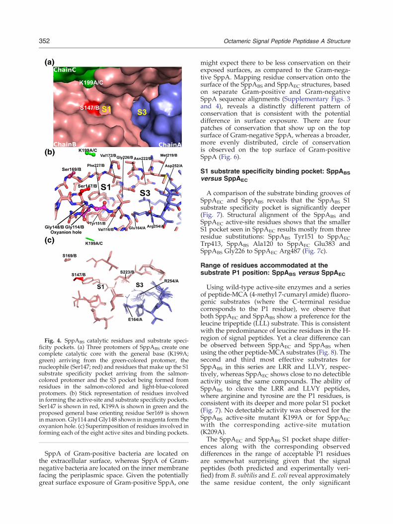
Fig. 4. SppABS catalytic residues and substrate speci-ficity pockets. (a) Three protomers of SppABS create onecomplete catalytic core with the general base (K199A;green) arriving from the green-colored protomer, thenucleophile (Ser147; red) and residues that make up the S1substrate specificity pocket arriving from the salmon-colored protomer and the S3 pocket being formed fromresidues in the salmon-colored and light-blue-coloredprotomers. (b) Stick representation of residues involvedin forming the active-site and substrate specificity pockets.Ser147 is shown in red, K199A is shown in green and theproposed general base orienting residue Ser169 is showninmaroon. Gly114 and Gly148 shown inmagenta form theoxyanion hole. (c) Superimposition of residues involved informing each of the eight active sites and binding pockets.
352 Octameric Signal Peptide Peptidase A Structure
SppA of Gram-positive bacteria are located onthe extracellular surface, whereas SppA of Gram-negative bacteria are located on the inner membranefacing the periplasmic space. Given the potentiallygreat surface exposure of Gram-positive SppA, one
might expect there to be less conservation on theirexposed surfaces, as compared to the Gram-nega-tive SppA. Mapping residue conservation onto thesurface of the SppABS and SppAEC structures, basedon separate Gram-positive and Gram-negativeSppA sequence alignments (Supplementary Figs. 3and 4), reveals a distinctly different pattern ofconservation that is consistent with the potentialdifference in surface exposure. There are fourpatches of conservation that show up on the topsurface of Gram-negative SppA, whereas a broader,more evenly distributed, circle of conservationis observed on the top surface of Gram-positiveSppA (Fig. 6).
S1 substrate specificity binding pocket: SppABSversus SppAEC
A comparison of the substrate binding grooves ofSppAEC and SppABS reveals that the SppABS S1substrate specificity pocket is significantly deeper(Fig. 7). Structural alignment of the SppABS andSppAEC active-site residues shows that the smallerS1 pocket seen in SppAEC results mostly from threeresidue substitutions: SppABS Tyr151 to SppAECTrp413, SppABS Ala120 to SppAEC Glu383 andSppABS Gly226 to SppAEC Arg487 (Fig. 7c).
Range of residues accommodated at thesubstrate P1 position: SppABS versus SppAEC
Using wild-type active-site enzymes and a seriesof peptide-MCA (4-methyl 7-cumaryl amide) fluoro-genic substrates (where the C-terminal residuecorresponds to the P1 residue), we observe thatboth SppAEC and SppABS show a preference for theleucine tripeptide (LLL) substrate. This is consistentwith the predominance of leucine residues in the H-region of signal peptides. Yet a clear difference canbe observed between SppAEC and SppABS whenusing the other peptide-MCA substrates (Fig. 8). Thesecond and third most effective substrates forSppABS in this series are LRR and LLVY, respec-tively, whereas SppAEC shows close to no detectibleactivity using the same compounds. The ability ofSppABS to cleave the LRR and LLVY peptides,where arginine and tyrosine are the P1 residues, isconsistent with its deeper and more polar S1 pocket(Fig. 7). No detectable activity was observed for theSppABS active-site mutant K199A or for SppAECwith the corresponding active-site mutation(K209A).The SppAEC and SppABS S1 pocket shape differ-
ences along with the corresponding observeddifferences in the range of acceptable P1 residuesare somewhat surprising given that the signalpeptides (both predicted and experimentally veri-fied) from B. subtilis and E. coli reveal approximatelythe same residue content, the only significant
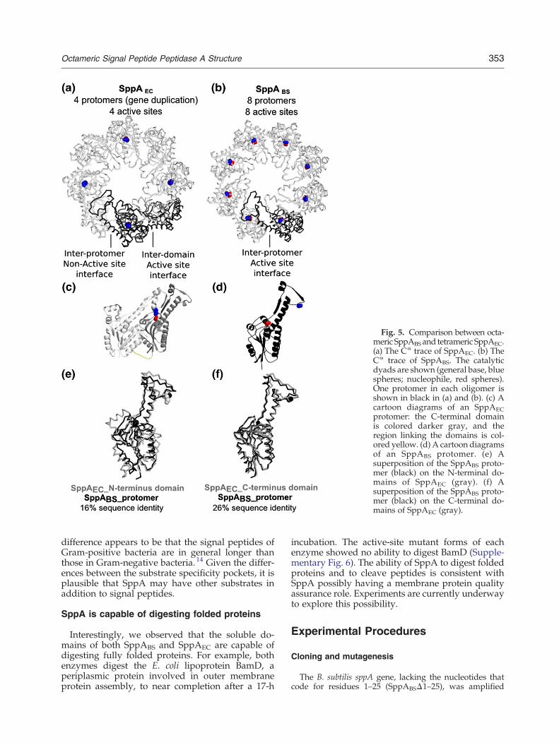
Fig. 5. Comparison between octa-meric SppABS and tetrameric SppAEC.(a) The Cα trace of SppAEC. (b) TheCα trace of SppABS. The catalyticdyads are shown (general base, bluespheres; nucleophile, red spheres).One protomer in each oligomer isshown in black in (a) and (b). (c) Acartoon diagrams of an SppAECprotomer: the C-terminal domainis colored darker gray, and theregion linking the domains is col-ored yellow. (d) A cartoon diagramsof an SppABS protomer. (e) Asuperposition of the SppABS proto-mer (black) on the N-terminal do-mains of SppAEC (gray). (f) Asuperposition of the SppABS proto-mer (black) on the C-terminal do-mains of SppAEC (gray).
353Octameric Signal Peptide Peptidase A Structure
difference appears to be that the signal peptides ofGram-positive bacteria are in general longer thanthose in Gram-negative bacteria.14 Given the differ-ences between the substrate specificity pockets, it isplausible that SppA may have other substrates inaddition to signal peptides.
SppA is capable of digesting folded proteins
Interestingly, we observed that the soluble do-mains of both SppABS and SppAEC are capable ofdigesting fully folded proteins. For example, bothenzymes digest the E. coli lipoprotein BamD, aperiplasmic protein involved in outer membraneprotein assembly, to near completion after a 17-h
incubation. The active-site mutant forms of eachenzyme showed no ability to digest BamD (Supple-mentary Fig. 6). The ability of SppA to digest foldedproteins and to cleave peptides is consistent withSppA possibly having a membrane protein qualityassurance role. Experiments are currently underwayto explore this possibility.
Experimental Procedures
Cloning and mutagenesis
The B. subtilis sppA gene, lacking the nucleotides thatcode for residues 1–25 (SppABSΔ1–25), was amplified

Fig. 6. Surface residue conserva-tion of Gram-negative SppA versusGram-positive SppA. Residue con-servation of Gram-negative SppAand Gram-positive SppA aremapped onto the surface of theSppAEC and SppABS structures, asviewed from the top external sur-face. The conservation is based onseparate Gram-positive and Gramnegative SppA sequence alignments(Supplementary Figs. 3 and 4).Completely conserved residues areshown in maroon, while highlyvariable residues are shown in cyan.
354 Octameric Signal Peptide Peptidase A Structure
using PCR. The oligonucleotides used to amplify thisconstruct were forward primer 5′ gccatatgagtttcttt-gaaagcgtcaaaggc 3′ and reverse primer 5′ ctcgagctacttcg-catagagatacatcattct 3′. The SppABSΔ1–25 construct wascloned into the expression vector pET28b+ (Novagen)using the NdeI and XhoI restriction sites. Using the aboveconstruct as a template, we individually mutated thecodons for the proposed active-site residues Lys199 andSer147 to a codon for alanine following the QuikChangesite-directed mutagenesis procedure. The oligonucleotidesused to perform the K199A site-directed mutagenesis wereforward primer 5′ agcggggcccatgcggacattatgtct 3′ andreverse primer 5′ agacataatgtccgcatgggccccgct 3′. Theoligonucleotides used to perform the S147A site-directedmutagenesis were forward primer 5′ tcgatggcagcagcag-gaggctattac 3′ and reverse primer 5′ gtaatagcctcctgctgctgc-catcga 3′. The sequences of the native active site, the K199Amutant and the S147A mutant constructs were confirmedby DNA sequencing. The expressed proteins have a hexa-histidine affinity tag followed by a thrombin cleavage sitesuch that the amino-terminus of the expressed protein hasthe following sequence preceding the 26th residue of the B.subtilis sppA gene (MGSSHHHHHHSSGLVPRGSH). Theexpressed K199A mutant protein (including 6× His tag
and linker/thrombin site) has a calculated molecularmass of 36,182 Da and a theoretical isoelectric point(pI) of 6.9.In addition, another construct of the B. subtilis sppA
gene was prepared, and this construct lacks the nucleo-tides that code for residues 2–54 (SppABSΔ2–54). Thiswild-type active-site construct gave a sufficient expres-sion level for the enzyme to be purified and for activityassays to be performed. The construct was amplifiedusing PCR. The oligonucleotides used to amplify thisconstruct were forward primer 5′ catatgagtccctcaag-taaaattgccg 3′ and reverse primer 5′ ctcgagctacttcgcata-gagatacatcattct 3′. The SppABSΔ2–54 construct wascloned into the expression vector pET28a+ (Novagen)using the NdeI and XhoI restriction sites. Active-sitemutations (S147A and K199A) in this construct wereprepared as described above for the SppABSΔ1–25construct. The sequences were confirmed by DNAsequencing. The expressed proteins have a hexa-histidineaffinity tag followed by a thrombin cleavage site suchthat the amino-terminus of the expressed protein has thefollowing sequence preceding the 55th residue of the B.subtilis sppA gene (MGSSHHHHHHSSGLVPRGSH). Theexpressed native active-site protein (including 6× His tag
Fig. 7. Substrate specificity pocketcomparison between SppABS andSppAEC. (a) A cross-section of thesubstrate specificity groove surfacein SppABS. (b) A cross-section of thesubstrate specificity groove surfacein SppAEC. (c) A superimposition ofthe binding-site residues in SppABS(black) and SppAEC (red), renderedin stick.
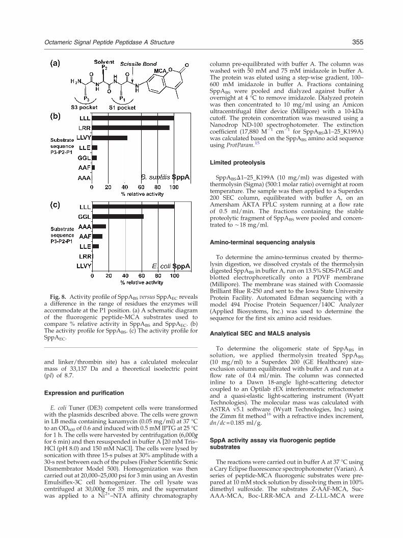
Fig. 8. Activity profile of SppABS versus SppAEC revealsa difference in the range of residues the enzymes willaccommodate at the P1 position. (a) A schematic diagramof the fluorogenic peptide-MCA substrates used tocompare % relative activity in SppABS and SppAEC. (b)The activity profile for SppABS. (c) The activity profile forSppAEC.
355Octameric Signal Peptide Peptidase A Structure
and linker/thrombin site) has a calculated molecularmass of 33,137 Da and a theoretical isoelectric point(pI) of 8.7.
Expression and purification
E. coli Tuner (DE3) competent cells were transformedwith the plasmids described above. The cells were grownin LB media containing kanamycin (0.05 mg/ml) at 37 °Cto an OD600 of 0.6 and induced with 0.5 mM IPTG at 25 °Cfor 1 h. The cells were harvested by centrifugation (6,000gfor 6 min) and then resuspended in buffer A [20 mM Tris–HCl (pH 8.0) and 150 mM NaCl]. The cells were lysed bysonication with three 15-s pulses at 30% amplitude with a30-s rest between each of the pulses (Fisher Scientific SonicDismembrator Model 500). Homogenization was thencarried out at 20,000–25,000 psi for 3 min using an AvestinEmulsiflex-3C cell homogenizer. The cell lysate wascentrifuged at 30,000g for 35 min, and the supernatantwas applied to a Ni2+–NTA affinity chromatography
column pre-equilibrated with buffer A. The column waswashed with 50 mM and 75 mM imidazole in buffer A.The protein was eluted using a step-wise gradient, 100–600 mM imidazole in buffer A. Fractions containingSppABS were pooled and dialyzed against buffer Aovernight at 4 °C to remove imidazole. Dialyzed proteinwas then concentrated to 10 mg/ml using an Amiconultracentrifugal filter device (Millipore) with a 10-kDacutoff. The protein concentration was measured using aNanodrop ND-100 spectrophotometer. The extinctioncoefficient (17,880 M−1 cm−1 for SppABSΔ1–25_K199A)was calculated based on the SppABS amino acid sequenceusing ProtParam.15
Limited proteolysis
SppABSΔ1–25_K199A (10 mg/ml) was digested withthermolysin (Sigma) (500:1 molar ratio) overnight at roomtemperature. The sample was then applied to a Superdex200 SEC column, equilibrated with buffer A, on anAmersham ÄKTA FPLC system running at a flow rateof 0.5 ml/min. The fractions containing the stableproteolytic fragment of SppABS were pooled and concen-trated to ∼18 mg/ml.
Amino-terminal sequencing analysis
To determine the amino-terminus created by thermo-lysin digestion, we dissolved crystals of the thermolysindigested SppABS in buffer A, run on 13.5% SDS-PAGE andblotted electrophoretically onto a PDVF membrane(Millipore). The membrane was stained with CoomassieBrilliant Blue R-250 and sent to the Iowa State UniversityProtein Facility. Automated Edman sequencing with amodel 494 Procise Protein Sequencer/140C Analyzer(Applied Biosystems, Inc.) was used to determine thesequence for the first six amino acid residues.
Analytical SEC and MALS analysis
To determine the oligomeric state of SppABS insolution, we applied thermolysin treated SppABS(10 mg/ml) to a Superdex 200 (GE Healthcare) size-exclusion column equilibrated with buffer A and run at aflow rate of 0.4 ml/min. The column was connectedinline to a Dawn 18-angle light-scattering detectorcoupled to an Optilab rEX interferometric refractometerand a quasi-elastic light-scattering instrument (WyattTechnologies). The molecular mass was calculated withASTRA v5.1 software (Wyatt Technologies, Inc.) usingthe Zimm fit method16 with a refractive index increment,dn/dc=0.185 ml/g.
SppA activity assay via fluorogenic peptidesubstrates
The reactions were carried out in buffer A at 37 °C usinga Cary Eclipse fluorescence spectrophotometer (Varian). Aseries of peptide-MCA fluorogenic substrates were pre-pared at 10 mM stock solution by dissolving them in 100%dimethyl sulfoxide. The substrates Z-AAF-MCA, Suc-AAA-MCA, Boc-LRR-MCA and Z-LLL-MCA were

Table 1. Data collection and refinement statistics
Crystal parametersSpace group P212121a, b, c (Å) 87.8, 131.1, 207.3
Data collection statisticsWavelength (Å) 0.9795Resolution (Å) 50.0–2.4 (2.5–2.4)a
Total reflections 709,425Unique reflections 96,535 (9520)Rmerge
b 0.085 (0.294)Mean (I)/σ(I) 52.7 (7.8)Completeness (%) 99.8 (99.8)Redundancy 7.4 (6.9)
356 Octameric Signal Peptide Peptidase A Structure
purchased from the Peptide Institute. The substrates Suc-LLVY-MCA and Z-LLE-MCA were purchased fromCalbiochem. The substrate Z-GGL-MCA was purchasedfrom Bachem. The abbreviations for the amino-terminalmodifications are as follows: Z- is benzyloxycarbonyl, Suc-is succinyl and Boc- is t-butyloxycarbonyl. For comparisonof the relative activity between SppABSΔ2–54 and SppA-ECΔ2–46, a single substrate concentration was utilized(10 μM for SppABSΔ2–54 and 2 μM for SppAECΔ2–46) ineach 500-μl reaction containing 40 nM enzyme in a1 cm×1 cm cuvette. The final dimethyl sulfoxide concen-tration was 2%. The excitation and emission wavelengthsused were 380 nm and 460 nm, respectively.
Refinement statisticsProtein molecules (chains) in
asymmetric unit8
Residues 1803Water molecules 257Total number of atoms 13,957Rcryst
c/Rfreed (%) 20.6/24.1
Average B-factor (Å2) (all atoms) 49.0r.m.s.d. on angles (°) 1.135r.m.s.d. on bonds (Å) 0.010
a The data collection statistics in parentheses are the values forthe highest-resolution shell.
b Rmerge =P
hklP
j j Ij hklð Þ − I hklð Þð Þ j = Phkl
Pj Ij hklð Þ, where
Ij(hkl) is the intensity of an individual reflection and is the meanintensity of that reflection.
c R =P j jF j − jF j j = P jF j , where F and
Crystallization
The thermolysin digested SppABS crystals were grownusing the sitting-drop vapor diffusion method. Thedetergent n-dodecyl-β-maltoside (0.1% final concentra-tion) was added to the protein before setting upcrystallization drops. The drop contained 1 μl of proteinand 1 μl of reservoir solution. The refined reservoircondition was 23% t-butanol, 0.1 M Tris–HCl (pH 8.5) and5% MPD. The drop was equilibrated against 1 ml ofreservoir solution at 18 °C. The cryo-solution contained20% MPD, 23% t-butanol and 0.1 M Tris–HCl (pH 8.5).The crystal was transferred to the cryo-solution and flash-cooled in liquid nitrogen.
cryst hkl obs calc hkl obs obsFcalc are the observed and calculated structure factor amplitudes,respectively.
d Rfree is calculated using 5% of the reflections randomlyexcluded from refinement.
Diffraction data collection
Diffraction images were collected on beamline 08B1-1 atthe Canadian Macromolecular Crystallography Facility ofthe Canadian Light Source (CLS), using a RayonixMX300HE X-ray detector. A total of 360 images werecollected at wavelength 0.9795 Å with 0.5° oscillations,and each image was exposed for 1 s. The crystal-to-detector distance was 250 mm. HKL2000 was used toprocess the diffraction images.17 The crystal belongs tospace group P212121 with unit cell dimensions of87.8 Å×131.1 Å×207.3 Å. There are eight molecules inthe asymmetric unit with aMatthews coefficient of 2.67 Å3
Da−1 (54.0% solvent), which was calculated with theMatthews Probabilities calculator18 using the SppABSmolecular mass after limited proteolysis (28,000 Da). SeeTable 1 for crystal parameters, data collection andrefinement statistics.
Structure determination and refinement
Phase estimates were obtained by molecular replace-ment using the program Phaser.19 A search model wasbuilt using the C-terminal domain of SppAEC (ProteinData Bank ID: 3BF0; chain A) as a template. The homologymodel was built using the program CHAINSAW.20 Theside chains of conserved residues were included, andnon-conserved side chains were truncated to the Cβ
atom. The initial Rwork/Rfree before refinement was0.3484/0.4696. The program Autobuild within PHENIXversion 1.6.421 was used to build the side chains andrefine the initial structure. Restrained refinement wasperformed using the program REFMAC5,22 and manual
adjustments and manipulations were executed with theprogram Coot.23 The final round of refinement includedTLS and restrained refinement with five TLS groups foreach chain. Input files were obtained by the TLSmotion determination server.24,25 See Table 1 for crystalparameters, data collection statistics and refinementstatistics.
Structural analysis
The figures were created using PyMOL.26 CLUSTALW27
and ESPript v.2.228 were utilized for the sequencealignments. Signal peptide sequences of B. subtilis and E.coli were obtained from SPdb Signal Peptide Resource.29
The protein–protein interface between protomers wasanalyzed using PISA30 and ProtorP.31 The conservedresidues among Gram-negative and Gram-positive SppAwere mapped onto the structure using ConSurf.32–34 Thesubstrate specificity pockets were analyzed usingCASTp35
while the stereochemistry of the structure was analyzedwith the program PROCHECK.36 PROMOTIF37 was usedto identify and analyze the secondary structure and motifswithin in the protein.
Accession numbers
Coordinates and structure factors have been depositedin the Protein Data Bank with accession number 3RST.

357Octameric Signal Peptide Peptidase A Structure
Acknowledgements
This work was supported in part by the CanadianInstitute of Health Research (to M.P.), the NaturalScience and Engineering Research Council of Can-ada (to M.P.), the Michael Smith Foundation forHealth Research (to M.P.) and the CanadianFoundation of Innovation (to M.P.). We thank thestaff at beamline 08ID-1 at the CLS, Saskatoon,Canada (especially Shaunivan Labiuk and JulienCotelesage) for their help with data collection. CLSwas supported by Natural Science and EngineeringResearch Council of Canada, National ResearchCouncil, Canadian Institute of Health Research andthe University of Saskatchewan. Special thanks toDr. Jaeyong Lee for helpful discussions.
Supplementary Data
Supplementary data associated with this articlecan be found, in the online version, at doi:10.1016/j.jmb.2012.03.020
References
1. Paetzel, M., Karla, A., Strynadka, N. C. & Dalbey, R. E.(2002). Signal peptidases. Chem. Rev. 102, 4549–4580.
2. Yuan, J., Zweers, J. C., van Dijl, J. M. & Dalbey, R. E.(2010). Protein transport across and into cell mem-branes in bacteria and archaea. Cell. Mol. Life Sci. 67,179–199.
3. Bolhuis, A., Matzen, A., Hyyrylainen, H. L., Kontinen,V. P., Meima, R., Chapuis, J. et al. (1999). Signal peptidepeptidase- and ClpP-like proteins of Bacillus subtilisrequired for efficient translocation and processing ofsecretory proteins. J. Biol. Chem. 274, 24585–24592.
4. Lensch, M., Herrmann, R. G. & Sokolenko, A. (2001).Identification and characterization of SppA, a novellight-inducible chloroplast protease complex associat-ed with thylakoid membranes. J. Biol. Chem. 276,33645–33651.
5. Matsumi, R., Atomi, H. & Imanaka, T. (2006).Identification of the amino acid residues essential forproteolytic activity in an archaeal signal peptidepeptidase. J. Biol. Chem. 281, 10533–10539.
6. Novak, P. & Dev, I. K. (1988). Degradation of a signalpeptide by protease IV and oligopeptidase A. J.Bacteriol. 170, 5067–5075.
7. Kim, A. C., Oliver, D. C. & Paetzel, M. (2008). Crystalstructure of a bacterial signal peptide peptidase. J. Mol.Biol. 376, 352–366.
8. Weihofen, A., Binns, K., Lemberg, M. K., Ashman, K.& Martoglio, B. (2002). Identification of signal peptidepeptidase, a presenilin-type aspartic protease. Science,296, 2215–2218.
9. Hussain, M., Ozawa, Y., Ichihara, S. & Mizushima, S.(1982). Signal peptide digestion in Escherichia coli.Effect of protease inhibitors on hydrolysis of thecleaved signal peptide of the major outer-membranelipoprotein. Eur. J. Biochem. 129, 233–239.
10. Wang, P., Shim, E., Cravatt, B., Jacobsen, R.,Schoeniger, J., Kim, A. C. et al. (2008). Escherichiacoli signal peptide peptidase A is a serine–lysineprotease with a lysine recruited to the nonconservedamino-terminal domain in the S49 protease family.Biochemistry, 47, 6361–6369.
11. Schechter, I. & Berger, A. (1967). On the size of theactive site in proteases. I. Papain. Biochem. Biophys.Res. Commun. 27, 157–162.
12. Paetzel, M., Dalbey, R. E. & Strynadka, N. C. (2002).Crystal structure of a bacterial signal peptidaseapoenzyme: implications for signal peptide bindingand the Ser–Lys dyad mechanism. J. Biol. Chem. 277,9512–9519.
13. Feldman, A. R., Lee, J., Delmas, B. & Paetzel, M.(2006). Crystal structure of a novel viral protease witha serine/lysine catalytic dyad mechanism. J. Mol. Biol.358, 1378–1389.
14. van Roosmalen, M. L., Geukens, N., Jongbloed, J. D.,Tjalsma, H., Dubois, J. Y., Bron, S. et al. (2004). Type Isignal peptidases of Gram-positive bacteria. Biochim.Biophys. Acta, 1694, 279–297.
15. Gasteiger, E., Hoogland, C., Gattiker, A., Duvaud, S.,Wilkins, M. R., Appel, R. D. & Bairoch, A. (2005).Protein identification and analysis tools on the ExPASyserver. In The Proteomics Protocols Handbook (Walker,J. M., ed.), pp. 571–607, Humana Press, Totowa, NJ.
16. Zimm, B. H. (1948). The scattering of light and theradial distribution function of high polymer solutions.J. Chem. Phys. 16, 1093–1099.
17. Otwinowski, Z. (1993). Denzo. In Denzo (Sawyer, L.,Isaacs, N. & Baily, S., eds), pp. 56–62, DaresburyLaboratory, Warrington, UK.
18. Kantardjieff, K. A. & Rupp, B. (2003). Matthewscoefficient probabilities: improved estimates for unitcell contents of proteins, DNA, and protein–nucleicacid complex crystals. Protein Sci. 12, 1865–1871.
19. McCoy, A. J., Grosse-Kunstleve, R.W., Storoni, L. C. &Read, R. J. (2005). Likelihood-enhanced fast transla-tion functions. Acta Crystallogr., Sect. D: Biol. Crystal-logr. 61, 458–464.
20. Stein, N. (2008). CHAINSAW: a program for mutatingpdb files used as templates in molecular replacement.J. Appl. Crystallogr. 41, 641–643.
21. Adams, P. D., Afonine, P. V., Bunkoczi, G., Chen, V. B.,Davis, I. W., Echols, N. et al. (2010). PHENIX: acomprehensive Python-based system for macromolec-ular structure solution. Acta Crystallogr., Sect. D: Biol.Crystallogr. 66, 213–221.
22. Murshudov, G. N., Skubak, P., Lebedev, A. A., Pannu,N. S., Steiner, R. A., Nicholls, R. A. et al. (2011).REFMAC5 for the refinement of macromolecularcrystal structures. Acta Crystallogr., Sect. D: Biol.Crystallogr. 67, 355–367.
23. Emsley, P. & Cowtan, K. (2004). Coot: model-buildingtools for molecular graphics. Acta Crystallogr., Sect. D:Biol. Crystallogr. 60, 2126–2132.
24. Painter, J. & Merritt, E. A. (2006). Optimal descriptionof a protein structure in terms of multiple groupsundergoing TLS motion. Acta Crystallogr., Sect. D: Biol.Crystallogr. 62, 439–450.
25. Winn, M. D., Murshudov, G. N. & Papiz, M. Z.(2003). Macromolecular TLS refinement in REFMACat moderate resolutions. Methods Enzymol. 374,300–321.

358 Octameric Signal Peptide Peptidase A Structure
26. DeLano, W. L. (2002). The PyMOL Molecular GraphicsSystem. DeLano Scientific, San Carlos, CA.
27. Thompson, J. D., Higgins, D. G. & Gibson, T. J.(1994). CLUSTALW: improving the sensitivity ofprogressive multiple sequence alignment throughsequence weighting, position-specific gap penaltiesand weight matrix choice. Nucleic Acids Res. 22,4673–4680.
28. Gouet, P., Courcelle, E., Stuart, D. I. & Metoz, F.(1999). ESPript: multiple sequence alignments inPostScript. Bioinformatics, 14, 305–308.
29. Choo, K. H., Tan, T. W. & Ranganathan, S. (2005).SPdb—a signal peptide database. BMC Bioinformatics,6, 249.
30. Krissinel, E. & Henrick, K. (2007). Inference ofmacromolecular assemblies from crystalline state.J. Mol. Biol. 372, 774–797.
31. Reynolds, C., Damerell, D. & Jones, S. (2009). ProtorP:a protein–protein interaction analysis server. Bioinfor-matics, 25, 413–414.
32. Ashkenazy, H., Erez, E., Martz, E., Pupko, T. & Ben-Tal, N. (2010). ConSurf 2010: calculating evolutionary
conservation in sequence and structure of proteinsand nucleic acids. Nucleic Acids Res. 38, W529–W533.
33. Glaser, F., Pupko, T., Paz, I., Bell, R. E., Bechor-Shental, D., Martz, E. & Ben-Tal, N. (2003). ConSurf:identification of functional regions in proteins bysurface-mapping of phylogenetic information. Bioin-formatics, 19, 163–164.
34. Landau, M., Mayrose, I., Rosenberg, Y., Glaser, F.,Martz, E., Pupko, T. & Ben-Tal, N. (2005). ConSurf2005: the projection of evolutionary conservationscores of residues on protein structures. NucleicAcids Res. 33, W299–W302.
35. Liang, J., Edelsbrunner, H. & Woodward, C. (1998).Anatomy of protein pockets and cavities: measure-ment of binding site geometry and implications forligand design. Protein Sci. 7, 1884–1897.
36. Laskowski, R. A. (2001). PDBsum: summaries andanalyses of PDB structures. Nucleic Acids Res. 29,221–222.
37. Hutchinson, E. G. & Thornton, J. M. (1996). PROMO-TIF—a program to identify and analyze structuralmotifs in proteins. Protein Sci. 5, 212–220.