Embed Size (px)
Citation preview
Crystal Structure of Cytomegalovirus IE1 Protein RevealsTargeting of TRIM Family Member PML via Coiled-CoilInteractionsMyriam Scherer1., Stefan Klingl2., Madhumati Sevvana2, Victoria Otto1, Eva-Maria Schilling1,
Joachim D. Stump3, Regina Muller1, Nina Reuter1, Heinrich Sticht3, Yves A. Muller2*,
Thomas Stamminger1*
1 Institute for Clinical and Molecular Virology, University of Erlangen-Nuremberg, Erlangen, Germany, 2 Division of Biotechnology, University of Erlangen-Nuremberg,
Erlangen, Germany, 3 Division of Bioinformatics, Institute of Biochemistry, University of Erlangen-Nuremberg, Erlangen, Germany
Abstract
PML nuclear bodies (PML-NBs) are enigmatic structures of the cell nucleus that act as key mediators of intrinsic immunityagainst viral pathogens. PML itself is a member of the E3-ligase TRIM family of proteins that regulates a variety of innateimmune signaling pathways. Consequently, viruses have evolved effector proteins to modify PML-NBs; however, little isknown concerning structure-function relationships of viral antagonists. The herpesvirus human cytomegalovirus (HCMV)expresses the abundant immediate-early protein IE1 that colocalizes with PML-NBs and induces their dispersal, whichcorrelates with the antagonization of NB-mediated intrinsic immunity. Here, we delineate the molecular basis for thisantagonization by presenting the first crystal structure for the evolutionary conserved primate cytomegalovirus IE1 proteins.We show that IE1 consists of a globular core (IE1CORE) flanked by intrinsically disordered regions. The 2.3 A crystal structureof IE1CORE displays an all a-helical, femur-shaped fold, which lacks overall fold similarity with known protein structures, butshares secondary structure features recently observed in the coiled-coil domain of TRIM proteins. Yeast two-hybrid andcoimmunoprecipitation experiments demonstrate that IE1CORE binds efficiently to the TRIM family member PML, and is ableto induce PML deSUMOylation. Intriguingly, this results in the release of NB-associated proteins into the nucleoplasm, butnot of PML itself. Importantly, we show that PML deSUMOylation by IE1CORE is sufficient to antagonize PML-NB-institutedintrinsic immunity. Moreover, co-immunoprecipitation experiments demonstrate that IE1CORE binds via the coiled-coildomain to PML and also interacts with TRIM5a We propose that IE1CORE sequesters PML and possibly other TRIM familymembers via structural mimicry using an extended binding surface formed by the coiled-coil region. This mode ofinteraction might render the antagonizing activity less susceptible to mutational escape.
Member PML via Coiled-Coil Interactions. PLoS Pathog 10(11): e1004512. doi:10.1371/journal.ppat.1004512
Editor: Roger D. Everett, University of Glasgow, United Kingdom
Received July 4, 2014; Accepted October 9, 2014; Published November 20, 2014
Copyright: � 2014 Scherer et al. This is an open-access article distributed under the terms of the Creative Commons Attribution License, which permitsunrestricted use, distribution, and reproduction in any medium, provided the original author and source are credited.
Data Availability: The authors confirm that all data underlying the findings are fully available without restriction. Coordinates and structure factors for theIE1CORE structure have been deposited in the Protein Data Bank under accession code 4WID.
Funding: This work was supported by the Deutsche Forschungsgemeinschaft, SFB796 (projects A2, A3 and B3) and the Interdisziplinares Zentrum fur KlinischeForschung Erlangen (project A62). The funders had no role in study design, data collection and analysis, decision to publish, or preparation of the manuscript.
Competing Interests: The authors have declared that no competing intests exist.
* Email: [email protected] (YAM); [email protected] (TS)
. These authors contributed equally to this work.
Introduction
Promyelocytic leukemia protein PML is the organizer of small
nuclear matrix structures termed nuclear bodies (NBs) or nuclear
domain 10 (ND10) [1]. PML, also named TRIM19, is a member
of the tripartite motif (TRIM) family of proteins, which are
characterized by the presence of RING, B-box and coiled-coil
domains [2]. Recent studies showed that an unprecedented large
number of TRIMs positively regulate innate immune signaling
pathways by acting as E3-Ub ligases [3,4]. Additionally, a
subgroup of TRIMs, including PML, was demonstrated to exhibit
small ubiquitin related modifier (SUMO) E3 activity and PML
itself is covalently conjugated to SUMO on three lysine residues
[5,6]. This modification, which affects PML localization, stability
and interaction with other partners, is critical for NB functions [7].
In response to stimuli, PML-NBs recruit a number of proteins
implicated in different cellular processes such as DNA damage
response, apoptosis, senescence and protein degradation [8,9].
Accumulating evidence implicates this subnuclear structure as
an important component of intrinsic immunity against viruses
from different families including herpes-, adeno-, polyoma,
rhabdo- and retroviruses [10–12]. Unlike the innate and adaptive
immunity, the intrinsic immune response is mediated by cellular
restriction factors that are constitutively expressed and perma-
nently active, even before a pathogen enters the cell. Other
characteristics of intrinsic immune mechanisms are that they are
saturable and subject to viral countermeasures [13]. Besides PML,
a number of NB components, such as Sp100, hDaxx and ATRX,
function as cellular restriction factors. Recent evidence suggests
that NB proteins independently contribute to the repression of
PLOS Pathogens | www.plospathogens.org 1 November 2014 | Volume 10 | Issue 11 | e1004512
Citation: Scherer M, Klingl S, Sevvana M, Otto V, Schilling E-M, et al. (2014) Crystal Structure of Cytomegalovirus IE1 Protein Reveals Targeting of TRIM Family
herpesvirus replication, raising the concept that individual NB
components, rather than the PML-NB structure as a whole,
restrict viral infections [14–20]. Consequently, various viruses
have been shown to antagonize the intrinsic cellular defense via
the modification of NB proteins. For instance, the herpes simplex
virus type I immediate-early protein ICP0 has been described as a
viral ubiquitin ligase with preferential substrate specificity for
SUMO-modified isoforms of PML thus promoting the degrada-
tion of PML [21]. However, no structural information on this and
many other NB-antagonistic proteins is available, yet.
Human cytomegalovirus (HCMV), a ubiquitous beta-herpesvi-
rus causing serious disease in immunocompromised individuals,
encodes an abundant immediate-early protein termed IE1 that
modulates innate immune mechanisms as well as other cellular
processes (reviewed in [22]). Although IE1 is a major player in the
initiation of lytic HCMV infection and has been subject to
extensive studies over the last decades, structural data on this
protein are still limited. Four distinct regions have been identified
within the 491 amino acid IE1 protein: a short N-terminal region
that is required for nuclear import, a large core domain, an acidic
region near the C-terminus that harbors a SUMOylation site and
a 16 amino acid chromatin-tethering domain (CTD) at the
extreme C-terminus [23–26]. Recent results have suggested that
the acidic C-terminal region of IE1 is characterized by a lack of
well-defined three-dimensional structure, but contains a binding
motif for signal transducer and activator of transcription (STAT)
proteins. This interaction site enables IE1 to compromise STAT-
mediated interferon signaling, thereby interfering with a crucial
branch of the innate immune system and promoting viral
replication [27–29]. In addition to its effects on the innate
immune system, IE1 is required to overcome the PML-NB-
mediated intrinsic immunity that targets HCMV immediately
upon infection. IE1 transiently co-localizes with PML-NBs during
the first 2–4 hours after infection but subsequently induces
disruption of these structures [30–32]. NB dispersal correlates
with the functional activities of IE1 during infection and a
PML knock-down efficiently compensates for IE1 in promoting
replication of an IE1-deficient virus, establishing IE1 as an
important antagonist of PML-mediated cellular repression of viral
replication [15,16,33]. Studies on the mechanism of NB dispersal
have demonstrated that IE1 induces the loss of the SUMOylated
forms of PML, and also influences the SUMOylation state of
Sp100 [34,35]. However, in contrast to ICP0, this neither requires
proteasomal activity nor does IE1 affect the abundance of
unmodified PML [24,35]. In further studies, a physical interaction
between IE1 and PML, which requires the N-terminal TRIM
region of PML, has been detected as prerequisite for the transient
co-localization and subsequent disruption of PML-NB integrity.
The interaction site for PML has been mapped to the large core
region of IE1, since deletions or mutations affecting this domain
abrogate PML binding and NB disruption [23,35,36]. However, it
was noted in several reports that mutations in the core region often
result in unstable IE1 proteins, so that the molecular basis for the
IE1-PML interaction remains uncharacterized [37,38].
Here we report the crystal structure of the evolutionary
conserved globular core domain of primate cytomegalovirus IE1
proteins, determined to 2.3 A resolution. Unexpectedly, the
overall structure does not resemble any known protein fold, but
exhibits an unusual all a-helical, femur-like shape which shares
secondary structure features recently observed in the coiled-coil
domain of TRIM proteins. We show that this IE1CORE domain
binds with high affinity to PML via the coiled-coil domain. This
induces PML de-SUMOylation thus releasing the PML-associated
factors hDaxx, Sp100 and ATRX, while PML accumulations itself
are not dispersed. Since IE1CORE efficiently complements lytic
replication of an IE1-deleted HCMV, we conclude that seques-
tration of PML via IE1CORE is sufficient for antagonization of NB-
mediated intrinsic immunity. Thus, cytomegaloviruses may have
evolved a distinct structural fold to effectively bind and neutralize
an important cellular hub protein that exerts critical roles during
the regulation of innate immune responses as well as the control of
programmed cell death [8–10,12].
Results
IE1 consists of a globular core domain flanked byintrinsically disordered regions
In order to further clarify the mechanism of IE1-mediated PML
antagonization, we investigated the molecular architecture of the
IE1 proteins from human, chimpanzee and rhesus cytomegalovi-
rus (h-, c- and rhIE1) (Figure S1). As previously proposed by
Krauss et al. [28], in silico predictions using the web server IUPred
[39] suggested that the N- and C-terminal regions of all IE1
proteins display consistently high intrinsic disorder propensities
(Figure 1A). Based on these predictions as well as on sequence
conservation and the characterization of protease-resistant IE1
subdomains, we generated truncated IE1 constructs covering the
folded core (Figure 1B). Limited proteolysis of recombinant full-
length hIE1 as well as of C- or N/C-terminally truncated hIE1
proteins confirmed the in silico predictions (Figure 1C). These
studies revealed the existence of a stably folded IE1CORE domain
of about 360 residues (Figure 1C, hIE1 20-382) that is flanked at
the N- and C-termini by intrinsically disordered regions (IE1N-IDR
and IE1C-IDR). Circular dichroism (CD) spectroscopy [40,41] was
applied to investigate the secondary structure composition of the
IE1 variants. All hIE1 proteins produced typical a-helical spectra
with negative ellipticity above 200 nm and two distinct minima at
208 nm and 222 nm (Figure 1D and S2). The spectra of full-
length hIE1 and hIE1CORE differed in the region below 210 nm.
Calculation of the difference spectrum revealed strong random coil
Author Summary
Research of the last few years has revealed that microbialinfections are not only controlled by innate and adaptiveimmune mechanisms, but also by cellular restrictionfactors, which give cells the capacity to resist pathogens.PML nuclear bodies (PML-NBs) are dot-like nuclear struc-tures representing multiprotein complexes that consist ofthe PML protein, a member of the TRIM family of proteins,as well as a multitude of additional regulatory factors. PML-NB components act as a barrier against many viralinfections; however, viral antagonistic proteins haveevolved to modify PML-NBs, thus abrogating this cellulardefense. Here, we delineate the molecular basis forantagonization by the immediate-early protein IE1 of theherpesvirus human cytomegalovirus. We present the firstcrystal structure for the evolutionary conserved coredomain (IE1CORE) of primate cytomegalovirus IE1, whichexhibits a novel, unusual fold. IE1CORE modifies PML-NBs byreleasing other PML-NB proteins into the nucleoplasmwhich is sufficient to antagonize intrinsic immunity.Importantly, IE1CORE shares secondary structure featureswith the coiled-coil domain (CC) of TRIM factors, and wedemonstrate strong binding of IE1 to the PML-CC. Wepropose that IE1CORE sequesters PML and possibly otherTRIM family members via an extended binding surfaceformed by the coiled-coil domain.
Crystal Structure of Cytomegalovirus IE1 Protein
PLOS Pathogens | www.plospathogens.org 2 November 2014 | Volume 10 | Issue 11 | e1004512
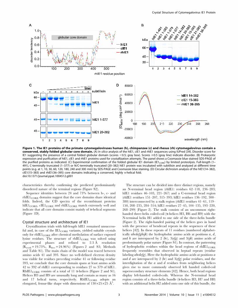
characteristics thereby confirming the predicted predominantly
disordered nature of the terminal regions (Figure S2).
Sequence identities between 24 and 73% between h-, c- and
rhIE1CORE domains suggest that the core domains share identical
folds. Indeed, the CD spectra of the recombinant proteins
hIE1CORE, cIE1CORE and rhIE1CORE match extremely well and
indicate that all core domains consist mainly of a-helical segments
(Figure 1D).
Crystal structure and architecture of IE1Crystallization trials with full-length hIE1 remained unsuccess-
ful and, in case of the IE1CORE variants, yielded suitable crystals
only for rhIE1CORE after chemical methylation of surface exposed
lysine residues. The structure of rhIE1CORE was solved using
experimental phases and refined to 2.3 A resolution
(Rwork = 19.73%, Rfree = 24.96%) (Figures 2 and S3; Methods
and Table S1). The main chain of the model was traced between
amino acids 41 and 393. Since no well-defined electron density
was visible for residues preceding residue 41 or following residue
393, we conclude that the core domain spans at least amino acids
42 to 392 of rhIE1 corresponding to residues 27 to 379 of hIE1.
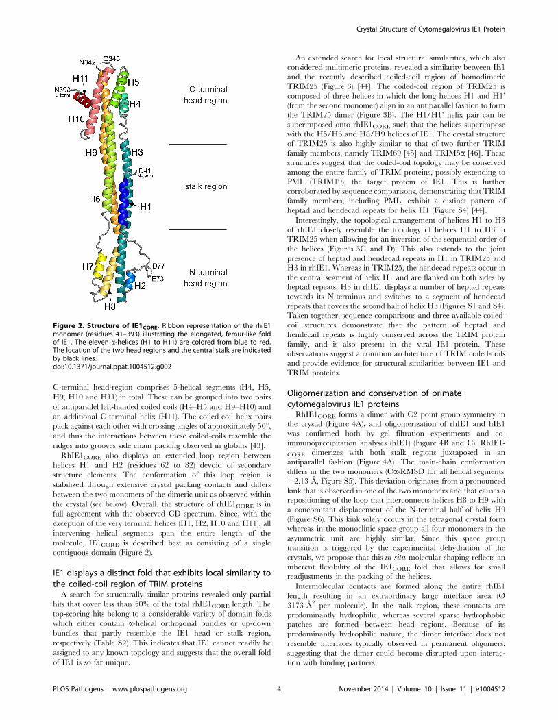
RhIE1CORE consists of a total of 11 a-helices (Figure 2 and S1).
Helices H3 and H9 are unusually long and contain as many as 16
and 17 helical turns, respectively. RhIE1CORE adopts an
elongated, femur-like shape with dimensions of 130625625 A3.
The structure can be divided into three distinct regions, namely
an N-terminal head region (rhIE1: residues 62–118, 236–283;
hIE1 residues 46–103, 221–267) and a C-terminal head region
(rhIE1 residues 151–207, 315–393; hIE1 residues 136–192, 300–
380) interconnected by a stalk region (rhIE1 residues 41–61, 119–
150, 208–235, 284–314; hIE1 residues 27–45, 104–135, 193–220,
268–299) (Figure 2). The stalk consists of an uncommon right-
handed three-helix coiled-coil (a-helices H3, H6 and H9) with the
N-terminal helix H1 added to one side of the three-helix bundle
(Figure 2). The right-handed pairing of the helices goes in hand
with the presence of hendecad repeats in the sequences of these
helices [42]. In these repeats of 11 residues (numbered alphabet-
ically abcdefghijk) the hydrophobic amino acids at positions a, d,
and h are interspaced by 2 (bc), 3(efg) and 3(ijk) amino acids of
predominantly polar nature (Figure S1). In contrast, the patterning
of hydrophobic residues within the head regions of rhIE1CORE
frequently resembles that observed in heptad repeats (residue
labeling abcdefg). Here the hydrophobic amino acids at positions aand d are interspaced by 2 (bc) and 3(efg) polar residues, and the
interdigitation of the a and d residues from neighboring helices
gives rise to more commonly observed left handed coiled-coil
supersecondary structure elements [42]. Hence, both head regions
display left-handed coiled-coils. Whereas the N-terminal head
region consists of a three-helix bundle (a-helices H3, H7 and H8)
with an additional helix H2 added onto one side of this bundle, the
Figure 1. The IE1 proteins of the primate cytomegaloviruses human (h), chimpanzee (c) and rhesus (rh) cytomegalovirus contain aconserved, stably folded globular core domain. (A) In silico analysis of the hIE1, cIE1 and rhIE1 sequences using IUPred [39]. Disorder score forIE1 suggesting the presence of a central folded globular domain (scores ,0.5; gray box). Scores $0.5 (gray line) indicate disorder. (B) Prokaryoticexpression and purification of hIE1, cIE1 and rhIE1 proteins used for crystallization attempts. The panel shows a Coomassie blue stained SDS-PAGE ofthe purified proteins as indicated. (C) Experimental confirmation of the folded globular IE1 domain (IE1CORE) by limited proteolysis. Full-length (1–491), C-terminally truncated (1–377) or N/C-terminally truncated (20–382) hIE1 protein was incubated with subtilisin and analyzed at different timepoints (e.g. at 1, 10, 30, 60, 120, 180, 240 and 300 min) by SDS-PAGE and Coomassie blue staining. (D) Circular dichroism analysis of the hIE1(14–382),cIE1(15–383) and rhIE1(36–395) core domains indicating a conserved, highly a-helical fold.doi:10.1371/journal.ppat.1004512.g001
Crystal Structure of Cytomegalovirus IE1 Protein
PLOS Pathogens | www.plospathogens.org 3 November 2014 | Volume 10 | Issue 11 | e1004512
C-terminal head-region comprises 5-helical segments (H4, H5,
H9, H10 and H11) in total. These can be grouped into two pairs
of antiparallel left-handed coiled coils (H4–H5 and H9–H10) and
an additional C-terminal helix (H11). The coiled-coil helix pairs
pack against each other with crossing angles of approximately 50u,and thus the interactions between these coiled-coils resemble the
ridges into grooves side chain packing observed in globins [43].
RhIE1CORE also displays an extended loop region between
helices H1 and H2 (residues 62 to 82) devoid of secondary
structure elements. The conformation of this loop region is
stabilized through extensive crystal packing contacts and differs
between the two monomers of the dimeric unit as observed within
the crystal (see below). Overall, the structure of rhIE1CORE is in
full agreement with the observed CD spectrum. Since, with the
exception of the very terminal helices (H1, H2, H10 and H11), all
intervening helical segments span the entire length of the
molecule, IE1CORE is described best as consisting of a single
contiguous domain (Figure 2).
IE1 displays a distinct fold that exhibits local similarity tothe coiled-coil region of TRIM proteins
A search for structurally similar proteins revealed only partial
hits that cover less than 50% of the total rhIE1CORE length. The
top-scoring hits belong to a considerable variety of domain folds
which either contain a-helical orthogonal bundles or up-down
bundles that partly resemble the IE1 head or stalk region,
respectively (Table S2). This indicates that IE1 cannot readily be
assigned to any known topology and suggests that the overall fold
of IE1 is so far unique.
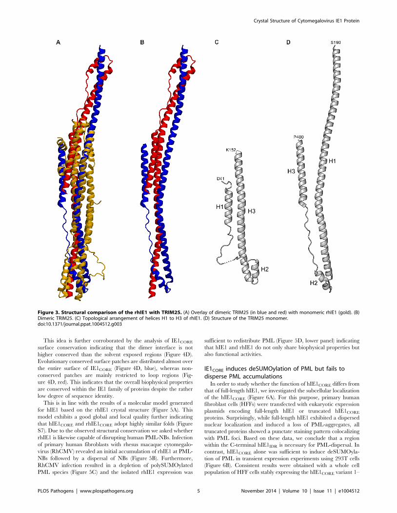
An extended search for local structural similarities, which also
considered multimeric proteins, revealed a similarity between IE1
and the recently described coiled-coil region of homodimeric
TRIM25 (Figure 3) [44]. The coiled-coil region of TRIM25 is
composed of three helices in which the long helices H1 and H1’
(from the second monomer) align in an antiparallel fashion to form
the TRIM25 dimer (Figure 3B). The H1/H1’ helix pair can be
superimposed onto rhIE1CORE such that the helices superimpose
with the H5/H6 and H8/H9 helices of IE1. The crystal structure
of TRIM25 is also highly similar to that of two further TRIM
family members, namely TRIM69 [45] and TRIM5a [46]. These
structures suggest that the coiled-coil topology may be conserved
among the entire family of TRIM proteins, possibly extending to
PML (TRIM19), the target protein of IE1. This is further
corroborated by sequence comparisons, demonstrating that TRIM
family members, including PML, exhibit a distinct pattern of
heptad and hendecad repeats for helix H1 (Figure S4) [44].
Interestingly, the topological arrangement of helices H1 to H3
of rhIE1 closely resemble the topology of helices H1 to H3 in
TRIM25 when allowing for an inversion of the sequential order of
the helices (Figures 3C and D). This also extends to the joint
presence of heptad and hendecad repeats in H1 in TRIM25 and
H3 in rhIE1. Whereas in TRIM25, the hendecad repeats occur in
the central segment of helix H1 and are flanked on both sides by
heptad repeats, H3 in rhIE1 displays a number of heptad repeats
towards its N-terminus and switches to a segment of hendecad
repeats that covers the second half of helix H3 (Figures S1 and S4).
Taken together, sequence comparisons and three available coiled-
coil structures demonstrate that the pattern of heptad and
hendecad repeats is highly conserved across the TRIM protein
family, and is also present in the viral IE1 protein. These
observations suggest a common architecture of TRIM coiled-coils
and provide evidence for structural similarities between IE1 and
TRIM proteins.
Oligomerization and conservation of primatecytomegalovirus IE1 proteins
RhIE1CORE forms a dimer with C2 point group symmetry in
the crystal (Figure 4A), and oligomerization of rhIE1 and hIE1
was confirmed both by gel filtration experiments and co-
immunoprecipitation analyses (hIE1) (Figure 4B and C). RhIE1-
CORE dimerizes with both stalk regions juxtaposed in an
antiparallel fashion (Figure 4A). The main-chain conformation
differs in the two monomers (Ca-RMSD for all helical segments
= 2.13 A, Figure S5). This deviation originates from a pronounced
kink that is observed in one of the two monomers and that causes a
repositioning of the loop that interconnects helices H8 to H9 with
a concomitant displacement of the N-terminal half of helix H9
(Figure S6). This kink solely occurs in the tetragonal crystal form
whereas in the monoclinic space group all four monomers in the
asymmetric unit are highly similar. Since this space group
transition is triggered by the experimental dehydration of the
crystals, we propose that this in situ molecular shaping reflects an
inherent flexibility of the IE1CORE fold that allows for small
readjustments in the packing of the helices.
Intermolecular contacts are formed along the entire rhIE1
length resulting in an extraordinary large interface area (Ø
3173 A2 per molecule). In the stalk region, these contacts are
predominantly hydrophilic, whereas several sparse hydrophobic
patches are formed between head regions. Because of its
predominantly hydrophilic nature, the dimer interface does not
resemble interfaces typically observed in permanent oligomers,
suggesting that the dimer could become disrupted upon interac-
tion with binding partners.
Figure 2. Structure of IE1CORE. Ribbon representation of the rhIE1monomer (residues 41–393) illustrating the elongated, femur-like foldof IE1. The eleven a-helices (H1 to H11) are colored from blue to red.The location of the two head regions and the central stalk are indicatedby black lines.doi:10.1371/journal.ppat.1004512.g002
Crystal Structure of Cytomegalovirus IE1 Protein
PLOS Pathogens | www.plospathogens.org 4 November 2014 | Volume 10 | Issue 11 | e1004512
This idea is further corroborated by the analysis of IE1CORE
surface conservation indicating that the dimer interface is not
higher conserved than the solvent exposed regions (Figure 4D).
Evolutionary conserved surface patches are distributed almost over
the entire surface of IE1CORE (Figure 4D, blue), whereas non-
conserved patches are mainly restricted to loop regions (Fig-
ure 4D, red). This indicates that the overall biophysical properties
are conserved within the IE1 family of proteins despite the rather
low degree of sequence identity.
This is in line with the results of a molecular model generated
for hIE1 based on the rhIE1 crystal structure (Figure 5A). This
model exhibits a good global and local quality further indicating
that hIE1CORE and rhIE1CORE adopt highly similar folds (Figure
S7). Due to the observed structural conservation we asked whether
rhIE1 is likewise capable of disrupting human PML-NBs. Infection
of primary human fibroblasts with rhesus macaque cytomegalo-
virus (RhCMV) revealed an initial accumulation of rhIE1 at PML-
NBs followed by a dispersal of NBs (Figure 5B). Furthermore,
RhCMV infection resulted in a depletion of polySUMOylated
PML species (Figure 5C) and the isolated rhIE1 expression was
sufficient to redistribute PML (Figure 5D, lower panel) indicating
that hIE1 and rhIE1 do not only share biophysical properties but
also functional activities.
IE1CORE induces deSUMOylation of PML but fails todisperse PML accumulations
In order to study whether the function of hIE1CORE differs from
that of full-length hIE1, we investigated the subcellular localization
of the hIE1CORE (Figure 6A). For this purpose, primary human
fibroblast cells (HFFs) were transfected with eukaryotic expression
plasmids encoding full-length hIE1 or truncated hIE1CORE
proteins. Surprisingly, while full-length hIE1 exhibited a dispersed
nuclear localization and induced a loss of PML-aggregates, all
truncated proteins showed a punctate staining pattern colocalizing
with PML foci. Based on these data, we conclude that a region
within the C-terminal hIE1IDR is necessary for PML-dispersal. In
contrast, hIE1CORE alone was sufficient to induce deSUMOyla-
tion of PML in transient expression experiments using 293T cells
(Figure 6B). Consistent results were obtained with a whole cell
population of HFF cells stably expressing the hIE1CORE variant 1–
Figure 3. Structural comparison of the rhIE1 with TRIM25. (A) Overlay of dimeric TRIM25 (in blue and red) with monomeric rhIE1 (gold). (B)Dimeric TRIM25. (C) Topological arrangement of helices H1 to H3 of rhIE1. (D) Structure of the TRIM25 monomer.doi:10.1371/journal.ppat.1004512.g003
Crystal Structure of Cytomegalovirus IE1 Protein
PLOS Pathogens | www.plospathogens.org 5 November 2014 | Volume 10 | Issue 11 | e1004512
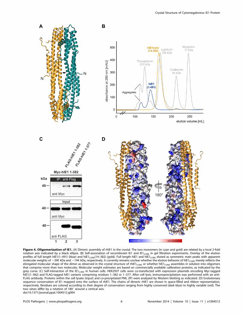
Figure 4. Oligomerization of IE1. (A) Dimeric assembly of rhIE1 in the crystal. The two monomers (in cyan and gold) are related by a local 2-foldrotation axis indicated by a black ellipse. (B) Self-association of recombinant IE1 and IE1CORE in gel filtration experiments. Overlay of the elutionprofiles of full length hIE1(1–491) (blue) and hIE1CORE(14–382) (gold). Full length hIE1 and hIE1CORE eluted as symmetric main peaks with apparentmolecular weights of ,390 kDa and ,194 kDa, respectively. It currently remains unclear whether the elution behavior of hIE1CORE merely reflects theelongated molecular shape of the dimer as observed in the crystal structure of rhIE1CORE or whether hIE1CORE assembles in solution into oligomersthat comprise more than two molecules. Molecular weight estimates are based on commercially available calibration proteins, as indicated by thegrey curve. (C) Self-interaction of the IE1CORE in human cells. HEK293T cells were co-transfected with expression plasmids encoding Myc-taggedhIE1(1–382) and FLAG-tagged hIE1 variants comprising residues 1–382 or 1–377. After cell lysis, immunoprecipitation was performed with an anti-FLAG antibody. Proteins within the cell lysate (input) and co-precipitated PML (IP) were analyzed by Western blotting as indicated. (D) Evolutionarysequence conservation of IE1 mapped onto the surface of rhIE1. The chains of dimeric rhIE1 are shown in space-filled and ribbon representation,respectively. Residues are colored according to their degree of conservation ranging from highly conserved (dark blue) to highly variable (red). Thetwo views differ by a rotation of 180u around a vertical axis.doi:10.1371/journal.ppat.1004512.g004
Crystal Structure of Cytomegalovirus IE1 Protein
PLOS Pathogens | www.plospathogens.org 6 November 2014 | Volume 10 | Issue 11 | e1004512
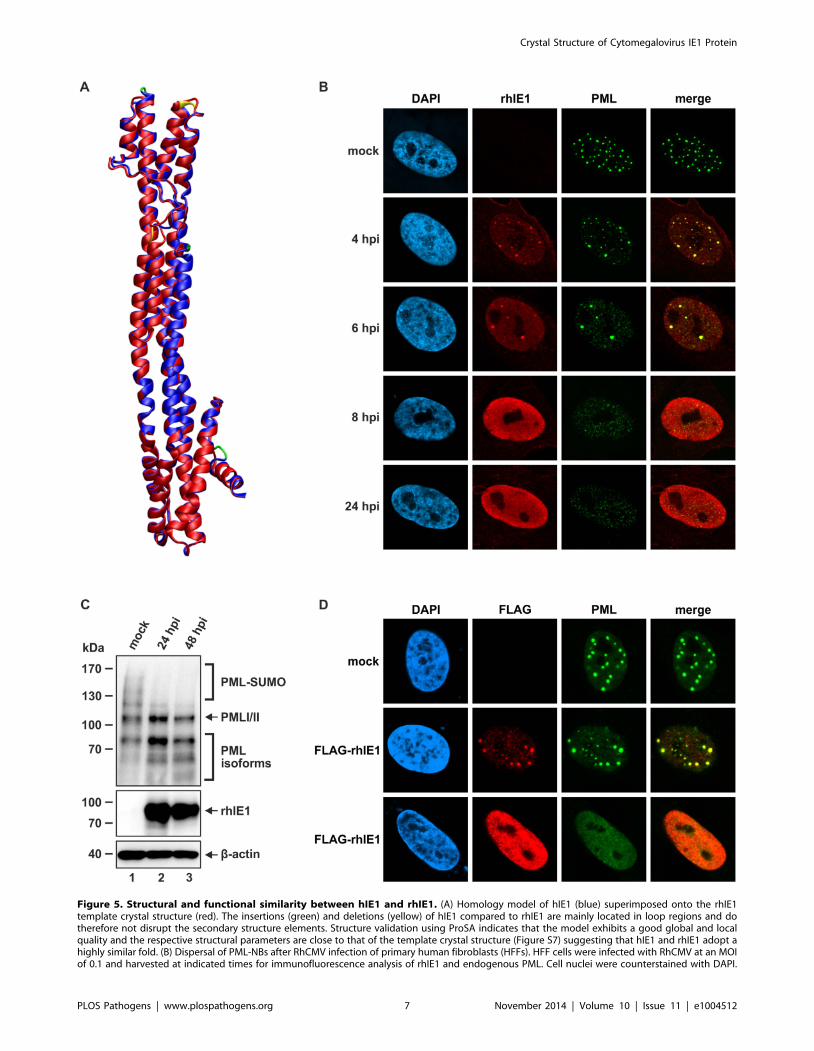
Figure 5. Structural and functional similarity between hIE1 and rhIE1. (A) Homology model of hIE1 (blue) superimposed onto the rhIE1template crystal structure (red). The insertions (green) and deletions (yellow) of hIE1 compared to rhIE1 are mainly located in loop regions and dotherefore not disrupt the secondary structure elements. Structure validation using ProSA indicates that the model exhibits a good global and localquality and the respective structural parameters are close to that of the template crystal structure (Figure S7) suggesting that hIE1 and rhIE1 adopt ahighly similar fold. (B) Dispersal of PML-NBs after RhCMV infection of primary human fibroblasts (HFFs). HFF cells were infected with RhCMV at an MOIof 0.1 and harvested at indicated times for immunofluorescence analysis of rhIE1 and endogenous PML. Cell nuclei were counterstained with DAPI.
Crystal Structure of Cytomegalovirus IE1 Protein
PLOS Pathogens | www.plospathogens.org 7 November 2014 | Volume 10 | Issue 11 | e1004512

382 (Figure 6C). Given that SUMO modification of PML is a
prerequisite for the recruitment of other NB components like
Sp100, hDaxx and ATRX, it was important to explore the
subcellular localization of these factors after expression of
hIE1CORE. Interestingly, while PML was detected in a dot-like
pattern, Sp100, hDaxx and ATRX were released from NBs in the
presence of hIE1CORE (Figure 6C). Taken together, these data
demonstrate that hIE1CORE is sufficient to sequester and
deSUMOylate PML resulting in the dissociation of other NB
components.
IE1CORE efficiently binds to PML via the coiled-coildomain
Due to the accumulation of hIE1CORE at PML foci, it was
attractive to speculate that the two proteins might strongly interact
with each other, which was investigated by co-immunoprecipita-
tion (Figure 7A). Intriguingly, while only a trace amount of PML
was associated with full-length hIE1, PML was efficiently
coprecipitated with hIE1CORE variants. An increased affinity of
hIE1CORE for PML was also confirmed by yeast two-hybrid
experiments (Figure 7B), which is in line with previous results by
Lee et al. (2004) that show an enhanced interaction of PML with
an IE1 variant lacking the acidic C-terminus (IE1 1–420) [35].
Having observed a structural similarity between IE1 and coiled-
coil regions of TRIM proteins, we asked whether this domain of
PML is required for binding of IE1. In a yeast two-hybrid analysis
utilizing a series of C-terminal PML deletion mutants we observed
that a truncation of the coiled-coil domain abrogated the
interaction with IE1 (Figure 7C). To further confirm this finding,
coimmunoprecipitation analyses were performed with additional
N- and/or C-terminal PML deletions. Importantly, this experi-
ment revealed that the coiled-coil domain of PML was sufficient to
mediate an interaction with IE1 (Figure 7D, lower panel, lane 4).
Furthermore, we observed that IE1 was also able to bind to
TRIM5asuggesting that IE1 targets additional TRIM factors via
coiled-coil interactions (Figure 7E).
IE1CORE antagonizes PML-mediated intrinsic immunityduring viral infection
Having shown that hIE1CORE binds with high affinity to and
deSUMOylates PML, but fails to disrupt PML accumulations, it
was important to investigate whether this is sufficient to antagonize
PML-NB mediated repression of viral infection. We constructed a
recombinant HCMV expressing hIE1 lacking the C-terminal
IE1IDR (Figure 8A) and could observe that this virus exhibited a
severe defect to disperse PML after infection of HFFs (Figure 8B).
Consistent with our results obtained after isolated expression of
hIE1CORE, deSUMOylation of both PML and Sp100 was fully
preserved (Figure 8C). Most importantly, however, the hIE1CORE-
expressing virus replicated nearly as efficient as wild-type virus
while an hIE1-deleted virus exhibited a severe growth defect
(Figure 8D). The approximately 10fold growth reduction observed
for the hIE1CORE-expressing virus at 4 and 6 dpi is in line with
previously published results on viruses lacking the C-terminal
acidic domain which binds STAT2 thus antagonizing the
interferon response [28,29]. This was also confirmed in a
complementation experiment after infection of either hIE1- or
hIE1CORE-expressing HFFs with an hIE1-deleted HCMV, finally
demonstrating that hIE1CORE can efficiently substitute for full-
length hIE1 during lytic HCMV infection (Figure 8E).
Discussion
The immediate-early protein IE1 of human cytomegalovirus
that directly binds to PML is known as an important herpesviral
antagonist of PML-NB-mediated intrinsic immunity
[15,22,33,36]. However, the structural basis for its function has
remained elusive due to the paucity of high-resolution structural
information. Here, we present the first crystal structure for the
evolutionary conserved primate cytomegalovirus IE1 proteins and
demonstrate that a structurally conserved IE1CORE domain is
sufficient to antagonize PML-mediated intrinsic immunity. The
structure of IE1CORE consists of a femur-shaped bundle of helices,
which surprisingly does not share any overall fold similarity with
known protein structures. IE1CORE binds with high affinity to
PML and efficiently abrogates PML SUMOylation, but fails to
disrupt PML accumulations itself. Only upon inclusion of the C-
terminal, intrinsically disordered region (IE1C-IDR) PML dispersal
is observed. Thus, our study demonstrates that PML deSUMOy-
lation can be discriminated from PML dispersal. Whereas the first
activity is achieved by a distinctly folded IE1CORE domain, the
second activity requires inclusion of C-terminal sequences of the
IE1C-IDR region that is highly susceptible to proteolytic degrada-
tion and for which we did not observe any stable secondary
structure formation. As it has been observed for many intrinsically
disordered proteins, folding of the natively disordered IE1C-IDR
region may occur upon binding to a specific interaction partner.
First evidence for this comes from a recent study predicting that
the chromatin-tethering domain (CTD) at the extreme C-terminus
of IE1 forms a b-hairpin when bound to histone proteins [47].
Importantly, IE1CORE is able to release other NB-components like
Sp100, hDaxx and ATRX into the nucleoplasm and this
correlates with antagonization of NB-mediated repression. This
shows that PML dispersal as observed during infection with herpes
simplex virus type I and HCMV is not a prerequisite to antagonize
the repressive effects of this cellular multiprotein complex on viral
gene expression [24,48]. As also suggested by recent findings on
the c-herpesviruses Herpesvirus saimiri and Kaposi sarcoma
herpesvirus as well as on the polyomavirus BKV more subtle
modifications like the release or degradation of individual NB-
components appear to be sufficient [49–51].
IE1CORE resembles the action of the SUMO-specific protease
SENP-1 which also abrogates the SUMOylation of PML but
leaves most PML aggregated [52]. It was previously speculated
that IE1 could harbor an intrinsic SUMO protease activity itself or
recruit SUMO-specific proteases to the NBs [53]. However, our
structural analysis of IE1CORE provides no evidence for the
presence of a potential active site with hydrolase activity.
Furthermore, in earlier studies no interaction of IE1 with SENPs
could be detected, but it was reported that full-length hIE1 could
still disassemble foci formed by a PML protein with all
(C) DeSUMOylation of PML after RhCMV infection of HFF cells. Cell lysates harvested either from mock infected cells or from cells infected with RhCMVat an MOI of 3 for indicated times (24, 48 h) were separated by SDS-PAGE and analyzed by Western blotting for expression of PML (upper panel),rhIE1 (middle panel) and b-actin as internal loading control (lower panel). (D) Dispersal of PML-NBs after transient expression of rhIE1 in HFFs. HFFswere transfected with a eukaryotic expression vector encoding rhIE1 fused to an N-terminal FLAG-tag and, after 48 h, subjected to indirectimmunofluorescence analysis of rhIE1 using an anti-FLAG antibody and of endogenous PML; cell nuclei were stained with DAPI. Upper panel: mocktransfected cells. Middle panel: colocalization of rhIE1 and PML, as observed in only few transfected cells. Lower panel: dispersed pattern of rhIE1 andPML, as observed in the majority of transfected cells.doi:10.1371/journal.ppat.1004512.g005
Crystal Structure of Cytomegalovirus IE1 Protein
PLOS Pathogens | www.plospathogens.org 8 November 2014 | Volume 10 | Issue 11 | e1004512
Crystal Structure of Cytomegalovirus IE1 Protein
PLOS Pathogens | www.plospathogens.org 9 November 2014 | Volume 10 | Issue 11 | e1004512
SUMOylation sites mutated [53]. Based on this observation, the
authors raised the idea that SUMO-independent interference with
PML oligomerization followed by exposure of SUMOylated PML
to cellular SUMO proteases may account for NB disruption.
However, the results of our study argue against such a scenario,
since abrogation of PML SUMOylation by IE1CORE was detected
while PML aggregates were still present. Thus, IE1 affects its
targets via direct, SUMO-independent substrate interaction and
this suggests that IE1 does not directly or indirectly act as a
hydrolase that specifically targets SUMOylated PML.
Importantly, our study revealed structural similarities between
IE1CORE and the crystal structure of the tripartite motif coiled-coil
that appears to act as a critical scaffold organizing the biochemical
activities of TRIM proteins [44,45]. Moreover, we were able to
confirm that the coiled-coil of PML is sufficient for strong binding
to IE1. Increasing evidence suggests that TRIM proteins function
as E3-ubiquitin ligases in agreement with the family-wide presence
of several conserved domains, namely a RING domain followed
by two B-boxes and a coiled-coil region [3]. Based on the recently
solved crystal structure of the TRIM25 coiled-coil it was shown
that TRIM proteins dimerize by forming interdigitating antipar-
allel helical hairpins that position the N-terminal catalytic RING
domain at opposite ends of the dimer and the C-terminal
substrate-binding domains at the center [44]. For some of the
TRIM members, and among these PML, E3-SUMO instead of
E3-ubiquitin ligase activity has been reported [5]. Thus, we would
like to propose that IE1CORE, via its strong interaction with the
PML coiled-coil, may inhibit an E3-SUMO ligase activity of PML
that is required for auto-SUMOylation. Alternatively, IE1 binding
to the coiled-coil might block the accessibility of PML for other
components of the cellular SUMOylation machinery. Thus, the
results of our study favor a model whereby IE1 primarily affects
the on-rate of SUMO modification which is also supported by the
slow kinetics of IE1-mediated loss of PML SUMOylation [54].
This is different from the ICP0 protein of herpes simplex virus type
I which induces the rapid degradation of SUMO-conjugated
proteins by acting as a SUMO-targeted ubiquitin ligase (STUbL)
[21]. Similar to IE1, the adenoviral E4-ORF3 protein which has
been shown to form a multivalent matrix via extensive self-
interactions, appears to inactivate PML via tight binding [55].
This specific assembly of E4-ORF3 creates avidity-driven inter-
actions that capture PML as well as other tumor suppressors thus
disrupting PML bodies. However, in contrast to IE1, the recently
solved crystal structure of E4-ORF3 revealed the molecular
mechanism of multimerization, but not the exact mode of PML
recognition [55].
In this context it should be noted that the nonstructural NS1
protein of influenza A virus has also been shown to target a TRIM
protein, TRIM25, via interaction with the coiled-coil domain to
inhibit its E3 ligase function [56]. Since TRIM25 catalyzes a
critical ubiquitination of the viral RNA sensor RIG-I this
constitutes a mechanism by which influenza virus inhibits the
host IFN response. Interestingly, we detected that IE1CORE not
only binds to PML but also to TRIM5a and a recent publication
reported an interaction with TRIM33 [57]. Thus, the unique
structure of IE1core may have developed during evolution to
target an extended spectrum of members of the TRIM family via
the conserved coiled-coil domain of these factors [44,45]. This is
also supported by our analysis of evolutionary conserved surface
patches of IE1CORE. When assuming that sites of protein-protein
interaction are reflected by conserved surface patches, our
observation that conserved residues are distributed evenly over
the entire IE1CORE protein surface suggests that rather large parts
of the IE1 surface are involved in recognition of the PML coiled-
coil. Consequently, the helical structure of IE1CORE might have
evolved as a decoy that, by means of extensive helix-helix
interactions might either pair up with the coiled-coil region of
PML or substitute for one of the PML monomers within the PML
dimer interface. In this respect, the similarity between the
topological arrangement of helices H1 to H3 of IE1 and of
predicted helices H3 to H1 of PML in combination with the joint
occurrence of regions with extended hendecad repeats might
facilitate the formation of heteromeric assemblies. The formation
of extended coiled-coil interactions would also readily offer an
explanation for the finding that single mutations within the
conserved surface patches of IE1 only moderately affect its
interaction properties with PML. In contrast, mutations affecting
the overall tertiary structure (e.g. IE1 L174P) abrogate the
functionality.
Furthermore, it agrees with our observation that rhIE1 can
substitute for hIE1 during infection of human cells despite low
overall sequence identity. Thus, the size and unique elongated fold
of the IE1CORE could have developed during evolution to
accommodate efficient binding of PML and possibly other TRIM
factors via an extended surface involving coiled-coil interactions.
This feature might render the interaction less amenable to
mutational escape.
Materials and Methods
Recombinant protein production and purificationAll variants of h-, c- and rhIE1 were recombinantly produced in
E.coli strain BL21(DE3) (Novagen) as GST-tagged fusion proteins
for in vitro experiments and crystallization. LB media (Carl Roth
GmbH + Co. KG, Karlsruhe, Germany) were inoculated with
freshly transformed E. coli colonies, and cell cultures grown at
either 30u or 37uC. Seleno-methionine labeling of rhIE1(residues
36–395) was achieved by incubation of the cells with non-inducing
PAG medium (pre-culture) and auto-inducing PASM-5052
medium (main culture). Cell pellets were resuspended in
phosphate buffer and lysed by sonication. Protein purification
was achieved by the following steps: a first affinity chromatography
(Glutathione sepharose, GE Healthcare, Freiburg), proteolytic
cleavage with PreScission protease, a second affinity chromatog-
raphy and a final size exclusion chromatography (Superdex 200
prepgrade, GE Healthcare). The gel filtration column was pre-
equilibrated in 25 mM Tris, 150 mM NaCl, 10 mM DTT,
pH 7.5. The samples were separated with an isocratic gradient of
1.2 column volumes (CV) of the same buffer at a flow rate of
1.5 mL/min. The column was calibrated utilizing the elution
peaks of thyroglobulin (670 kDa), bovine c-globulin (158 kDa),
chicken ovalbumin (44 kDa) and equine myoglobin (17 kDa) of
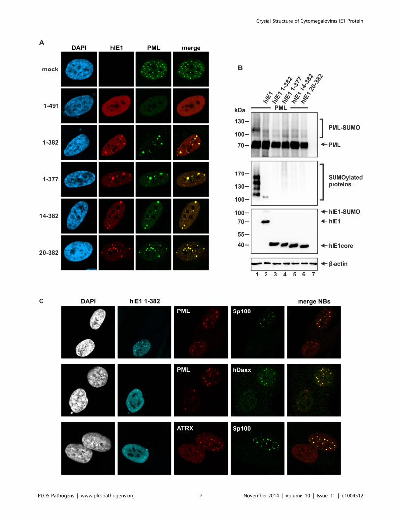
Figure 6. Effect of IE1CORE on the integrity of PML-NBs and the SUMOylation of PML. (A) Dispersal of PML requires the IE1IDRc. HFF cellswere transiently transfected with expression plasmids encoding FLAG-tagged full-length hIE1(1–491) or truncated hIE1 variants followed byimmunodetection of hIE1 proteins using an anti-FLAG antibody and of endogenous PML. (B) DeSUMOylation of PML by IE1CORE. HEK293T cells werecotransfected with expression plasmids encoding FLAG-PML (isoform VI), FLAG-hIE1 variants as indicated and Myc-SUMO3. PML (upper panel),SUMOylated proteins (second panel), IE1 (third panel) and b-actin (lower panel) were detected by Western blot analysis. (C) Release of Sp100, hDaxxand ATRX from PML foci by IE1CORE. The hIE1CORE(1–382) was stably expressed in HFFs followed by immunodetection of hIE1, PML, Sp100, hDaxx orATRX by triple staining as indicated.doi:10.1371/journal.ppat.1004512.g006
Crystal Structure of Cytomegalovirus IE1 Protein
PLOS Pathogens | www.plospathogens.org 10 November 2014 | Volume 10 | Issue 11 | e1004512
the Bio-Rad gel filtration standard (Bio-Rad Laboratories,
Munich, Germany). The molecular weight of the samples was
determined by linear regression. The Kav coefficients of the
standard proteins were plotted vs the logarithm of their molecular
weights to obtain the calibration curve, with Kav = (Ve-V0)/(Vc-
V0), where V0 is the column void volume, Ve is the elution
volume and Vc is the geometric column volume. All purification
steps were performed in the presence of 10 mM DTT. For the
crystallization of variant rhIE1(36–395) the protein was chemically
modified by lysine methylation prior to the final size exclusion
chromatography step. A 1 mg/mL IE1 protein solution was
incubated on ice with 20 mL of 1 M dimethylamine borane
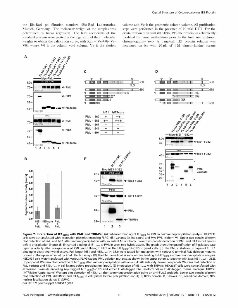
Figure 7. Interaction of IE1CORE with PML and TRIM5a. (A) Enhanced binding of IE1CORE to PML in coimmunoprecipitation analysis. HEK293Tcells were cotransfected with expression plasmids encoding FLAG-hIE1 variants (as indicated) and Myc-PML (isoform VI). Upper two panels: Westernblot detection of PML and hIE1 after immunoprecipitation with an anti-FLAG antibody. Lower two panels: detection of PML and hIE1 in cell lysatesbefore precipitation (input). (B) Enhanced binding of IE1CORE to PML in yeast two-hybrid assays. The graph shows the quantification of b-galactosidasereporter activity after coexpression of PML and full-length hIE1 or the hIE1CORE(14–382) in yeast cells. (C) The PML coiled-coil is required for IE1binding in yeast two-hybrid assays. Full-length hIE1 and hIE1CORE(14–382) were tested for interaction with various C-terminal PML deletion mutants(shown in the upper scheme) by XGal filter lift assays. (D) The PML coiled-coil is sufficient for binding to hIE1CORE in coimmunoprecipitation analysis.HEK293T cells were transfected with various FLAG-tagged PML deletion mutants, as shown in the upper scheme, together with Myc-hIE1CORE(1–382).Upper panel: Western blot detection of hIE1CORE after immunoprecipitation with an anti-FLAG antibody. Lower two panels: Western blot detection ofPML variants and hIE1CORE in cell lysates before precipitation (input). (E) Interaction of hIE1CORE with TRIM5a. HEK293T cells were cotransfected withexpression plasmids encoding Myc-tagged hIE1CORE(1–382) and either FLAG-tagged PML (isoform VI) or FLAG-tagged rhesus macaque TRIM5a(rhTRIM5a). Upper panel: Western blot detection of hIE1CORE after coimmunoprecipitation using an anti-FLAG antibody. Lower two panels: Westernblot detection of PML, rhTRIM5a and hIE1CORE in cell lysates before precipitation (input). R, RING domain; B, B-boxes; CC, coiled-coil domain; NLS,nuclear localization signal; S, SUMO.doi:10.1371/journal.ppat.1004512.g007
Crystal Structure of Cytomegalovirus IE1 Protein
PLOS Pathogens | www.plospathogens.org 11 November 2014 | Volume 10 | Issue 11 | e1004512
(DMAB) and 40 mL of 1 M formalin per mL of IE1 solution. After
two hours the addition of DMAB and formalin was repeated and,
following an additional two-hour incubation, 10 mL of 1 M
DMAB per mL of IE1 solution were added, and the solution
was incubated at 4uC overnight. The reaction was quenched by
adding 125 mL 1 M Tris/HCl, pH 7.5 per mL of IE1 solution,
and the protein was stabilized by addition of 10 mM DTT [58].
Following the final chromatography step, the protein samples were
concentrated to 20 mg/ml and stored at 220uC in 25 mM Tris/
HCl, 1.5 mM NaCl, 15 mM DTT, 1 mM EDTA, pH 7.4 before
further usage.
Limited proteolysisLimited proteolysis was performed in order to probe the
conformational architecture of the protein [59]. The assay was
conducted at 21uC with protein concentrations between 0.2 and
0.5 mg/mL and 0.014 mU subtilisin (Sigma-Aldrich) per mg IE1
protein. Aliquots of 10 mL were taken at different time points, for
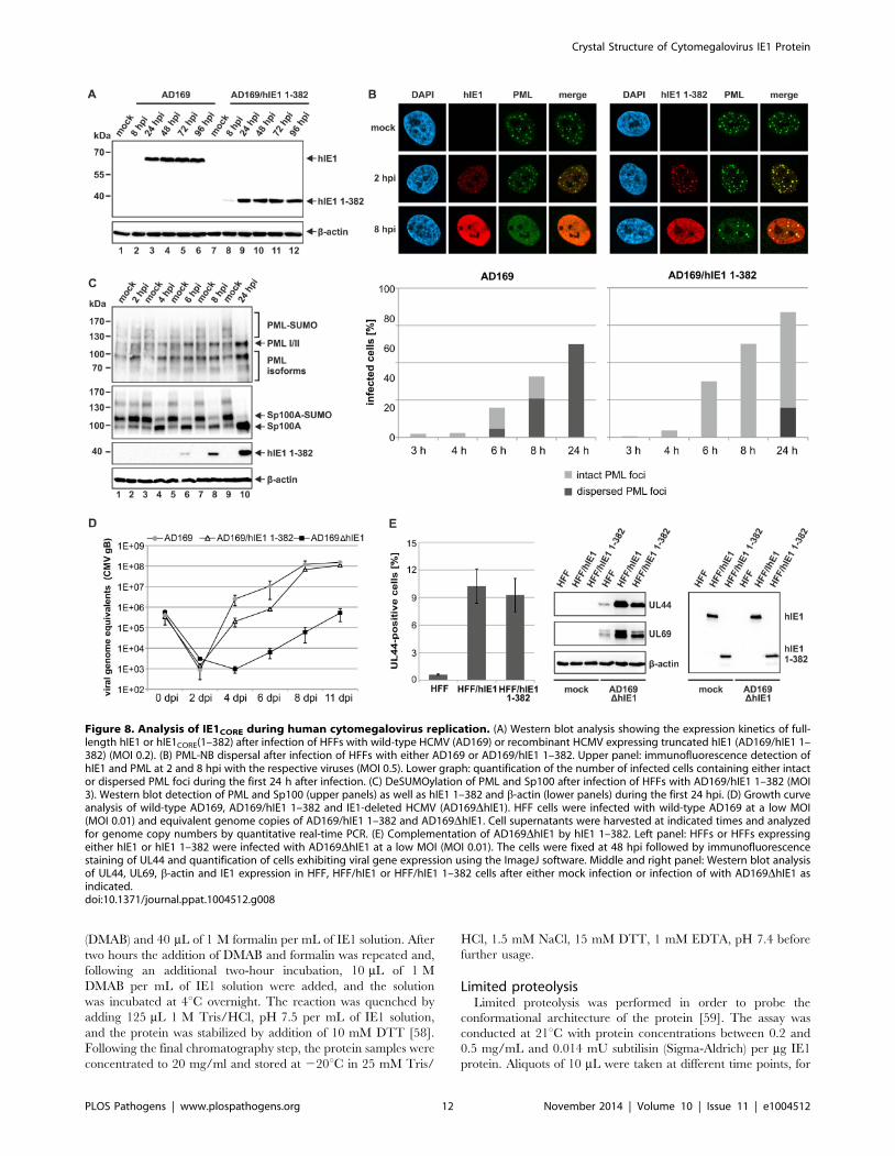
Figure 8. Analysis of IE1CORE during human cytomegalovirus replication. (A) Western blot analysis showing the expression kinetics of full-length hIE1 or hIE1CORE(1–382) after infection of HFFs with wild-type HCMV (AD169) or recombinant HCMV expressing truncated hIE1 (AD169/hIE1 1–382) (MOI 0.2). (B) PML-NB dispersal after infection of HFFs with either AD169 or AD169/hIE1 1–382. Upper panel: immunofluorescence detection ofhIE1 and PML at 2 and 8 hpi with the respective viruses (MOI 0.5). Lower graph: quantification of the number of infected cells containing either intactor dispersed PML foci during the first 24 h after infection. (C) DeSUMOylation of PML and Sp100 after infection of HFFs with AD169/hIE1 1–382 (MOI3). Western blot detection of PML and Sp100 (upper panels) as well as hIE1 1–382 and b-actin (lower panels) during the first 24 hpi. (D) Growth curveanalysis of wild-type AD169, AD169/hIE1 1–382 and IE1-deleted HCMV (AD169DhIE1). HFF cells were infected with wild-type AD169 at a low MOI(MOI 0.01) and equivalent genome copies of AD169/hIE1 1–382 and AD169DhIE1. Cell supernatants were harvested at indicated times and analyzedfor genome copy numbers by quantitative real-time PCR. (E) Complementation of AD169DhIE1 by hIE1 1–382. Left panel: HFFs or HFFs expressingeither hIE1 or hIE1 1–382 were infected with AD169DhIE1 at a low MOI (MOI 0.01). The cells were fixed at 48 hpi followed by immunofluorescencestaining of UL44 and quantification of cells exhibiting viral gene expression using the ImageJ software. Middle and right panel: Western blot analysisof UL44, UL69, b-actin and IE1 expression in HFF, HFF/hIE1 or HFF/hIE1 1–382 cells after either mock infection or infection of with AD169DhIE1 asindicated.doi:10.1371/journal.ppat.1004512.g008
Crystal Structure of Cytomegalovirus IE1 Protein
PLOS Pathogens | www.plospathogens.org 12 November 2014 | Volume 10 | Issue 11 | e1004512
example at 1 min, 10 min, 30 min, 60 min, 120 min, 180 min,
240 min and 300 min, mixed with 3.3 mL 46SDS loading buffer
and boiled at 95uC for 5 min to stop the cleavage reaction.
Circular dichroism spectroscopyCircular dichroism spectra were recorded between 185 and
260 nm from protein samples containing 1.5 mM or 2 mM protein
for full-length or truncated IE1, respectively. The measurements
were performed in 10 mM potassium phosphate buffer, pH 7.5
with a Jasco J-815 spectropolarimeter (Jasco, Tokyo, Japan) at
20uC with standard sensitivity. The cuvette had a path length of
0.1 cm, the band width was 1.0 nm, the scan speed 20 nm*sec21,
data integration time 1 sec and the data pitch 0.1 nm. All
measurements were accumulated ten times and corrected for the
sample buffer. Conversion of the data to concentration-indepen-
dent mean residual weight (MRW) ellipticities [h]MRW was done as
described previously [40].
Crystallization of rhIE1(36–395)Initial crystallization conditions were identified with a sparse
matrix screening approach (Index Screen, Hampton Research,
Aliso Viejo, USA) and a Phoenix protein crystallization robot (Art
Robbins Instruments, Sunnyvale, USA) [60]. The crystallization
conditions were optimized in a hanging drop vapor diffusion setup
and involved microseeding. Crystals of diffraction quality were
obtained by mixing 1 mL of protein solution with 1 mL of reservoir
solution and equilibrating the droplet of 2 mL against 700 mL
reservoir solution [0.4 M magnesium formate, 15% (w/v) PEG
3350]. The crystals were soaked in cryo-solution [0.4 M magne-
sium formate, 15% (w/v) PEG 3350, 15% (v/v) ethylenglycol or
20% (v/v) dimethyl sulfoxide (DMSO)] prior to flash-cooling in
liquid nitrogen.
Crystallographic data collection and crystal dehydrationThe crystal structure of rhIE1(36–395) was initially solved in
space group P21 using the following diffraction datasets collected
at 100 K at beamline BL14.1 at BESSY II synchrotron
(Helmholtz Zentrum Berlin): a native dataset 1 extending to
2.85 A resolution, a MAD dataset (peak, inflection point, remote)
recorded from a gold-soaked crystal diffracting to 3.5 A and a
peak dataset from a seleno-methionine derivatized protein crystal
diffracting to 3.1 A resolution (Table S1). The gold-soaked crystal
was prepared by incubating crystals for three days in cryo-
solution containing DMSO and 2.5 mM KAu(CN)2. Before flash
cooling, the crystals were back-soaked in cryo-solution for several
minutes to remove unspecifically bound heavy atoms. All
monoclinic datasets are highly isomorphous. The Matthews
coefficient is 2.96 (58.42% solvent) when assuming the presence
of four rhIE1(36–395) molecules in the asymmetric unit [61]. The
final refinement of rhIE1(36–395) was performed against a
dataset with space group symmetry P43 extending to 2.3 A (Table
S1). The increase in resolution and concomitant space group
change were obtained upon controlled dehydration of the
monoclinic crystals from above with the HC1c crystal humidifier
device at beamline BL14.3 at BESSY II (Helmholtz Zentrum
Berlin). Crystals were first equilibrated against 98% relative
humidity before decreasing the humidity in steps of 4% and
10 min equilibration time to a final value of 86% humidity. Upon
observation of an increase in diffraction power, the crystals were
flash cooled and transferred to beamline BL14.1 for the recording
of a complete dataset extending to 2.3 A resolution (Table S1).
All diffraction datasets were processed with XDS and scaled with
XSCALE [62].
Structure determination and refinementInitial protein phases were derived for the monoclinic crystal
form using the MAD dataset collected from a gold-soaked crystal
(Table S1). The positions of the gold atoms could be readily
located with program SHELXD [63]. The non-crystallographic
symmetry (NCS) relationship between the 4 monomers, i.e. the
presence of two IE1 dimers with C2 point group symmetry,
became apparent upon visualization of the gold positions in
program COOT and the inspection of the initial electron density
maps calculated with program SHELXE [64,65]. The NCS
relationship was corroborated by the self-rotation function,
calculated with program POLARRFN from the CCP4 program
suite [66]. The quality of these initial electron density maps could
be significantly improved upon phase calculation with program
SHARP/autoSHARP [67,68] and density averaging with pro-
gram DM [69]. The improved phases also allowed for the
identification of the selenium positions in the peak dataset of the
seleno-methionine-labeled protein crystal and the inclusion of this
dataset in the calculation of the experimental protein phases. An
initial atomic model covering a single monomer was then
manually built starting from protein fragments derived with
program autoSHARP and following the lead of electron density
maps calculated with either program autoSHARP or
MLPHARE/DM [66,68]. The registration of the protein
sequence was obtained from the shape of the local electron
density and the positions of the selenium atoms as visualized by an
anomalous difference map. These considerations also showed that
one gold cation is bound via a free cysteine side chain in each IE1
monomer chain. The model was then stepwise completed,
extended to four molecules in the asymmetric unit and refined
with program PHENIX [70]. Convergence of the refinement at
2.8 A in space group P21 was facilitated upon inclusion of NCS
weights and secondary structure restraints in program PHENIX
[70]. The final model of rhIE1(36–395) was obtained after
transferring the monoclinic model into the tetragonal unit cell
with program PHASER and upon refinement against the 2.3 A
dataset in space group P43 (Table S1) [71]. Refinement converged
at crystallographic R-factors of 19.73% (Rwork) and 24.96% (Rfree).
The space group change that took place upon dehydration can be
easily explained by small readjustments in the packing of the IE1
dimers in the crystals.
PDB accession numbersCoordinates and structure factors for the IE1CORE structure
have been deposited in the Protein Data Bank under accession
code 4WID.
Bioinformatic analysesThe size of the dimerization interface between the two
rhIE1CORE monomers was calculated with the program PISA
[72]. The reported value is the average of the buried surface of
both chains. The rhIE1CORE monomers shown in Figure S5 were
superposed with the program LSQKAB [66]. Only the a-helical
segments of the protein as defined in Figure S1 were superim-
posed. Comparative modeling of hIE1 was performed with
MODELLER 9.9 [73] and the resulting model was validated
using ProSA [74,75]. Searches for structurally similar proteins
were performed with PDBeFold [76]. Since standard parameters
did not result in any hits, the following search options were set. (i)
The threshold of 70% for the lowest acceptable match in query
and target was reduced to 30% and 60%, respectively. (ii) The
search was extended to proteins with a different connectivity of
their secondary structure elements. Searches were performed
independently for chain A and chain B of rhIE1, and the list of hits
Crystal Structure of Cytomegalovirus IE1 Protein
PLOS Pathogens | www.plospathogens.org 13 November 2014 | Volume 10 | Issue 11 | e1004512
was merged. For reasons of clarity, duplicate hits and hits related
closely in sequence (.90% identity) were removed from the list.
The normRMSD was calculated according to the following
equation [77]: normRMSD = [RMSD N max(L1,L2)]/Naln.
Where RMSD is the root mean square deviation of the
superposition of query and target, max(L1,L2) is the number of
amino acids of the largest chain in the superposition, and Naln is
defined by the number of structurally equivalent residue pairs.
Sequence conservation was calculated based on a Blosum30
matrix using the MultiSeq Plugin [78] of VMD [79]. The IE1
sequences from the following viruses served as input: human CMV
(strain AD169), Rhesus-CMV, Baboon-CMV, Simian-CMV, and
Panine-HV2/Chimpanzee CMV (Uniprot-accession-numbers
P13202, Q2FAE9, D0UZW7, Q98682, Q8QRY6). Prediction of
intrinsically disordered regions in hIE1, cIE1 and rhIE1 was
performed with IUPred [39] using the prediction type ‘‘short
disorder’’. The disorder tendencies in the three IE1 homologs
were plotted in one diagram using the rhIE1 amino acid
numbering. Multiple sequence alignment was performed with
TCoffee (http://www.ebi.ac.uk/Tools/services/web/toolform.
ebi?tool=tcoffee).
Oligonucleotides and plasmid constructsThe oligonucleotide primers used for this study were purchased
from Biomers GmbH (Ulm, Germany) and are listed in Table S3.
All prokaryotic expression plasmids were generated by PCR
amplification of the respective codon-optimized IE1 sequences and
subsequent cloning into pGEX-6P-1 (GE Healthcare Bio-Sciences
AB, Uppsala, Sweden). The synthetic, codon-optimized hIE1
cDNA (strain AD169) was obtained from Mr. Gene GmbH
(Regensburg, Germany). The codon-optimized cDNAs of cIE1
(NP_612746.1) and rhIE1 (Q2FAE9) were synthesized by
GENEART gene synthesis service (Regensburg, Germany). The
eukaryotic expression plasmids encoding full length or truncated
hIE1 were generated via PCR amplification of the respective
fragments using pHM494 [80] as template, followed by insertion
into pHM971 (pcDNA3.1-FLAG) [80], pHM1580 (pcDNA3.1-
Myc) [80], or into the yeast expression vectors pGBT9 and
pGAD424 (Clontech, Mountain View, CA). The synthetic gene
coding for rhIE1 (Q2FAE9) was obtained from GENEART gene
synthesis service (Regensburg, Germany). The rhIE1 coding
sequence was subcloned into pHM971 (pcDNA3.1-FLAG) [80]
using BamHI and XhoI. Full length PML, isoform VI, and
truncated PML variants were amplified from pAS-PML (a gift
from G.G. Maul, Philadelphia, USA) and inserted into pHM1580
(pcDNA3.1-Myc) [81], pHM971 (pcDNA3.1-FLAG) [80],
pHM972 (pcDNA3.1-FLAG-NLS), or yeast expression vectors
pGBT9 and pGAD424 (Clontech, Mountain View, CA). The
eukaryotic expression plasmid encoding rhesus TRIM5a was a gift
from T. Gramberg (Erlangen, Germany). For transduction
experiments, hIE1 variants were amplified utilizing pHM494
[80] as template and inserted into a pLKO-based lentiviral vector
(a gift of R. Everett, Glasgow, UK).
Cells, infections and virusesHEK293T cells and primary human foreskin fibroblast (HFF)
cells (obtained from Life Technologies) or telomerase-immortal-
ized HFFs (HFFi) were cultured as described previously [54,82].
HFFs were infected with either the HCMV laboratory strain
AD169, a recombinant HCMV expressing hIE1 1–382 (AD169/
hIE1 1–382), an IE1-deficient virus (AD169DhIE1), or rhesus
macaque CMV (RhCMV) at specified multiplicities of infection
(MOI). Titers of wild-type (wt) AD169 and the AD169 recombi-
nants were determined by UL112/113 fluorescence. For this
purpose, HFFs were infected with various dilutions of virus stocks.
After 72 h of incubation, cells were fixed and stained with a
monoclonal antibody directed against UL112/113. Subsequently,
the number of UL112/113-positive cells was determined and was
used to calculate viral titers. RhCMV was titrated via rhIE1
fluorescence, which was analyzed 24 h postinfection.
The AD169-based HCMV bacterial artificial chromosome
(BAC) HB15 was used for recombination-based genetic engineer-
ing of AD169/hIE1 1–382 and AD169DhIE1. AD169/hIE1 1–
382 was constructed by introducing a stop codon into the hIE1
gene replacing residue 383. For this purpose, the two-step red-
mediated recombination technique was utilized [83], which uses
the kanamycin gene as a first selection marker. The linear
recombination fragment was generated by PCR using primers
59BAC_short and 3’BAC_hIE1_382 (Table S3), and pEPkan-S
(kindly provided by K. Osterrieder, Berlin) as template DNA. The
PCR product was treated with DpnI, gel purified, and subjected to
a second round of PCR amplification using primers 59BA-
C_hIE1_382 and 39BAC_hIE1_382_short (Table S3). For
homologous recombination, the PCR fragment was transformed
into Escherichia coli strain GS1783 (a gift of M. Mach, Erlangen)
already harboring HB15, and bacteriophage l red-mediated
recombination was conducted as described elsewhere [83]. To
identify positive transformants, the bacteria were plated on agar
plates containing 30 mg/mL kanamycin (first recombination) or
30 mg/mL chloramphenicol and 1% arabinose (second recombi-
nation) and incubated at 32uC for 2 days. BAC DNA was purified
from bacterial colonies growing on these plates and was further
analyzed by PCR, restriction enzyme digestion and direct
sequencing. For construction of the AD169DhIE1 BAC by
homologous recombination, a linear recombination fragment,
comprising a kanamycin resistance marker along with 59 and 39
genomic sequences, was generated by PCR amplification using
pKD13 as template and primers 59Intron3/pKD13 and 39Exon
4/pkd13 (Table S3). This fragment was used for electroporation of
competent Escherichia coli strain DH10B harboring HB15 and
recombination was performed as described previously in order to
delete exon 4 of the IE1 gene [84]. The integrity of the resulting
recombinant BAC was confirmed by PCR, restriction enzyme
digestion and direct sequencing.
For reconstitution of recombinant AD169, HFFs seeded in six-
well dishes (36105 cells/well) were cotransfected with 1 mg of
purified BAC DNA, 0.5 mg of the pp71 expression plasmid pCB6-
pp71, and 0.5 mg of a vector encoding the Cre recombinase using
FuGENE6 transfection reagent (Promega, Mannheim, Germany).
Transfected HFFs were propagated until viral plaques appeared,
and the supernatants from these cultures were used for further
virus propagation.
Lentivirus transduction and selection of stablytransduced cells
For the generation of HFF cells stably expressing full length
hIE1 or hIE1 1–382, replication-deficient lentiviruses were
generated using pLKO-based expression vectors. For this purpose,
HEK293T cells seeded in 10 cm dishes (56106 cells/dish) were
cotransfected with a pLKO vector encoding either full length hIE1
or hIE1 1–382 together with packaging plasmids pLP1, pLP2, and
pLP/VSV-G using the Lipofectamine 2000 reagent (Invitrogen,
Karlsruhe, Germany). Viral supernatants were harvested 48 h
after transfection, cleared by centrifugation, filtered, and stored at
280uC. Primary HFFs or telomerase-immortalized HFFs were
incubated for 24 h with lentivirus supernatants in the presence of
7.5 mg/mL polybrene (Sigma-Aldrich, Deisenhofen, Germany).
Crystal Structure of Cytomegalovirus IE1 Protein
PLOS Pathogens | www.plospathogens.org 14 November 2014 | Volume 10 | Issue 11 | e1004512
Stably transduced cell populations were selected by adding
500 mg/mL geneticin to the cell culture medium.
TransfectionHFF cells were transfected with the DNA transfection reagent
FuGENE6 (Promega, Mannheim, Germany). One day before
transfection, 36105 cells were seeded into six-well dishes. DNA
content and transfection procedure were according to the
instructions of the manufacturer. 48 hours after transfection, cells
were harvested for further analyses. HEK293T cells were
transfected by applying the standard calcium phosphate precip-
itation method. For this, 56105 to 56106 HEK293T cells were
seeded into six-well dishes or 10 cm dishes one day before
transfection. For Western blot analyses and coimmunoprecipita-
tions, 1 to 10 mg of plasmid DNA were used for each transfection
reaction. At about 16 hours later, the cells were washed two times
with PBSo and provided with fresh medium. 48 hours after
transfection, cells were harvested for further analyses.
AntibodiesMonoclonal antibodies used for immunofluorescence and
Western blot analyses were: a-IE1 CH443 (Santa Cruz Biotech-
nology, Santa Cruz, CA, USA), a-UL112/113 M23, a-UL44
BS510 (kindly provided by B. Plachter, Mainz, Germany), a-UL69
69–66, a-FLAG M2 (Sigma-Aldrich, Deisenhofen, Germany), a-
Myc 9E10, a-b-actin AC-15 (Sigma-Aldrich), a-PML PG-M3
(Santa Cruz). Polyclonal antibodies used for immunofluorescence
and Western blot analyses were: a-rhesus IE1 (a kind gift from M.
Mach, Erlangen, Germany), a-PML #4 (a kind gift from P.
Hemmerich, Jena, Germany), a-PML H238 (Santa Cruz), a-PML
A301–167A (Bethyl Laboratories, Montgomery, TX, USA), a-
PML A301–168A (Bethyl Laboratories), a-Sp100 #2 (a kind gift
from P. Hemmerich, Jena, Germany), a-Sp100 GH3 (kindly
provided by H. Will, Hamburg, Germany), a-hDaxx C-20 (Santa
Cruz), a-ATRX H-300 (Santa Cruz). Secondary antibodies used
for immunofluorescence and Western blot analyses were: Alexa
Fluor 488-/555-/647-conjugated secondary antibodies for indirect
immunofluorescence experiments were purchased from Molecular
Probes (Karlsruhe, Germany), horseradish peroxidase-conjugated
anti-mouse/-rabbit secondary antibodies for Western blot analyses
were obtained from Dianova (Hamburg, Germany).
Indirect immunofluorescenceHFF cells grown on coverslips in six-well dishes (36105 cells/
well) were washed twice with PBSo at 48 hours after transfection
or at various times after virus infection. Cells were fixed with a 4%
paraformaldehyde solution for 10 min at room temperature (RT)
and then washed for two times. Permeabilization of the cells was
achieved by incubation with 0.2% Triton X-100 in PBSo on ice
for 20 min. Cells were washed again with PBSo over a time period
of 5 min and incubated with the appropriate primary antibody
diluted in PBSo-1% FCS for 30 min at 37uC. Excessive antibodies
were removed by washing four times with PBSo, followed by
incubation with the corresponding fluorescence-coupled secondary
antibody diluted in PBSo-1% FCS for 30 min at 37uC. The cells
were mounted using the DAPI-containing Vectashield mounting
medium (Alexis, Grunberg, Germany) and analyzed using a Leica
TCS SP5 confocal microscope, with 488 nm, 543 nm, and
633 nm laser lines, scanning each channel separately under image
capture conditions that eliminated channel overlap. The images
were exported, processed with Adobe Photoshop CS5 and
assembled using CorelDraw 65. In order to quantify PML-NB
disruption in infected HFFs, 150 cells were analyzed for the
presence of PML dots.
ImmunoblottingLysates from transfected or infected cells were prepared in a
sodium dodecyl sulfate-polyacrylamide gel electrophoresis (SDS-
PAGE) loading buffer, separated on sodium dodecyl sulfate-
containing 8 to 15% polyacrylamide gels, and transferred to
nitrocellulose membranes. Chemiluminescence was detected
according to the manufacturer’s protocol (ECL Western blot
detection kit; Amersham Pharmacia Biotech).
CoimmunoprecipitationTransfected HEK293T cells (16106 or 56106 were lysed for 20
to 40 min at 4uC in 800 mL of CoIP buffer (50 mM Tris-HCl
[pH 8.0], 150 mM NaCl, 5 mM EDTA, 0.5% NP-40, 1 mM
PMSF, 2 mg/mL of aprotinin, 2 mg/mL of leupeptin, and 2 mg/
mL of pepstatin). After centrifugation, aliquots of each sample
were taken as input controls and the remaining supernatant was
incubated with anti-FLAG antibody M2 coupled to protein-A-
sepharose beads for 2 h at 4uC. The sepharose beads were
collected by centrifugation and washed five times in 1 mL CoIP
buffer. Finally, the immunoprecipitated proteins were recovered
by boiling in 46 SDS sample buffer and protein complexes were
analyzed by SDS-PAGE and Western blotting.
Yeast two-hybrid analysisSaccharomyces cerevisiae Y153 was used in a two-hybrid system.
Both the plasmid pGBT9 (Clontech, Mountain View, CA)
encoding the GAL4-DB (Trp+) fusion and the plasmid pGAD424
(Clontech, Mountain View, CA) encoding the GAL4-A fusion
(Leu+) were introduced into Y153 cells using a modified lithium
acetate (LiAc) method. For this, cells were grown overnight in
YAPD medium, pelleted and treated with LP-mix (40% w/v PEG
4000, 0.15 M LiAc, 10 mM Tris/HCL pH 7.5, 1 mM EDTA
pH 8.0) and DMSO. Single-stranded carrier-DNA as well as both
plasmids were added to the yeast cells. This step was followed by
incubation at room temperature and subsequent incubation at
42uC. Thereafter, the cells were plated on minimal selection agar
lacking Trp and Leu. For rapid in situ assays of lacZ expression
from yeast colonies, an XGal filter assay was used. Nitrocellulose
filters were laid onto the plate and allowed to wet completely, then
lifted off the plate and placed in liquid nitrogen to permeabilize the
cells. The filters were removed and placed cell side up in a petri
dish containing Whatman Paper soaked with Z buffer containing
b-Mercaptoethanol and XGal. The filters were incubated at 30uCand constantly analyzed for the development of a positive blue
color. For quantitation of the ß-galactosidase activity in the yeast
cells three colonies were picked and grown in medium also lacking
Trp and Leu. The next day, the optical density was measured at
600 nm. After pelleting the culture, the cells were resuspended in
Z buffer and permeabilized using chloroform and 0.1% SDS. The
ß-galactosidase activity within the cells was assayed by the
standard method using o-nitrophenyl-ß-D-galactopyranoside
(ONPG) as substrate. The reaction was stopped by adding
Na2CO3 and the absorbance was measured at OD405. The unit
of ß-galactosidase was defined as (1.0006OD405)/(t6v6OD600) (t,
reaction time [min]; v, culture volume [mL]). The ß-galactosidase
activity for each sample was corrected for background by
subtracting the signal of the empty vectors.
Multistep growth curve analysisHFF cells were seeded into six-well dishes at a density of 36105
cells/well and infected the following day with wt AD169 at an
MOI of 0.01 and equivalent genome copies of AD169/hIE1 1–
382 and AD169DhIE1. Triplicate samples of the infected cell
Crystal Structure of Cytomegalovirus IE1 Protein
PLOS Pathogens | www.plospathogens.org 15 November 2014 | Volume 10 | Issue 11 | e1004512
supernatants were harvested at 2, 4, 6, 8 and 11 days after
inoculation and subjected to lysis by proteinase K treatment.
Thereafter, all samples were analyzed for the amount of genome
copy numbers by quantitative real-time PCR (TaqMan-PCR)
using an Applied Biosystems 7500 Real-Time PCR System
(Applied Biosystems, Foster City, CA, USA) together with the
corresponding software SDS (sequence detection system) [85]. For
quantification of the viral DNA load, a sequence region within the
gB gene locus was amplified using primers 5’gB_forw and
3’gB_rev along with the fluorescence labeled probe CMV gB
FAM/TAMRA also directed against the gB gene region. In
parallel, the cellular DNA amount was quantified using primers
59Alb and 39 Alb together with a fluorescence labeled probe, Alb
FAM/TAMRA, specific for the cellular albumin gene. Real-time
PCR was performed in 96-well plates being compatible with the
ABI Prism sequence detector. For the determination of reference
CT values (cycle threshold), serial dilutions of the respective
standards (107–101 DNA molecules of gB or albumin) were
examined by PCR reactions in parallel. The 20 mL reaction mix
contained 5 mL sample or standard DNA solution together with
10 mL 26 TaqMan PCR Mastermix (Applied Biosystems, Foster
City, CA, USA), 1.5 mL of each primer (5 mM stock solution),
0.4 mL of probe (10 mM stock solution), and 1.6 mL of H2O. The
thermal cycling conditions consisted of two initial steps of 2 min at
50uC and 10 min at 95uC followed by 40 amplification cycles
(15 sec at 95uC, 1 min 60uC). The viral genome copy numbers
and albumin copy numbers were subsequently calculated using the
sample-specific CT value when set into relation to the standard
serial dilutions.
Complementation assayFor analysis of complementation by immunofluorescence
staining, control HFFi cells as well as HFFi cells expressing hIE1
or hIE1 1–382 were infected with AD169DhIE1 at an MOI of
0.01. Triplicate samples of infected cells were fixed at 48 h post
infection and subjected to immunostaining of UL44. Images of
approximately 500 cells per sample were taken and the number of
UL44-positive cells was determined via measuring of mean gray
values using the ImageJ software (version 1.47). For analysis of
complementation by Western blotting, normal HFFi cells as well
as HFFi cells expressing hIE1 or hIE1 1–382 were infected with
AD169DhIE1 at an MOI of 0.05 in triplicate, and were harvested
72 h later for detection of UL44, UL69, IE1 and b-actin.
Supporting Information
Figure S1 Sequence alignment of homologous IE1 proteins:
hIE1 of HCMV (strain AD169, AC146999), cIE1 of chimpanzee
CMV (panine herpesvirus 2, NC003521.1), and rhIE1 of rhesus
macaque CMV (ceropithecine herpesvirus 8, DQ120516.1).
Sequence conservation is indicated by gray shading (dark gray:
fully conserved residues, middle gray: strongly similar residues,
light gray: weakly similar residues). Residues as resolved by the
rhIE1 crystal structure are marked by a black border. The
positions of the a-helices within this region, which are numbered
consecutively from the N- to the C-terminus, were determined on
the basis of the structural data. The positions of hydrophobic
amino acids within hendecad and heptad repeats of helices 1 and 3
are indicated below the amino acid sequence (a, d, h).
(TIF)
Figure S2 The CD spectra of full-length hIE1(1–491) and
hIE1(14–382) are shown in blue and gold, respectively (A). The
difference spectrum obtained by subtracting the data of hIE1(14–
382) from hIE1(1–491) is shown in green (B). The difference
spectrum exhibits strong random coil characteristics with its
minimum at a wavelength around 200 nm and therefore confirms
the intrinsically disordered nature of the terminal regions.
(TIF)
Figure S3 Stereo image of a representative section of the
experimentally phased electron density map used for model
building. Protein phases in space group P21 were derived from a
3.5 A MAD dataset collected from a gold-soaked crystal and
combined with 3.1 A data from a SeMet SAD dataset collected at
the peak wave-length of Se (Table S1). Extensive four-fold non-
crystallographic symmetry averaging was applied. The protein
density (contoured at 1s) is shown in blue and the anomalous
density calculated from the anomalous difference signal in the
SeMet peak dataset (contoured at 3s) in orange. The helical
structure of the protein is clearly visible and the positions of the Se-
atoms coincide with the locations of the peaks in the anomalous
density map.
(TIF)
Figure S4 Multiple sequence alignment of the coiled-coil region
of different TRIM proteins. The a and d position of the heptad
and hendecad repeats and the h position present only in the
hendecad repeats are highlighted.
(TIF)
Figure S5 Stereorepresentation showing the superposition of the
two monomers that are present in dimeric rhIE1CORE in the final
refined structure in space group P43. When considering the Capositions from the a-helical segments, the two monomers can be
superimposed with an r.m.s. deviation of 2.13 A. The area with
the highest deviation, namely, the loop between helices H8 and
H9, is marked by a red box (see also Figure S6). The molecules
were superposed with the program LSQKAB [66].
(TIF)
Figure S6 Simulated annealed OMIT difference density map
illustrating a difference in the kink of the loop formed between
helices H8 and H9 in the two monomers (panel A and B) of
dimeric rhIE1CORE (see also Figure S5). The maps were generated
with program PHENIX and residues Glu276 to Thr292 omitted
during refinement and subsequent difference density calculation.
The sA-weighted mFo-DFc electron density is displayed at a 3slevel.
(TIF)
Figure S7 Structure validation of the hIE1 model and
comparison of the structural parameters to the rhIE1 template
crystal structure. (A, B) Overall structure quality as indicated by
the Z-score. The Z-scores of rhIE1 (A) and hIE1 (B) are displayed
in a plot that contains the Z-scores of all experimentally
determined protein chains in the PDB. In this plot, groups of
structures from different sources (X-ray, NMR) are distinguished
by different colors. Z-scores for IE1 chain A and B are denoted by
a dot and a rectangle symbol, respectively. Note that the Z-scores
of the crystal structure and model are very similar and well within
the range expected for protein structures of this size. (C)–(F) Local
structure quality evidenced by a knowledge-based energy as a
function of amino acid sequence position. The plots are smoothed
by calculating the average energy over each 10- residue fragment
(thin line) or 40-residue fragment (thick line) along the peptide
chain. The latter plots are similar for rhIE1 (C, D) and hIE1 (E, F)
and exhibit favorable negative energy values along the entire
peptide chains. This indicates that the modeling procedure has not
placed residues in an unfavorable environment.
(TIF)
Crystal Structure of Cytomegalovirus IE1 Protein
PLOS Pathogens | www.plospathogens.org 16 November 2014 | Volume 10 | Issue 11 | e1004512
Table S1 Data collection, phasing and refinement statistics.
(DOC)
Table S2 List of structurally similar proteins of IE1 sorted by
their Z-score in descending order. For each hit the PDB code, the
RMSD of the overlay, and the number of structurally equivalent
residues (Nalgn) is given. Generally, the number of equivalent
residues is rather low covering less than 50% of the total length of
IE1. This demonstrates that the overall fold in IE1 is rather unique
and there are only local structural similarities to other helical
proteins. These local hits were further inspected based on their
CATH-classification. Most of the hits from the present search are
classified as a-helical orthogonal bundle (code 1.10.) or up-down
bundle (code 1.20.). Comparison of the third number of the
CATH-code shows that these proteins belong to at least 18
different folds indicating that IE1 cannot readily be assigned to any
known topology. This is further corroborated by the calculation of
a normalized rmsd (normRMSD): For all hits listed in Table S2
the normRMSD values are significantly larger than the threshold
of 5 A [77], which indicates that a protein is significantly
structurally different from those proteins previously deposited in
the PDB.
(DOC)
Table S3 List of oligonucleotides.
(DOC)
Acknowledgments
We thank P. Hemmerich (Jena, Germany), R. Everett (Glasgow, UK), H.
Will (Hamburg, Germany), B. Plachter (Mainz, Germany), M. Mach
(Erlangen, Germany) and T. Gramberg (Erlangen, Germany) for providing
reagents for this study. Furthermore, we would like to thank U. Muller and
M. Weiss from BESSY II synchrotron Berlin for help with diffraction data
collection.
Author Contributions
Conceived and designed the experiments: TS YAM. Performed the
experiments: MSc SK VO EMS RM NR MSe YAM JDS HS. Analyzed
the data: SK MSe YAM JDS HS MSc EMS NR TS. Wrote the paper:
MSc SK HS YAM TS.
References
1. Ishov AM, Sotnikov AG, Negorev D, Vladimirova OV, Neff N, et al. (1999)PML is critical for ND10 formation and recruits the PML-interacting protein
daxx to this nuclear structure when modified by SUMO-1. J Cell Biol 147: 221–234.
2. Jensen K, Shiels C, Freemont PS (2001) PML protein isoforms and the RBCC/
TRIM motif. Oncogene 20: 7223–7233.
3. Rajsbaum R, Garcia-Sastre A, Versteeg GA (2014) TRIMmunity: the roles of
the TRIM E3-ubiquitin ligase family in innate antiviral immunity. J Mol Biol
426: 1265–1284.
4. Versteeg GA, Rajsbaum R, Sanchez-Aparicio MT, Maestre AM, Valdiviezo J,
et al. (2013) The E3-ligase TRIM family of proteins regulates signaling pathwaystriggered by innate immune pattern-recognition receptors. Immunity 38: 384–
398.
5. Chu Y, Yang X (2011) SUMO E3 ligase activity of TRIM proteins. Oncogene30: 1108–1116.
6. Kamitani T, Kito K, Nguyen HP, Wada H, Fukuda-Kamitani T, et al. (1998)
Identification of three major sentrinization sites in PML. J Biol Chem 273:26675–26682.
7. Lallemand-Breitenbach V, Zhu J, Puvion F, Koken M, Honore N, et al. (2001)
Role of promyelocytic leukemia (PML) sumolation in nuclear body formation,11S proteasome recruitment, and As2O3-induced PML or PML/retinoic acid
receptor alpha degradation. J Exp Med 193: 1361–1371.
8. Bernardi R, Pandolfi PP (2003) Role of PML and the PML-nuclear body in the
control of programmed cell death. Oncogene 22: 9048–9057.
9. Bernardi R, Pandolfi PP (2007) Structure, dynamics and functions ofpromyelocytic leukaemia nuclear bodies. Nat Rev Mol Cell Biol 8: 1006–1016.
10. Tavalai N, Stamminger T (2008) New insights into the role of the subnuclear
structure ND10 for viral infection. Biochim Biophys Acta 1783: 2207–2221.
11. Everett RD, Chelbi-Alix MK (2007) PML and PML nuclear bodies: implications
in antiviral defence. Biochimie 89: 819–830.
12. El Asmi F, Maroui MA, Dutrieux J, Blondel D, Nisole S, et al. (2014)Implication of PMLIV in Both Intrinsic and Innate Immunity. PLoS Pathog 10:
e1003975.
13. Bieniasz PD (2004) Intrinsic immunity: a front-line defense against viral attack.
Nat Immunol 5: 1109–1115.
14. Woodhall DL, Groves IJ, Reeves MB, Wilkinson G, Sinclair JH (2006) HumanDaxx-mediated repression of human cytomegalovirus gene expression correlates
with a repressive chromatin structure around the major immediate early
promoter. J Biol Chem 281: 37652–37660.
15. Tavalai N, Papior P, Rechter S, Leis M, Stamminger T (2006) Evidence for a
role of the cellular ND10 protein PML in mediating intrinsic immunity againsthuman cytomegalovirus infections. J Virol 80: 8006–8018.
16. Tavalai N, Papior P, Rechter S, Stamminger T (2008) Nuclear domain 10
components promyelocytic leukemia protein and hDaxx independentlycontribute to an intrinsic antiviral defense against human cytomegalovirus
infection. J Virol 82: 126–137.
17. Lukashchuk V, McFarlane S, Everett RD, Preston CM (2008) Humancytomegalovirus protein pp71 displaces the chromatin-associated factor ATRX
from nuclear domain 10 at early stages of infection. J Virol 82: 12543–12554.
18. Adler M, Tavalai N, Muller R, Stamminger T (2011) Human cytomegalovirus
immediate-early gene expression is restricted by the nuclear domain 10
component Sp100. J Gen Virol 92: 1532–1538.
19. Glass M, Everett RD (2013) Components of promyelocytic leukemia nuclear
bodies (ND10) act cooperatively to repress herpesvirus infection. J Virol 87:
2174–2185.
20. Saffert RT, Kalejta RF (2006) Inactivating a cellular intrinsic immune defensemediated by Daxx is the mechanism through which the human cytomegalovirus
pp71 protein stimulates viral immediate-early gene expression. J Virol 80: 3863–
3871.
21. Boutell C, Cuchet-Lourenco D, Vanni E, Orr A, Glass M, et al. (2011) A viralubiquitin ligase has substrate preferential SUMO targeted ubiquitin ligase
activity that counteracts intrinsic antiviral defence. PLoS Pathog 7: e1002245.
22. Scherer M, Stamminger T (2014) The human cytomegalovirus IE1 protein -
past and present developments. Future Virology 9: 415–430.
23. Wilkinson GW, Kelly C, Sinclair JH, Rickards C (1998) Disruption of PML-associated nuclear bodies mediated by the human cytomegalovirus major
immediate early gene product. J Gen Virol 79 (Pt 5): 1233–1245.
24. Xu Y, Ahn JH, Cheng M, apRhys CM, Chiou CJ, et al. (2001) Proteasome-independent disruption of PML oncogenic domains (PODs), but not covalent
modification by SUMO-1, is required for human cytomegalovirus immediate-
early protein IE1 to inhibit PML-mediated transcriptional repression. J Virol 75:10683–10695.
25. Reinhardt J, Smith GB, Himmelheber CT, zizkhan-Clifford J, Mocarski ES
(2005) The carboxyl-terminal region of human cytomegalovirus IE1491aacontains an acidic domain that plays a regulatory role and a chromatin-tethering
domain that is dispensable during viral replication. J Virol 79: 225–233.
26. Lee HR, Huh YH, Kim YE, Lee K, Kim S, et al. (2007) N-terminal
determinants of human cytomegalovirus IE1 protein in nuclear targeting anddisrupting PML-associated subnuclear structures. Biochem Biophys Res
Commun 356: 499–504.
27. Paulus C, Krauss S, Nevels M (2006) A human cytomegalovirus antagonist of
type I IFN-dependent signal transducer and activator of transcription signaling.Proc Natl Acad Sci U S A 103: 3840–3845.
28. Krauss S, Kaps J, Czech N, Paulus C, Nevels M (2009) Physical requirements
and functional consequences of complex formation between the cytomegalovirusIE1 protein and human STAT2. J Virol 83: 12854–12870.
29. Huh YH, Kim YE, Kim ET, Park JJ, Song MJ, et al. (2008) Binding STAT2 bythe acidic domain of human cytomegalovirus IE1 promotes viral growth and is
negatively regulated by SUMO. J Virol 82: 10444–10454.
30. Kelly C, van DR, Wilkinson GW (1995) Disruption of PML-associated nuclearbodies during human cytomegalovirus infection. J Gen Virol 76 (Pt 11): 2887–
2893.
31. Korioth F, Maul GG, Plachter B, Stamminger T, Frey J (1996) The nuclear
domain 10 (ND10) is disrupted by the human cytomegalovirus gene productIE1. Exp Cell Res 229: 155–158.
32. Ahn JH, Hayward GS (1997) The major immediate-early proteins IE1 and IE2
of human cytomegalovirus colocalize with and disrupt PML-associated nuclearbodies at very early times in infected permissive cells. J Virol 71: 4599–4613.
33. Ahn JH, Hayward GS (2000) Disruption of PML-associated nuclear bodies byIE1 correlates with efficient early stages of viral gene expression and DNA
replication in human cytomegalovirus infection. Virology 274: 39–55.
34. Muller S, Dejean A (1999) Viral immediate-early proteins abrogate themodification by SUMO-1 of PML and Sp100 proteins, correlating with nuclear
body disruption. J Virol 73: 5137–5143.
35. Lee HR, Kim DJ, Lee JM, Choi CY, Ahn BY, et al. (2004) Ability of the human
cytomegalovirus IE1 protein to modulate sumoylation of PML correlates with itsfunctional activities in transcriptional regulation and infectivity in cultured
fibroblast cells. Journal of Virology 78: 6527–6542.
36. Ahn JH, Brignole EJ, III, Hayward GS (1998) Disruption of PML subnuclear
domains by the acidic IE1 protein of human cytomegalovirus is mediated
Crystal Structure of Cytomegalovirus IE1 Protein
PLOS Pathogens | www.plospathogens.org 17 November 2014 | Volume 10 | Issue 11 | e1004512
through interaction with PML and may modulate a RING finger-dependent
cryptic transactivator function of PML. Mol Cell Biol 18: 4899–4913.
37. Hayhurst GP, Bryant LA, Caswell RC, Walker SM, Sinclair JH (1995) CCAATbox-dependent activation of the TATA-less human DNA polymerase alpha
promoter by the human cytomegalovirus 72-kilodalton major immediate-early
protein. J Virol 69: 182–188.
38. LaFemina RL, Pizzorno MC, Mosca JD, Hayward GS (1989) Expression of theacidic nuclear immediate-early protein (IE1) of human cytomegalovirus in stable
cell lines and its preferential association with metaphase chromosomes. Virology172: 584–600.
39. Dosztanyi Z, Csizmok V, Tompa P, Simon I (2005) IUPred: web server for the
prediction of intrinsically unstructured regions of proteins based on estimatedenergy content. Bioinformatics 21: 3433–3434.
40. Kelly SM, Jess TJ, Price NC (2005) How to study proteins by circular dichroism.
Biochim Biophys Acta 1751: 119–139.
41. Greenfield NJ (2006) Using circular dichroism spectra to estimate protein
secondary structure. Nat Protoc 1: 2876–2890.
42. Offer G, Hicks MR, Woolfson DN (2002) Generalized Crick equations formodeling noncanonical coiled coils. J Struct Biol 137: 41–53.
43. Chothia C, Levitt M, Richardson D (1977) Structure of proteins: packing of
alpha-helices and pleated sheets. Proc Natl Acad Sci U S A 74: 4130–4134.
44. Sanchez JG, Okreglicka K, Chandrasekaran V, Welker JM, Sundquist WI, et al.(2014) The tripartite motif coiled-coil is an elongated antiparallel hairpin dimer.
Proc Natl Acad Sci U S A 111: 2494–2499.
45. Li Y, Wu H, Wu W, Zhuo W, Liu W, et al. (2014) Structural insights into theTRIM family of ubiquitin E3 ligases. Cell Res 24: 762–765.
46. Goldstone DC, Walker PA, Calder LJ, Coombs PJ, Kirkpatrick J, et al. (2014)
Structural studies of postentry restriction factors reveal antiparallel dimers that
enable avid binding to the HIV-1 capsid lattice. Proc Natl Acad Sci U S A 111:9609–9614.
47. Mucke K, Paulus C, Bernhardt K, Gerrer K, Schon K, et al. (2014) Human
cytomegalovirus major immediate early 1 protein targets host chromosomes bydocking to the acidic pocket on the nucleosome surface. J Virol 88: 1228–1248.
48. Everett RD, Freemont P, Saitoh H, Dasso M, Orr A, et al. (1998) The disruption
of ND10 during herpes simplex virus infection correlates with the Vmw110- andproteasome-dependent loss of several PML isoforms. J Virol 72: 6581–6591.
49. Full F, Reuter N, Zielke K, Stamminger T, Ensser A (2012) Herpesvirus saimiri
antagonizes nuclear domain 10-instituted intrinsic immunity via an ORF3-
mediated selective degradation of cellular protein Sp100. J Virol 86: 3541–3553.
50. Full F, Jungnickl D, Reuter N, Bogner E, Brulois K, et al. (2014) Kaposi’ssarcoma associated herpesvirus tegument protein ORF75 is essential for viral
lytic replication and plays a critical role in the antagonization of ND10-institutedintrinsic immunity. PLoS Pathog 10: e1003863.
51. Jiang M, Entezami P, Gamez M, Stamminger T, Imperiale MJ (2011)
Functional reorganization of promyelocytic leukemia nuclear bodies duringBK virus infection. MBio 2: e00281–10.
52. Gong L, Millas S, Maul GG, Yeh ET (2000) Differential regulation of
sentrinized proteins by a novel sentrin-specific protease. J Biol Chem 275: 3355–
3359.
53. Kang H, Kim ET, Lee HR, Park JJ, Go YY, et al. (2006) Inhibition of SUMO-independent PML oligomerization by the human cytomegalovirus IE1 protein.
J Gen Virol 87: 2181–2190.
54. Tavalai N, Adler M, Scherer M, Riedl Y, Stamminger T (2011) Evidence for adual antiviral role of the major nuclear domain 10 component Sp100 during the
immediate-early and late phases of the human cytomegalovirus replication cycle.J Virol 85: 9447–9458.
55. Ou HD, Kwiatkowski W, Deerinck TJ, Noske A, Blain KY, et al. (2012) A
structural basis for the assembly and functions of a viral polymer that inactivates
multiple tumor suppressors. Cell 151: 304–319.
56. Gack MU, Albrecht RA, Urano T, Inn KS, Huang IC, et al. (2009) Influenza Avirus NS1 targets the ubiquitin ligase TRIM25 to evade recognition by the host
viral RNA sensor RIG-I. Cell Host Microbe 5: 439–449.
57. Martinez FP, Tang Q (2014) Identification of cellular proteins that interact withhuman cytomegalovirus immediate-early protein 1 by protein array assay.
Viruses 6: 89–105.
58. Walter TS, Meier C, Assenberg R, Au KF, Ren J, et al. (2006) Lysinemethylation as a routine rescue strategy for protein crystallization. Structure 14:
1617–1622.
59. Fontana A, de Laureto PP, Spolaore B, Frare E, Picotti P, et al. (2004) Probing
protein structure by limited proteolysis. Acta Biochim Pol 51: 299–321.60. Jancarik J, Kim S-H (1991) Sparse matrix sampling: a screening method for
crystallization of proteins. J Appl Cryst 24: 409–411.
61. Matthews BW (1968) Solvent content of protein crystals. J Mol Biol 33: 491–497.
62. Kabsch W (2010) XDS. Acta Crystallogr D Biol Crystallogr 66: 125–132.63. Schneider TR, Sheldrick GM (2002) Substructure solution with SHELXD. Acta
Crystallogr D Biol Crystallogr 58: 1772–1779.
64. Sheldrick GM (2010) Experimental phasing with SHELXC/D/E: combiningchain tracing with density modification. Acta Crystallogr D Biol Crystallogr 66:
479–485.65. Emsley P, Lohkamp B, Scott WG, Cowtan K (2010) Features and development
of Coot. Acta Crystallogr D Biol Crystallogr 66: 486–501.66. Winn MD, Ballard CC, Cowtan KD, Dodson EJ, Emsley P, et al. (2011)
Overview of the CCP4 suite and current developments. Acta Crystallogr D Biol
Crystallogr 67: 235–242.67. Bricogne G, Vonrhein C, Flensburg C, Schiltz M, Paciorek W (2003)
Generation, representation and flow of phase information in structuredetermination: recent developments in and around SHARP 2.0. Acta Crystal-
logr D Biol Crystallogr 59: 2023–2030.
68. Vonrhein C, Blanc E, Roversi P, Bricogne G (2007) Automated structuresolution with autoSHARP. Methods Mol Biol 364: 215–230.
69. Cowtan K.D. (1994) ’dm’: an automated procedure for phase improvement bydensity modification. Joint CCP4 and ESF-EACBM Newsletter on Protein
Crystallography 31: 34–38.70. Adams PD, Afonine PV, Bunkoczi G, Chen VB, Davis IW, et al. (2010)
PHENIX: a comprehensive Python-based system for macromolecular structure
solution. Acta Crystallogr D Biol Crystallogr 66: 213–221.71. McCoy AJ, Grosse-Kunstleve RW, Adams PD, Winn MD, Storoni LC, et al.
(2007) Phaser crystallographic software. J Appl Crystallogr 40: 658–674.72. Krissinel E, Henrick K (2007) Inference of macromolecular assemblies from
crystalline state. J Mol Biol 372: 774–797.
73. Sali A, Blundell TL (1993) Comparative protein modelling by satisfaction ofspatial restraints. J Mol Biol 234: 779–815.
74. Sippl MJ (1993) Recognition of errors in three-dimensional structures ofproteins. Proteins 17: 355–362.
75. Wiederstein M, Sippl MJ (2007) ProSA-web: interactive web service for therecognition of errors in three-dimensional structures of proteins. Nucleic Acids
Res 35: W407–W410.
76. Krissinel E, Henrick K (2004) Secondary-structure matching (SSM), a new toolfor fast protein structure alignment in three dimensions. Acta Crystallogr D Biol
Crystallogr 60: 2256–2268.77. Dessailly BH, Nair R, Jaroszewski L, Fajardo JE, Kouranov A, et al. (2009) PSI-
2: structural genomics to cover protein domain family space. Structure 17: 869–
881.78. Roberts E, Eargle J, Wright D, Luthey-Schulten Z (2006) MultiSeq: unifying
sequence and structure data for evolutionary analysis. BMC Bioinformatics 7:382.
79. Humphrey W, Dalke A, Schulten K (1996) VMD: visual molecular dynamics.J Mol Graph 14: 33–38.
80. Hofmann H, Floss S, Stamminger T (2000) Covalent modification of the
transactivator protein IE2-p86 of human cytomegalovirus by conjugation to theubiquitin-homologous proteins SUMO-1 and hSMT3b. J Virol 74: 2510–2524.
81. Hofmann H, Sindre H, Stamminger T (2002) Functional interaction betweenthe pp71 protein of human cytomegalovirus and the PML-interacting protein
human Daxx. J Virol 76: 5769–5783.
82. Scherer M, Reuter N, Wagenknecht N, Otto V, Sticht H, et al. (2013) Smallubiquitin-related modifier (SUMO) pathway-mediated enhancement of human
cytomegalovirus replication correlates with a recruitment of SUMO-1/3proteins to viral replication compartments. J Gen Virol 94: 1373–1384.
83. Tischer BK, von EJ, Kaufer B, Osterrieder N (2006) Two-step red-mediated
recombination for versatile high-efficiency markerless DNA manipulation inEscherichia coli. Biotechniques 40: 191–197.
84. Datsenko KA, Wanner BL (2000) One-step inactivation of chromosomal genesin Escherichia coli K-12 using PCR products. Proc Natl Acad Sci U S A 97:
6640–6645.85. Heid CA, Stevens J, Livak KJ, Williams PM (1996) Real time quantitative PCR.
Genome Res 6: 986–994.
Crystal Structure of Cytomegalovirus IE1 Protein
PLOS Pathogens | www.plospathogens.org 18 November 2014 | Volume 10 | Issue 11 | e1004512