Embed Size (px)
Citation preview
Crystal Structure of Homoserine Transacetylase fromHaemophilus influenzaeReveals a New Family ofR/â-Hydrolases†,‡
I. Ahmad Mirza,§ Ishac Nazi,| Magdalena Korczynska,§ Gerard D. Wright,| and Albert M. Berghuis*,§
Department of Biochemistry and Department of Microbiology and Immunology, McGill UniVersity, Montreal, Quebec,Canada H3A 1A4, and Antimicrobial Research Centre and Department of Biochemistry and Biomedical Sciences,
McMaster UniVersity, Hamilton, Ontario, Canada L8N 3Z5
ReceiVed September 27, 2005
ABSTRACT: Homoserine transacetylase catalyzes one of the required steps in the biosynthesis of methioninein fungi and several bacteria. We have determined the crystal structure of homoserine transacetylase fromHaemophilus influenzaeto a resolution of 1.65 Å. The structure identifies this enzyme to be a memberof the R/â-hydrolase structural superfamily. The active site of the enzyme is located near the end of adeep tunnel formed by the juxtaposition of two domains and incorporates a catalytic triad involving Ser143,His337, and Asp304. A structural basis is given for the observed double displacement kinetic mechanismof homoserine transacetylase. Furthermore, the properties of the tunnel provide a rationale for howhomoserine transacetylase catalyzes a transferase reaction vs hydrolysis, despite extensive similarity inactive site architecture to hydrolytic enzymes.
The aspartate pathway is one of the critical amino acidbiosynthetic pathways found in all kingdoms except animals(1). In bacteria, plants, and fungi this pathway converts Aspinto the essential amino acids Ile, Met, and Thr. Furthermore,in bacteria the aspartate pathway also forms the starting pointfor the synthesis of Lys (2). In brief, metabolic processingof Asp is initiated via phosphorylation of the C4 carboxyl,followed by two successive two-electron reduction stepsgenerating the corresponding C4-alcohol. The resultingproduct, homoserine, is then situated at a key metabolicbranch point where it can be either phosphorylated andsubsequently further converted into Thr and Ile or acylated,directing it toward Met biosynthesis. In fungi and manyclinically important bacterial species (e.g.,Pseudomonasaeruginosa, Mycobacterium tuberculosis, andHaemophilusinfluenzae) the enzyme that commits homoserine toward Metbiosynthesis is homoserine transacetylase (HTA).
The critical nature of the aspartate pathway is not onlythe result of the necessity of maintaining sufficient pools ofamino acids for protein synthesis but it also affects numerousother metabolic processes as Met is the precursor to theessential biological methyl donor,S-adenosylmethionine (3).As a consequence of this critical biochemical role and theabsence of a homologous pathway in humans, enzymes ofthe aspartate pathway have been considered as potential drugtargets for antimicrobial drug development. The viability of
exploiting these enzymes as drug targets has been demon-strated in the antifungal/antimycobacterial compound 5-hy-droxy-4-oxonorvaline, which targets the enzyme upstreamof HTA, homoserine dehydrogenase (4).
Given the potential relevance of enzymes of the aspartatepathway in antimicrobial drug discovery, it is not surprisingthat they have been the subject of extensive studies includingstructural characterization. For example, the pathway hasbeen probed by genetic tools to identify factors affecting itsflux (5); inhibitors have been identified through high-throughput screening methods and other approaches (4, 6,7); and enzymological studies have been reported for all ofthe enzymes that make up the aspartate pathway (2). Crystalstructures have been determined for four of the enzymesinvolved in the aspartate pathway, i.e., the two enzymespreceding HTA in the pathway, aspartate semialdehydedehydrogenase (8) and homoserine dehydrogenase (9), andthe two enzymes that follow HTA toward Met synthesis,cystathioneγ-synthase (10) and cystathionineâ-lyase (11).
HTA has thus far resisted structural characterization buthas been kinetically characterized. Blanchard and co-workershave reported studies for HTA fromH. influenzae, and wehave studied theSchizosaccharomyces pombehomologue(12, 13). These studies show that HTA catalyzes the transferof the acetyl group from coenzyme A to homoserineemploying a double displacement (ping-pong) mechanism,involving an acyl-enzyme intermediate. Here we present thecrystal structure of HTA fromH. influenza to 1.65 Åresolution and discuss its mechanistic implications.
EXPERIMENTAL PROCEDURES
Cloning of HTA from H. influenzae.The met2 geneencoding HTA was amplified fromH. influenzae genomicDNA using the oligonucleotides ML662, 5′-GGGAATTC-CATATGTCTGTGCAAAATGTAGTG, and ML663, 5′-
† This work was supported by the Canadian Institutes of HealthResearch (Grant MOP-77824), Crompton Corp./Cie, and by CanadaResearch Chairs in Structural Biology (to A.M.B.) and AntibioticBiochemistry (to G.D.W.).
‡ Atomic coordinates for HTA have been deposited in the ProteinData Bank (accession number 2B61).
* Corresponding author: phone, (514) 398-8795; fax, (514) 398-2036; e-mail, [email protected].
§ McGill University.| McMaster University.
15768 Biochemistry2005,44, 15768-15773
10.1021/bi051951y CCC: $30.25 © 2005 American Chemical SocietyPublished on Web 11/05/2005

TCCCCCCGGGAAGCTTTTAATTACCTGCCAAACCA-TC. The amplicon was cloned into the pET28 vector(Novagen) at theNdeI andHindIII restriction enzyme sitesusing standard techniques, and the DNA sequence wasverified. The resulting plasmid was transformed intoEs-cherichia coliB834 DE3 competent cells, allowing for theexpression of HTA with an N-terminal hexahistidine tag.
Expression and Purification of Selenomethionine-LabeledHTA. Transformed cells were grown at 37°C in 2 L of M9minimal media supplemented with selenomethionine to anOD600nm of 1.0, at which time induction was achieved bythe addition of isopropylâ-D-thiogalactopyranoside to 1 mM.Cells were then grown for an additional 9 h, harvested bycentrifugation at 6000g, and resuspended in 30 mL of 50mM HEPES, pH 7.5, 500 mM NaCl, and 20 mM imidazole.Lysis was performed by sonication and cleared by centrifu-gation at 21000g for 45 min. The supernatant was subse-quently passaged through a 0.22µm filter and applied ontoa 5 mL HiTrap Chelating HP column (Amersham). Proteinwas eluted by using an increasing gradient of imidazole toa final concentration of 200 mM. Fractions showing absor-bance at 280 nm were analyzed by SDS-polyacrylamidegel electrophoresis, and fractions containing HTA werepooled. After the pooled fractions were concentrated, thesample was applied to a Sephacryl S200 sizing column andeluted using 50 mM HEPES, pH 7.5, 500 mM NaCl, and20 mM imidazole. Fractions corresponding to single bandson a Coomassie-stained polyacrylamide gel were subse-quently assayed for enzyme activity (12, 13), pooled, andconcentrated to 10 mg/mL.
Crystallization, Data Collection, and Data Processing.Crystals of HTA were grown by the hanging drop vapordiffusion method using a 1:1 protein to well solution ratiofor a total drop size of 4µL. Diffraction quality crystals wereobtained in 1-2 days when using 20% PEG 8000 and 100mM HEPES, pH 7.5, as the well solution. In preparation fordata collection crystals were briefly soaked in PEG 200 andflash frozen. Diffraction data from two crystals, collected atone and two different wavelengths, were acquired at the X29and X8C beamlines of the National Synchrotron LightSource, Brookhaven National Laboratories, Upton, NY,respectively. Data were processed using the HKL2000software suite (14). Details of data collection statistics areprovided in Table 1.
Structure Determination and Refinement.Diffraction dataoriginating from the two crystals were separately used forstructure determination, employing Solve/Resolve (15) fol-lowed by automated model building with ARP/wARP (16).In both cases, Solve readily identified eight out of a possiblenine selenium sites present in the asymmetric unit. The twoindependent models resulting from this automated procedurerevealed remarkable agreement with an rmsd of 0.121 Å for341 CR atoms (94% complete model; Figure 1). Given thehigher (1.65 Å) resolution of the data measured at the X29beamline, this model was further refined using refmac (17).The final model of HTA consists of residues 2-358 and425 solvent molecules but excludes the N-terminal hexa-His tag as no density for this peptide was observed. Phasingand refinement statistics are provided in Table 1.
RESULTS AND DISCUSSION
OVerall Fold of HTA.The structure of HTA reveals a two-domain organization, where one domain consists of residues1-167 and 281-358 and the second domain is composedof residues 173-276 (Figure 2a,b). The first domain consistsof an eight-stranded strongly twistedâ-sheet, which ispredominantly parallel in topology. Connectivity of thisâ-sheet is achieved by two flankingR-helices parallel to thesheet on one side and three helices on the opposite face.The second domain is inserted betweenâ-strand 6 and helixD and is composed of fiveR-helices. The orientation of thisdomain is perpendicular to theâ-sheet and forms a canopyover the first domain. The intervening residues between thetwo domains (residues 168-172 and 277-281) do notdisplay any secondary structure.
The group of Dr. Blanchard has suggested that HTA fromH. influenzaeexists physiologically as a dimer (12). Thecrystal structure determined here has only one HTA moleculeper asymmetric unit. However, examination of crystal
Table 1: Data Collection, Data Processing, Phasing, andRefinement Statisticsa
Data CollectionX-ray source BNL-X29 BNL-X8Cspace group P3121 P3121cell dimensions (Å) a ) b ) 85.3,
c ) 120.3a ) b ) 85.8,
c ) 120.8wavelength (Å) 0.9788 0.9800 0.9184
Data Processingresolution (Å) 50.0-1.65
(1.71-1.65)50.0-2.19
(2.27-2.19)50.0-2.06
(2.13-2.06)completeness (%) 98.8 (98.6) 97.5 (94.8) 99.8 (99.3)Rmerge(%) 7.1 (32.9) 9.7 (16.5) 7.1 (21.9)I/σ 10.8 (4.0) 9.3 (6.3) 12.8 (5.1)
PhasingFOM (prior den. mod.) 0.34 0.48FOM (post den. mod.) 0.75 0.77
Refinementunique reflections 57586Rfactor (%) 16.4Rfree (%) 18.7rmsd bonds (Å) 0.010rmsd angles (deg) 1.155
a Values for the highest resolution shell are shown in parentheses.
FIGURE 1: Electron density map obtained immediately followingdensity modification and automated model building with ARP/wARP. The map is contoured at 1σ. Superposed on the electrondensity map is the final refined model of HTA. The portion of themolecule shown includes residues Ser143, Asp304, and His337.This figure and similar following figures were prepared usingPyMOL (27).
Crystal Structure of Homoserine Transacetylase Biochemistry, Vol. 44, No. 48, 200515769
packing contacts reveals that two HTA molecules can forma physiological dimer species through interactions betweenthe two all-R-helical domains (Figure 2c). Helices La andLb from one monomer are found to interact in an antiparallelorientation with the complementary helices of the adjacentmolecule. Together they form a classical four-helix bundlearrangement in which the core is completely hydrophobic.Additionally, helices Lc and Ld perpendicularly flank thefour-helix bundle, thereby further strengthening dimericinteractions by providing both hydrogen bonds and van derWaals contacts. As a result, the two HTA monomers forman arrangement resembling a handshake. The total extent ofthe dimer interface is 2200 Å2 (per monomer) and ispredominantly hydrophobic in character, in full agreementwith what has been observed for physiologically stable
dimers (18). Note that the dimer interactions are limited toresidues from one domain, leaving the possibility of domainmovement within one HTA monomer.
The most striking feature of the molecular surface of HTAis a deep tunnel formed by the juxtaposition of the twodomains (Figure 2d). The tunnel is cylindrically shaped witha length of∼14 Å and a diameter of∼7 Å. The overallvolume of the tunnel is approximately 650 Å3. The residueslining the wall of the tunnel are predominantly polar andinclude residues Thr50, Asp52, Arg61, Ser143, Arg212,Tyr219, Tyr294, His337, and Asp338. Intriguingly, Ser143is the only residue in HTA which displays strainedφ/ψangles despite having very well defined density, suggestingits strained conformation is important for function (Figure1).
FIGURE 2: Structure of HTA fromH. influenzae. (a) TOPS diagram of HTA in which the two-domain architecture of enzyme is highlightedby boxes (28). The secondary structure nomenclature follows the canonical labeling scheme used forR/â-hydrolase enzymes. (b) Cartoonrepresentation of HTA colored according to (a). (c) Quaternary structure of HTA. (d) Cross section of the enzyme illustrating the size anddepth of the active site tunnel.
15770 Biochemistry, Vol. 44, No. 48, 2005 Mirza et al.
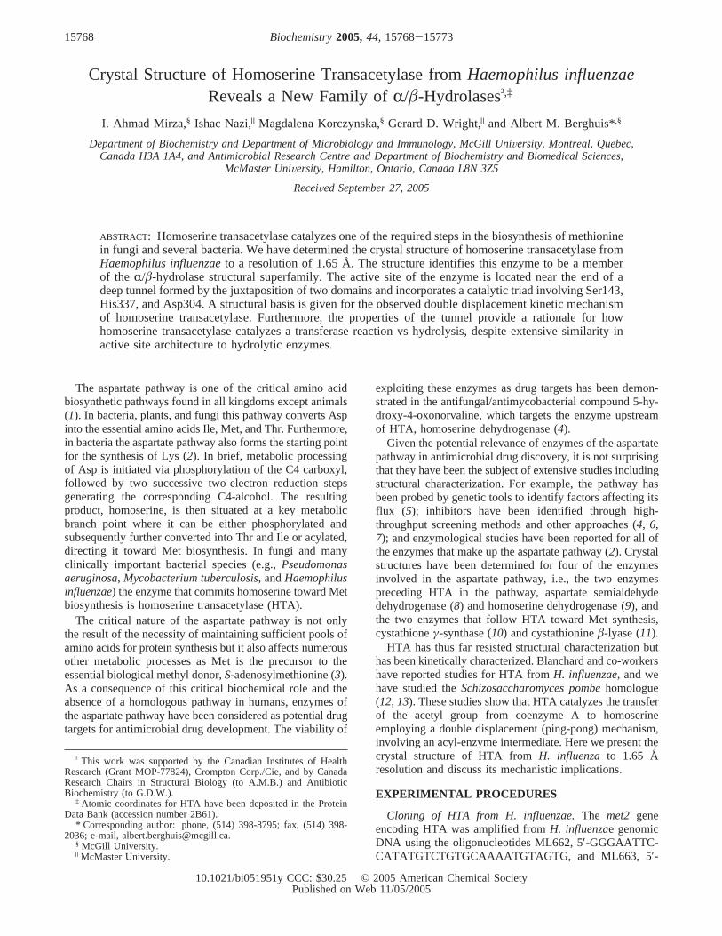
HTA Is a Member of theR/â-Hydrolase Superfamily.VAST analysis (19) reveals that the second all-R-helicaldomain possesses a unique fold. However, the firstâ-sheet-containing domain displays a fold which identifies HTA asa member of theR/â-hydrolase structural superfamily (Figure3a). TheR/â-hydrolase superfamily has been well studiedfor over 15 years (20). The canonical fold consists of aneight-stranded mainly parallelâ-sheet, in which the secondstrand is oriented in the antiparallel direction, surroundedby a total of six R-helices (21). Significant amino acidsequence similarity among the various members of thissuperfamily is effectively nonexistent, implying substantialevolutionary divergence (22). This divergence is also re-flected in the plethora of chemical transformations catalyzedby R/â-hydrolase superfamily members. The followingcatalytic activities have been observed in members of thissuperfamily: hydrolase, thioesterase, haloperoxidase, de-halogenase, and C-C bond breaking (23). HTA’s â-sheet-containing domain, hereafter referred to as theR/â-hydrolase(ABH) domain, is structurally most similar to a putativeserine hydrolase (Ydr428C) fromSaccharomyces cereVisiae(PDB code 1VKH), with an rmsd of 3.9 Å for 204 alignedresidues. When the second domain of HTA, hereafter referredto as the lid domain, is included in the structural comparison,proline iminopeptidase fromXanthomonas campestris(PDBcode 1AZW) is identified as the nearest structural homologuein the current Protein Data Bank (rmsd of 3.4 Å for 259residues). The ABH domain differs most significantly fromthe canonicalR/â-hydrolase fold in the absence ofR-helixA, which is normally located betweenâ-strands 2 and 3 butwhich is replaced in HTA by a loop that lacks secondarystructure.
It is somewhat unexpected that HTA is a member of theR/â-hydrolase superfamily. BLAST search of the amino acidsequence against the current Protein Data Bank does notreveal any significant hits. The structure-based sequencealignments of HTA to its nearest structural neighbors alsoshow percent sequence identity of 10% or less. However,bioinformatics analysis using the Conserved Domain Data-base does predict HTA to be a member of theR/â-hydrolasesuperfamily (24). As a result, several groups including ourshave speculated that HTA may possess theR/â-hydrolasefold (13, 25, 26).
Structural Basis for the Catalytic Mechanism of HTA.Comparison of all HTA sequence data presently availableindicates that the average pairwise percent identity is∼30-40% (Figure 3b). Specifically, the following residues thatare adjacent to the tunnel are absolutely conserved: Ser143,Arg212, Asp304, His337, and Asp338. Furthermore, someof these residues (Ser143, Asp304, and His337) are strongly,though not absolutely, conserved amongR/â-hydrolasesuperfamily members. Note that HTA’s from some organ-isms are significantly longer than theH. influenzaenzyme,possessing inserts between helices Lc and Ld or Le and D,which are unlikely to impact activity. Previous kinetic studiesof HTA from H. influenzaeandS. pombeshow that HTA-catalyzed acetylation of homoserine follows a double dis-placement (ping-pong) mechanism where acetyl-CoA donatesthe acetyl group to a nucleophilic residue, thus forming aninitial acyl-enzyme intermediate, followed by a transfer ofthe acetyl group to homoserine (12, 13). Mutagenesis studieswith theS. pombeenzyme have provided additional informa-tion on the nature of the nucleophilic residue and possiblemechanism of activating the nucleophile; specifically, Ser143
FIGURE 3: Comparison of HTA with members of theR/â-hydrolase superfamily and other HTA enzymes. (a) rmsd comparison of HTA vs33 diverse family members from SCOP database (29). Residues that are structurally conserved are colored blue, and those that possess nostructurally equivalent residues in otherR/â-hydrolase superfamily members are colored yellow. The scoring results are presented both asa cartoon representation and as a fingerprint (30). Structural alignments were carried out using Lsqman (31). (b) Similar to (a) except thathere sequence similarity among 24 unique HTA sequences in GenBank is mapped onto the structure. Unique sequences used in this comparisonwere obtained using Blastp. Alignments were carried out with ClustalX (32), and scoring of residues was assessed using a Gonnet 350matrix (33).
Crystal Structure of Homoserine Transacetylase Biochemistry, Vol. 44, No. 48, 200515771
was identified as the catalytic nucleophile, and residuesequivalent to H. influenzae His337 and Asp304 werepredicted to complete a catalytic triad for acyl transfer (13).By incorporating these results with the structural datapresented here and exploiting similarities with other enzymesof theR/â-hydrolase superfamily, a detailed catalytic mech-anism can be proposed.
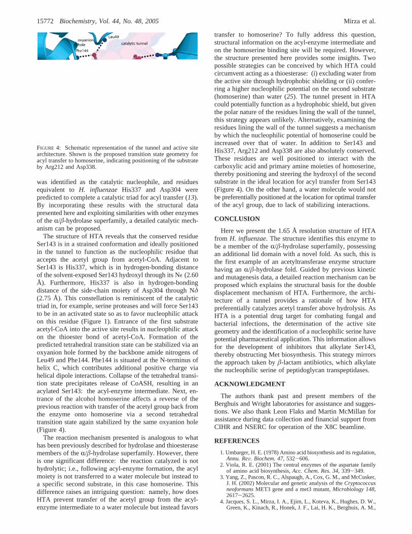
The structure of HTA reveals that the conserved residueSer143 is in a strained conformation and ideally positionedin the tunnel to function as the nucleophilic residue thataccepts the acetyl group from acetyl-CoA. Adjacent toSer143 is His337, which is in hydrogen-bonding distanceof the solvent-exposed Ser143 hydroxyl through its Nε (2.60Å). Furthermore, His337 is also in hydrogen-bondingdistance of the side-chain moiety of Asp304 through Nδ(2.75 Å). This constellation is reminiscent of the catalytictriad in, for example, serine proteases and will force Ser143to be in an activated state so as to favor nucleophilic attackon this residue (Figure 1). Entrance of the first substrateacetyl-CoA into the active site results in nucleophilic attackon the thioester bond of acetyl-CoA. Formation of thepredicted tetrahedral transition state can be stabilized via anoxyanion hole formed by the backbone amide nitrogens ofLeu49 and Phe144. Phe144 is situated at the N-terminus ofhelix C, which contributes additional positive charge viahelical dipole interactions. Collapse of the tetrahedral transi-tion state precipitates release of CoASH, resulting in anacylated Ser143: the acyl-enzyme intermediate. Next, en-trance of the alcohol homoserine affects a reverse of theprevious reaction with transfer of the acetyl group back fromthe enzyme onto homoserine via a second tetrahedraltransition state again stabilized by the same oxyanion hole(Figure 4).
The reaction mechanism presented is analogous to whathas been previously described for hydrolase and thioesterasemembers of theR/â-hydrolase superfamily. However, thereis one significant difference: the reaction catalyzed is nothydrolytic; i.e., following acyl-enzyme formation, the acylmoiety is not transferred to a water molecule but instead toa specific second substrate, in this case homoserine. Thisdifference raises an intriguing question: namely, how doesHTA prevent transfer of the acetyl group from the acyl-enzyme intermediate to a water molecule but instead favors
transfer to homoserine? To fully address this question,structural information on the acyl-enzyme intermediate andon the homoserine binding site will be required. However,the structure presented here provides some insights. Twopossible strategies can be conceived by which HTA couldcircumvent acting as a thioesterase: (i) excluding water fromthe active site through hydrophobic shielding or (ii) confer-ring a higher nucleophilic potential on the second substrate(homoserine) than water (25). The tunnel present in HTAcould potentially function as a hydrophobic shield, but giventhe polar nature of the residues lining the wall of the tunnel,this strategy appears unlikely. Alternatively, examining theresidues lining the wall of the tunnel suggests a mechanismby which the nucleophilic potential of homoserine could beincreased over that of water. In addition to Ser143 andHis337, Arg212 and Asp338 are also absolutely conserved.These residues are well positioned to interact with thecarboxylic acid and primary amine moieties of homoserine,thereby positioning and steering the hydroxyl of the secondsubstrate in the ideal location for acyl transfer from Ser143(Figure 4). On the other hand, a water molecule would notbe preferentially positioned at the location for optimal transferof the acyl group, due to lack of stabilizing interactions.
CONCLUSION
Here we present the 1.65 Å resolution structure of HTAfrom H. influenzae. The structure identifies this enzyme tobe a member of theR/â-hydrolase superfamily, possessingan additional lid domain with a novel fold. As such, this isthe first example of an acetyltransferase enzyme structurehaving anR/â-hydrolase fold. Guided by previous kineticand mutagenesis data, a detailed reaction mechanism can beproposed which explains the structural basis for the doubledisplacement mechanism of HTA. Furthermore, the archi-tecture of a tunnel provides a rationale of how HTApreferentially catalyzes acetyl transfer above hydrolysis. AsHTA is a potential drug target for combating fungal andbacterial infections, the determination of the active sitegeometry and the identification of a nucleophilic serine havepotential pharmaceutical application. This information allowsfor the development of inhibitors that alkylate Ser143,thereby obstructing Met biosynthesis. This strategy mirrorsthe approach taken byâ-lactam antibiotics, which alkylatethe nucleophilic serine of peptidoglycan transpeptidases.
ACKNOWLEDGMENT
The authors thank past and present members of theBerghuis and Wright laboratories for assistance and sugges-tions. We also thank Leon Flaks and Martin McMillan forassistance during data collection and financial support fromCIHR and NSERC for operation of the X8C beamline.
REFERENCES
1. Umbarger, H. E. (1978) Amino acid biosynthesis and its regulation,Annu. ReV. Biochem. 47, 532-606.
2. Viola, R. E. (2001) The central enzymes of the aspartate familyof amino acid biosynthesis,Acc. Chem. Res. 34, 339-349.
3. Yang, Z., Pascon, R. C., Alspaugh, A., Cox, G. M., and McCusker,J. H. (2002) Molecular and genetic analysis of theCryptococcusneoformansMET3 gene and a met3 mutant,Microbiology 148,2617-2625.
4. Jacques, S. L., Mirza, I. A., Ejim, L., Koteva, K., Hughes, D. W.,Green, K., Kinach, R., Honek, J. F., Lai, H. K., Berghuis, A. M.,
FIGURE 4: Schematic representation of the tunnel and active sitearchitecture. Shown is the proposed transition state geometry foracyl transfer to homoserine, indicating positioning of the substrateby Arg212 and Asp338.
15772 Biochemistry, Vol. 44, No. 48, 2005 Mirza et al.
and Wright, G. D. (2003) Enzyme-assisted suicide: molecularbasis for the antifungal activity of 5-hydroxy-4-oxonorvaline bypotent inhibition of homoserine dehydrogenase,Chem. Biol. 10,989-995.
5. Arevalo-Rodriguez, M., Pan, X., Boeke, J. D., and Heitman, J.(2004) FKBP12 controls aspartate pathway flux inSaccharomycescereVisiaeto prevent toxic intermediate accumulation,EukaryoticCell 3, 1287-1296.
6. Bareich, D. C., Nazi, I., and Wright, G. D. (2003) Simultaneousin vitro assay of the first four enzymes in the fungal aspartatepathway identifies a new class of aspartate kinase inhibitor,Chem.Biol. 10, 967-973.
7. Ejim, L., Mirza, I. A., Capone, C., Nazi, I., Jenkins, S., Chee, G.L., Berghuis, A. M., and Wright, G. D. (2004) New phenolicinhibitors of yeast homoserine dehydrogenase,Bioorg. Med. Chem.12, 3825-3830.
8. Hadfield, A., Kryger, G., Ouyang, J., Petsko, G. A., Ringe, D.,and Viola, R. (1999) Structure of aspartate-beta-semialdehydedehydrogenase fromEscherichia coli, a key enzyme in theaspartate family of amino acid biosynthesis,J. Mol. Biol. 289,991-1002.
9. DeLaBarre, B., Thompson, P. R., Wright, G. D., and Berghuis,A. M. (2000) Crystal structures of homoserine dehydrogenasesuggest a novel catalytic mechanism for oxidoreductases,Nat.Struct. Biol. 7, 238-244.
10. Clausen, T., Huber, R., Prade, L., Wahl, M. C., and Messerschmidt,A. (1998) Crystal structure ofEscherichia colicystathioninegamma-synthase at 1.5 A resolution,EMBO J. 17, 6827-6838.
11. Clausen, T., Huber, R., Laber, B., Pohlenz, H. D., and Messer-schmidt, A. (1996) Crystal structure of the pyridoxal-5′-phosphatedependent cystathionine beta-lyase fromEscherichia coliat 1.83Å, J. Mol. Biol. 262, 202-224.
12. Born, T. L., Franklin, M., and Blanchard, J. S. (2000) Enzyme-catalyzed acylation of homoserine: mechanistic characterizationof theHaemophilus influenzaemet2-encoded homoserine transacety-lase,Biochemistry 39, 8556-8564.
13. Nazi, I., and Wright, G. D. (2005) Catalytic mechanism of fungalhomoserine transacetylase,Biochemistry 44, 13560-13566.
14. Otwinowski, Z., and Minor, W. (1997) Processing of X-raydiffraction data collected in oscillation mode, inMethods inEnzymology, Vol. 276: Macromolecular Crystallography(Carter,C. W., Jr., and Sweet, R. M., Eds.) Part A, pp 307-326, AcademicPress, New York.
15. Terwilliger, T. C. (2003) Automated main-chain model buildingby template matching and iterative fragment extension,ActaCrystallogr., Sect. D: Biol. Crystallogr. 59, 38-44.
16. Morris, R. J., Perrakis, A., and Lamzin, V. S. (2003) ARP/wARPand automatic interpretation of protein electron density maps,Methods Enzymol. 374, 229-244.
17. Murshudov, G. N., Vagin, A. A., Lebedev, A., Wilson, K. S., andDodson, E. J. (1999) Efficient anisotropic refinement of macro-molecular structures using FFT,Acta Crystallogr., Sect. D: Biol.Crystallogr. 55(Part 1), 247-255.
18. Janin, J. (1995) Principles of protein-protein recognition fromstructure to thermodynamics,Biochimie 77, 497-505.
19. Gibrat, J. F., Madej, T., and Bryant, S. H. (1996) Surprisingsimilarities in structure comparison,Curr. Opin. Struct. Biol. 6,377-385.
20. Heikinheimo, P., Goldman, A., Jeffries, C., and Ollis, D. L. (1999)Of barn owls and bankers: a lush variety of alpha/beta hydrolases,Struct. Folding Des. 7, R141-R146.
21. Ollis, D. L., Cheah, E., Cygler, M., Dijkstra, B., Frolow, F.,Franken, S. M., Harel, M., Remington, S. J., Silman, I., Schrag,J., et al. (1992) The alpha/beta hydrolase fold,Protein Eng. 5,197-211.
22. Nardini, M., and Dijkstra, B. W. (1999) Alpha/beta hydrolase foldenzymes: the family keeps growing,Curr. Opin. Struct. Biol. 9,732-737.
23. Holmquist, M. (2000) Alpha/beta-hydrolase fold enzymes: struc-tures, functions and mechanisms,Curr. Protein Pept. Sci. 1, 209-235.
24. Marchler-Bauer, A., Anderson, J. B., Cherukuri, P. F., DeWeese-Scott, C., Geer, L. Y., Gwadz, M., He, S., Hurwitz, D. I., Jackson,J. D., Ke, Z., Lanczycki, C. J., Liebert, C. A., Liu, C., Lu, F.,Marchler, G. H., Mullokandov, M., Shoemaker, B. A., Simonyan,V., Song, J. S., Thiessen, P. A., Yamashita, R. A., Yin, J. J., Zhang,D., and Bryant, S. H. (2005) CDD: a conserved domain databasefor protein classification,Nucleic Acids Res. 33, D192-D196.
25. Milkowski, C., and Strack, D. (2004) Serine carboxypeptidase-like acyltransferases,Phytochemistry 65, 517-524.
26. Johnson, C. M., Roderick, S. L., and Cook, P. F. (2005) The serineacetyltransferase reaction: acetyl transfer from an acylpantothenyldonor to an alcohol,Arch. Biochem. Biophys. 433, 85-95.
27. DeLano, W. L. (2002)The PyMOL Molecular Graphics System,DeLano Scientific, San Carlos, CA.
28. Gilbert, D., Westhead, D., Viksna, J., and Thornton, J. (2001) Acomputer system to perform structure comparison using TOPSrepresentations of protein structure,Comput. Chem. 26, 23-30.
29. Murzin, A. G., Brenner, S. E., Hubbard, T., and Chothia, C. (1995)SCOP: a structural classification of proteins database for theinvestigation of sequences and structures,J. Mol. Biol. 247, 536-540.
30. Beitz, E. (2000) TeXshade: shading and labeling of multiplesequence alignments using LaTeX2e,Bioinformatics 16, 135-139.
31. Sierk, M. L., and Kleywegt, G. J. (2004) Deja vu all over again:finding and analyzing protein structure similarities,Structure(Camridge) 12, 2103-2111.
32. Jeanmougin, F., Thompson, J. D., Gouy, M., Higgins, D. G., andGibson, T. J. (1998) Multiple sequence alignment with ClustalX, Trends Biochem. Sci. 23, 403-405.
33. Gonnet, G. H., Cohen, M. A., and Benner, S. A. (1992) Exhaustivematching of the entire protein sequence database,Science 256,1443-1445.
BI051951Y
Crystal Structure of Homoserine Transacetylase Biochemistry, Vol. 44, No. 48, 200515773