Embed Size (px)
Citation preview
Crystal Surface Adhesion Crystal Surface Adhesion Explains the Pathological Activity Explains the Pathological Activity of Calcium Oxalate Hydrates in of Calcium Oxalate Hydrates in
Kidney Stone FormationKidney Stone Formation
Experimented by Xiaoxia Sheng*, Experimented by Xiaoxia Sheng*, Michael D. Ward*Michael D. Ward*, Jeffrey A. Wesson^, Jeffrey A. Wesson^•Department of Chemical Engineering and Materials Science, University of Minnesota, Department of Chemical Engineering and Materials Science, University of Minnesota,
Minneapolis, Minnesota; and ^ Nephrology Division, Department of Veteran Affairs medical Center Minneapolis, Minnesota; and ^ Nephrology Division, Department of Veteran Affairs medical Center and the Medical College Wisconsin, Milwaukee, Wisconsinand the Medical College Wisconsin, Milwaukee, Wisconsin
•http://jasn.asnjournals.org/cgi/content/full/16/7/1904http://jasn.asnjournals.org/cgi/content/full/16/7/1904
PowerPoint done
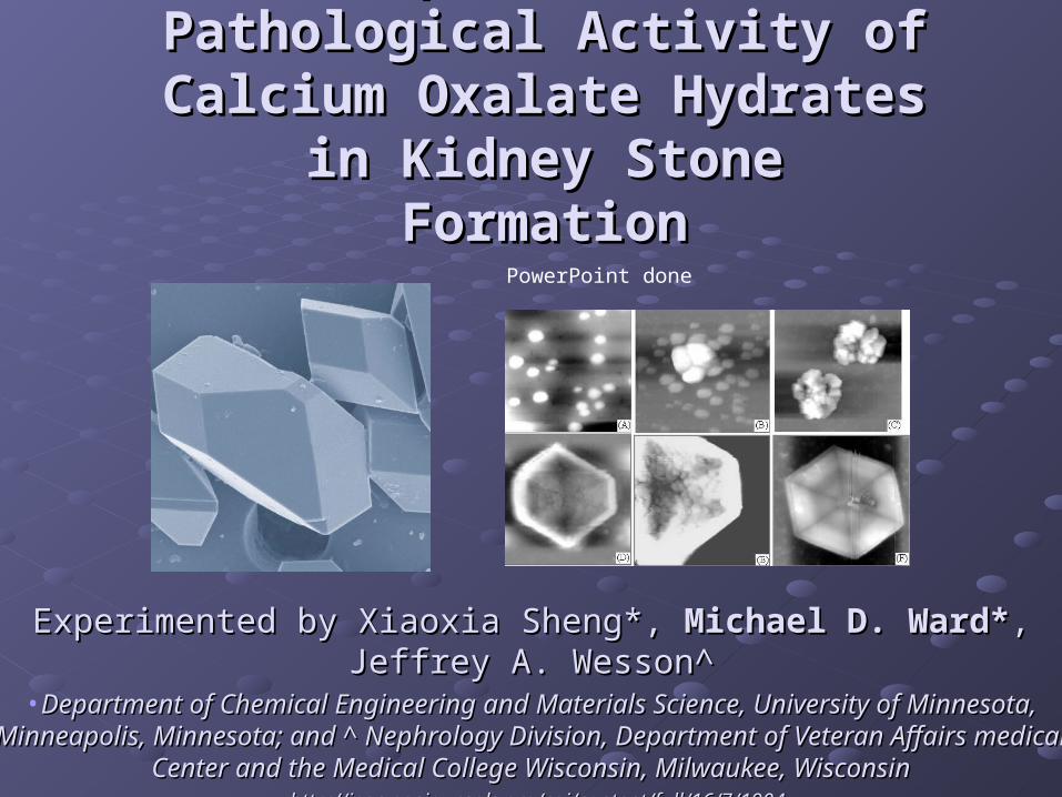
ANALYSIS OF TITLEANALYSIS OF TITLECrystal Surface Adhesion Explains the Pathological Activity Crystal Surface Adhesion Explains the Pathological Activity
of Calcium Oxalate Hydrates in Kidney Stone Formationof Calcium Oxalate Hydrates in Kidney Stone Formation
In layman terms:In layman terms:
The findings of this experiment concludes that the cluster of The findings of this experiment concludes that the cluster of crystal surfaces of calcium oxalate hydrates in the distal crystal surfaces of calcium oxalate hydrates in the distal nephrons of the kidney contributes to the process of kidney nephrons of the kidney contributes to the process of kidney stone formation. stone formation.
Kidney Stone DiseaseKidney Stone DiseaseKidney stone disease is prevalent in more than Kidney stone disease is prevalent in more than
10% of the people in the United States. It can 10% of the people in the United States. It can cause excruciating pain and occasionally, renal cause excruciating pain and occasionally, renal failure. failure.
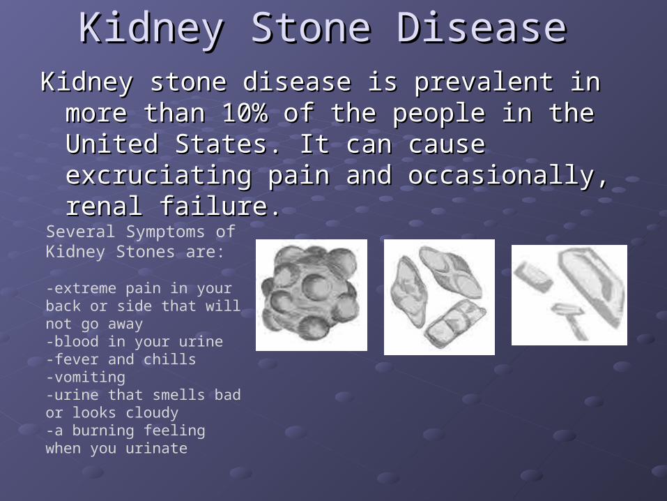
Several Symptoms of Kidney Stones are:
-extreme pain in your back or side that will not go away-blood in your urine-fever and chills-vomiting-urine that smells bad or looks cloudy-a burning feeling when you urinate
Calcium Oxalate HydratesCalcium Oxalate HydratesThere are two predominant types of Calcium Oxalates:There are two predominant types of Calcium Oxalates:
Calcium Oxalate Monohydrate (COM) is the more common of the two that Calcium Oxalate Monohydrate (COM) is the more common of the two that helps form the kidney stones. These oxalates are inorganic and are picked helps form the kidney stones. These oxalates are inorganic and are picked up from our food. Kidney stones are aggregates, or a cluster of something. up from our food. Kidney stones are aggregates, or a cluster of something. These stones have proteins that make them adhesive with one another and These stones have proteins that make them adhesive with one another and this is what causes the formation of kidney stones.this is what causes the formation of kidney stones.
The less predominant of the two oxalates found in the kidney is Calcium The less predominant of the two oxalates found in the kidney is Calcium Oxalate Dihydrate (COD). These types of oxalates are actually the benign Oxalate Dihydrate (COD). These types of oxalates are actually the benign ones when compared to the monohydrates. The crystal of a COD is the size ones when compared to the monohydrates. The crystal of a COD is the size of a single micron. This actually prevents kidney stone development of a single micron. This actually prevents kidney stone development because it cannot form stable aggregates and strong adhesions to the renal because it cannot form stable aggregates and strong adhesions to the renal walls. walls.
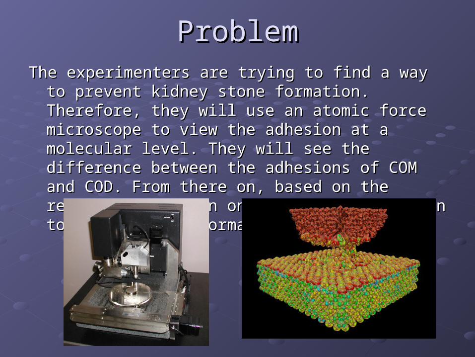
ProblemProblemThe experimenters are trying to find a way to prevent The experimenters are trying to find a way to prevent
kidney stone formation. Therefore, they will use an kidney stone formation. Therefore, they will use an atomic force microscope to view the adhesion at a atomic force microscope to view the adhesion at a molecular level. They will see the difference between the molecular level. They will see the difference between the adhesions of COM and COD. From there on, based on adhesions of COM and COD. From there on, based on the results, they plan on finding a resolution to kidney the results, they plan on finding a resolution to kidney stone formation. stone formation.
MaterialsMaterials
COM crystals used for adhesion force measurements were grownCOM crystals used for adhesion force measurements were grown in vitroin vitro
COD crystalsCOD crystals with large (100) faces were grown in aqueous solutions with large (100) faces were grown in aqueous solutions containingcontaining 1.0 mM CaOx, 10 µg/ml poly(acrylic acid), 10 mM HEPES1.0 mM CaOx, 10 µg/ml poly(acrylic acid), 10 mM HEPES buffer, buffer, and 150 mM sodium chlorideand 150 mM sodium chloride
COD crystals with large (101) faces were grown in 1.0 mMCOD crystals with large (101) faces were grown in 1.0 mM CaOx and 5 CaOx and 5 µg/ml poly(acrylic acid), in the absence ofµg/ml poly(acrylic acid), in the absence of buffer and saltbuffer and salt
DigitalDigital Instruments Nanoscope IIIa Multimode systemInstruments Nanoscope IIIa Multimode system
aqueousaqueous solutions saturated with CaOx at approximately pH 7solutions saturated with CaOx at approximately pH 7
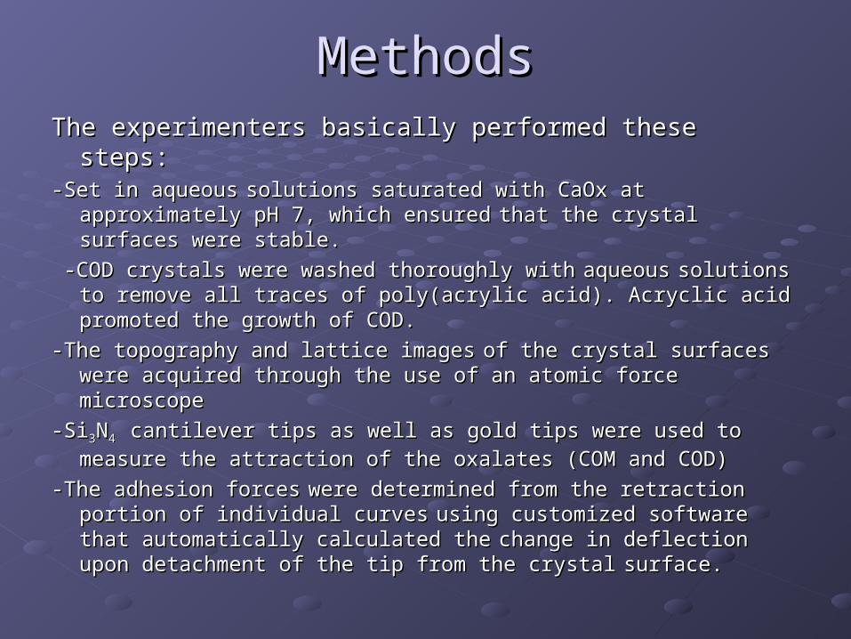
MethodsMethodsThe experimenters basically performed these steps:The experimenters basically performed these steps:-Set in aqueous-Set in aqueous solutions saturated with CaOx at approximately pH 7, solutions saturated with CaOx at approximately pH 7,
which ensuredwhich ensured that the crystal surfaces were stable.that the crystal surfaces were stable.
-COD crystals were washed thoroughly with-COD crystals were washed thoroughly with aqueousaqueous solutions to solutions to remove all traces of poly(acrylic acid). Acryclic acid promoted the remove all traces of poly(acrylic acid). Acryclic acid promoted the growth of COD.growth of COD.
-The topography and lattice images-The topography and lattice images of the crystal surfaces were of the crystal surfaces were acquired through the use of an atomic force microscopeacquired through the use of an atomic force microscope
-Si-Si33NN44 cantilever tips as well as gold tips were used to measure the cantilever tips as well as gold tips were used to measure the
attraction of the oxalates (COM and COD)attraction of the oxalates (COM and COD)
-The adhesion forces-The adhesion forces were determined from the retraction portion of were determined from the retraction portion of individual curvesindividual curves using customized software that automatically using customized software that automatically calculated thecalculated the change in deflection upon detachment of the tip from change in deflection upon detachment of the tip from the crystalthe crystal surface.surface.
Experimental and Control GroupsExperimental and Control Groups
Experimental Group:Experimental Group:
The experimental groups were the types of calcium oxalate hydrates. These The experimental groups were the types of calcium oxalate hydrates. These included that of Calcium Oxalate Monohydrate(COM) and Calcium Oxalate included that of Calcium Oxalate Monohydrate(COM) and Calcium Oxalate Dihydrate (COD). These were the materials being tested and the results Dihydrate (COD). These were the materials being tested and the results came directly from the testing of these materials.came directly from the testing of these materials.
Control Groups:Control Groups:
The control groups were in vitro systems with no COD present. Also, what was The control groups were in vitro systems with no COD present. Also, what was used as a control were the aqueousused as a control were the aqueous solutions saturated with CaOx at solutions saturated with CaOx at approximately pH 7, which ensuredapproximately pH 7, which ensured that the crystal surfaces were stable. that the crystal surfaces were stable.

ResultsResults
This diagram is a product of the atomic force microscope and the lower right depicts a contact between a calcium oxalate (CaOx) surface and an idealized hemispherical gold-coated AFM tip modified with a monolayer of organosulfur molecules. The radius of curvature of the tip and the gold thickness are approximately 75 and 50 nm, respectively, and the adhesion force measurements were performed in aqueous solutions saturated with CaOx.
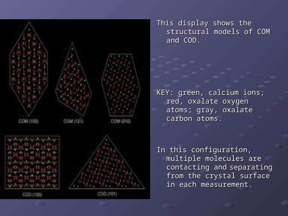
This display shows the structural This display shows the structural models of COM and COD. models of COM and COD.
KEY: green, calcium ions; red, KEY: green, calcium ions; red, oxalate oxygen atoms; gray, oxalate oxygen atoms; gray, oxalate carbon atoms. oxalate carbon atoms.
In this configuration, multiple In this configuration, multiple molecules are contacting andmolecules are contacting and
separating from the crystal separating from the crystal surface in each surface in each measurement.measurement.
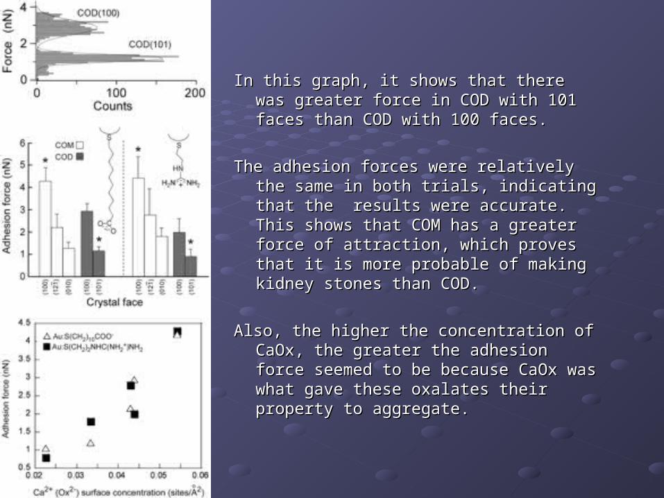
In this graph, it shows that there was In this graph, it shows that there was greater force in COD with 101 faces than greater force in COD with 101 faces than COD with 100 faces. COD with 100 faces.
The adhesion forces were relatively the The adhesion forces were relatively the same in both trials, indicating that the same in both trials, indicating that the results were accurate. This shows that results were accurate. This shows that COM has a greater force of attraction, COM has a greater force of attraction, which proves that it is more probable of which proves that it is more probable of making kidney stones than COD.making kidney stones than COD.
Also, the higher the concentration of CaOx, Also, the higher the concentration of CaOx, the greater the adhesion force seemed to the greater the adhesion force seemed to be because CaOx was what gave these be because CaOx was what gave these oxalates their property to aggregate. oxalates their property to aggregate.
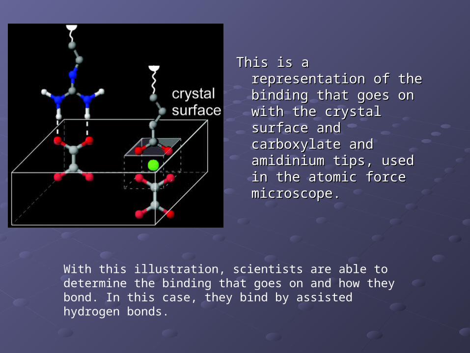
This is a representation of This is a representation of the binding that goes on the binding that goes on with the crystal surface with the crystal surface and carboxylate and and carboxylate and amidinium tips, used in amidinium tips, used in the atomic force the atomic force microscope.microscope.
With this illustration, scientists are able to determine the binding that goes on and how they bond. In this case, they bind by assisted hydrogen bonds.
DiscussionDiscussion
This experiment shows that COM aggregates more easily than This experiment shows that COM aggregates more easily than COD. COM is the primary cause of kidney stone formation. COD. COM is the primary cause of kidney stone formation. COD is a micro sized crystal that is easily released when one is COD is a micro sized crystal that is easily released when one is urinating. This oxalate is very benign and can seldom cause urinating. This oxalate is very benign and can seldom cause any complications. any complications.
Some questions that were present were: Some questions that were present were:
How were the COMs and CODs consistent to the ones found in How were the COMs and CODs consistent to the ones found in the kidneys? the kidneys?
Surprises: I was surprised that the COM (100) was the pnly Surprises: I was surprised that the COM (100) was the pnly oxalate responsible of having a great adhesion force when oxalate responsible of having a great adhesion force when compared to all other types of oxalates.compared to all other types of oxalates.
ConclusionConclusion
Michael D. Ward and his team concluded from the experiment that the Michael D. Ward and his team concluded from the experiment that the agent responsible for truly forming kidney stones was COM. The agent responsible for truly forming kidney stones was COM. The COD is a micro sized crystal that has little or nothing to do with the COD is a micro sized crystal that has little or nothing to do with the aggregation of kidney stone formation. COM with 100 faces is the aggregation of kidney stone formation. COM with 100 faces is the most potent of aggregating with each other and this is the leading most potent of aggregating with each other and this is the leading method of kidney stone formation. The main determinant of method of kidney stone formation. The main determinant of adhesion is the number of binding sites there are for the crystals to adhesion is the number of binding sites there are for the crystals to
aggregate.aggregate.
COD aggregates are able to protect against kidney COD aggregates are able to protect against kidney stone formation because of their weak adhesive stone formation because of their weak adhesive attraction and because if its instability (less attraction and because if its instability (less tendency to form stones).tendency to form stones).
Further ExperimentationFurther Experimentation
What to do next:What to do next:- Measure the adhesion force of the COM and COD with increasing amounts Measure the adhesion force of the COM and COD with increasing amounts
of CaOx solution. To see if there is a relationship between the two materials. of CaOx solution. To see if there is a relationship between the two materials.
- Place the COM and COD crystals in different pH solutions. The experiment Place the COM and COD crystals in different pH solutions. The experiment was set in 7.0 pH, neutrality. But if we test the pH differentiation with the was set in 7.0 pH, neutrality. But if we test the pH differentiation with the crystal aggregation, then there may be different adhesive forces based on crystal aggregation, then there may be different adhesive forces based on acidity or base.acidity or base.
- Perform this experiment with different urinary species to see if the species Perform this experiment with different urinary species to see if the species have anything to do with the COM or COD aggregates into kidney stone have anything to do with the COM or COD aggregates into kidney stone formation. This would help to identify the key factors responsible for stone formation. This would help to identify the key factors responsible for stone disease and can help create therapies. disease and can help create therapies.

CitationsCitations
No other sources usedNo other sources used


![Reduction of Oxalate Levels in Tomato Fruit and … of Oxalate Levels in Tomato Fruit and Consequent Metabolic Remodeling Following Overexpression of a Fungal Oxalate Decarboxylase1[W]](https://img.pdfslide.net/doc/110x75/5af8e5787f8b9aff288c704b/reduction-of-oxalate-levels-in-tomato-fruit-and-of-oxalate-levels-in-tomato.jpg)