Embed Size (px)
Citation preview
University of KentuckyUKnowledge
Molecular and Cellular Biochemistry FacultyPatents Molecular and Cellular Biochemistry
11-4-2008
Crystallization and Structure of a Plant PeptideDeformylaseRobert L. HoutzUniversity of Kentucky, [email protected]
David W. RodgersUniversity of Kentucky, [email protected]
Lynnette M. A. DirkUniversity of Kentucky, [email protected]
Mark A. WilliamsUniversity of Kentucky, [email protected]
Right click to open a feedback form in a new tab to let us know how this document benefits you.
Follow this and additional works at: https://uknowledge.uky.edu/biochem_patents
Part of the Medical Biochemistry Commons
This Patent is brought to you for free and open access by the Molecular and Cellular Biochemistry at UKnowledge. It has been accepted for inclusion inMolecular and Cellular Biochemistry Faculty Patents by an authorized administrator of UKnowledge. For more information, please [email protected].
Recommended CitationHoutz, Robert L.; Rodgers, David W.; Dirk, Lynnette M. A.; and Williams, Mark A., "Crystallization and Structure of a Plant PeptideDeformylase" (2008). Molecular and Cellular Biochemistry Faculty Patents. 3.https://uknowledge.uky.edu/biochem_patents/3
(12) United States Patent Houtz et al.
US007445923B2
US 7,445,923 B2 Nov. 4, 2008
(10) Patent N0.: (45) Date of Patent:
(54) CRYSTALLIZATION AND STRUCTURE OF A PLANT PEPTIDE DEFORMYLASE
(75) Inventors: Robert L. Houtz, Lexington, KY (US); David W. Rodgers, Versailles, KY (U S); Lynette M. A. Dirk, Lexington, KY (US); Mark A. Williams, Lexington, KY (Us)
(73) Assignee: University of Kentucky Research Foundation, Lexington, KY (U S)
( * ) Notice: Subject to any disclaimer, the term of this patent is extended or adjusted under 35 U.S.C. 154(b) by 0 days.
(21) Appl.No.: 11542989
(22) Filed: Oct. 3, 2006
(65) Prior Publication Data
US 2008/0124808 A1 May 29, 2008
Related U.S. Application Data
(60) Provisional application No. 60/835,823, ?led on Aug. 4, 2006.
(51) Int. Cl. C12N 9/78 (2006.01) G01N 31/00 (2006.01)
(52) U.S. Cl. ......................................... .. 435/227; 436/4
(58) Field of Classi?cation Search ..................... .. None
See application ?le for complete search history.
(56) References Cited
U.S. PATENT DOCUMENTS
5,985,273 A 11/1999 Reed et al. 6,730,634 B1 5/2004 HoutZ et al. 6,864,080 B2 3/2005 Baldwin et al.
2004/0088755 A1 5/2004 HoutZ et al.
OTHER PUBLICATIONS
A Hypertext Book of Crystallographic Space Group Diagrams and Tables, Birkbeck College, University of London, 1997-1999, Retrieved from the Internet <URL: http://img.chem.ucl.ac.uk/sgp/ mainmenu.htm>.*
McPherson et al., Eur. J. Biochem. 189:1-23, 1990.* KierZek et al., Biophys Chem 91 :1-20, 2001.* Branden et al., “Introduction to Protein Structure Second Edition”, Garland Publishing Inc., New York, 1999* Drenth, “Principles of X-ray Crystallography,” Springer, New York, 1995* Fieulaine et al., The crystal structure of mitochondrial (Type IA) peptide deformylase provides clear guidelines for the design of inhibitors speci?c for the bacterial forms, Journal of Biological Chemistry, published Sep. 28, 2005, vol. 280, No. 51, p. 42315 42624.* Dardel et al., Solution structure of nickel-peptide deformylase, J. Mol. Biol. (1998) 280, 501-513.* Chan et al., Crystal Structure of the Escherichia coli Peptide Deformylase, Biochemistry, 1997, 36, 13904-13909.* De?nition of “ligand” Retrieved from the Internet <URL:http:// stedmans.com/> on Oct. 29, 2007.* Akers, Alan, “Molecular Biology as Virtual Biology: Limitations of Molecular Biology in Pesticide Discovery”, 1996, pp. 85-91, vol. 46, Pesticide Science, Great Britain. Braun, et al. Puri?cation and sequencing of cytochrome b from potato reveals methionine cleavage of mitochondrially encoded protein, FEBS Letters 316: 128-132, 1993. Chen, et a., “Actinonin, a Naturally Occurring Antibacterial Agent, Is a Potent Deformylase Inhibitor”, Biochemistry 2000, vol. 39, pp. 1256-1262, American Cancer Society, Washington, DC. Giglione et al., “Identi?cation of eukaryotic peptide deformylases reveals universality of N-terminal protein processing mechanisms”, The EMBO Journal, Sep. 13, 2000, pp. 5916-5929, vol. 19, No. 21, European Molecular Biology Organization, France. Giglione, et al. “Peptide deformylase as a target for new generation, broad spectrum antimicrobial agents”, Microreview, Molecular Microbiology, 2000, 36(6):1197-1205, Blackwell Science, Ltd., Oxford, England. Meinnel, T., “Peptide deformylasae of Eukaryotic Protists: A Target for New Antiparasitic Agents?” Parasitology Today 200, pp. 156 168, vol. 16, No. 4 Elsevier Science, Ltd., Oxford, England.
* cited by examiner
Primary ExamineriDavid J. Steadman Assistant ExamineriJae W Lee (74) Attorney, Agent, or Firm4CroWell & Moring, LLP
(57) ABSTRACT
This invention relates to the crystal structure of a plant peptide deformylase polypeptide and methods of using the structure to design compounds that modulate the activity of the polypeptide.
4 Claims, 15 Drawing Sheets
US. Patent Nov. 4, 2008 Sheet 2 0f 15 US 7,445,923 B2
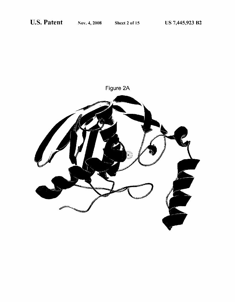
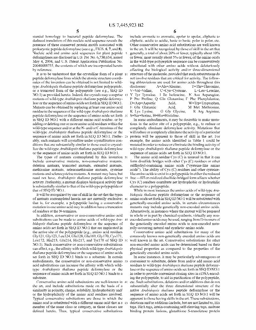
Figure 2A
US. Patent Nov. 4, 2008 Sheet 3 0f 15 US 7,445,923 B2
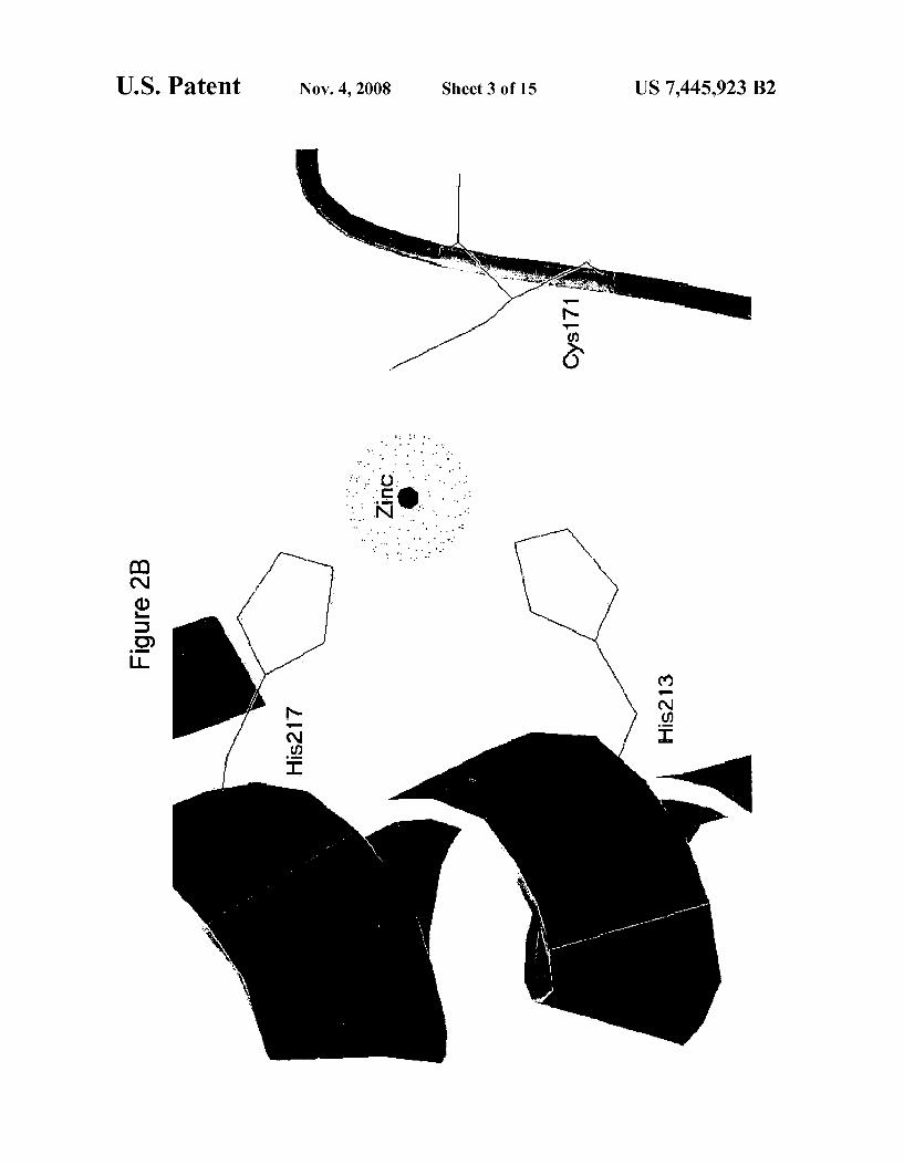
Figure 2B
US. Patent Nov. 4, 2008 Sheet 4 0f 15 US 7,445,923 B2
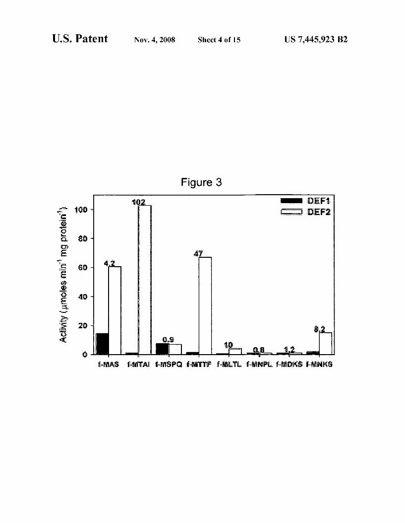
Figure 3
180*
me a
a — q it“ $.32
US. Patent US 7,445,923 B2
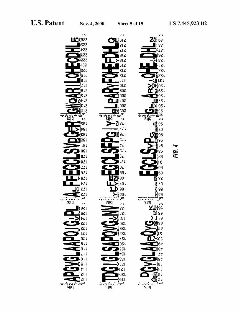
FIG. 4
US. Patent Nov. 4, 2008 Sheet 6 0f 15 US 7,445,923 B2
US. Patent Nov. 4, 2008 Sheet 11 0f 15 US 7,445,923 B2
m .525 M 6t
5
E
@Q
Q
55.6 55m
Q
m m >
5
5
5 5
H 5m: 25 a 85% 553% E 2:52
9:
m2
at
ME
mt
E
mt
E
Q:
m2
SF
32 $8
$352 55% 8533 mm _ 3533 $52
US. Patent Nov. 4, 2008 Sheet 12 0f 15 US 7,445,923 B2
u .ESE N GE 0 Nn mm mm mm mm 8 IN In IN IN IN IN IN IN IN I IBIQ 55w
2 I >
F >>
N F >
N
N F I P m
N In N I I
N In N I
IN I
_ N z
I
I IN 5 ._
N I
I P _
3 In _ I
m: I
IN In F m I
I IN I I
IN N I
Q
g N . <
> I I I I I > I I I I I < I I w m
Nm mm mm mm IN IN IN IN IN In In 3” Em EN 3m
68? IE a 85% $0553 s 252
INN EN EN tN IN IN 3N 2N NFN :N ON @IN 2N 8N EON EN EIIEI
EEC? 8533 NI _ 85:88:?
_= 522
US. Patent N v. 4, 2008 Sheet 13 0f 15 US 7,445,923 B2
444 444 444 444 444 444 444 444 444 444 444 444 444 44 44 4 4 444444444
4 4 4 4
N 4 44 4 44 44 4 4 44 >
4 44 4 4 N 4
4N 4 4 4 4 4 4
4 4 4N 4
44 4 4 44 4 N 4
4N 44 44 4
4 4 44 44 4 4 4
4 N 4 44
4N 44 44 44 44 44 4
44 N 4 4 N 4
4 44 44 44 44 44 N 4 _
4 N 4 4 4 4
4 44 N 4 44 44 4 4
N 4
4 4 4 44 4 4
4 44 44 4
4 N. N 4
4 4 4 44 N4 4 4 4 44 4
4 4 4 4 4 4 4 4 _ 4 4 4
444 444 444 444 444 444 444 444 444 444 444 444 _444 _44 _44 _44 44
444444444 4444 44 4444444 444444444 44 4444442
44 44 44 44 N4 44 44 44 44 44 44 44 _44 _44 _N4 _44 4444444444444 4444444444444 4444444444 44444 4444 4 444444 444444444444 44444 4444 44 44444444 4444444 444.444 4444 4444 44244 44 4444444444 44444444 444 4444 44444 44444 444 444 4 444445 4444444444 44444444 444
_ _ _ _ _ _ _ _ _ 4444444444444 444 44 444444444 444 444444 4444444 444 44444 444444 44444 44444 _ _4444-444>N4
4444244
US 7,445,923 B2 1
CRYSTALLIZATION AND STRUCTURE OF A PLANT PEPTIDE DEFORMYLASE
CROSS REFERENCE TO RELATED APPLICATIONS
This application claims priority to US. Provisional Appli cation Ser. No. 60/835,823 ?led Aug. 4, 2006, the disclosure of Which is incorporated herein by reference.
STATEMENTS REGARDING FEDERALLY SPONSORED RESEARCH
The invention Was funded in part by Grant No. MCB MCB-0240165 aWarded by the National Science Foundation (NSF). The government may have certain rights in the inven tion.
TECHNICAL FIELD
This invention relates to the crystallization and structure of plant peptide deformylase and methods of using the structure.
BACKGROUND
Peptide deformylase (DEF; EC 3.5.1.88) is a metallopep tidase that catalyzes the removal of an N-formyl group from N-formyl methionine, Which is the initiating amino acid resi due for prokaryotically translated proteins. DEF is an essen tial enzyme and mutations, deletions, or insertions in the DEF gene, or inhibition of enzymatic activity, are lethal to prokary otic organisms. For decades DEF Was believed to be exclu sively restricted to prokaryotes because protein translation in eukaryotic organisms initiates With an unformylated methionine residue. The restriction to prokaryotic organisms and the essentiality of DEF have made this enzyme the molecular target of many research efforts directed toWards the development of broad-spectrum antibiotics, Which Would have little or no mammalian toxicity. In 2000 the existence of DEF in the chloroplasts of higher plants Was reported, and it Was also discovered that actinonin, a potent inhibitor of DEF, Was phytotoxic to all plant species. The lethality of actinonin to a Wide range of plants, including many agriculturally sig ni?cant Weed species, suggests that DEF is an essential and highly conserved enzyme in plants, and inhibitors targeting this enzyme could potentially serve as a neW class of broad spectrum herbicides as Well as selectable markers.
Accordingly, plant peptide deformylase (DEF) polypep tides provide an attractive target for crystallization and struc tural studies Which can lead to the identi?cation and synthesis of neW broad-spectrum herbicides and selectable markers With high speci?city toWards plant DEF.
SUMMARY
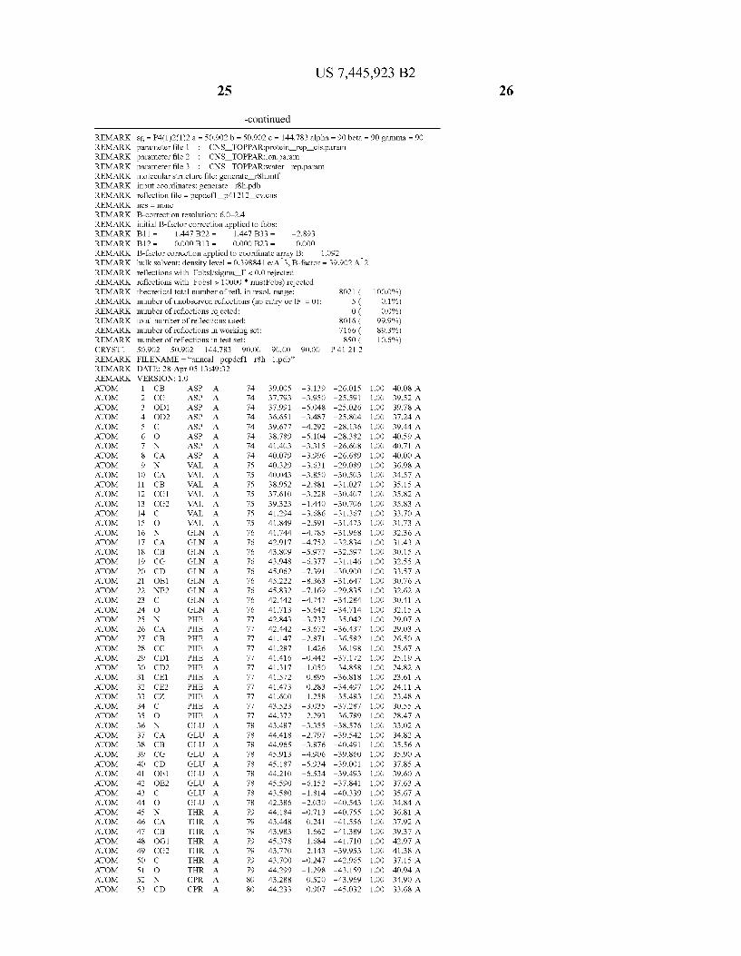
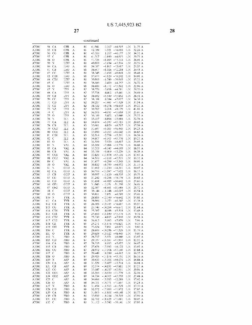
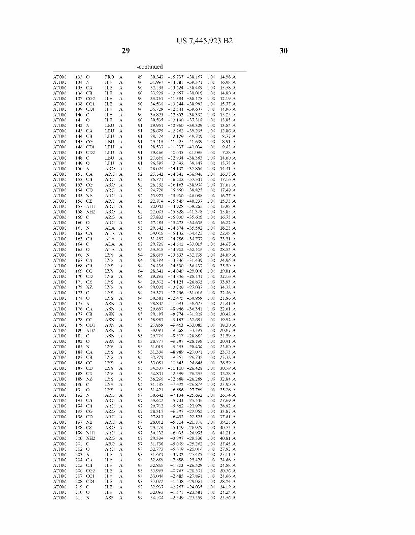
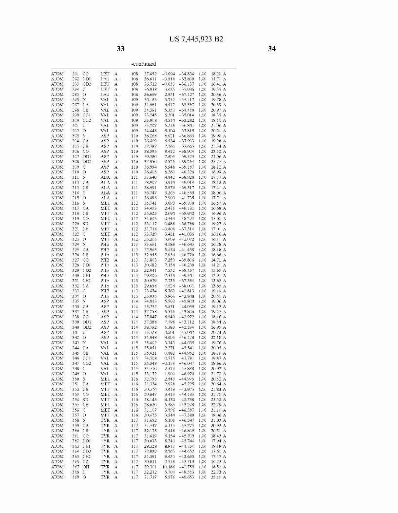
Provided herein are crystalline forms of a peptide deformy lase, and atomic coordinates derived therefrom, useful for designing and identifying compounds that modulate the activity of the peptide deformylase. Accordingly, in one embodiment, a crystalline form of a polypeptide comprising the amino acid residues of SEQ ID N011, is provided. In some aspects, the crystalline form includes a structure character ized by tetragonal space group symmetry P412l2 and unit cell of dimensions a, b, and c. In some aspects, a is about 40 A to about 60 A, b is about 40 A to about 60 A, and c is about 120 A to about 160 A. In other aspects, (XIBIYI9OO. In some aspects, the polypeptide is a peptide deformylase isolated from Arabidopsis Zhaliana.
20
25
30
35
40
45
50
55
60
65
2 In some embodiments, the crystalline form includes a coor
dinated metal ion selected from the group of consisting of Fe, Zn, and Ni, and any combination thereof. In one aspect, the metal ion is coordinated by amino acid residues Cys171, His2l3, and His2l7 of SEQ ID N011.
In another embodiment, a crystalline form of a polypeptide including a structure de?ned by one or more structure coor
dinates of Arabidopsis Zhaliana peptide deformylase amino acid residues Gly121, Glyl23, Leul24, Gln128, Glul69, Gly170, Cys171, Leul72, His2l3, Glu214, His2l7, and Tyr178 according to Table 1, is provided. In general, struc tures derived from these crystalline forms encompass struc tures having coordinates that differ by a root mean square deviation ofless than about 1.5 A, 0.75 A, or 0.35 A, or any deviation in this range, When superimposed on the non-hy drogen atom positions of the corresponding atomic coordi nates of Table 1. In some aspects, amino acid residues Gly121, Gly123,Leu124,Gln128, Glul69, Gly170,Cys171, Leul72, His2l3, Glu2l4, His2l7, and Tyr178 include the active site of the peptide deformylase. In some aspects, the polypeptide includes an amino acid sequence having at least 75%, at least 85%, or at least 95%, or any percent in this range, amino acid sequence identity to SEQ ID N011.
In other embodiments, a crystalline form of a polypeptide provided herein also includes a ligand complexed With the polypeptide. In some aspects, the ligand is a small molecule.
In another embodiment, a crystalline form of a polypeptide that includes the amino acid residues of SEQ ID N011 and an atomic structure characterized by the coordinates of Table 1, is provided.
In yet another embodiment, a machine-readable medium embedded With information that corresponds to a three-di mensional structural representation of a crystalline form of a polypeptide as provided herein.
In one embodiment, a computer system including a data base containing information on the three dimensional struc ture of a crystalline form of an Arabidopsis Zhaliana peptide deformylase polypeptide and a user interface to vieW the information, is provided. In some aspects, the computer sys tem includes information related to diffraction data obtained from a crystalline form comprising SEQ ID N011. In other aspects, the computer system of includes information related to an electron density map of a crystal comprising SEQ ID N011.
In another aspect, a computer system provided herein includes information related to the structure coordinates of Table 1 or homologous structure coordinates for the amino acid residues of SEQ ID N011 that have a root mean square deviation of non-hydrogen atoms of less than about 1.5 A, 0.75 A, 0.35 A, or any percent in this range, When superim posed on the non-hydrogen atom positions of the correspond ing atomic coordinates of Table 1.
In other aspects, a computer system provided herein includes information related to the structure coordinates for one or more amino acid residues Gly121, Glyl23, Leul24, Glnl28,Glul69,Gly170,Cys171,Leu172,His213,Glu214, His2l7, and Tyr178 according to Table 1, or similar structure coordinates for the amino acids including a root mean square deviation of non-hydrogen atoms of less than about 1.5 A, 0.75 A, 0.35 A, or any percent in this range, When superim posed on the non-hydrogen atom positions of the correspond ing atomic coordinates of Table 1.
In another embodiment, a method of identifying a candi date compound that binds to the active site of Arabidopsis Zhaliana peptide deformylase polypeptide, is provided. The method includes comparing the atomic structure of the com pound With a three-dimensional structure of a crystalline
US 7,445,923 B2 3
form of an Arabidopsis Zhaliana peptide deformylase polypeptide and computationally identifying a candidate compound for an ability to bind to the Arabidopsis Zhaliana peptide deformylase. In some aspects, the candidate com pound binds to the active site of the Arabidopsis Zhaliana peptide deformylase. In other aspects, comparing the atomic structure of the compound With a three-dimensional structure of a crystalline form of an Arabidopsis Zhaliana peptide deformylase polypeptide includes employing a computa tional means to perform a ?tting operation betWeen the com pound and at least one binding site of the peptide deformy lase.
In some embodiments, the candidate compound identi?ed by a computational method provided herein can be synthe siZed and screened for the ability to bind a plant peptide deformylase in vitro or in vivo. In some aspects, the com pound is an herbicide.
In another embodiment, a method of identifying a candi date compound that binds to the active site of Arabidopsis Zhaliana peptide deformylase polypeptide, is provided. The method includes comparing the atomic structure of the com pound With a three-dimensional structural representation of a crystalline form provided herein and computationally identi fying a candidate compound for an ability to bind to the active site of Arabidopsis Zhaliana peptide deformylase.
In yet another embodiment, a method of computationally designing a candidate compound that binds to Arabidopsis Zhaliana peptide deformylase polypeptide, is provided. The method includes comparing the atomic structure of chemical entities, or fragments thereof, With a three-dimensional struc tural representation of a crystalline form of a polypeptide provided herein; identifying chemical entities capable of associating With the three-dimensional structural representa tion of a crystalline form of a polypeptide; and assembling the chemical entities, or fragments thereof, into a single molecule to provide the structure of the candidate compound. In some aspects, the candidate compound binds to the active site of Arabidopsis Zhaliana peptide deformylase.
In another embodiment, a method of identifying a region of Arabidopsis Zhaliana peptide deformylase polypeptide that contacts a compound, is provided. The method includes obtaining X-ray diffraction data for a crystal of Arabidopsis Zhaliana peptide deformylase; obtaining X-ray diffraction data for a complex of a Arabidopsis Zhaliana peptide deformylase and the compound; subtracting the X-ray dif fraction data from the peptide deformylase With the X-ray diffraction data obtained from the complex to obtain the dif ference in the X-ray diffraction data; obtaining phases that correspond to X-ray diffraction data obtained for the peptide deformylase; correlating the data to generate a difference Fourier image of the compound; and locating the region of Arabidopsis Zhaliana peptide deformylase contacted by the compound. In some aspects, the compound is actinonin.
In another embodiment, a method of modifying an inhibi tor of Arabidopsis Zhaliana peptide deformylase activity, is provided. The method includes obtaining a crystal including an Arabidopsis Zhaliana peptide deformylase polypeptide and an inhibitor; obtaining the atomic coordinates of the crystal; correlating the atomic coordinate data With one or more molecular modeling techniques; identifying at least one modi?cation predicted to effect the interaction of the inhibi tor With the polypeptide; and modifying the inhibitor based on the prediction. In one aspect, the modi?cation is a com puter generated modi?cation. In other aspects, the modi?ca tion is a physical modi?cation made to the structure of the inhibitor. In one aspect, the crystal comprises the amino acid residues of SEQ ID NO: 1.
20
25
30
35
40
45
50
55
60
65
4 In other aspects, the one or more molecular modeling tech
niques are selected from the group consisting of graphic molecular modeling and computational chemistry. In another aspect, obtaining the atomic coordinates of the crystal includes detecting the interaction of the inhibitor to one or more amino acid residues Glyl 2 l, Glyl23, Leul 24, Glnl28, Glul69, Glyl70, Cys17l, Leu172, His2l3, Glu2l4, His2l7, and Tyrl78 of SEQ ID NO:l. The details of one or more embodiments of the disclosure
are set forth in the accompanying draWings and the descrip tion beloW. Other features, objects, and advantages Will be apparent from the description and draWings, and from the claims.
BRIEF DESCRIPTION OF DRAWINGS
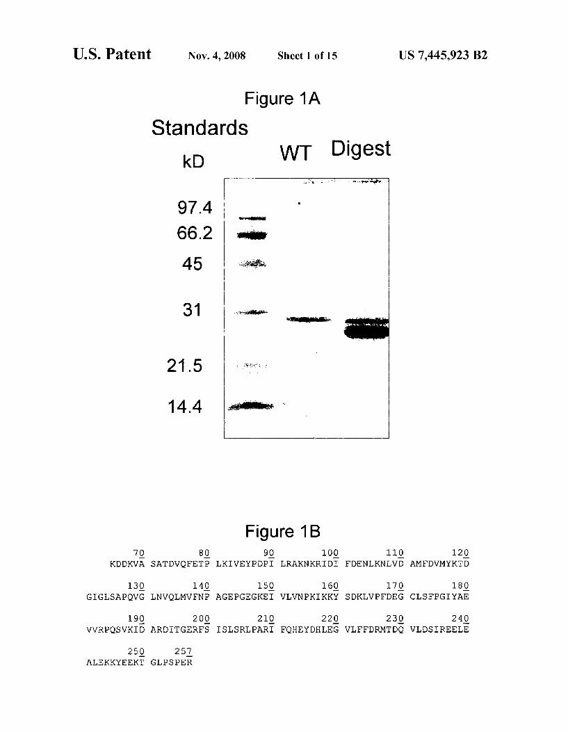
FIG. 1A depicts a polyacrylamide gel shoWing that limited trypsinolysis creates a core protein that retained activity and remained soluble in the absence of high salt concentrations.
FIG. 1B depicts the amino acid sequence of the AtDEF peptide (SEQ ID NO: 1).
FIG. 2A depicts a ribbon representation of crystallized AtDEF2. The cylinders represent helices and the arroWs rep resent sheets.
FIG. 2B depicts a slab vieW of the ribbon representation of trypsinolyZed AtDEF2 highlighting the active-site-metal binding ligands (l Cys and 2 His) from motifs II and III, respectively (EGCLS and QHEXXH) (SEQ ID NOS 15-16).
FIG. 3 depicts a graph of substrate speci?city comparison of AtDEFl and AtDEF2.
FIG. 4 depicts a comparison of amino acid sequence con servation of the three motifs in AtDEFl and 2 and bacterial DEFs (SEQ ID NOS 2-5).
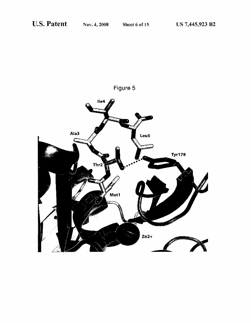
FIG. 5 depicts a molecular model of the N-terminal resi dues from the D1 polypeptide docked into the active site of Arabidopsis Zhaliana peptide deformylase.
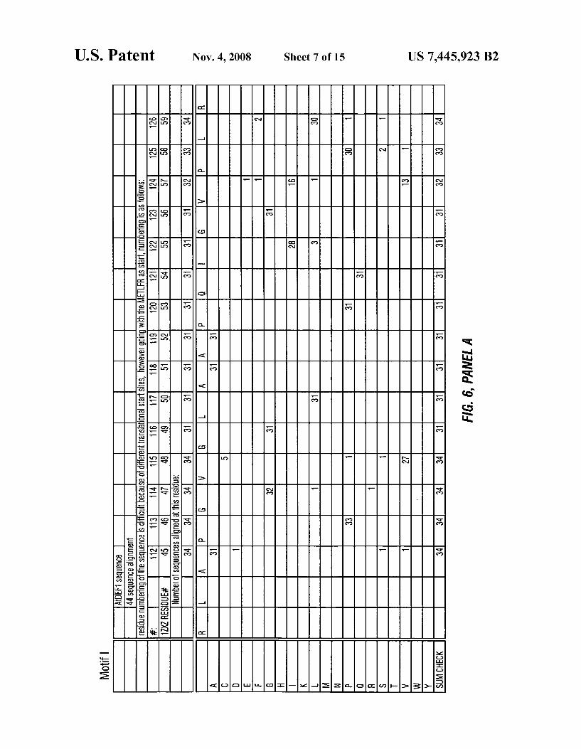
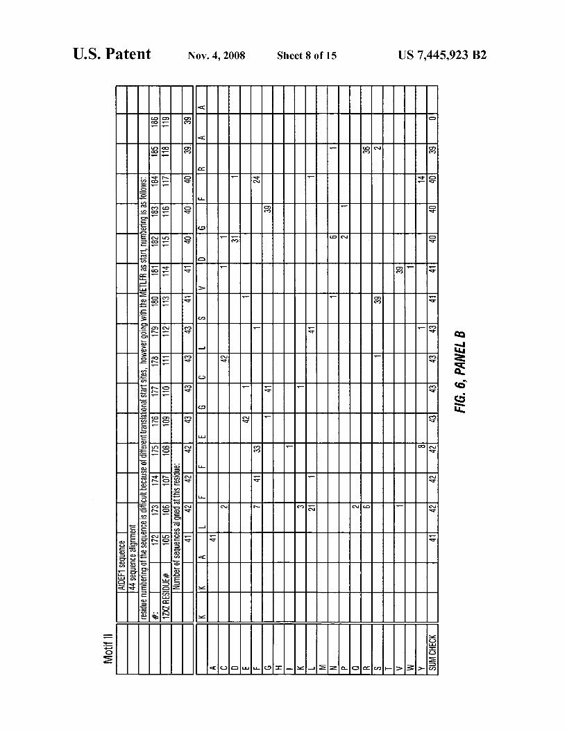
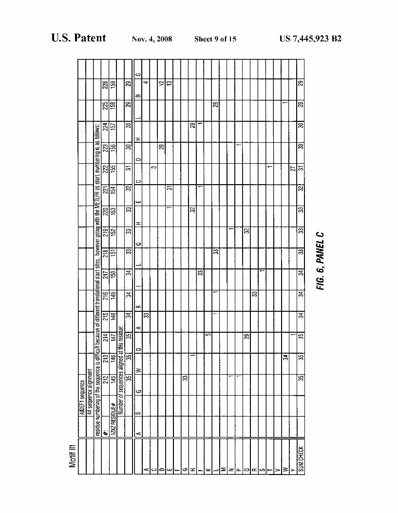
FIG. 6 depicts a phylogenetic analyses comparing Motif l (SEQ ID NO: 6), Motif 2 (SEQ ID NO: 7) and Motif 3 (SEQ ID NO: 8) of plant AtDEFl peptide deformylase With the amino acid sequence of otherpeptide deformylase sequences.
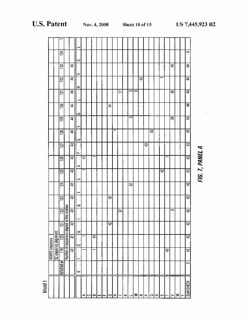
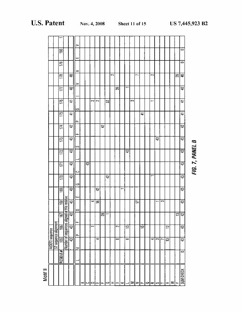
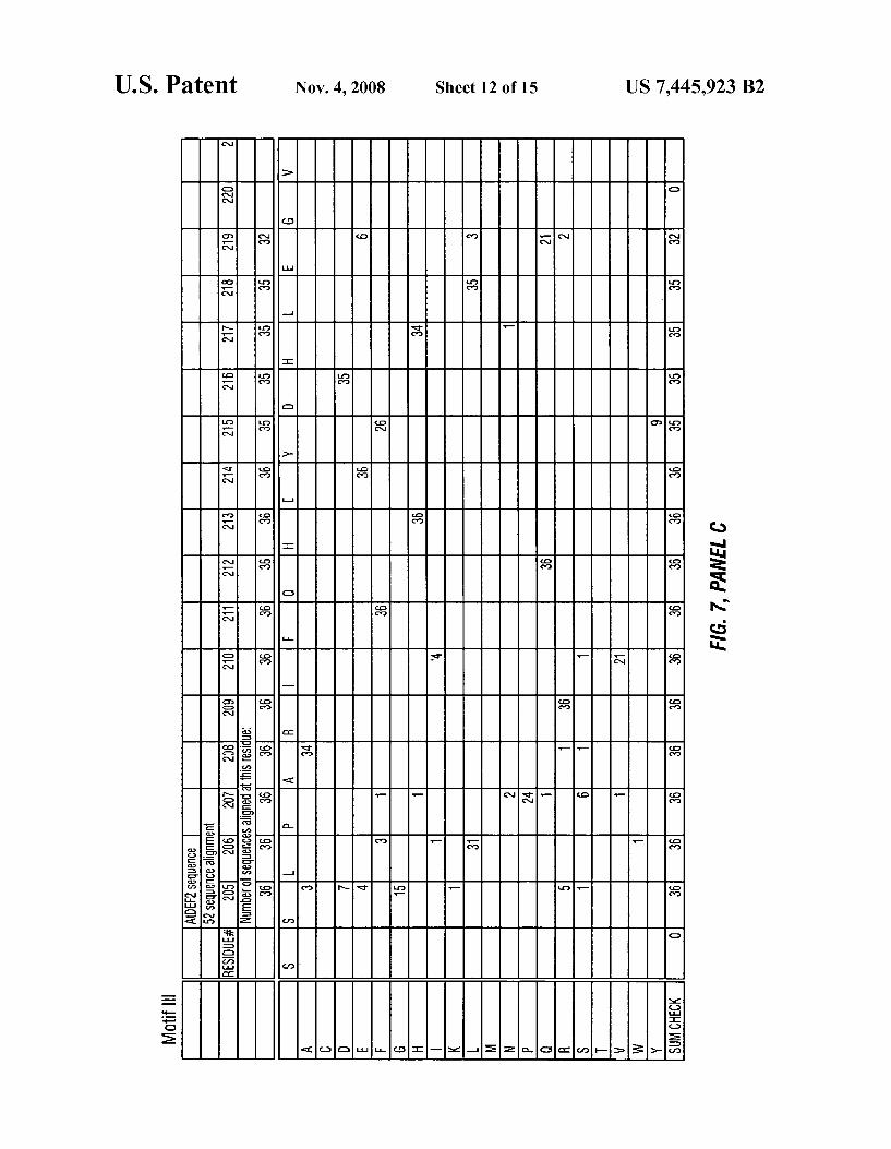
FIG. 7 depicts a phylogenetic analyses comparing Motif l (SEQ ID NO: 9), Motif2 (SEQ ID NO: 10) and Motif3 (SEQ ID NO: 11) of plant AtDEF2 peptide deformylase With the amino acid sequence of otherpeptide deformylase sequences.
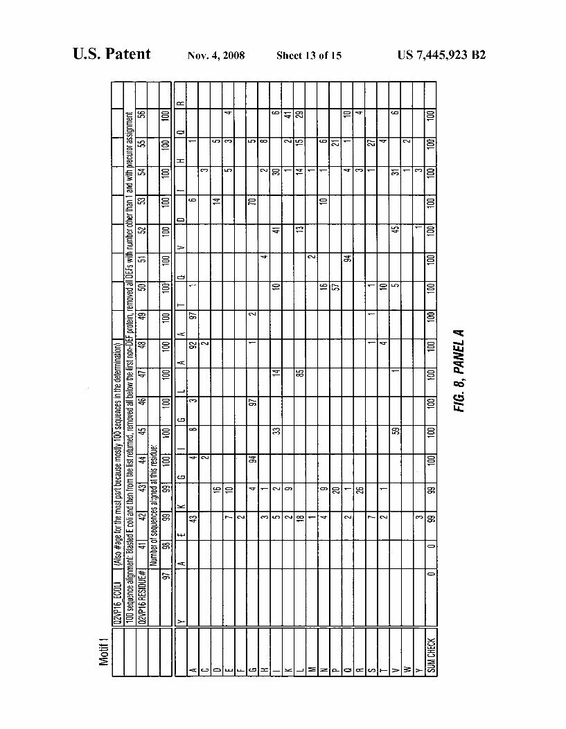
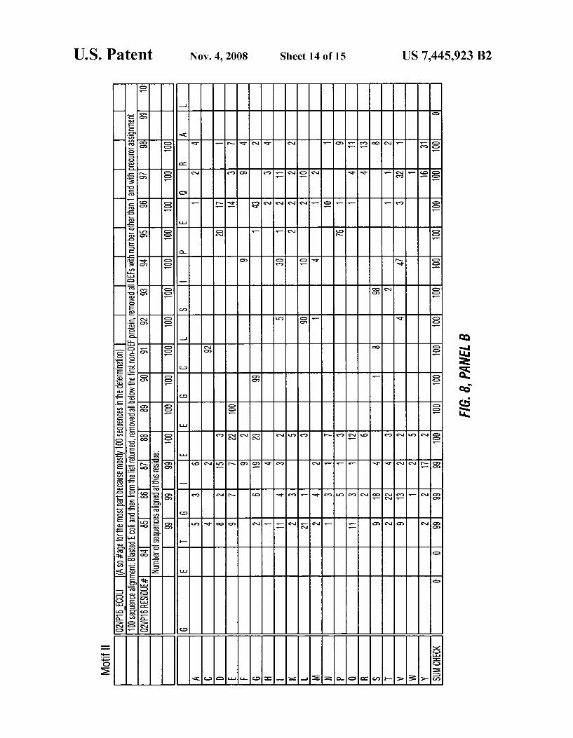
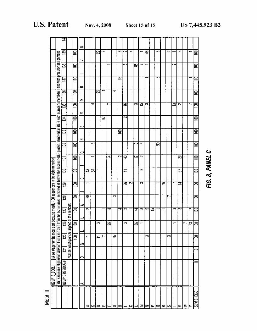
FIG. 8 depicts a phylogenetic analyses comparing Motif l (SEQ ID NO: 12), Motif 2 (SEQ ID NO: 13) and Motif 3 (SEQ ID NO: 14) of various peptide deformylase amino acid sequences.
Like reference symbols in the various draWings indicate like elements.
DETAILED DESCRIPTION
Provided herein are novel crystalline forms of peptide deformylase polypeptides and atomic coordinate information related to such crystals. Also provided are methods of using such information to identify, design and/or modify com pounds that modulate the activity of a peptide deformylase. In addition, computer systems that include such information are provided. The crystal structures and information derived therefrom are suitable for designing and identifying, for example, broad spectrum herbicides. Such herbicides can be used, for example, to inhibit or prevent the groWth of unde sirable vegetation. The crystal structure is based, at least in part, on the dis
covery of a plant nuclear gene that encodes a chloroplast targeted peptide deformylase polypeptide. The gene has sub
US 7,445,923 B2 5
stantial homology to bacterial peptide deformylase. The deduced translation of this nucleic acid sequence reveals the presence of three conserved protein motifs associated With prokaryotic peptide deformylase (see e.g., FIGS. 6, 7, and 8). Nucleic acid and amino acid sequences for plant peptide defor'mylases are disclosed in US. Pat. No. 6,730,634, issued May 4, 2004, and US. Patent Application Publication No. 20040088755, the contents of Which are incorporated herein by reference.
It is to be understood that the crystalline form of a plant peptide deformylase from Which the atomic structure coordi nates of the invention can be obtained is not limited to Wild type Arabidopsis Zhaliana peptide deformylase polypeptide, or a truncated form of the polypeptide (see e.g., SEQ ID NO: 1) as provided herein. Indeed, the crystals may comprise mutants of Wild-type Arabidopsis Zhaliana peptide deformy lase or the sequence of amino acids set forth in SEQ ID NO:1 . Mutants can be obtained by replacing at least one amino acid residue in the sequence of the Wild-type Arabidopsis Zhaliana peptide deformylase or the sequence of amino acids set forth in SEQ ID NO:1 With a different amino acid residue, or by adding or deleting one or more amino acid residues Within the Wild-type sequence and/or at the N- and/ or C-terminus of the Wild-type Arabidopsis Zhaliana peptide deformylase or the sequence of amino acids set forth in SEQ ID NO:1. Prefer ably, such mutants Will crystallize under crystallization con ditions that are substantially similar to those used to crystal lize the Wild-type Arabidopsis Zhaliana peptide deformylase or the sequence of amino acids set forth in SEQ ID NO:1.
The types of mutants contemplated by this invention include conservative mutants, non-conservative mutants, deletion mutants, truncated mutants, extended mutants, methionine mutants, selenomethionine mutants, cysteine mutants and selenocysteine mutants. A mutant may have, but need not have, Arabidopsis Zhaliana peptide deformylase activity. Preferably, a mutant displays biological activity that is substantially similar to that of the Wild-type polypeptide or that of SEQ ID NO:1.
It Will be recognized by one of skill in the art that the types of mutants contemplated herein are not mutually exclusive; that is, for example, a polypeptide having a conservative mutation in one amino acid may in addition have a truncation of residues at the N-terminus.
In addition, conservative or non-conservative amino acid substitutions can be made to amino acids of Wild-type Ara bidopsis Zhaliana peptide deformylase or the sequence of amino acids set forth in SEQ ID NO:1 that are implicated in the active site of the polypeptide (e.g., amino acid residues Glyl2l, Glyl23, Leu124,Gln128,Glu169, Gly170,Cys171, Leu172, His213, Glu2l4, His217, and Tyr178 of SEQ ID NO: 1). Such conservative or non-conservative substitutions can affect, e.g., the a?inity With Which wild-typeArabidopsis Zhaliana peptide deformylase or the sequence of amino acids set forth in SEQ ID NO:1 binds to a substrate. In certain embodiments, the conservative or non-conservative amino acid substitutions can increase the a?inity With Which Wild type Arabidopsis Zhaliana peptide deformylase or the sequence of amino acids set forth in SEQ ID NO:1 binds to a substrate.
Conservative amino acid substitutions are Well-knoWn in the art, and include substitutions made on the basis of a similarity in polarity, charge, solubility, hydrophobicity and/ or the hydrophilicity of the amino acid residues involved. Typical conservative substitutions are those in Which the amino acid is substituted With a different amino acid that is a member of the same class or category, as those classes are de?ned herein. Thus, typical conservative substitutions
20
25
30
35
40
45
50
55
60
65
6 include aromatic to aromatic, apolar to apolar, aliphatic to aliphatic, acidic to acidic, basic to basic, polar to polar, etc. Other conservative amino acid substitutions are Well knoWn in the art. It Will be recognized by those of skill in the art that generally, a total of about 20% or feWer, typically about 10% or feWer, most usually about 5% or feWer, of the amino acids in the Wild-type polypeptide sequence can be conservatively substituted With other amino acids Without deleteriously affecting the biological activity and/or three-dimensional structure of the molecule, provided that such substitutions do not involve residues that are critical for activity. The folloW ing abbreviations are used for amino acids throughout this disclosure: A:Ala:Alanine, T:Thr:Threonine, V:Val:Valine, CICysICysteine, LILeuILeucine, YITyFTyrosine, I:Ile:Isoleucine, NIAsnIAsparagine, P:Pro:Proline, Q:Gln:Glutamine, FIPheIPhenyIaIanine, DIAspIAspartic Acid, W:Trp:Tryptophan, E:Glu:Glutamic Acid, M:Met:Methionine, KILysILysine, G:Gly:Glycine, RIArgIArginine, SISerISerine, HIHisIHistidine.
In some embodiments, it may be desirable to make muta tions in the active site of a polypeptide, e.g., to reduce or completely eliminate deformylase activity. Mutations that Will reduce or completely eliminate the activity of a particular protein Will be apparent to those of skill in the art. For example, the amino acids identi?ed in Table 1 could be mutated in order to reduce or eliminate the binding activity of Wild-type Arabidopsis Zhaliana peptide deformylase or the sequence of amino acids set forth in SEQ ID NO: 1. The amino acid residue Cys (C) is unusual in that it can
form disul?de bridges With other Cys (C) residues or other sulfhydryl-containing amino acids (“cysteine-like amino acids”). The ability of Cys (C) residues and other cysteine like amino acids to exist in a polypeptide in either the reduced free iSH or oxidized disul?de-bridged form affects Whether Cys (C) residues contribute net hydrophobic or hydrophilic character to a polypeptide.
While in most instances the amino acids of Wild-typeAra bidopsis Zhaliana peptide deformylase or the sequence of amino acids set forth in SEQ ID NO:1 Will be substituted With genetically-encoded amino acids, in certain circumstances mutants may include genetically non-encoded amino acids. Alternatively, in instances Where the mutant Will be prepared in Whole or in part by chemical synthesis, virtually any non encoded amino acids may be used, ranging from D-isomers of the genetically encoded amino acids to non-encoded natu rally-occurring natural and synthetic amino acids.
Conservative amino acid substitutions for many of the commonly knoWn non-genetically encoded amino acids are Well knoWn in the art. Conservative substitutions for other non-encoded amino acids can be determined based on their physical properties as compared to the properties of the genetically encoded amino acids.
In some instances, it may be particularly advantageous or convenient to substitute, delete from and/ or add amino acid residues to wild-typeArabidopsis Zhaliana peptide deformy lase or the sequence of amino acids set forth in SEQ ID NO:1 in order to provide convenient cloning sites in cDNA encod ing the polypeptide, to aid in puri?cation of the polypeptide, etc. Such substitutions, deletions and/or additions that do not substantially alter the three dimensional structure of the native Arabidopsis Zhaliana peptide deformylase or the sequence of amino acids set forth in SEQ ID NO:1 Will be apparent to those having skills in the art. These substitutions, deletions and/ or additions include, but are not limited to, His tags, BirA tags, intein-containing self-cleaving tags, maltose binding protein fusions, glutathione S-transferase protein
US 7,445,923 B2 7
fusions, antibody fusions, green ?uorescent protein fusions, signal peptide fusions, biotin accepting peptide fusions, and the like.
Mutations may also be introduced into a polypeptide sequence Where there are residues, e.g., cysteine residues, that interfere With crystallization. Such cysteine residues can be substituted With an appropriate amino acid that does not readily form covalent bonds With other amino acid residues under crystallization conditions; e. g., by substituting the cys teine With Ala, Ser or Gly. Any cysteine located in a non helical or non-beta-stranded segment, based on secondary structure assignments, are good candidates for replacement.
It should be noted that the mutants contemplated herein need not exhibit deformylase activity. Indeed, amino acid substitutions, additions or deletions that interfere With the binding activity of Wild-type Arabidopsis Zhaliana peptide deformylase or the sequence of amino acids set forth in SEQ ID NO:1 are speci?cally contemplated by the invention. Such crystalline polypeptides, or the atomic structure coordinates obtained therefrom, can be used to provide phase information to aid the determination of the three-dimensional X-ray struc tures of other related or non-related crystalline polypeptides.
Also contemplated are homologs of the Arabidopsis Zhaliana peptide deformylase. The present invention provides a computer-assisted method for homology modeling an Ara bidopsis Zhaliana peptide deformylase homolog including: aligning the amino acid sequence of anArabidopsis Zhaliana peptide deformylase homolog With the amino acid sequence of Arabidopsis Zhaliana peptide deformylase SEQ ID N011 and incorporating the sequence of the Arabidopsis Zhaliana peptide deformylase homolog into a model of Arabidopsis thaliana peptide deformylase derived from structure coordi nates set forth in Table 1 to yield a preliminary model of the Arabidopsis Zhaliana peptide deformylase homolog; subject ing the preliminary model to energy minimization to yield an energy minimized model; remodeling regions of the energy minimized model Where stereochemistry restraints are vio lated to yield a ?nal model of the Arabidopsis Zhaliana pep tide deformylase homolog. As used herein, the term “homolog” refers to the polypep
tide molecule or the nucleic acid molecule Which encodes the polypeptide, or a functional domain from said polypeptide from a ?rst source having at least about 30%, 40% or 50% sequence identity, or at least about 60%, 70% or 75% sequence identity, or at least about 80% sequence identity, or more preferably at least about 85% sequence identity, or even more preferably at least about 90% sequence identity, and most preferably at least about 95%, 97% or 99% amino acid or nucleotide sequence identity, With the polypeptide, encod ing nucleic acid molecule or any functional domain thereof, from a second source. The second source may be a version of the molecule from the ?rst source that has been genetically altered by any available means to change the primary amino acid or nucleotide sequence or may be from the same or a different species than that of the ?rst source. Homology mod eling is further discussed beloW.
Accordingly, provided herein are crystalline forms of a plant peptide deformylase. Referring to FIG. 1A, limited trypsinolysis creates a core protein that retained activity and remained soluble in the absence of high salt concentrations. Analysis of Wild-type and proteolyzed AtDEF2 on an 8- 1 6% gradient SDS-PAGE. Trypsinolysis produces a truncated DEF2 With a mobility shift corresponding to a 3 kDa loss in molecular mass from AtDEF2, a 24.598 kDa enzyme. The truncated DEF2, Which loses its hexahistidyl sequence, Was subsequently separated from undigested DEF2 by loading the digested sample onto a HiTrap® a?inity column (Amersham
20
25
30
35
40
45
50
55
60
65
8 Pharmacia) and collecting the ?oWthrough. Undigested DEF2 remained bound to the column. Referring to FIG. 1B, the amino acid sequence of the truncated DEF2 polypeptide is provided.
It is understood that the term “crystalline form” includes a polypeptide associated With a plant peptide deformylase can include just the polypeptide, or the polypeptide complexed With a metal, a ligand, or any other chemical entity suitable for crystallization With the polypeptide. An exemplary polypeptide includes Arabidopsis Zhaliana peptide deformy lase, or fragments thereof, suitable for crystallization. Such fragments include optionally, the crystal may include a coor dinated metal ion selected from the group of consisting of Fe, Zn, Ni, or combinations thereof. Thus, “crystalline form” and “crystal” refer to a composition comprising a polypeptide complex in crystalline form. The term “crystal” includes native crystals, heavy-atom derivative crystals and poly-crys tals. “Native Crystal” refers to a crystal Wherein the polypep tide complex is substantially pure.
Referring to FIG. 2A, the crystal structure of DEF2 Was determined by molecular replacement and re?ned to a reso lution of 2.7 A. “Molecular Replacement” refers to the method of calculating initial phases for a neW crystal of a polypeptide Whose structure coordinates are unknown by ori enting and positioning a polypeptide Whose structure coordi nates are knoWn Within the unit cell of the neW crystal so as to best account for the observed diffraction pattern of the neW crystal. Phases are then calculated from the oriented and positioned polypeptide and combined With observed ampli tudes to provide an approximate Fourier synthesis of the structure of the polypeptides comprising the neW crystal (Jones et al., 1991, Acta Crystallogr. 47:753-70; Brunger et al., 1998, Acta Crystallogr. D. Biol. Crystallogr. 541905-21). The overall fold of the enzyme resembles the 0t+[3 confor
mation of knoWn bacterial peptide deformylases, With an r.m.s deviation of about 1.04 A on main chain atoms relative to the E. coli enzyme. The largest differences occur in the orientation of the C-terminal helix (helix 3) and the confor mation of the loop betWeen [3 strands 2 and 3, Which form part of the ?ve-stranded central sheet. Motif I, II and III are col ored blue, green and pink, respectively. The active site metal, modeled as zinc due to the conditions of crystallization, is a space-?lled sphere in the middle of the structure. Referring to FIG. 2B, a slab vieW of the ribbon representation of trypsi nolyzed AtDEF2 highlighting the active-site-metal binding ligands (1 Cys and 2 His) from motifs II and III, respectively (EGCLS (SEQ ID NO: 2) and QHEXXH (SEQ ID NO: 3) is provided. As used herein, the term “active site” refers to regions on a protein or a structural motif of a protein that are directly involved in the function or activity of the peptide deformylase. As used herein, the terms “binding site” or “binding
pocket” refer to a region of a polypeptide or a molecular complex comprising the polypeptide that, as a result of the primary amino acid sequence of the polypeptide and/or its three-dimensional shape, favorably associates With another chemical entity or compound including ligands or inhibitors. The crystalline form can include the tetragonal space group
symmetry P4l212 and includes a unit cell having dimensions a, b, and c; Wherein a is about 40 A to about 60 A, b is about 40 A to about 60 A, and c is about 120 A to about 160 A; and Wherein alpha:beta:gamma:90 degree. In some aspects, a is about 49 A to about 52 A, b is about 49 A to about 52 A, and c is about 143 A to about 147 A.
“Unit Cell” refers to the smallest and simplest volume element (i.e., parallel piped-shaped block) of a crystal that is completely representative of the unit or pattern of the crystal,