Embed Size (px)
Citation preview
research papers
Acta Cryst. (2015). D71, 427–441 doi:10.1107/S1399004714025061 427
Received 18 August 2014
Accepted 15 November 2014
‡ Present address: Mechanical Engineering
Department, Stanford University, 440 Escondido
Mall, Stanford, CA 94305-3030, USA.
§ Present address: Department of Materials
Science and Engineering, University of
Pennsylvania, 3231 Walnut Street, Philadelphia,
PA 19104-6272, USA.
} Present address: Albert Einstein College of
Medicine, Yeshiva University, 1300 Morris Park
Avenue, Bronx, NY 10461-1900, USA.
Keywords: MPD; crystallization additives;
precipitants; high-resolution protein structures;
chirality.
PDB references: lysozyme, crystallized with
(RS)-2-methyl-2,4-pentanediol, 4b4j; crystal-
lized with (R)-2-methyl-2,4-pentanediol, 4b4e;
crystallized with (S)-2-methyl-2,4-pentanediol,
4b4i; crystallized without 2-methyl-2,4-
pentanediol, 4b49
Supporting information: this article has
supporting information at journals.iucr.org/d
Crystallization of lysozyme with (R)-, (S)- and(RS)-2-methyl-2,4-pentanediol
Mark Stauber,a,b‡ Jean Jakoncic,c Jacob Berger,a,b§ Jerome M. Karp,a,b} Ariel
Axelbaum,a,b Dahniel Sastow,a,b Sergey V. Buldyrev,a Bruce J. Hrnjezd and Neer
Asheriea,b*
aDepartment of Physics, Yeshiva University, 2495 Amsterdam Avenue, New York, NY 10033-3312, USA, bDepartment of
Biology, Yeshiva University, 2495 Amsterdam Avenue, New York, NY 10033-3312, USA, cNational Synchrotron Light
Source, Brookhaven National Laboratory, Building 725D, Upton, NY 11973-5000, USA, and dCollegiate School, 260
West 78th Street, New York, NY 10024-6559, USA. *Correspondence e-mail: [email protected]
Chiral control of crystallization has ample precedent in the small-molecule
world, but relatively little is known about the role of chirality in protein
crystallization. In this study, lysozyme was crystallized in the presence of the
chiral additive 2-methyl-2,4-pentanediol (MPD) separately using the R and S
enantiomers as well as with a racemic RS mixture. Crystals grown with (R)-MPD
had the most order and produced the highest resolution protein structures. This
result is consistent with the observation that in the crystals grown with (R)-MPD
and (RS)-MPD the crystal contacts are made by (R)-MPD, demonstrating that
there is preferential interaction between lysozyme and this enantiomer. These
findings suggest that chiral interactions are important in protein crystallization.
1. Introduction
Proteins are difficult to crystallize. According to the most
recently available statistics from the Structural Biology
Knowledgebase (Gabanyi et al., 2011), fewer than one in
eight purified proteins produces diffraction-quality crystals.
Furthermore, this success rate has been decreasing over the
past decade (Chayen, 2002, 2004; Chayen & Saridakis, 2008);
one explanation offered is that the proteins which are easy to
crystallize were tackled first (Pusey et al., 2005).
A protein will crystallize when the solution conditions are
thermodynamically and kinetically favorable (Candoni et al.,
2012). As there is currently no way to predict these favorable
conditions, protein crystallization remains essentially a brute-
force endeavor: many different conditions are examined in the
hope that at least one of them will produce crystals (Chan et
al., 2013; Wilson & DeLucas, 2014). One common way to alter
the solution conditions is through the use of additives. These
additives are typically salts, small organic molecules and
polymers (Dumetz et al., 2009; McPherson et al., 2011),
although other additives have been used, such as silicon-based
surfaces that promote nucleation (Chayen et al., 2001; Ghatak
& Ghatak, 2011; Tsekova et al., 2012).
Much has been carried out to understand the role
of protein–additive interactions in crystal formation
(McPherson, 1999). While the detailed mechanisms through
which additives promote protein crystallization are often not
known, a few general features of protein–additive interactions
are understood. For example, additives can form favorable
crystal contacts leading to a stable and highly ordered crys-
talline arrangement of proteins (McPherson et al., 2011).
ISSN 1399-0047
We are investigating an aspect of protein–additive inter-
actions that is relatively unexplored in the context of protein
crystallization: chirality. Chiral control of crystallization has
ample precedent in the small-molecule world (Addadi et al.,
1982; Amharar et al., 2012; Blackmond, 2011; Brittain, 2013;
Eicke et al., 2013; Gou et al., 2012; Levilain et al., 2012; Lorenz
& Seidel-Morgenstern, 2014), but most of the work on chiral
interactions between proteins and additives has focused on
how such interactions control protein function (Brooks et al.,
2011). Typically, when a protein binds a small chiral molecule,
it interacts differently with one enantiomer than with the other
because the protein itself is chiral. In principle, such chiral
interactions may affect not only protein function but also
protein phase behaviour, including crystallization.
Our previous work on thaumatin and sodium tartrate
demonstrated that the chirality of the additive has a
substantial effect on the habit, packing, solubility and growth
of protein crystals (Asherie, Ginsberg, Blass et al., 2008;
Asherie, Ginsberg, Greenbaum et al., 2008; Asherie et al.,
2009). Furthermore, by working with enantiomerically pure
additives, we were able to determine the highest resolution
(0.94 A) thaumatin structure currently available (Asherie et
al., 2009).
To examine the generality of our findings, we are studying
other pairs of proteins and chiral precipitants. Here, we discuss
our results for the crystallization of lysozyme with 2-methyl-
2,4-pentanediol (MPD; C6H14O2). We chose this protein–
precipitant pair for three reasons. Firstly, lysozyme is the most
widely examined protein in crystallization and structural
analysis studies (Chayen & Saridakis, 2001; Liang et al., 2013;
Magay & Yoon, 2011; Tu et al., 2014). Secondly, MPD is a
chiral molecule that is one of the most common additives in
protein crystallization (Anand et al., 2002), although it has
been used exclusively as the racemate. Thirdly, lysozyme has
previously been crystallized with (RS)-MPD, but the results of
these investigations are contradictory: Weiss and coworkers
found only (R)-MPD in the crystal (Weiss et al., 2000), whereas
Michaux and coworkers found only (S)-MPD (Michaux et al.,
2008).
In the current study, we crystallized lysozyme using the
individual R and S enantiomers of MPD separately as well as
the racemate, and determined the X-ray structures of the
resultant crystals. We also determined the X-ray structure of
lysozyme crystals grown without MPD. All structures were
obtained to high resolution (1.25 A or better), allowing a
detailed comparison of the protein structures and, in principle,
an unambiguous assignment of the absolute configuration (R
or S) of the MPD molecules. This assignment, however, is
complicated by the fact that the MPD molecule can adopt
different conformations (Anand et al., 2002). We therefore
performed a detailed conformational analysis of MPD using
quantum-chemical (QC) calculations and molecular-dynamics
(MD) simulations. Consequently, we were able to use the
stable conformer that is likely to dominate the relative
conformer population in our analysis of the protein
crystal structures. Ambiguity in the analysis is thereby
diminished.
We find that crystals grown with (R)-MPD had the least
disorder (as measured by the mosaicity and B factor) and
produced the highest resolution protein structures. This
finding is consistent with the observation that co-crystal-
lization with either (R)- or (RS)-MPD gives crystal contacts
made exclusively by (R)-MPD, demonstrating that there is
preferential interaction between lysozyme and this enan-
tiomer. These results support the hypothesis that chiral
interactions may be important in protein crystallization with
chiral additives.
2. Materials and methods
2.1. Materials
Lysozyme (catalog No. 2933, lot No. 36P9210) was
purchased from Worthington Biochemical Corporation,
Lakewood, New Jersey, USA. (R)-MPD and (S)-MPD were
synthesized by Reuter Chemische Apparatebau KG, Freiburg,
Germany. (RS)-MPD (catalog No. 68340, lot No. 1345630) was
purchased from Sigma–Aldrich, St Louis, Missouri, USA.
Tris base (catalog No. BP512-500), sodium azide (catalog No.
S227I-500) and hydrochloric acid (catalog No. A144S-500)
were purchased from Fisher Scientific, Pittsburgh, Pennsyl-
vania, USA. All materials were used without further purifi-
cation. The purity of the protein and the chemical and
enantiomeric purity of (R)-, (S)- and (RS)-MPD were deter-
mined as described in the Supporting Information. Deionized
water was obtained from an Integral 3 deionization system
(Millipore, Billerica, Massachusetts, USA). Solutions were
filtered through a Nalgene disposable 0.22 mm filter unit
(Nalge Nunc International, Rochester, New York, USA) prior
to use.
Concentration measurements were carried out by UV–Vis
extinction spectroscopy on a Beckman–Coulter DU800
spectrophotometer. The extinction coefficient of lysozyme at
280 nm was taken to be "0.1% = 2.64 mg ml�1 cm�1 (Aune &
Tanford, 1969). Conductivity and pH measurements were
performed using an Orion 4-Star conductivity and pH meter
with a DuraProbe conductivity cell and a ROSS Sure-Flow pH
electrode (Thermo Fisher Scientific, Waltham, Massachusetts,
USA).
2.2. Protein crystallization
Lysozyme was dissolved in 200 mM Tris (titrated to pH 8.0
with HCl; � = 9.90 mS cm�1), washed three times in the same
buffer in an Amicon Ultra-4 centrifugal filter device with a
3 kDa molecular-weight cutoff (Millipore, Billerica, Massa-
chusetts, USA) and then concentrated to approximately
35 mg ml�1. Crystals were grown using the hanging-drop
vapor-diffusion method in the EasyXtal 15-Well Tool (catalog
No. 132006; Qiagen, Valencia, California, USA). Drops were
made by mixing 10 ml protein solution with 10 ml reservoir
solution. This mixture was vortexed briefly and three 5 ml
drops were then dispensed onto the crystallization supports.
The reservoir solutions (300 ml) were 60%(v/v) (R)-, (S)- or
(RS)-MPD in water. A control experiment with only water in
research papers
428 Stauber et al. � Crystallization of lysozyme with 2-methyl-2,4-pentanediol Acta Cryst. (2015). D71, 427–441
the reservoir was also carried out. The crystallization trays
were left at 4.0 � 0.5�C and inspected periodically by bright-
field microscopy with an AxioImager A1m microscope (Carl
Zeiss, Gottingen, Germany). Crystals of roughly 200 mm in
size grew in about 5 d with MPD (Supplementary Fig. S1);
crystals of similar size took about two weeks to grow in the
control. The crystals were harvested with mounted cryoloops
(Hampton Research, Aliso Viejo, California, USA). No
cryoprotectant was used, except for the crystals grown in the
control, which were dipped into Paratone N (Hampton
Research, Aliso Viejo, California, USA) immediately before
the diffraction measurements.
2.3. Data collection, refinement and structural analysis
All X-ray diffraction data were recorded on beamline X6A
at the National Synchrotron Light Source, Brookhaven
National Laboratory, Upton, New York, USA between 13.5
and 15.1 keV. All data were recorded at 100 K using an ADSC
Q270 CCD detector (ADCS, Poway, California, USA). Data
were indexed, integrated and scaled in HKL-2000 (Otwi-
nowski & Minor, 1997). Lysozyme crystal structures were
solved by molecular replacement using MOLREP (Vagin &
Teplyakov, 2010) and the model from PDB entry 1iee (Sauter
et al., 2001). Each model was refined by restrained maximum-
likelihood refinement with REFMAC (Murshudov et al., 2011;
Winn et al., 2011) with individual anisotropic temperature
factors and manual building performed in Coot (Emsley et al.,
2010). After the final refinement, the stereochemistry of the
structures was assessed with PROCHECK (Laskowski et al.,
1993). All figures were prepared with PyMOL (http://
www.pymol.org).
2.4. Quantum-chemical calculations and molecular-dynamics simulations
Ab initio electronic structure calculations for the nine
(R)-MPD conformers 1–3 (see Fig. 2) were performed with
Gaussian09 (Frisch et al., 2009). Stationary points were located
on the local potential surfaces for these nine conformers at a
modest level of quantum-chemical theory, using the second-
order Møller–Plesset perturbation (MP2) method (Møller &
Plesset, 1934) and the 6-311++G(d,p) medium-sized basis
(Clark et al., 1983) set for C, H and O atoms. The program
package defaults, including criteria for wavefunction conver-
gence and locations of stationary points on potential surfaces,
were used. The nine initial input structures were the result of
a qualitative conformational analysis of the all-staggered
conformational possibilities for (R)-MPD. Conformer 1a was
assumed to be intramolecularly hydrogen-bonded; we were
able to focus on a single conformer because the geometry-
optimized energies of different intramolecularly hydrogen-
bonded rotamers about C—O bonds differed by less than
1 kJ mol�1. As expected, inversion of configuration at C4 to
give (S)-MPD gave identical computational results.
We examined the effect of water solvation on the relative
conformer energies at the same level of quantum-chemical
theory, incorporating the default integral equation formalism
variant (IEF) within the polarizable continuum model (PCM)
for placing a solute in a cavity within the solvent reaction field
(SCRF; Tomasi et al., 2005).
Molecular-dynamics simulations of both (R)-MPD and (S)-
MPD were performed using GROMACS v.4.0.5 (Hess et al.,
2008). The initial coordinates for (R)- and (S)-MPD were
taken from PDB entries 4b4e and 4b4i, respectively, with
H atoms added using the pdb2gmx utility of GROMACS.
Simulations were run in vacuo with one molecule of either
(R)- or (S)-MPD placed in a cubic box of length 3.0 nm with
periodic boundary conditions. Since MPD has no net charge,
no counterions were added. The topology files were
constructed using parameters for OPLS-AA atom types
(Jorgensen et al., 1996; Jorgensen & Tirado-Rives, 1988). The
properties of the atoms used in the topology file for both
enantiomers are shown in Supplementary Table S1.
The OPLS-AA all-atom force field was used in running
simulations. Each simulation box was subjected to energy
minimization using the steepest-descent method. Simulations
were run using the NVT ensemble, and the temperature was
held constant at 300 or 370 K using the V-rescale thermostat
(Bussi et al., 2009) with a coupling constant of 0.1. Electro-
static interactions were treated using the particle mesh Ewald
algorithm (Essmann et al., 1995) using electrostatic, van der
Waals and neighbor-list cutoffs of 0.9 nm. The SHAKE
constraint algorithm (Ryckaert et al., 1977) was used to
constrain all bonds with a tolerance of 0.0002. Simulations
were run for 100 ns, using 1 fs time steps and saving coordi-
nates and energies every 1 ps. The first 10 ns of each simula-
tion were considered to be equilibration time and were not
used in subsequent analysis.
Annealing simulations were run with (R)-MPD in vacuo.
The systems were prepared as above. In each of 100 simula-
tions, the initial velocities were independently randomly
generated. The initial temperature was 370 K and the simu-
lation was run for 200 ps. Over every subsequent 200 ps, the
temperature was decreased linearly with time by 5 K. Thus,
14.6 ns after the beginning the simulations the temperature
reached 5 K. During the next 200 ps, the temperature was
decreased linearly with time to 0.1 K. The simulation
continued at this temperature until it had run for a total of
20 ns.
Trajectory analysis was performed with the GROMACS
utilities package. For clarity, we report torsion angles in the
range 0 to 360� instead of the customary �180 to 180� (torsion
angles greater than 180� can be converted to the usual nega-
tive torsion angles by subtracting 360�).
2.5. Database analysis
MPD conformations were extracted from the RCSB Protein
Data Bank (PDB; http://www.pdb.org; Berman et al., 2000)
and the Cambridge Structural Database (CSD; Allen, 2002).
The PDB was searched using the chemical IDs for either
(R)-MPD (MRD) or (S)-MPD (MPD) together with two
additional requirements: X-ray resolution between 0 and
1.5 A and sequence homology of less than 90% to other
research papers
Acta Cryst. (2015). D71, 427–441 Stauber et al. � Crystallization of lysozyme with 2-methyl-2,4-pentanediol 429
macromolecules. This search yielded 49 protein structure hits
for MRD and 89 protein structure hits for MPD. Some of these
hits contained both enantiomers, yielding 117 unique protein
structures with (R)- or (S)-MPD. [We note that numerous
protein structures had more than one (R)- or (S)-MPD
molecule associated with them.] These molecules were
inspected using Coot (Emsley et al., 2010) with the structure
and electron-density maps (2Fo� Fc and Fo� Fc) downloaded
through the Uppsala Electron Density Server (Kleywegt et al.,
2004). Hits that had no density or no structure factors were
discarded. The quality of the electron-density map, the local
hydrogen bonding and the atomic B factors of the molecules
were used to check whether the assigned model (R)- or
(S)-MPD structure was acceptable as is, i.e. whether the
enantiomer and conformer selected were supported by the
data. Acceptable structures were kept, while unacceptable
structures were either discarded (because the torsion angles
of the molecule could not be determined unambiguously) or
reassigned to achieve a better agreement between the model
and the electron density. The torsion angles of the acceptable
and reassigned structures were measured using the built-in
function of Coot. A total of 221 molecules were retained: 109
were (R)-MPD and 112 were (S)-MPD.
The CSD was searched with ConQuest (Bruno et al., 2002)
using the chemical formula of MPD (C6H14O2). The resulting
28 hits were inspected and only the seven hits that corre-
sponded to 2-methyl-2,4-pentanediol were retained (reference
codes BACXIM10, FALDUS, KOFPAW, NIRQIO, NOSVOG,
PIVYEZ and TECYIJ). The hits, some of which contained
multiple molecules of both enantiomers, were examined using
Mercury (Bruno et al., 2002). Since no structure-factor infor-
mation was available, the assigned model (R)-MPD or (S)-
MPD structures were accepted as is, unless the local hydrogen
bonding suggested that the structure be reassigned. The
torsion angles of the acceptable and reassigned structures
were measured using the built-in function of Mercury. A total
of ten molecules were retained: three were (R)-MPD and
seven were (S)-MPD.
3. Results and discussion
3.1. Protein and precipitant purity
Protein purity is a crucial factor in crystallization
(McPherson, 1999). Indeed, the deleterious effect of impu-
rities on the crystallization of lysozyme has been studied
extensively (Dold et al., 2006; Judge et al., 1998; Lorber et al.,
1993; Parmar et al., 2007; Thomas et al., 1996). We chose
to work with lysozyme from Worthington Biochemical
Corporation because it has been shown to produce high-
quality crystals (Parmar et al., 2007). Our characterization
of the protein by size-exclusion and cation-exchange high-
performance chromatography, quasi-elastic light scattering
and electrospray ionization mass spectroscopy confirmed its
high purity (Supplementary Figs. S2 and S3 and Table S2).
For the precipitant, both chemical and enantiomeric purity
must be controlled. We purchased (RS)-MPD from Sigma–
Aldrich, but decided against using the commercially available
(R)-MPD from the same manufacturer (catalog No. 252840)
because of its lower chemical purity and the limited infor-
mation about its enantiomeric purity (only the specific rota-
tion is given). For (S)-MPD, a commercial supplier was not an
option. To our knowledge, this enantiomer is not available as a
common chemical.
We therefore commissioned the custom syntheses of (R)-
MPD and (S)-MPD by Reuter Chemische Apparatebau KG
(the structures of the two enantiomers and the associated
nomenclature are shown in Fig. 1). Since (S)-MPD has not
been characterized previously and only partial information
is available for (R)-MPD, we analyzed these enantiomers and
the racemate (RS)-MPD by 1H and 13C NMR, tandem mass
spectrometry and optical activity (Supplementary Figs. S4–S9
and Tables S3 and S4). For completeness, we also provide the
gas-chromatography results of (R)-MPD and (S)-MPD that
were given to us by the manufacturer (Supplementary Figs.
S10 and S11). These results confirm the chemical identity
of the molecules synthesized and demonstrate their high
chemical and enantiomeric purity; the key results are
summarized in Table 1.
3.2. MPD conformation
An important step in the analysis of the X-ray diffraction
data is the proper assignment of any MPD molecules that are
present in the crystal structure. This assignment involves
research papers
430 Stauber et al. � Crystallization of lysozyme with 2-methyl-2,4-pentanediol Acta Cryst. (2015). D71, 427–441
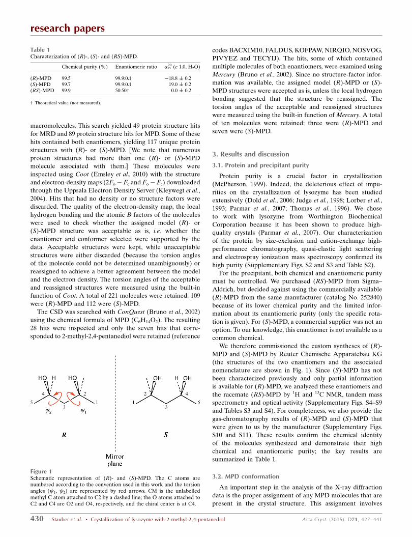
Figure 1Schematic representation of (R)- and (S)-MPD. The C atoms arenumbered according to the convention used in this work and the torsionangles ( 1, 2) are represented by red arrows. CM is the unlabelledmethyl C atom attached to C2 by a dashed line; the O atoms attached toC2 and C4 are O2 and O4, respectively, and the chiral center is at C4.
Table 1Characterization of (R)-, (S)- and (RS)-MPD.
Chemical purity (%) Enantiomeric ratio �D20 (c 1.0, H2O)
(R)-MPD 99.5 99.9:0.1 �18.8 � 0.2(S)-MPD 99.7 99.9:0.1 19.0 � 0.2(RS)-MPD 99.9 50:50† 0.0 � 0.2
† Theoretical value (not measured).
selecting the enantiomer(s) and conformer(s) of the molecule
that best fit the electron density. For the experiments with pure
(R)- or (S)-MPD there is only one enantiomer to choose,
but for crystallization with (RS)-MPD the selection is less
straightforward.
If the electron-density map is of sufficiently high quality, it is
possible to distinguish the two enantiomers by inspecting the
shape of the map, even though the H atom on the chiral center
C4 (Fig. 1) is not visible in the X-ray data. An example is the
(R)-MPD molecule found near Phe34 by Weiss and coworkers
in the structure of lysozyme (PDB entry 1dpw) crystallized
with (RS)-MPD (Weiss et al., 2000). If the shape of the map is
inconclusive, knowledge of the most likely conformer can be
helpful in selecting the appropriate enantiomer.
Since H atoms contribute little to the electron density, the
conformation of MPD as obtained from the electron-density
map is completely determined by the torsion angles ( 1, 2),
which are defined by C atoms C1—C2—C3—C4 and C2—
C3—C4—C5, respectively (Fig. 1). Furthermore, for an
isolated molecule the stable conformers of one enantiomer
will be mirror images of the other. Energetic considerations
suggest that the expected values of these angles for an isolated
research papers
Acta Cryst. (2015). D71, 427–441 Stauber et al. � Crystallization of lysozyme with 2-methyl-2,4-pentanediol 431
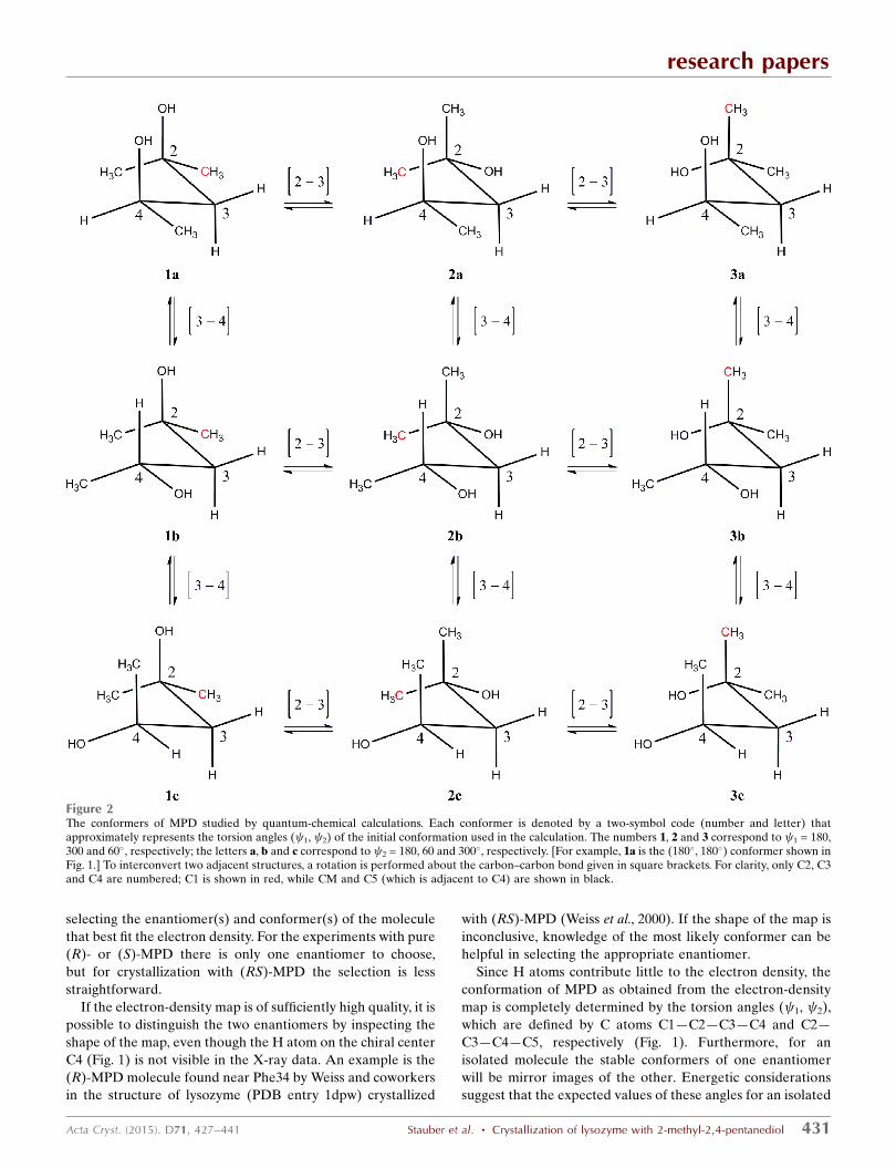
Figure 2The conformers of MPD studied by quantum-chemical calculations. Each conformer is denoted by a two-symbol code (number and letter) thatapproximately represents the torsion angles ( 1, 2) of the initial conformation used in the calculation. The numbers 1, 2 and 3 correspond to 1 = 180,300 and 60�, respectively; the letters a, b and c correspond to 2 = 180, 60 and 300�, respectively. [For example, 1a is the (180�, 180�) conformer shown inFig. 1.] To interconvert two adjacent structures, a rotation is performed about the carbon–carbon bond given in square brackets. For clarity, only C2, C3and C4 are numbered; C1 is shown in red, while CM and C5 (which is adjacent to C4) are shown in black.
(R)-MPD molecule are approximately (180�, 180�). This
conformation (shown in Fig. 1) allows the formation of an
intramolecular hydrogen bond (the distance between O2 and
O4 atoms is 2.8 A) and corresponds to a favorable arrange-
ment of the C1—C2—C3—C4—C5 backbone (Salam &
Deleuze, 2002). To verify these considerations, we carried out
both quantum-chemical calculations and molecular-dynamics
simulations on MPD.
We performed quantum-chemical (QC) calculations to
determine the relative energies of the nine conformers of
(R)-MPD shown schematically in Fig. 2. These all-staggered
conformers were chosen as the initial configurations for
geometry optimization; each of these is likely to be close to a
local minimum on the conformational potential energy surface
(Mo, 2010). The relative energies of the optimized geometries
for the nine conformers are listed in Table 2 (see also
research papers
432 Stauber et al. � Crystallization of lysozyme with 2-methyl-2,4-pentanediol Acta Cryst. (2015). D71, 427–441
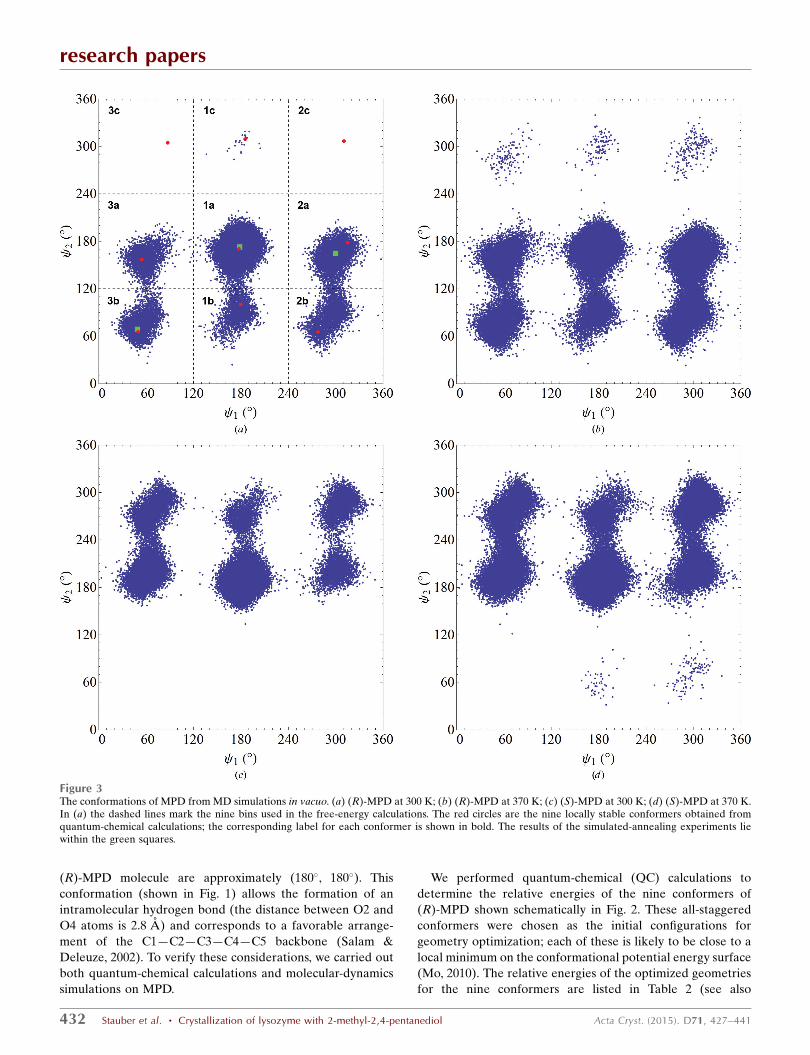
Figure 3The conformations of MPD from MD simulations in vacuo. (a) (R)-MPD at 300 K; (b) (R)-MPD at 370 K; (c) (S)-MPD at 300 K; (d) (S)-MPD at 370 K.In (a) the dashed lines mark the nine bins used in the free-energy calculations. The red circles are the nine locally stable conformers obtained fromquantum-chemical calculations; the corresponding label for each conformer is shown in bold. The results of the simulated-annealing experiments liewithin the green squares.
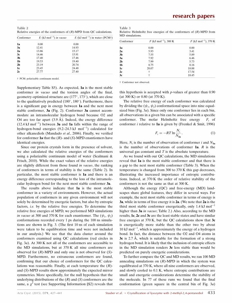
Supplementary Table S5). As expected, 1a is the most stable
conformer in vacuo and the torsion angles of the final,
geometry-optimized structure are (177�, 173�), which are close
to the qualitatively predicted (180�, 180�). Furthermore, there
is a significant gap in energy between 1a and the next most
stable conformer, 3a (Fig. 2). Conformer 3a cannot accom-
modate an intramolecular hydrogen bond because O2 and
O4 are too far apart (3.9 A). Indeed, the energy difference
(12.4 kJ mol�1) between 3a and 1a falls within the range of
hydrogen-bond energies (9.2–24.3 kJ mol�1) calculated for
other alkanediols (Mandado et al., 2006). Finally, we verified
for conformer 1a that the (R)- and (S)-MPD enantiomers have
identical energies.
Since our protein crystals form in the presence of solvent,
we also calculated the relative energies of the conformers
using a polarizable continuum model of water (Scalmani &
Frisch, 2010). While the exact values of the relative energies
are slightly different from those found in vacuo, the ranking
of conformers in terms of stability is the same (Table 2). In
particular, the most stable conformer is 1a and there is an
energy difference corresponding to the loss of the intramole-
cular hydrogen bond for the next most stable conformer 3a.
The results above indicate that 1a is the most stable
conformer in a variety of environments. However, the actual
population of conformers in any given environment will not
solely be determined by energetic factors, but also by entropic
factors, i.e. by the relative free energies. To determine the
relative free energies of MPD, we performed MD simulations
in vacuo at 300 and 370 K for each enantiomer. The ( 1, 2)
conformations recorded every 1 ps during the 100 ns simula-
tions are shown in Fig. 3. (The first 10 ns of each simulation
were taken to be equilibration time and were not included
in our analysis.) We see that the data cluster around the
conformers examined using QC calculations (red circles in
Fig. 3a). At 300 K not all of the conformers are accessible to
the MD simulations, but at 370 K all nine conformers are
observed for (R)-MPD and only one is not observed for (S)-
MPD. Furthermore, no extraneous conformers are found,
confirming that our choice of conformers for the QC calcu-
lations was reasonable. Finally, at each temperature the (R)-
and (S)-MPD results show approximately the expected mirror
symmetries. More specifically, for the null hypothesis that the
underlying distributions of the (R) and (S) conformers are the
same, a �2 test (see Supporting Information xS2) reveals that
this hypothesis is accepted with p-values of greater than 0.90
(at 300 K) or 0.80 (at 370 K).
The relative free energy of each conformer was calculated
by dividing the ( 1, 2) conformational space into nine equal-
sized bins (Fig. 3a). Since only one conformer lies in each bin,
all observations in a given bin can be associated with a specific
conformer. The molar Helmholtz free energy �Fi of
conformer i relative to 1a is given by (Frenkel & Smit, 1996)
�Fi ¼ �RT lnNi
N1a
: ð1Þ
Here, Ni is the number of observation of conformer i and N1a
is the number of observations of conformer 1a. R is the
universal gas constant and T is the absolute temperature.
As we found with our QC calculations, the MD simulations
reveal that 1a is the most stable conformer and that there is
a gap to the next most stable conformer (Table 3). When the
temperature is changed from 300 to 370 K this gap decreases,
illustrating the increased importance of entropic contribu-
tions. Indeed, at 370 K the order of relative stability of the
conformers is not the same as that at 300 K.
Although the energy (QC) and free-energy (MD) land-
scapes share global features, they differ in several ways. For
example, the next most stable conformer in terms of energy is
3a, while in terms of free energy it is 2a. (We note that 2a is the
third most stable conformer energetically, only 1.4 kJ mol�1
higher than 3a in vacuo; Table 2.) Also, according to the MD
results, 1c, 2c and 3c are the least stable states and have similar
free energies at 370 K, but the QC calculations show that 3c
is energetically more stable than the other two by about
10 kJ mol�1, which is approximately the energy of a hydrogen
bond. In fact, the distance between the O2 and O4 atoms in
3c is 2.7 A, which is suitable for the formation of a strong
hydrogen bond. It is likely that the inclusion of entropic effects
in the MD simulation renders 3c less stable than would be
predicted on purely energetic considerations.
To further compare the QC and MD results, we ran 100 MD
annealing simulations on (R)-MPD in which the system was
equilibrated at 370 K, where all nine conformers are observed,
and slowly cooled to 0.1 K, where entropic contributions are
small and energetic considerations determine the stability of
the molecule. In 77 of these runs we found that the final
conformation (green square in the central bin of Fig. 3a)
research papers
Acta Cryst. (2015). D71, 427–441 Stauber et al. � Crystallization of lysozyme with 2-methyl-2,4-pentanediol 433
Table 3Relative Helmholtz free energies of the conformers of (R)-MPD fromMD simulations.
Conformer �F (kJ mol�1), 300 K �F (kJ mol�1), 370 K
1a 0.00 0.002a 5.93 3.413b 7.55 5.893a 7.92 6.462b 7.99 5.731b 8.75 8.161c 20.07 18.882c † 17.543c † 18.45
† Conformer not observed.
Table 2Relative energies of the conformers of (R)-MPD from QC calculations.
Conformer �E (kJ mol�1) in vacuo �E (kJ mol�1) in water (PCM†)
1a 0.00 0.003a 12.41 14.932a 13.86 15.173c 14.46 15.913b 17.40 17.461b 19.53 19.402b 23.19 20.741c 23.45 24.482c 27.77 27.40
† PCM: polarizable continuum model.
coincided with the 1a conformation of the QC calculations
(red circle in the central bin of Fig. 3a), which is consistent
with our previous result that 1a is a global energy minimum.
In 20 of the remaining 23 runs, (R)-MPD reached a local
minimum in the 2a bin (the next most stable state in the MD
simulations after 1a; Table 3), while in three runs it reached
the 3b conformation (the third most stable state). Based on
the QC results we would have expected the order of states
in the low-temperature limit to be 1a < 3a < 2a (in terms of
increasing energy; Table 2). The difference may reflect the
limitations of a classical force field in capturing the quantum-
mechanical aspects of atomic interactions.
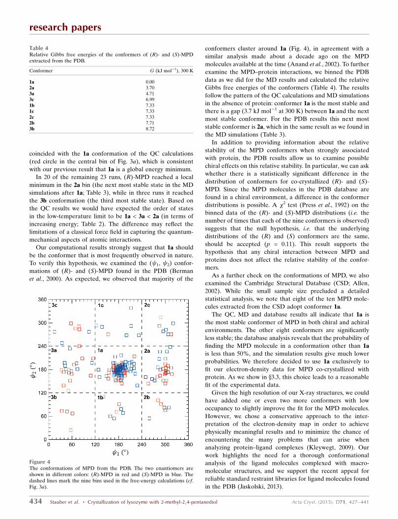
Our computational results strongly suggest that 1a should
be the conformer that is most frequently observed in nature.
To verify this hypothesis, we examined the ( 1, 2) confor-
mations of (R)- and (S)-MPD found in the PDB (Berman
et al., 2000). As expected, we observed that majority of the
conformers cluster around 1a (Fig. 4), in agreement with a
similar analysis made about a decade ago on the MPD
molecules available at the time (Anand et al., 2002). To further
examine the MPD–protein interactions, we binned the PDB
data as we did for the MD results and calculated the relative
Gibbs free energies of the conformers (Table 4). The results
follow the pattern of the QC calculations and MD simulations
in the absence of protein: conformer 1a is the most stable and
there is a gap (3.7 kJ mol�1 at 300 K) between 1a and the next
most stable conformer. For the PDB results this next most
stable conformer is 2a, which in the same result as we found in
the MD simulations (Table 3).
In addition to providing information about the relative
stability of the MPD conformers when strongly associated
with protein, the PDB results allow us to examine possible
chiral effects on this relative stability. In particular, we can ask
whether there is a statistically significant difference in the
distribution of conformers for co-crystallized (R)- and (S)-
MPD. Since the MPD molecules in the PDB database are
found in a chiral environment, a difference in the conformer
distributions is possible. A �2 test (Press et al., 1992) on the
binned data of the (R)- and (S)-MPD distributions (i.e. the
number of times that each of the nine conformers is observed)
suggests that the null hypothesis, i.e. that the underlying
distributions of the (R) and (S) conformers are the same,
should be accepted (p = 0.11). This result supports the
hypothesis that any chiral interaction between MPD and
proteins does not affect the relative stability of the confor-
mers.
As a further check on the conformations of MPD, we also
examined the Cambridge Structural Database (CSD; Allen,
2002). While the small sample size precluded a detailed
statistical analysis, we note that eight of the ten MPD mole-
cules extracted from the CSD adopt conformer 1a.
The QC, MD and database results all indicate that 1a is
the most stable conformer of MPD in both chiral and achiral
environments. The other eight conformers are significantly
less stable; the database analysis reveals that the probability of
finding the MPD molecule in a conformation other than 1a
is less than 50%, and the simulation results give much lower
probabilities. We therefore decided to use 1a exclusively to
fit our electron-density data for MPD co-crystallized with
protein. As we show in x3.3, this choice leads to a reasonable
fit of the experimental data.
Given the high resolution of our X-ray structures, we could
have added one or even two more conformers with low
occupancy to slightly improve the fit for the MPD molecules.
However, we chose a conservative approach to the inter-
pretation of the electron-density map in order to achieve
physically meaningful results and to minimize the chance of
encountering the many problems that can arise when
analyzing protein–ligand complexes (Kleywegt, 2009). Our
work highlights the need for a thorough conformational
analysis of the ligand molecules complexed with macro-
molecular structures, and we support the recent appeal for
reliable standard restraint libraries for ligand molecules found
in the PDB (Jaskolski, 2013).
research papers
434 Stauber et al. � Crystallization of lysozyme with 2-methyl-2,4-pentanediol Acta Cryst. (2015). D71, 427–441
Table 4Relative Gibbs free energies of the conformers of (R)- and (S)-MPDextracted from the PDB.
Conformer �G (kJ mol�1), 300 K
1a 0.002a 3.703a 4.713c 6.991b 7.331c 7.332c 7.332b 7.713b 8.72
Figure 4The conformations of MPD from the PDB. The two enantiomers areshown in different colors: (R)-MPD in red and (S)-MPD in blue. Thedashed lines mark the nine bins used in the free-energy calculations (cf.Fig. 3a).
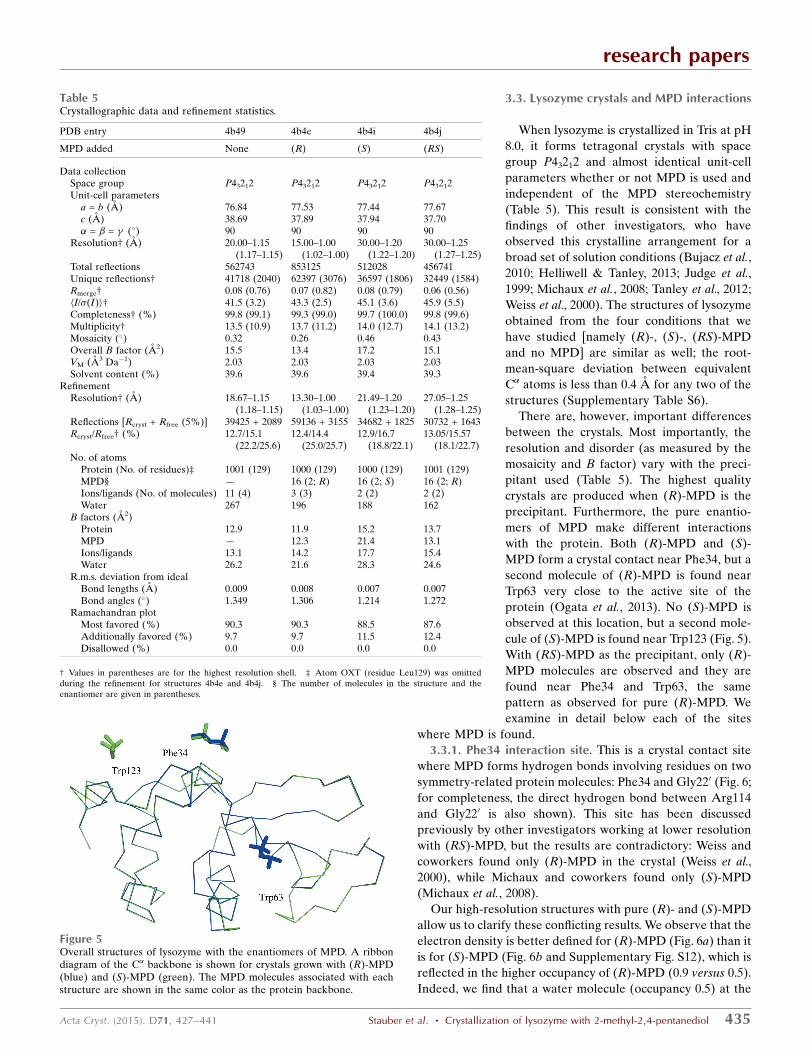
3.3. Lysozyme crystals and MPD interactions
When lysozyme is crystallized in Tris at pH
8.0, it forms tetragonal crystals with space
group P43212 and almost identical unit-cell
parameters whether or not MPD is used and
independent of the MPD stereochemistry
(Table 5). This result is consistent with the
findings of other investigators, who have
observed this crystalline arrangement for a
broad set of solution conditions (Bujacz et al.,
2010; Helliwell & Tanley, 2013; Judge et al.,
1999; Michaux et al., 2008; Tanley et al., 2012;
Weiss et al., 2000). The structures of lysozyme
obtained from the four conditions that we
have studied [namely (R)-, (S)-, (RS)-MPD
and no MPD] are similar as well; the root-
mean-square deviation between equivalent
C� atoms is less than 0.4 A for any two of the
structures (Supplementary Table S6).
There are, however, important differences
between the crystals. Most importantly, the
resolution and disorder (as measured by the
mosaicity and B factor) vary with the preci-
pitant used (Table 5). The highest quality
crystals are produced when (R)-MPD is the
precipitant. Furthermore, the pure enantio-
mers of MPD make different interactions
with the protein. Both (R)-MPD and (S)-
MPD form a crystal contact near Phe34, but a
second molecule of (R)-MPD is found near
Trp63 very close to the active site of the
protein (Ogata et al., 2013). No (S)-MPD is
observed at this location, but a second mole-
cule of (S)-MPD is found near Trp123 (Fig. 5).
With (RS)-MPD as the precipitant, only (R)-
MPD molecules are observed and they are
found near Phe34 and Trp63, the same
pattern as observed for pure (R)-MPD. We
examine in detail below each of the sites
where MPD is found.
3.3.1. Phe34 interaction site. This is a crystal contact site
where MPD forms hydrogen bonds involving residues on two
symmetry-related protein molecules: Phe34 and Gly220 (Fig. 6;
for completeness, the direct hydrogen bond between Arg114
and Gly220 is also shown). This site has been discussed
previously by other investigators working at lower resolution
with (RS)-MPD, but the results are contradictory: Weiss and
coworkers found only (R)-MPD in the crystal (Weiss et al.,
2000), while Michaux and coworkers found only (S)-MPD
(Michaux et al., 2008).
Our high-resolution structures with pure (R)- and (S)-MPD
allow us to clarify these conflicting results. We observe that the
electron density is better defined for (R)-MPD (Fig. 6a) than it
is for (S)-MPD (Fig. 6b and Supplementary Fig. S12), which is
reflected in the higher occupancy of (R)-MPD (0.9 versus 0.5).
Indeed, we find that a water molecule (occupancy 0.5) at the
research papers
Acta Cryst. (2015). D71, 427–441 Stauber et al. � Crystallization of lysozyme with 2-methyl-2,4-pentanediol 435
Figure 5Overall structures of lysozyme with the enantiomers of MPD. A ribbondiagram of the C� backbone is shown for crystals grown with (R)-MPD(blue) and (S)-MPD (green). The MPD molecules associated with eachstructure are shown in the same color as the protein backbone.
Table 5Crystallographic data and refinement statistics.
PDB entry 4b49 4b4e 4b4i 4b4j
MPD added None (R) (S) (RS)
Data collectionSpace group P43212 P43212 P43212 P43212Unit-cell parameters
a = b (A) 76.84 77.53 77.44 77.67c (A) 38.69 37.89 37.94 37.70� = � = � (�) 90 90 90 90
Resolution† (A) 20.00–1.15(1.17–1.15)
15.00–1.00(1.02–1.00)
30.00–1.20(1.22–1.20)
30.00–1.25(1.27–1.25)
Total reflections 562743 853125 512028 456741Unique reflections† 41718 (2040) 62397 (3076) 36597 (1806) 32449 (1584)Rmerge† 0.08 (0.76) 0.07 (0.82) 0.08 (0.79) 0.06 (0.56)hI/�(I)i† 41.5 (3.2) 43.3 (2.5) 45.1 (3.6) 45.9 (5.5)Completeness† (%) 99.8 (99.1) 99.3 (99.0) 99.7 (100.0) 99.8 (99.6)Multiplicity† 13.5 (10.9) 13.7 (11.2) 14.0 (12.7) 14.1 (13.2)Mosaicity (�) 0.32 0.26 0.46 0.43Overall B factor (A2) 15.5 13.4 17.2 15.1VM (A3 Da�1) 2.03 2.03 2.03 2.03Solvent content (%) 39.6 39.6 39.4 39.3
RefinementResolution† (A) 18.67–1.15
(1.18–1.15)13.30–1.00
(1.03–1.00)21.49–1.20
(1.23–1.20)27.05–1.25
(1.28–1.25)Reflections [Rcryst + Rfree (5%)] 39425 + 2089 59136 + 3155 34682 + 1825 30732 + 1643Rcryst/Rfree† (%) 12.7/15.1
(22.2/25.6)12.4/14.4
(25.0/25.7)12.9/16.7
(18.8/22.1)13.05/15.57
(18.1/22.7)No. of atoms
Protein (No. of residues)‡ 1001 (129) 1000 (129) 1000 (129) 1001 (129)MPD§ — 16 (2; R) 16 (2; S) 16 (2; R)Ions/ligands (No. of molecules) 11 (4) 3 (3) 2 (2) 2 (2)Water 267 196 188 162
B factors (A2)Protein 12.9 11.9 15.2 13.7MPD — 12.3 21.4 13.1Ions/ligands 13.1 14.2 17.7 15.4Water 26.2 21.6 28.3 24.6
R.m.s. deviation from idealBond lengths (A) 0.009 0.008 0.007 0.007Bond angles (�) 1.349 1.306 1.214 1.272
Ramachandran plotMost favored (%) 90.3 90.3 88.5 87.6Additionally favored (%) 9.7 9.7 11.5 12.4Disallowed (%) 0.0 0.0 0.0 0.0
† Values in parentheses are for the highest resolution shell. ‡ Atom OXT (residue Leu129) was omittedduring the refinement for structures 4b4e and 4b4j. § The number of molecules in the structure and theenantiomer are given in parentheses.
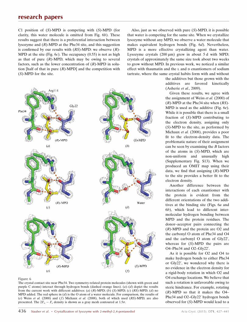
C1 position of (S)-MPD is competing with (S)-MPD (for
clarity, this water molecule is omitted from Fig. 6b). These
results suggest that there is a preferential interaction between
lysozyme and (R)-MPD at the Phe34 site, and this suggestion
is confirmed by our results with (RS)-MPD: we observe (R)-
MPD at the site (Fig. 6c). The occupancy (0.55) is not as high
as that of pure (R)-MPD, which may be owing to several
factors, such as the lower concentration of (R)-MPD in solu-
tion [half of that in pure (R)-MPD] and the competition with
(S)-MPD for the site.
Also, just as we observed with pure (S)-MPD, it is possible
that water is competing for the same site. When we crystallize
lysozyme without any MPD, we observe a water molecule that
makes equivalent hydrogen bonds (Fig. 6d). Nevertheless,
MPD is a more effective crystallizing agent than water.
Lysozyme crystals (200 mm) grew in about 5 d with MPD;
crystals of approximately the same size took about two weeks
to grow without MPD. In previous work, we noticed a similar
effect with thaumatin and the l and d enantiomers of sodium
tartrate, where the same crystal habits form with and without
the additives but those grown with the
additives are favored kinetically
(Asherie et al., 2009).
Given these results, we agree with
the assignment of Weiss et al. (2000) of
(R)-MPD at the Phe34 site when (RS)-
MPD is used as the additive (Fig. 6e).
While it is possible that there is a small
fraction of (S)-MPD contributing to
the electron density, assigning only
(S)-MPD to the site, as performed by
Michaux et al. (2008), provides a poor
fit to the electron-density data. The
problematic nature of their assignment
can be seen by examining the B factors
of the atoms in (S)-MPD, which are
non-uniform and unusually high
(Supplementary Fig. S13). When we
produced an OMIT map using their
data, we find that assigning (R)-MPD
to the site provides a better fit to the
electron density.
Another difference between the
interactions of each enantiomer with
the protein is evident from the
different orientations of the two addi-
tives at the binding site (Figs. 6a and
6b), which lead to different inter-
molecular hydrogen bonding between
MPD and the protein residues. The
donor–acceptor pairs connecting the
(R)-MPD and the protein are O2 and
the carbonyl O atom of Phe34 and O4
and the carbonyl O atom of Gly220,
whereas for (S)-MPD the pairs are
O4–Phe34 and O2–Gly220.
As it is possible for O2 and O4 to
make hydrogen bonds to either Phe34
or Gly220, we wondered why there is
no evidence in the electron density for
a rigid-body rotation in which O2 and
O4 exchange locations. We believe that
such a rotation is unfavorable owing to
steric hindrance. For example, rotating
(R)-MPD so that it makes the O4–
Phe34 and O2–Gly220 hydrogen bonds
observed for (S)-MPD would lead to a
research papers
436 Stauber et al. � Crystallization of lysozyme with 2-methyl-2,4-pentanediol Acta Cryst. (2015). D71, 427–441
Figure 6The crystal contact site near Phe34. Two symmetry-related protein molecules (shown with green andpurple C atoms) interact through hydrogen bonds (dashed orange lines). (a)–(d) depict the resultsfrom the current work with different additives: (a) (R)-MPD; (b) (S)-MPD; (c) (RS)-MPD; (d) noMPD added. The red sphere in (d) is the O atom of a water molecule. For comparison, the results of(e) Weiss et al. (2000) and ( f ) Michaux et al. (2008), both of which used (RS)-MPD, are alsopresented. The 2Fo � Fc density is shown as a gray mesh contoured at 1.5�.
strong repulsion between the methyl group CM and the
carbonyl O atom of Lys33.
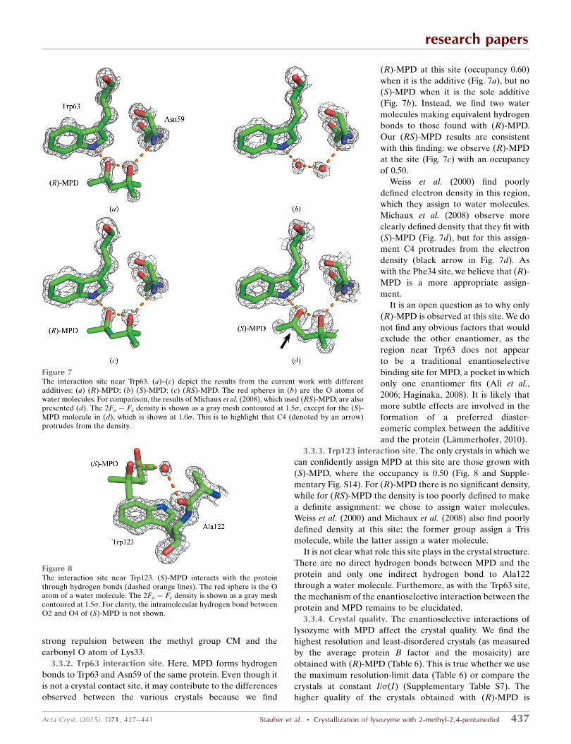
3.3.2. Trp63 interaction site. Here, MPD forms hydrogen
bonds to Trp63 and Asn59 of the same protein. Even though it
is not a crystal contact site, it may contribute to the differences
observed between the various crystals because we find
(R)-MPD at this site (occupancy 0.60)
when it is the additive (Fig. 7a), but no
(S)-MPD when it is the sole additive
(Fig. 7b). Instead, we find two water
molecules making equivalent hydrogen
bonds to those found with (R)-MPD.
Our (RS)-MPD results are consistent
with this finding: we observe (R)-MPD
at the site (Fig. 7c) with an occupancy
of 0.50.
Weiss et al. (2000) find poorly
defined electron density in this region,
which they assign to water molecules.
Michaux et al. (2008) observe more
clearly defined density that they fit with
(S)-MPD (Fig. 7d), but for this assign-
ment C4 protrudes from the electron
density (black arrow in Fig. 7d). As
with the Phe34 site, we believe that (R)-
MPD is a more appropriate assign-
ment.
It is an open question as to why only
(R)-MPD is observed at this site. We do
not find any obvious factors that would
exclude the other enantiomer, as the
region near Trp63 does not appear
to be a traditional enantioselective
binding site for MPD, a pocket in which
only one enantiomer fits (Ali et al.,
2006; Haginaka, 2008). It is likely that
more subtle effects are involved in the
formation of a preferred diaster-
eomeric complex between the additive
and the protein (Lammerhofer, 2010).
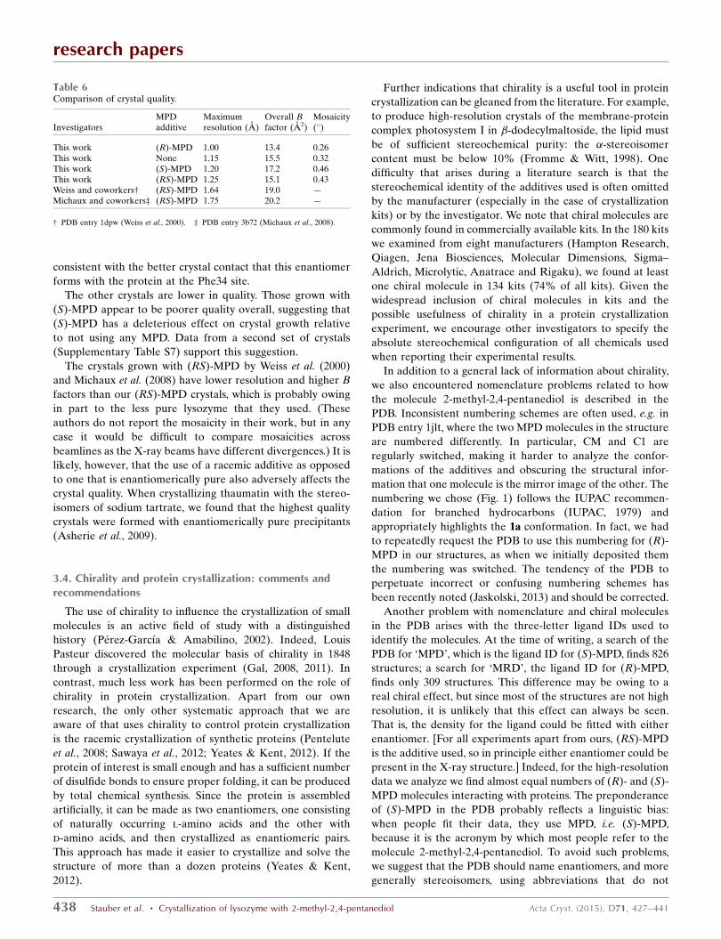
3.3.3. Trp123 interaction site. The only crystals in which we
can confidently assign MPD at this site are those grown with
(S)-MPD, where the occupancy is 0.50 (Fig. 8 and Supple-
mentary Fig. S14). For (R)-MPD there is no significant density,
while for (RS)-MPD the density is too poorly defined to make
a definite assignment: we chose to assign water molecules.
Weiss et al. (2000) and Michaux et al. (2008) also find poorly
defined density at this site; the former group assign a Tris
molecule, while the latter assign a water molecule.
It is not clear what role this site plays in the crystal structure.
There are no direct hydrogen bonds between MPD and the
protein and only one indirect hydrogen bond to Ala122
through a water molecule. Furthemore, as with the Trp63 site,
the mechanism of the enantioselective interaction between the
protein and MPD remains to be elucidated.
3.3.4. Crystal quality. The enantioselective interactions of
lysozyme with MPD affect the crystal quality. We find the
highest resolution and least-disordered crystals (as measured
by the average protein B factor and the mosaicity) are
obtained with (R)-MPD (Table 6). This is true whether we use
the maximum resolution-limit data (Table 6) or compare the
crystals at constant I/�(I) (Supplementary Table S7). The
higher quality of the crystals obtained with (R)-MPD is
research papers
Acta Cryst. (2015). D71, 427–441 Stauber et al. � Crystallization of lysozyme with 2-methyl-2,4-pentanediol 437
Figure 8The interaction site near Trp123. (S)-MPD interacts with the proteinthrough hydrogen bonds (dashed orange lines). The red sphere is the Oatom of a water molecule. The 2Fo � Fc density is shown as a gray meshcontoured at 1.5�. For clarity, the intramolecular hydrogen bond betweenO2 and O4 of (S)-MPD is not shown.
Figure 7The interaction site near Trp63. (a)–(c) depict the results from the current work with differentadditives: (a) (R)-MPD; (b) (S)-MPD; (c) (RS)-MPD. The red spheres in (b) are the O atoms ofwater molecules. For comparison, the results of Michaux et al. (2008), which used (RS)-MPD, are alsopresented (d). The 2Fo � Fc density is shown as a gray mesh contoured at 1.5�, except for the (S)-MPD molecule in (d), which is shown at 1.0�. This is to highlight that C4 (denoted by an arrow)protrudes from the density.
consistent with the better crystal contact that this enantiomer
forms with the protein at the Phe34 site.
The other crystals are lower in quality. Those grown with
(S)-MPD appear to be poorer quality overall, suggesting that
(S)-MPD has a deleterious effect on crystal growth relative
to not using any MPD. Data from a second set of crystals
(Supplementary Table S7) support this suggestion.
The crystals grown with (RS)-MPD by Weiss et al. (2000)
and Michaux et al. (2008) have lower resolution and higher B
factors than our (RS)-MPD crystals, which is probably owing
in part to the less pure lysozyme that they used. (These
authors do not report the mosaicity in their work, but in any
case it would be difficult to compare mosaicities across
beamlines as the X-ray beams have different divergences.) It is
likely, however, that the use of a racemic additive as opposed
to one that is enantiomerically pure also adversely affects the
crystal quality. When crystallizing thaumatin with the stereo-
isomers of sodium tartrate, we found that the highest quality
crystals were formed with enantiomerically pure precipitants
(Asherie et al., 2009).
3.4. Chirality and protein crystallization: comments andrecommendations
The use of chirality to influence the crystallization of small
molecules is an active field of study with a distinguished
history (Perez-Garcıa & Amabilino, 2002). Indeed, Louis
Pasteur discovered the molecular basis of chirality in 1848
through a crystallization experiment (Gal, 2008, 2011). In
contrast, much less work has been performed on the role of
chirality in protein crystallization. Apart from our own
research, the only other systematic approach that we are
aware of that uses chirality to control protein crystallization
is the racemic crystallization of synthetic proteins (Pentelute
et al., 2008; Sawaya et al., 2012; Yeates & Kent, 2012). If the
protein of interest is small enough and has a sufficient number
of disulfide bonds to ensure proper folding, it can be produced
by total chemical synthesis. Since the protein is assembled
artificially, it can be made as two enantiomers, one consisting
of naturally occurring l-amino acids and the other with
d-amino acids, and then crystallized as enantiomeric pairs.
This approach has made it easier to crystallize and solve the
structure of more than a dozen proteins (Yeates & Kent,
2012).
Further indications that chirality is a useful tool in protein
crystallization can be gleaned from the literature. For example,
to produce high-resolution crystals of the membrane-protein
complex photosystem I in �-dodecylmaltoside, the lipid must
be of sufficient stereochemical purity: the �-stereoisomer
content must be below 10% (Fromme & Witt, 1998). One
difficulty that arises during a literature search is that the
stereochemical identity of the additives used is often omitted
by the manufacturer (especially in the case of crystallization
kits) or by the investigator. We note that chiral molecules are
commonly found in commercially available kits. In the 180 kits
we examined from eight manufacturers (Hampton Research,
Qiagen, Jena Biosciences, Molecular Dimensions, Sigma–
Aldrich, Microlytic, Anatrace and Rigaku), we found at least
one chiral molecule in 134 kits (74% of all kits). Given the
widespread inclusion of chiral molecules in kits and the
possible usefulness of chirality in a protein crystallization
experiment, we encourage other investigators to specify the
absolute stereochemical configuration of all chemicals used
when reporting their experimental results.
In addition to a general lack of information about chirality,
we also encountered nomenclature problems related to how
the molecule 2-methyl-2,4-pentanediol is described in the
PDB. Inconsistent numbering schemes are often used, e.g. in
PDB entry 1jlt, where the two MPD molecules in the structure
are numbered differently. In particular, CM and C1 are
regularly switched, making it harder to analyze the confor-
mations of the additives and obscuring the structural infor-
mation that one molecule is the mirror image of the other. The
numbering we chose (Fig. 1) follows the IUPAC recommen-
dation for branched hydrocarbons (IUPAC, 1979) and
appropriately highlights the 1a conformation. In fact, we had
to repeatedly request the PDB to use this numbering for (R)-
MPD in our structures, as when we initially deposited them
the numbering was switched. The tendency of the PDB to
perpetuate incorrect or confusing numbering schemes has
been recently noted (Jaskolski, 2013) and should be corrected.
Another problem with nomenclature and chiral molecules
in the PDB arises with the three-letter ligand IDs used to
identify the molecules. At the time of writing, a search of the
PDB for ‘MPD’, which is the ligand ID for (S)-MPD, finds 826
structures; a search for ‘MRD’, the ligand ID for (R)-MPD,
finds only 309 structures. This difference may be owing to a
real chiral effect, but since most of the structures are not high
resolution, it is unlikely that this effect can always be seen.
That is, the density for the ligand could be fitted with either
enantiomer. [For all experiments apart from ours, (RS)-MPD
is the additive used, so in principle either enantiomer could be
present in the X-ray structure.] Indeed, for the high-resolution
data we analyze we find almost equal numbers of (R)- and (S)-
MPD molecules interacting with proteins. The preponderance
of (S)-MPD in the PDB probably reflects a linguistic bias:
when people fit their data, they use MPD, i.e. (S)-MPD,
because it is the acronym by which most people refer to the
molecule 2-methyl-2,4-pentanediol. To avoid such problems,
we suggest that the PDB should name enantiomers, and more
generally stereoisomers, using abbreviations that do not
research papers
438 Stauber et al. � Crystallization of lysozyme with 2-methyl-2,4-pentanediol Acta Cryst. (2015). D71, 427–441
Table 6Comparison of crystal quality.
InvestigatorsMPDadditive
Maximumresolution (A)
Overall Bfactor (A2)
Mosaicity(�)
This work (R)-MPD 1.00 13.4 0.26This work None 1.15 15.5 0.32This work (S)-MPD 1.20 17.2 0.46This work (RS)-MPD 1.25 15.1 0.43Weiss and coworkers† (RS)-MPD 1.64 19.0 —Michaux and coworkers‡ (RS)-MPD 1.75 20.2 —
† PDB entry 1dpw (Weiss et al., 2000). ‡ PDB entry 3b72 (Michaux et al., 2008).
introduce bias. This is particularly important given the large
number of chiral molecules in the PDB.
There are 90 chiral molecules in the top 200 PDB ligands
(ranked by ligand hits, i.e. the number of times the ligand
is reported in a PDB structure) and approximately 20% of
ligand hits involve chiral molecules. We believe that since
chiral molecules are common, possible chiral effects in protein
crystallization should be explored in detail. Furthermore, it
seems reasonable to explore more general stereochemical
effects beyond enantiomerism. Given the prevalence of sugars
as ligands in the PDB, we consider the stereoisomerism of
sugars as an interesting possibility to consider when crystal-
lizing proteins.
While the PDB protein structures and ligand list offer a
useful starting point for choosing candidate chiral ligands, they
provide only a partial view of the role of chiral molecules in
protein crystallization. It is possible for chiral effects to be
present in solution during protein nucleation but that the final
crystal does not incorporate the chiral additive. Indeed, we
have observed this with thaumatin and tartrate. The addition
of l-tartrate to thaumatin produces bipyramidal crystals that
incorporate the additive into the lattice. The crystals have
normal solubility and a tetragonal space group. Addition of
d-tartrate leads instead to the formation of prismatic crystals
with retrograde solubility and an orthorhombic space group;
these crystals do not contain any tartrate (Asherie, Ginsberg,
Blass et al., 2008; Asherie, Ginsberg, Greenbaum et al., 2008;
Asherie et al., 2009).
We focus here on high-resolution structures (resolution of
better than 1.5 A) because they allow us to determine the
stereochemistry and conformation of MPD with minimal
uncertainty and therefore we are able to analyze chiral effects
in detail. By doing so, we do not mean to imply that chiral
effects are confined only to high-resolution structures. On the
contrary, chiral effects span the range from the obvious (some
of which can be seen with the naked eye) to the subtle. This
is well known in small-molecule systems and by studying
different protein–additive pairs we expect to find that it holds
for protein systems as well.
Further work is needed to fully understand the mechanism
by which chirality affects protein crystallization. Chiral effects
are often clear at crystal contacts, but these only account for
only a small part of protein–additive interactions: we estimate
the fraction of protein structures with at least one crystal
contact by a chiral molecule to be about 5% (Carugo &
Djinovic-Carugo, 2014). A more common situation is one in
which two enantiomers interact with the protein at different
sites, but these are not crystal contact sites; this is the case for
the Trp63 and Trp123 interaction sites discussed in this work.
Also, as we mentioned above, chiral molecules can have an
effect in the solution phase.
We appreciate that working with enantiomerically pure
additives is expensive. Cost is one possible reason why crys-
tallization experiments with MPD have thus far been carried
out only with the racemate. At the time of writing, the cost per
gram of 99% pure (R)-MPD from Sigma–Aldrich (catalog No.
252840) is more than 3000 times that of (RS)-MPD of similar
purity (catalog No. 112100). Nevertheless, given the potential
benefits, some way to assess chiral effects should be incorpo-
rated into a crystallization experiment, and we expect that cost
will diminish with increased demand for enantiomerically pure
additives. If a full screening of crystallization conditions with
the separate enantiomers of the additive under study is
prohibitively expensive, we suggest that the initial screen be
carried out with the cheaper racemate; promising conditions
may then be optimized with the pure enantiomers. We are
happy to provide small amounts of pure (R)- and (S)-MPD to
members of the community.
4. Conclusions
We crystallized lysozyme with (R)-, (S)- and (RS)-MPD. We
also grew crystals without MPD under similar conditions. All
four crystalline arrangements obtained have the same space
group and almost identical unit-cell parameters. The crystals
grown with (R)-MPD have the highest resolution and least
disorder, suggesting a preferential interaction between lyso-
zyme and this enantiomer of MPD. This idea is confirmed by
the X-ray structures, which show that the two enantiomers
interact differently with the protein. Our findings support the
hypothesis that chiral interactions with chiral additives are
important in protein crystallization.
5. Related literature
The following references are cited in the Supporting Infor-
mation for this article: Ewing et al. (1996) and Lomakin et al.
(2005).
Acknowledgements
We thank Charles Ginsberg and Samuel Blass for help in the
initial stages of this project and Shekhar Garde and Amish
Patel for guidance on setting up molecular-dynamics simula-
tions. We also thank Jianfeng Jiang and Mark Harris for
helpful discussions. NA gratefully acknowledges financial
support from the National Science Foundation (DMR
1206416) and Yeshiva University. Computational resources
were provided by the Dr Bernard W. Gamson Computational
Science Center at Yeshiva University. We thank the staff of
the National Synchrotron Light Source, Brookhaven National
Laboratory for their continuous support. The NSLS is
supported by the US Department of Energy, Office of Basic
Energy Sciences under contract No. DE-AC02-98CH10886.
The NIGMS East Coast Structural Biology Facility, the X6A
beamline, is funded under contract No. GM-0080.
References
Addadi, L., Berkovitch-Yellin, Z., Domb, N., Gati, E., Lahav, M. &Leiserowitz, L. (1982). Nature (London), 296, 21–26.
Ali, I., Kumerer, K. & Aboul-Enein, H. Y. (2006). Chromatographia,63, 295–307.
Allen, F. H. (2002). Acta Cryst. B58, 380–388.Amharar, Y., Grandeury, A., Sanselme, M., Petit, S. & Coquerel, G.
(2012). J. Phys. Chem. B, 116, 6027–6040.
research papers
Acta Cryst. (2015). D71, 427–441 Stauber et al. � Crystallization of lysozyme with 2-methyl-2,4-pentanediol 439
Anand, K., Pal, D. & Hilgenfeld, R. (2002). Acta Cryst. D58, 1722–1728.
Asherie, N., Ginsberg, C., Blass, S., Greenbaum, A. & Knafo, S.(2008). Cryst. Growth Des. 8, 1815–1817.
Asherie, N., Ginsberg, C., Greenbaum, A., Blass, S. & Knafo, S.(2008). Cryst. Growth Des. 8, 4200–4207.
Asherie, N., Jakoncic, J., Ginsberg, C., Greenbaum, A., Stojanoff, V.,Hrnjez, B. J., Blass, S. & Berger, J. (2009). Cryst. Growth Des. 9,4189–4198.
Aune, K. C. & Tanford, C. (1969). Biochemistry, 8, 4579–4585.Berman, H. M., Westbrook, J., Feng, Z., Gilliland, G., Bhat, T. N.,
Weissig, H., Shindyalov, I. N. & Bourne, P. E. (2000). Nucleic AcidsRes. 28, 235–242.
Blackmond, D. G. (2011). Philos. Trans. R. Soc. Lond B Biol. Sci. 366,2878–2884.
Brittain, H. G. (2013). Chirality, 25, 8–15.Brooks, W. H., Guida, W. C. & Daniel, K. G. (2011). Curr. Top. Med.
Chem. 11, 760–770.Bruno, I. J., Cole, J. C., Edgington, P. R., Kessler, M., Macrae, C. F.,
McCabe, P., Pearson, J. & Taylor, R. (2002). Acta Cryst. B58,389–397.
Bujacz, G., Wrzesniewska, B. & Bujacz, A. (2010). Acta Cryst. D66,789–796.
Bussi, G., Zykova-Timan, T. & Parrinello, M. (2009). J. Chem. Phys.130, 074101.
Candoni, N., Grossier, R., Hammadi, Z., Morin, R. & Veesler, S.(2012). Protein Pept. Lett. 19, 714–724.
Carugo, O. & Djinovic-Carugo, K. (2014). J. Appl. Cryst. 47, 458–461.Chan, M., Fazio, V. J. & Newman, J. (2013). Cryst. Growth Des. 13,
1290–1294.Chayen, N. E. (2002). Trends Biotechnol. 20, 98.Chayen, N. E. (2004). Curr. Opin. Struct. Biol. 14, 577–583.Chayen, N. E. & Saridakis, E. (2001). J. Cryst. Growth, 232, 262–
264.Chayen, N. E. & Saridakis, E. (2008). Nature Methods, 5, 147–153.Chayen, N. E., Saridakis, E., El-Bahar, R. & Nemirovsky, Y. (2001). J.
Mol. Biol. 312, 591–595.Clark, T., Chandrasekhar, J., Spitznagel, G. W. & Schleyer, P. V. R.
(1983). J. Comput. Chem. 4, 294–301.Dold, P., Ono, E., Tsukamoto, K. & Sazaki, G. (2006). J. Cryst.
Growth, 293, 102–109.Dumetz, A. C., Chockla, A. M., Kaler, E. W. & Lenhoff, A. M. (2009).
Cryst. Growth Des. 9, 682–691.Eicke, M. J., Levilain, G. & Seidel-Morgenstern, A. (2013). Cryst.
Growth Des. 13, 1638–1648.Emsley, P., Lohkamp, B., Scott, W. G. & Cowtan, K. (2010). Acta
Cryst. D66, 486–501.Essmann, U., Perera, L., Berkowitz, M. L., Darden, T., Lee, H. &
Pedersen, L. G. (1995). J. Chem. Phys. 103, 8577–8593.Ewing, F. L., Forsythe, E. L., van der Woerd, M. & Pusey, M. L.
(1996). J. Cryst. Growth, 160, 389–397.Frenkel, D. & Smit, B. (1996). Understanding Molecular Simulation:
From Algorithms to Applications. San Diego: Academic Press.Frisch, M. J. et al. (2009). Gaussian09. Gaussian Inc., Pittsburgh,
Pennsylvania, USA.Fromme, P. & Witt, H. T. (1998). Biochim. Biophys. Acta, 1365,
175–184.Gabanyi, M. J. et al. (2011). J. Struct. Funct. Genomics, 12, 45–54.Gal, J. (2008). Chirality, 20, 5–19.Gal, J. (2011). Chirality, 23, 1–16.Ghatak, A. S. & Ghatak, A. (2011). Ind. Eng. Chem. Res. 50, 12984–
12989.Gou, L., Lorenz, H. & Seidel-Morgenstern, A. (2012). Cryst. Growth
Des. 12, 5197–5202.Haginaka, J. (2008). J. Chromatogr. B, 875, 12–19.Helliwell, J. R. & Tanley, S. W. M. (2013). Acta Cryst. D69, 121–125.Hess, B., Kutzner, C., van der Spoel, D. & Lindahl, E. (2008). J. Chem.
Theory Comput. 4, 435–447.
IUPAC (1979). Nomenclature of Organic Chemistry. Oxford:Pergamon Press.
Jaskolski, M. (2013). Acta Cryst. D69, 1865–1866.Jorgensen, W. L., Maxwell, D. S. & Tirado-Rives, J. (1996). J. Am.
Chem. Soc. 118, 11225–11236.Jorgensen, W. L. & Tirado-Rives, J. (1988). J. Am. Chem. Soc. 110,
1657–1666.Judge, R. A., Forsythe, E. L. & Pusey, M. L. (1998). Biotechnol.
Bioeng. 59, 776–785.Judge, R. A., Jacobs, R. S., Frazier, T., Snell, E. H. & Pusey, M. L.
(1999). Biophys. J. 77, 1585–1593.Kleywegt, G. J. (2009). Acta Cryst. D65, 134–139.Kleywegt, G. J., Harris, M. R., Zou, J., Taylor, T. C., Wahlby, A. &
Jones, T. A. (2004). Acta Cryst. D60, 2240–2249.Lammerhofer, M. (2010). J. Chromatogr. A, 1217, 814–856.Laskowski, R. A., MacArthur, M. W., Moss, D. S. & Thornton, J. M.
(1993). J. Appl. Cryst. 26, 283–291.Levilain, G., Eicke, M. J. & Seidel-Morgenstern, A. (2012). Cryst.
Growth Des. 12, 5396–5401.Liang, M., Jin, F., Liu, R., Yu, Y., Su, R., Wang, L., Qi, W. & He, Z.
(2013). Bioprocess Biosyst. Eng. 36, 91–99.Lomakin, A., Teplow, D. B. & Benedek, G. B. (2005). Methods Mol.
Biol. 299, 153–173.Lorber, B., Skouri, M., Munch, J. P. & Giege, R. (1993). J. Cryst.
Growth, 128, 1203–1211.Lorenz, H. & Seidel-Morgenstern, A. (2014). Angew. Chem. Int. Ed.
53, 1218–1250.Magay, E. & Yoon, T.-S. (2011). J. Appl. Cryst. 44, 252–253.Mandado, M., Mosquera, R. A. & Alsenoy, C. V. (2006). Tetrahedron,
62, 4243–4252.McPherson, A. (1999). Crystallizaion of Biological Macromolecules.
New York: Cold Spring Harbor Laboratory Press.McPherson, A., Nguyen, C., Cudney, R. & Larson, S. B. (2011). Cryst.
Growth Des. 11, 1469–1474.Michaux, C., Pouyez, J., Wouters, J. & Prive, G. G. (2008). BMC Struct.
Biol. 8, 29–36.Mo, Y. (2010). J. Org. Chem. 75, 2733–2736.Møller, C. & Plesset, M. S. (1934). Phys. Rev. 46, 618–622.Murshudov, G. N., Skubak, P., Lebedev, A. A., Pannu, N. S., Steiner,
R. A., Nicholls, R. A., Winn, M. D., Long, F. & Vagin, A. A. (2011).Acta Cryst. D67, 355–367.
Ogata, M., Umemoto, N., Ohnuma, T., Numata, T., Suzuki, A., Usui,T. & Fukamizo, T. (2013). J. Biol. Chem. 288, 6072–6082.
Otwinowski, Z. & Minor, W. (1997). Methods Enzymol. 276, 307–326.Parmar, A. S., Gottschall, P. E. & Muschol, M. (2007). Biophys. Chem.
129, 224–234.Pentelute, B. L., Gates, Z. P., Tereshko, V., Dashnau, J. L.,
Vanderkooi, J. M., Kossiakoff, A. A. & Kent, S. B. H. (2008). J.Am. Chem. Soc. 130, 9695–9701.
Press, W. H., Teukolsky, S. A., Vetterling, W. T. & Flannery, B. P.(1992). Numerical Recipes in C: The Art of Scientific Computing,2nd ed. Cambridge University Press.
Perez-Garcıa, L. & Amabilino, D. B. (2002). Chem. Soc. Rev. 31,342–356.
Pusey, M. L., Liu, Z.-J., Tempel, W., Praissman, J., Lin, D., Wang,B.-C., Gavira, J. A. & Ng, J. D. (2005). Prog. Biophys. Mol. Biol. 88,359–386.
Ryckaert, J. P., Ciccotti, G. & Berendsen, H. J. C. (1977). J. Comput.Phys. 23, 327–341.
Salam, A. & Deleuze, M. S. (2002). J. Chem. Phys. 116, 1296–1302.Sauter, C., Otalora, F., Gavira, J.-A., Vidal, O., Giege, R. & Garcıa-
Ruiz, J. M. (2001). Acta Cryst. D57, 1119–1126.Sawaya, M. R., Pentelute, B. L., Kent, S. B. H. & Yeates, T. O. (2012).
Acta Cryst. D68, 62–68.Scalmani, G. & Frisch, M. J. (2010). J. Chem. Phys. 132, 114110.Tanley, S. W. M., Schreurs, A. M. M., Kroon-Batenburg, L. M. J.,
Meredith, J., Prendergast, R., Walsh, D., Bryant, P., Levy, C. &Helliwell, J. R. (2012). Acta Cryst. D68, 601–612.
research papers
440 Stauber et al. � Crystallization of lysozyme with 2-methyl-2,4-pentanediol Acta Cryst. (2015). D71, 427–441
Thomas, B. R., Vekilov, P. G. & Rosenberger, F. (1996). Acta Cryst.D52, 776–784.
Tomasi, J., Mennucci, B. & Cammi, R. (2005). Chem. Rev. 105, 2999–3094.
Tsekova, D. S., Williams, D. R. & Heng, J. Y. Y. (2012). Chem. Eng.Sci. 77, 201–206.
Tu, J.-R., Miura, A., Yuyama, K., Masuhara, H. & Sugiyama, T.
(2014). Cryst. Growth Des. 14, 15–22.Vagin, A. & Teplyakov, A. (2010). Acta Cryst. D66, 22–25.Weiss, M. S., Palm, G. J. & Hilgenfeld, R. (2000). Acta Cryst. D56,
952–958.Wilson, W. W. & DeLucas, L. J. (2014). Acta Cryst. F70, 543–554.Winn, M. D. et al. (2011). Acta Cryst. D67, 235–242.Yeates, T. O. & Kent, S. B. H. (2012). Annu. Rev. Biophys. 41, 41–61.
research papers
Acta Cryst. (2015). D71, 427–441 Stauber et al. � Crystallization of lysozyme with 2-methyl-2,4-pentanediol 441