Embed Size (px)
Citation preview
CT-GAN: Malicious Tampering of 3D Medical Imagery using Deep Learning
Yisroel Mirsky1, Tom Mahler1, Ilan Shelef2, and Yuval Elovici11Department of Information Systems Engineering, Ben-Gurion University, Israel
2Soroka University Medical Center, Beer-Sheva, [email protected], [email protected], [email protected], and [email protected]
Published in the 28th USENIX Security Symposium (USENIX Security 2019)
Demo video with pen-test: https://youtu.be/_mkRAArj-x0Source code and datasets: https://github.com/ymirsky/CT-GAN
AbstractIn 2018, clinics and hospitals were hit with numerous attacksleading to significant data breaches and interruptions inmedical services. An attacker with access to medical recordscan do much more than hold the data for ransom or sell it onthe black market.
In this paper, we show how an attacker can use deep-learning to add or remove evidence of medical conditionsfrom volumetric (3D) medical scans. An attacker may performthis act in order to stop a political candidate, sabotage research,commit insurance fraud, perform an act of terrorism, oreven commit murder. We implement the attack using a 3Dconditional GAN and show how the framework (CT-GAN)can be automated. Although the body is complex and 3Dmedical scans are very large, CT-GAN achieves realisticresults which can be executed in milliseconds.
To evaluate the attack, we focused on injecting andremoving lung cancer from CT scans. We show how threeexpert radiologists and a state-of-the-art deep learning AI arehighly susceptible to the attack. We also explore the attacksurface of a modern radiology network and demonstrate oneattack vector: we intercepted and manipulated CT scans in anactive hospital network with a covert penetration test.
1 Introduction
Medical imaging is the non-invasive process of producinginternal visuals of a body for the purpose of medical examina-tion, analysis, and treatment. In some cases, volumetric (3D)scans are required to diagnose certain conditions. The twomost common techniques for producing detailed 3D medicalimagery are Magnetic Resonance Imaging (MRI), and CT(Computed Tomography). Both MRI and CT scanner areessential tools in the medical domain. In 2016, there wereapproximately 38 million MRI scans and 79 million CT scansperformed in the United States [1].1
1245 CT scans and 118 MRI scans per 1,000 inhabitants.
MRI and CT scanners are similar in that they both create3D images by taking many 2D scans of the body over theaxial plane (from front to back) along the body. The differencebetween the two is that MRIs use powerful magnetic fieldsand CTs use X-Rays. As a result, the two modalities capturebody tissues differently: MRIs are used to diagnose issueswith bone, joint, ligament, cartilage, and herniated discs.CTs are used to diagnose cancer, heart disease, appendicitis,musculoskeletal disorders, trauma, and infectious diseases [2].
Today, CT and MRI scanners are managed though a picturearchiving and communication system (PACS). A PACS isessentially an Ethernet-based network involving a centralserver which (1) receives scans from connected imagingdevices, (2) stores the scans in a database for later retrieval,and (3) retrieves the scans for radiologists to analyze andannotate. The digital medical scans are sent and stored usingthe standardized DICOM format.2
1.1 The VulnerabilityThe security of health-care systems has been lagging behindmodern standards [3–6]. This is partially because health-caresecurity policies mostly address data privacy (access-control)but not data security (availability/integrity) [7]. Some PACSare intentionally or accidentally exposed to the Internetvia web access solutions. Some example products includeCentricity PACS (GE Healthcare), IntelliSpace (Philips),Synapse Mobility (FujiFilm), and PowerServer (RamSoft).A quick search on Shodan.io reveals 1,849 medical image(DICOM) servers and 842 PACS servers exposed to theInternet. Recently, a researcher at McAfee demonstratedhow these web portals can be exploited to view and modifya patient’s 3D DICOM imagery [8]. PACS which are notdirectly connected to the Internet are indirectly connected viathe facility’s internal network [9]. They are also vulnerable tosocial engineering attacks, physical access, and insiders [10].
2https://www.dicomstandard.org/about/
arX
iv:1
901.
0359
7v3
[cs
.CR
] 6
Jun
201
9
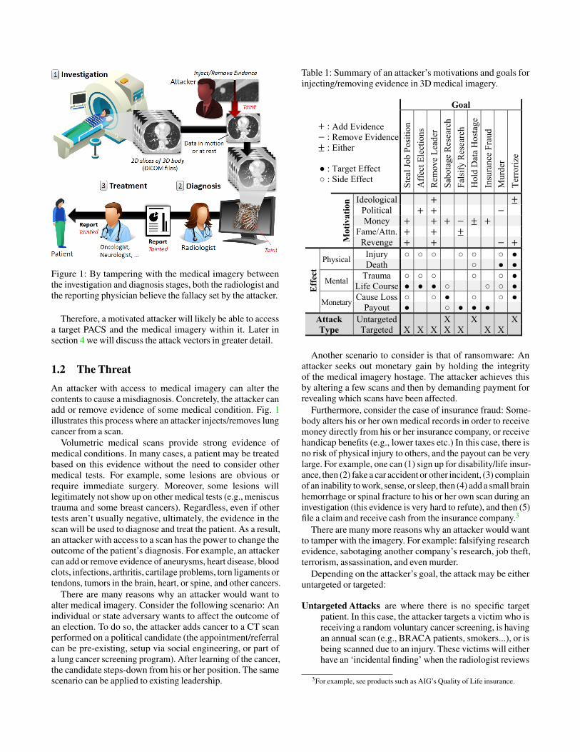
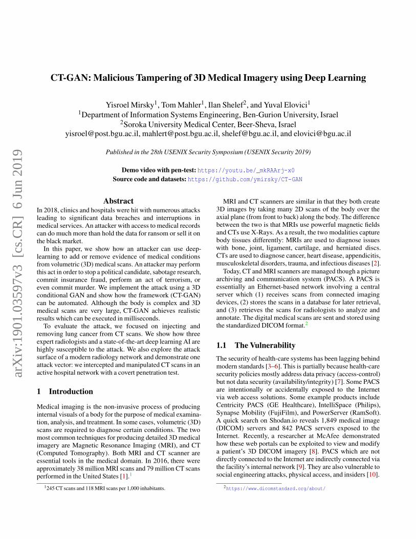
Figure 1: By tampering with the medical imagery betweenthe investigation and diagnosis stages, both the radiologist andthe reporting physician believe the fallacy set by the attacker.
Therefore, a motivated attacker will likely be able to accessa target PACS and the medical imagery within it. Later insection 4 we will discuss the attack vectors in greater detail.
1.2 The ThreatAn attacker with access to medical imagery can alter thecontents to cause a misdiagnosis. Concretely, the attacker canadd or remove evidence of some medical condition. Fig. 1illustrates this process where an attacker injects/removes lungcancer from a scan.
Volumetric medical scans provide strong evidence ofmedical conditions. In many cases, a patient may be treatedbased on this evidence without the need to consider othermedical tests. For example, some lesions are obvious orrequire immediate surgery. Moreover, some lesions willlegitimately not show up on other medical tests (e.g., meniscustrauma and some breast cancers). Regardless, even if othertests aren’t usually negative, ultimately, the evidence in thescan will be used to diagnose and treat the patient. As a result,an attacker with access to a scan has the power to change theoutcome of the patient’s diagnosis. For example, an attackercan add or remove evidence of aneurysms, heart disease, bloodclots, infections, arthritis, cartilage problems, torn ligaments ortendons, tumors in the brain, heart, or spine, and other cancers.
There are many reasons why an attacker would want toalter medical imagery. Consider the following scenario: Anindividual or state adversary wants to affect the outcome ofan election. To do so, the attacker adds cancer to a CT scanperformed on a political candidate (the appointment/referralcan be pre-existing, setup via social engineering, or part ofa lung cancer screening program). After learning of the cancer,the candidate steps-down from his or her position. The samescenario can be applied to existing leadership.
Table 1: Summary of an attacker’s motivations and goals forinjecting/removing evidence in 3D medical imagery.
Goal
: : :
● : ○ :
Add Evidence Remove Evidence Either Target Effect Side Effect
Ste
al J
ob P
osit
ion
Aff
ect E
lect
ions
Rem
ove
Lea
der
Sab
otag
e R
esea
rch
Fal
sify
Res
earc
h
Hol
d D
ata
Hos
tage
Insu
ranc
e F
raud
Mur
der
Ter
rori
ze
Mot
ivat
ion Ideological
Political Money
Fame/Attn. Revenge
Eff
ect
Physical Injury ○ ○ ○ ○ ○ ○ ●Death ○ ● ●
Mental Trauma ○ ○ ○ ○ ○ ●
Life Course ● ● ● ○ ○ ○ ●
Monetary Cause Loss ○ ○ ● ○ ○ ●
Payout ● ○ ● ● ●Attack Type
Untargeted X X XTargeted X X X X X X X
Another scenario to consider is that of ransomware: Anattacker seeks out monetary gain by holding the integrityof the medical imagery hostage. The attacker achieves thisby altering a few scans and then by demanding payment forrevealing which scans have been affected.
Furthermore, consider the case of insurance fraud: Some-body alters his or her own medical records in order to receivemoney directly from his or her insurance company, or receivehandicap benefits (e.g., lower taxes etc.) In this case, there isno risk of physical injury to others, and the payout can be verylarge. For example, one can (1) sign up for disability/life insur-ance, then (2) fake a car accident or other incident, (3) complainof an inability to work, sense, or sleep, then (4) add a small brainhemorrhage or spinal fracture to his or her own scan during aninvestigation (this evidence is very hard to refute), and then (5)file a claim and receive cash from the insurance company.3
There are many more reasons why an attacker would wantto tamper with the imagery. For example: falsifying researchevidence, sabotaging another company’s research, job theft,terrorism, assassination, and even murder.
Depending on the attacker’s goal, the attack may be eitheruntargeted or targeted:
Untargeted Attacks are where there is no specific targetpatient. In this case, the attacker targets a victim who isreceiving a random voluntary cancer screening, is havingan annual scan (e.g., BRACA patients, smokers...), or isbeing scanned due to an injury. These victims will eitherhave an ‘incidental finding’ when the radiologist reviews
3For example, see products such as AIG’s Quality of Life insurance.
the scan (injection) or are indeed sick but the evidencewon’t show (removal).
Targeted Attacks are where there is a specific target patient.In these attacks, the patient may be lured to the hospitalfor a scan. This can be accomplished by (1) addingan appointment in the system, (2) crafting a cancerscreening invite, (3) spoofing the patient’s doctor, or (4)tampering/appending the patient’s routine lab tests. Forexample, high-PSA in blood indicates prostate cancerleading to an abdominal MRI, high thyrotropin in bloodindicates a brain tumor leading to a head MRI, andmetanephrine in urine of hypertensive patients indicatescancer/tumor leading to a chest/abdominal CT
In this paper we will focus on the injection and removalof lung cancer from CT scans. Table 1 summarizes attacker’smotivations, goals, and effects by doing so. The reason weinvestigate this attack is because lung cancer is commonand has the highest mortality rate [11]. Therefore, due itsimpact, an attacker is likely to manipulate lung cancer toachieve his or her goal. We note that the threat, attack, andcountermeasures proposed in this paper also apply to MRIsand medical conditions other than those listed above.
1.3 The AttackWith the help of machine learning, the domain of imagegeneration has advanced significantly over the last tenyears [12]. In 2014, there was a breakthrough in the domainwhen Goodfellow et al. [13] introduced a special kind ofdeep neural network called a generative adversarial network(GAN). GANs consist of two neural networks which workagainst each other: the generator and the discriminator. Thegenerator creates fake samples with the aim of fooling thediscriminator, and the discriminator learns to differentiatebetween real and fake samples. When applied to images, theresult of this game helps the generator create fake imagerywhich are photo realistic. While GANs have been used forpositive tasks, researchers have also shown how they can beused for malicious tasks such as malware obfuscation [14, 15]and misinformation (e.g., deepfakes [16]).
In this paper, we show how an attacker can realisticallyinject and remove medical conditions with 3D CT scans. Theframework, called CT-GAN, uses two conditional GANs(cGAN) to perform in-painting (image completion) [17] on3D imagery. For injection, a cGAN is trained on unhealthysamples so that the generator will always complete the imagesaccordingly. Conversely, for removal, another cGAN is trainedon healthy samples only.
To make the process efficient and the output anatomicallyrealistic, we perform the following steps: (1) locate where theevidence should be inject/removed, (2) cut out a rectangularcuboid from the location, (3) interpolate (scale) the cuboid, (4)modify the cuboid with the cGAN, (5) rescale, and (6) pasteit back into the original scan. By dealing with a small portionof the scan, the problem complexity is reduced by focusingthe GAN on the relevant area of the body (as opposed to the
entire CT). Moreover, the algorithm complexity is reducedby processing fewer inputs4 (pixels) and concepts (anatomicalfeatures). This results in fast execution and high anatomicalrealism. The interpolation step is necessary because the scaleof a scan can be different between patients. To compensate forthe resulting interpolation blur, we mask the relevant contentaccording to water density in the tissue (Hounsfield units) andhide the smoothness by adding Gaussian white noise. In orderto assist the GAN in generating realistic features, histogramequalization is performed on the input samples. We found thatthis transformation helps the 3D convolutional neural networksin the GAN learn how to generate the subtle features foundin the human body. The entire process is automated, meaningsthat the attack can be deployed in an air gapped PACS.
To verify the threat of this attack, we trained CT-GANto inject/remove lung cancer and hired three radiologists todiagnose a mix of 70 tampered and 30 authentic CT scans.The radiologists diagnosed 99% of the injected patients withmalign cancer, and 94% of cancer removed patients as beinghealthy. After informing the radiologists of the attack, theystill misdiagnosed 60% of those with injections, and 87% ofthose with removals. In addition to the radiologists, we alsoshowed how CT-GAN is an effective adversarial machinelearning attack. We found that the state-of-the-art lung cancerscreening model misdiagnosed 100% of the tampered patients.Thus, cancer screening tools, used by some radiologists, arealso vulnerable to this attack.
This attack is a concern because infiltration of healthcarenetworks has become common [3], and internal networksecurity is often poor [18]. Moreover, for injection, the attackeris still likely to succeed even if medical treatment is notperformed. This is because many goals rely on simply scaringthe patient enough to affect his/her daily/professional life. Forexample, even if an immediate deletion surgery is not deemednecessary based on the scan and lab results, there will still beweekly/monthly follow-up scans to track the tumor’s growth.This will affect the patient’s life given the uncertainty of hisor her future.
1.4 The ContributionTo the best of our knowledge, it has not been shown how anattacker can maliciously alter the content of a 3D medical im-age in a realistic and automated way. Therefore, this is the firstcomprehensive research which exposes, demonstrates, and ver-ifies the threat of an attacker manipulating 3D medical imagery.In summary, the contributions of this paper are as follows:
The Attack Model We are the first to present how an attackercan infiltrate a PACS network and then use malwareto autonomously tamper 3D medical imagery. We alsoprovide a systematic overview of the attack, vulnerabilities,attack vectors, motivations, and attack goals. Finally,we demonstrate one possible attack vector through apenetration test performed on a hospital where we covertly
4A 3D CT scan can have over 157 million pixels whereas the latestadvances in GANs can only handle about 2 million pixels (HD images).
connect a man-in-the-middle device to an actual CT scanner.By performing this pen-test, we provide insights into thesecurity of a modern hospital’s internal network.
Attack Implementation We are the first to demonstratehow GANs, with the proper preprocessing, can be used toefficiently, realistically, and automatically inject/removelung cancer into/from large 3D CT scans. We also evaluatehow well the algorithm can deceive both humans andmachines: radiologists and state-of-the-art AI. We also showhow this implementation might be used by an attacker sinceit can be automated (in the case of an air gapped system)and is fast (in the case of an infected DICOM viewer).
Countermeasures We enumerate various countermeasureswhich can be used to mitigate the threat. We also providethe reader with best practices and configurations which canbe implemented immediately to help prevent this attack.
For reproducibility and further investigation, we havepublished our tampered datasets and source code online5
along with a pen-test video.6
The remainder of the paper is organized as follows: Firstwe present a short background on GANs. Then, in section 3,we review related works and contrast them ours. In section4 we present the attack model and demonstrate one of theattack vectors. In section 5, we present CT-GAN’s neuralarchitecture, its attack process, and some samples. In section6 we evaluate the quality of the manipulations and asses thethreat of the attack. Finally, in sections 7 and 8 we presentcountermeasures and our conclusion.
2 Background: GANs
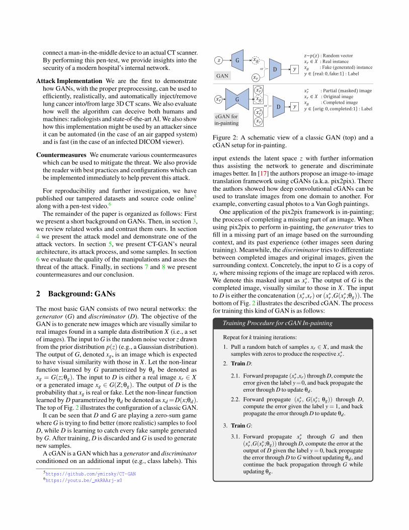
The most basic GAN consists of two neural networks: thegenerator (G) and discriminator (D). The objective of theGAN is to generate new images which are visually similar toreal images found in a sample data distribution X (i.e., a setof images). The input to G is the random noise vector z drawnfrom the prior distribution p(z) (e.g., a Gaussian distribution).The output of G, denoted xg, is an image which is expectedto have visual similarity with those in X . Let the non-linearfunction learned by G parametrized by θg be denoted asxg = G(z;θg). The input to D is either a real image xr ∈ Xor a generated image xg ∈ G(Z;θg). The output of D is theprobability that xg is real or fake. Let the non-linear functionlearned by D parametrized by θd be denoted as xd =D(x;θd).The top of Fig. 2 illustrates the configuration of a classic GAN.
It can be seen that D and G are playing a zero-sum gamewhere G is trying to find better (more realistic) samples to foolD, while D is learning to catch every fake sample generatedby G. After training, D is discarded and G is used to generatenew samples.
A cGAN is a GAN which has a generator and discriminatorconditioned on an additional input (e.g., class labels). This
5https://github.com/ymirsky/CT-GAN6https://youtu.be/_mkRAArj-x0
GAN
cGAN for in-painting
Random vectorReal instanceFake (generated) instance
Label
imageCompleted image
Label
Figure 2: A schematic view of a classic GAN (top) and acGAN setup for in-painting.
input extends the latent space z with further informationthus assisting the network to generate and discriminateimages better. In [17] the authors propose an image-to-imagetranslation framework using cGANs (a.k.a. pix2pix). Therethe authors showed how deep convolutional cGANs can beused to translate images from one domain to another. Forexample, converting casual photos to a Van Gogh paintings.
One application of the pix2pix framework is in-painting;the process of completing a missing part of an image. Whenusing pix2pix to perform in-painting, the generator tries tofill in a missing part of an image based on the surroundingcontext, and its past experience (other images seen duringtraining). Meanwhile, the discriminator tries to differentiatebetween completed images and original images, given thesurrounding context. Concretely, the input to G is a copy ofxr where missing regions of the image are replaced with zeros.We denote this masked input as x∗r . The output of G is thecompleted image, visually similar to those in X . The inputto D is either the concatenation (x∗r ,xr) or (x∗r ,G(x∗r ;θg)). Thebottom of Fig. 2 illustrates the described cGAN. The processfor training this kind of GAN is as follows:
Training Procedure for cGAN In-painting
Repeat for k training iterations:
1. Pull a random batch of samples xr ∈ X , and mask thesamples with zeros to produce the respective x∗r .
2. Train D:
2.1. Forward propagate (x∗r ,xr) through D, compute theerror given the label y=0, and back propagate theerror through D to update θd .
2.2. Forward propagate (x∗r , G(x∗r ; θg)) through D,compute the error given the label y = 1, and backpropagate the error through D to update θd .
3. Train G:
3.1. Forward propagate x∗r through G and then(x∗r ,G(x∗r ;θg)) through D, compute the error at theoutput of D given the label y = 0, back propagatethe error through D to G without updating θd , andcontinue the back propagation through G whileupdating θg.
Although pix2pix does not use a latent random input z, itavoids deterministic outputs by performing random dropoutsin the generator during training. this forces the network tolearn multiple representations of the data.
We note that there is a GAN called a CycleGAN [19] that candirectly translate images between two domains (e.g., benign↔malign). However, we found that the CycleGAN was unableto inject realistic cancer into 3D samples. Therefore, we optedto use the pix2pix model for in-painting because it producedmuch better results. This may be due to the complexity of theanatomy in the 3D samples and the fact that we had relativelyfew training samples. Since labeled datasets contain at most afew hundred scans, our approach is more likely to be used by anattacker. Another reason is that in-painting is arguably easierto perform than ‘style transfer’ when considering differentbodies. Regardless, in-painting ensures that the final image canbe seamlessly pasted back into the scan without border effects.
3 Related Work
The concept of tampering medical imagery, and the use ofGANs on medical imagery, is not new. In this section we brieflyreview these subjects and compare prior results to our work.
3.1 Tampering with Medical ImagesMany works have proposed methods for detecting forgeriesin medical images [20], but none have focused on the attackitself. The most common methods of image forgery are:copying content from one image to another (image splicing),duplicating content within the same image to cover up or addsomething (copy-move), and enhancing an image to give ita different feel (image retouching) [21].
Copy-move attacks can be used to cover up evidence or dupli-cate existing evidence (e.g., a tumor). However, duplicating evi-dence will raise suspicion because radiologists closely analyzeeach discovered instance. Image-splicing can be used to copyevidence from one scan to another. However, CT scanners havedistinct local noise patterns which are visually noticeable [22,23]. The copied patterns would not fit the local pattern and thusraise suspicion. More importantly, both copy-move and image-splicing techniques are performed using 2D image editing soft-ware such as Photoshop. These tools require a digital artist tomanually edit the scan. Even if the attacker has a digital artist, itis hard to accurately inject and remove cancer realistically. Thisis because human bodies are complex and diverse. For exam-ple, cancers and tumors are usually attached to nearby anatomy(lung walls, bronchi, etc.) which may be hard to alter accuratelyunder the scrutiny of expert radiologists. Another considerationis that CT scans are 3D and not 2D, which adds to the difficulty.It is also important to note that an attacker will likely need toautomate the entire process in a malware since (1) many PACSare not directly connected to the Internet and (2) the diagnosismay occur immediately after the scan is performed.
In contrast to the Photoshopping approach, CT-GAN(1) works on 3D medical imagery, which provide stronger
evidence than a 2D scans, (2) realistically alters the contentsof a 3D scan while considering nearby anatomy, and (3) can becompletely automated. The latter point is important because(1) some PACS are not directly connected to the Internet,(2) diagnosis can happen right after the actual scan, (3) themalware may be inside the radiologist’s viewing app.
3.2 GANs in Medical ImagerySince 2016, over 100 papers relating to GANs and medicalimaging have been published [24]. These publications mostlyrelate image reconstruction, denoising, image generation (syn-thesis), segmentation, detection, classification, and registration.We will focus on the use of GANs to generate medical images.
Due to privacy laws, it is hard to acquire medical scansfor training models and students. As a result, the main focusof GANs in this domain has been towards augmenting(expanding) datasets. One approach is to convert imageryfrom one modality to another. For example, in [25] the authorsused cGANs to convert 2D slices of CT images to PositronEmission Tomography (PET) images. In [26, 27] the authorsdemonstrated a similar concept using a fully convolutionalnetwork with a cGAN architecture. In [28], the authorsconverted MRI images to CT images using domain adaptation.In [29], the authors converted MRI to CT images and viceversa using a CycleGAN.
Another approach to augmenting medical datasets is thegeneration of new instances. In [30], the authors use a deepconvolutional GAN (DCGAN) to generate 2D brain MRIimages with a resolution of 220x172. In [31], the authorsused a DCGAN to generate 2D liver lesions with a resolutionof 64x64. In [32], the authors generated 3D blood vesselsusing a Wasserstien (WGAN). In [33], the authors use aLaplace GAN (LAPGAN) to generate skin lesion images with256x256 resolution. In [34], the authors train two DCGANsfor generating 2D chest X-rays (one for malign and the otherfor benign). However, in [34], the generated samples weredown sampled to 128x128 in resolution since this approachcould not be scaled to the original resolution of 2000x3000.In [35] the authors generated 2D images of pulmonary lungnodules (lung cancer) with 56x56 resolution. The author’smotivation was to create realistic datasets for doctors topractice on. The samples were generated using a DCGAN andtheir realism was assessed with help of two radiologists. Theauthors found that the radiologists were unable to accuratelydifferentiate between real and fake samples.
These works contrast to our work in the following ways:
1. We are the first to introduce the use of GANs as a wayto tamper with 3D imagery. The other works focusedon synthesizing cancer samples for boosting classifiers,experiments, and training students, but not for maliciousattacks. We also provide an overview of how the attackcan be accomplished in a modern medical system.
2. All of the above works either generate small regions ofa scan without the context of a surrounding body or gen-erate a full 2D scan with a very low resolution. Samples
which are generated without a context cannot be realis-tically ‘pasted’ back into any arbitrary medical scan. Wegenerate/remove content realistically within existing bod-ies. Moreover, very low-resolution images of full scanscannot replace existing ones without raising suspicion(especially if the body doesn’t match the actual person).Our approach can modify full resolution 3D scans,7 andthe approach can be easily extended to 2D as well.
3. We are the first to evaluate how well a GAN can foolexpert radiologists and state-of-the-art AI in full 3Dlung cancer screening. Moreover, in our evaluation, theradiologists and AI were able to consider how the cancerwas attached and placed within the surrounding anatomy.
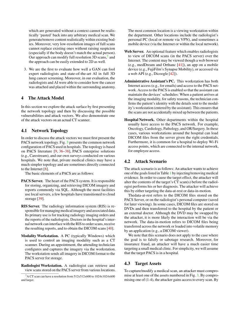
4 The Attack Model
In this section we explore the attack surface by first presentingthe network topology and then by discussing the possiblevulnerabilities and attack vectors. We also demonstrate oneof the attack vectors on an actual CT scanner.
4.1 Network TopologyIn order to discuss the attack vectors we must first present thePACS network topology. Fig. 3 presents the common networkconfiguration of PACS used in hospitals. The topology is basedon PACS literature [9, 36–38], PACS enterprise solutions(e.g., Carestream), and our own surveys conducted on varioushospitals. We note that, private medical clinics may have amuch simpler topology and are sometimes directly connectedto the Internet [8].
The basic elements of a PACS are as follows:
PACS Server. The heart of the PACS system. It is responsiblefor storing, organizing, and retrieving DICOM imagery andreports commonly via SQL. Although the most facilitiesuse local servers, a few hospitals have transitioned to cloudstorage [39].
RIS Server. The radiology information system (RIS) is re-sponsible for managing medical imagery and associated data.Its primary use is for tracking radiology imaging orders andthe reports of the radiologists. Doctors in the hospital’s inter-nal network can interface with the RIS to order scans, receivethe resulting reports, and to obtain the DICOM scans [40].
Modality Workstation. A PC (typically Windows) whichis used to control an imaging modality such as a CTscanner. During an appointment, the attending technicianconfigures and captures the imagery via the workstation.The workstation sends all imagery in DICOM format to thePACS server for storage.
Radiologist Workstation. A radiologist can retrieve andview scans stored on the PACS server from various locations.7A CT scan can have a resolution from 512x512x600 to 1024x1024x600
and larger.
The most common location is a viewing workstation withinthe department. Other locations include the radiologist’spersonal PC (local or remote via VPN), and sometimes amobile device (via the Internet or within the local network).
Web Server. An optional feature which enables radiologiststo view of DICOM scans (in the PACS server) over theInternet. The content may be viewed though a web browser(e.g., medDream and Orthanc [41]), an app on a mobiledevice (e.g., FujiFilm’s Synapse Mobility), or accessed viaa web API (e.g., Dicoogle [42]).
Administrative Assistant’s PC. This workstation has bothInternet access (e.g., for emails) and access to the PACS net-work. Access to the PACS is enabled so that the assistant canmaintain the devices’ schedules: When a patient arrives atthe imaging modality, for safety reasons, the technician con-firms the patient’s identity with the details sent to the modal-ity’s workstation (entered by the assistant). This ensures thatthe scans are not accidentally mixed up between the patients.
Hospital Network. Other departments within the hospitalusually have access to the PACS network. For example,Oncology, Cardiology, Pathology, and OR/Surgery. In thesecases, various workstations around the hospital can loadDICOM files from the server given the right credentials.Furthermore, it is common for a hospital to deploy Wi-Fiaccess points, which are connected to the internal network,for employee access.
4.2 Attack ScenarioThe attack scenario is as follows: An attacker wants to achieveone of the goals listed in Table 1 by injecting/removing medicalevidence. In order to cause the target effect, the attacker willalter the contents of the target’s CT scan(s) before the radiol-ogist performs his or her diagnosis. The attacker will achievethis by either targeting the data-at-rest or data-in-motion.
Thedata-at-rest refers to the DICOM files stored on thePACS Server, or on the radiologist’s personal computer (savedfor later viewing). In some cases, DICOM files are stored onDVDs and then transferred to the hospital by the patient oran external doctor. Although the DVD may be swapped bythe attacker, it is more likely the interaction will be via thenetwork. The data-in-motion refers to DICOM files beingtransferred across the network or loaded into volatile memoryby an application (e.g., a DICOM viewer).
We note that this scenario does not apply to the case wherethe goal is to falsify or sabotage research. Moreover, forinsurance fraud, an attacker will have a much easier timetargeting a small medical clinic. For simplicity, we will assumethat the target PACS is in a hospital.
4.3 Target AssetsTo capture/modify a medical scan, an attacker must compro-mise at least one of the assets numbered in Fig. 3. By compro-mising one of (1-4), the attacker gains access to every scan. By
Physician Workstation
DR D
evice
CT S
cann
erM
RIUl
tra S
ound
Web Server
Film Print Manager
Radiology Information System
Administration Terminal
Radiologist Workstations
InternetRemote
Site
Client Viewer
Modality Workstations
yons
1
4liCC
5
PACS Server/DB 3
stations
5
Hospital Network
5
2 PACS NetworkEthernet
VPN Router
Secretary PC
Oncology, Cardiology,
Surgery, Pathology…Ethernet
DICOM Firewall
WiFiNetworks
Figure 3: A network overview a PACS in a hospital. 1-3: points where an attacker can tamper with all scans. 4-5: points wherean attacker can tamper with a subset of scans.
compromising (5) or (6), the attacker only gains access to a sub-set of scans. The RIS (3) can give the attacker full control overthe PACS server (2), but only if the attacker can obtain the rightcredentials or exploit the RIS software. The network wiring be-tween the modalities and the PACS server (4) can be used to in-stall a man-in-the-middle device. This device can modify data-in-motion if it is not encrypted (or if the protocol is flawed).
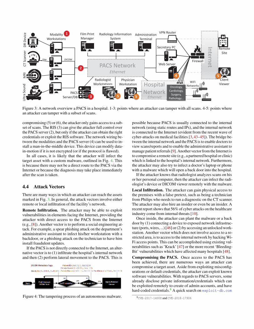
In all cases, it is likely that the attacker will infect thetarget asset with a custom malware, outlined in Fig. 4. Thisis because there may not be a direct route to the PACS via theInternet or because the diagnosis may take place immediatelyafter the scan is taken.
4.4 Attack VectorsThere are many ways in which an attacker can reach the assetsmarked in Fig. 3. In general, the attack vectors involve eitherremote or local infiltration of the facility’s network.Remote Infiltration. The attacker may be able to exploitvulnerabilities in elements facing the Internet, providing theattacker with direct access to the PACS from the Internet(e.g., [8]). Another vector is to perform a social engineering at-tack. For example, a spear phishing attack on the department’sadministrative assistant to infect his/her workstation with abackdoor, or a phishing attack on the technician to have himinstall fraudulent updates.
If the PACS is not directly connected to the Internet, an alter-native vector is to (1) infiltrate the hospital’s internal networkand then (2) perform lateral movement to the PACS. This is
Figure 4: The tampering process of an autonomous malware.
possible because PACS is usually connected to the internalnetwork (using static routes and IPs), and the internal networkis connected to the Internet (evident from the recent wave ofcyber-attacks on medical facilities [3, 43–45]). The bridge be-tween the internal network and the PACS is to enable doctors toview scans/reports and to enable the administrative assistant tomanage patient referrals [9]. Another vector from the Internet isto compromise a remote site (e.g., a partnered hospital or clinic)which is linked to the hospital’s internal network. Furthermore,the attacker may also try to infect a doctor’s laptop or phonewith a malware which will open a back door into the hospital.
If the attacker knows that radiologist analyzes scans on hisor her personal computer, then the attacker can infect the radi-ologist’s device or DICOM viewer remotely with the malware.Local Infiltration. The attacker can gain physical access tothe premises with a false pretext, such as being a technicianfrom Philips who needs to run a diagnostic on the CT scanner.The attacker may also hire an insider or even be an insider. Arecent report shows that 56% of cyber attacks on the healthcareindustry come from internal threats [10].
Once inside, the attacker can plant the malware or a backdoor by (1) connecting a device to exposed network infrastruc-ture (ports, wires, ...) [46] or (2) by accessing an unlocked work-station. Another vector which does not involve access to a re-stricted area, is to access to the internal network by hacking Wi-Fi access points. This can be accomplished using existing vul-nerabilities such as ’Krack’ [47] or the more recent ‘Bleeding-Bit’ vulnerabilities which have affected many hospitals [48].Compromising the PACS. Once access to the PACS hasbeen achieved, there are numerous ways an attacker cancompromise a target asset. Aside from exploiting misconfig-urations or default credentials, the attacker can exploit knownsoftware vulnerabilities. With regards to PACS servers, somealready disclose private information/credentials which canbe exploited remotely to create of admin accounts, and havehard-coded credentials.8 A quick search on exploit-db.com
8CVE-2017-14008 and CVE-2018-17906
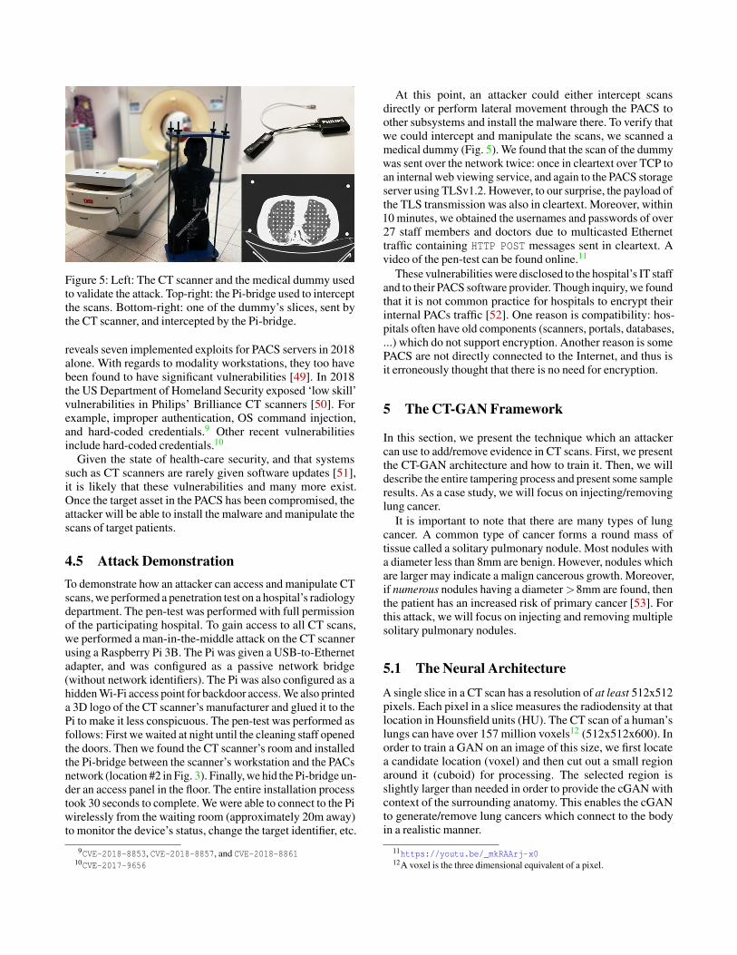
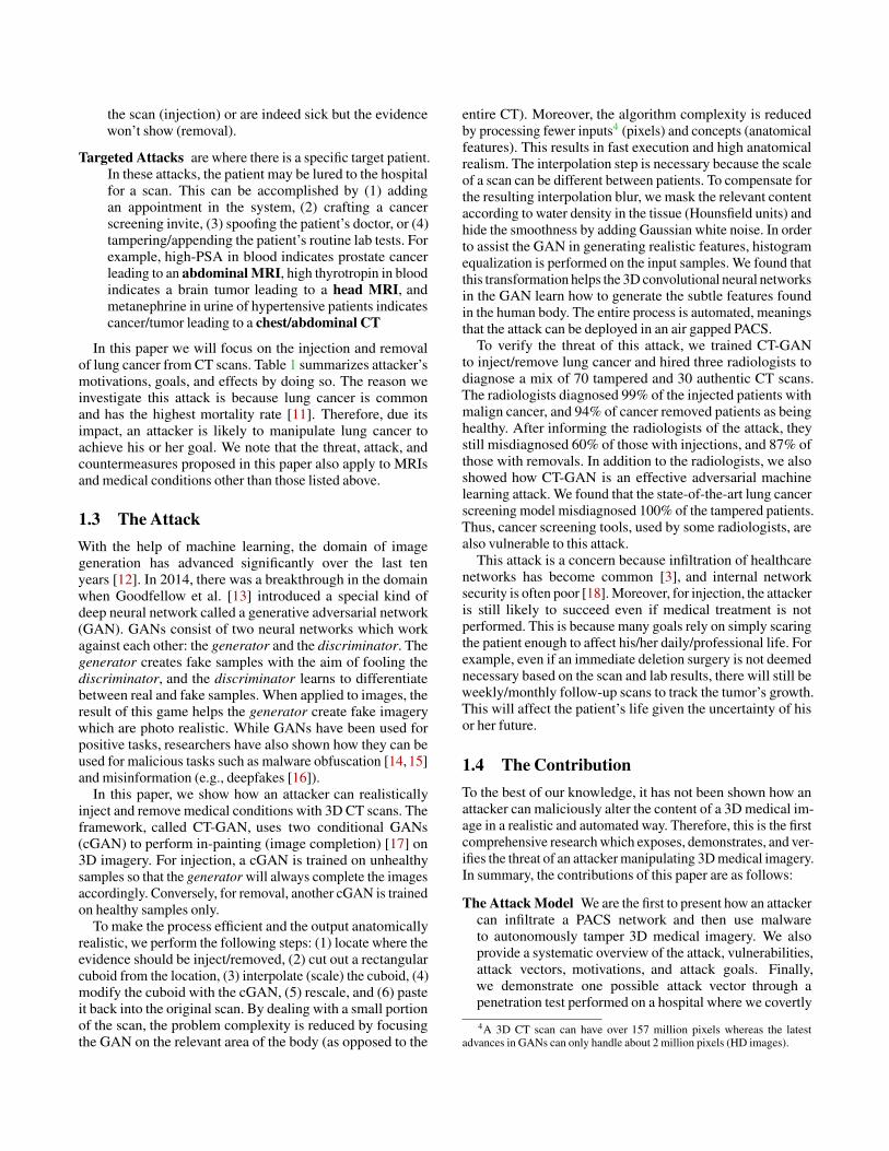
Figure 5: Left: The CT scanner and the medical dummy usedto validate the attack. Top-right: the Pi-bridge used to interceptthe scans. Bottom-right: one of the dummy’s slices, sent bythe CT scanner, and intercepted by the Pi-bridge.
reveals seven implemented exploits for PACS servers in 2018alone. With regards to modality workstations, they too havebeen found to have significant vulnerabilities [49]. In 2018the US Department of Homeland Security exposed ‘low skill’vulnerabilities in Philips’ Brilliance CT scanners [50]. Forexample, improper authentication, OS command injection,and hard-coded credentials.9 Other recent vulnerabilitiesinclude hard-coded credentials.10
Given the state of health-care security, and that systemssuch as CT scanners are rarely given software updates [51],it is likely that these vulnerabilities and many more exist.Once the target asset in the PACS has been compromised, theattacker will be able to install the malware and manipulate thescans of target patients.
4.5 Attack DemonstrationTo demonstrate how an attacker can access and manipulate CTscans, we performed a penetration test on a hospital’s radiologydepartment. The pen-test was performed with full permissionof the participating hospital. To gain access to all CT scans,we performed a man-in-the-middle attack on the CT scannerusing a Raspberry Pi 3B. The Pi was given a USB-to-Ethernetadapter, and was configured as a passive network bridge(without network identifiers). The Pi was also configured as ahidden Wi-Fi access point for backdoor access. We also printeda 3D logo of the CT scanner’s manufacturer and glued it to thePi to make it less conspicuous. The pen-test was performed asfollows: First we waited at night until the cleaning staff openedthe doors. Then we found the CT scanner’s room and installedthe Pi-bridge between the scanner’s workstation and the PACsnetwork (location #2 in Fig. 3). Finally, we hid the Pi-bridge un-der an access panel in the floor. The entire installation processtook 30 seconds to complete. We were able to connect to the Piwirelessly from the waiting room (approximately 20m away)to monitor the device’s status, change the target identifier, etc.
9CVE-2018-8853, CVE-2018-8857, and CVE-2018-886110CVE-2017-9656
At this point, an attacker could either intercept scansdirectly or perform lateral movement through the PACS toother subsystems and install the malware there. To verify thatwe could intercept and manipulate the scans, we scanned amedical dummy (Fig. 5). We found that the scan of the dummywas sent over the network twice: once in cleartext over TCP toan internal web viewing service, and again to the PACS storageserver using TLSv1.2. However, to our surprise, the payload ofthe TLS transmission was also in cleartext. Moreover, within10 minutes, we obtained the usernames and passwords of over27 staff members and doctors due to multicasted Ethernettraffic containing HTTP POST messages sent in cleartext. Avideo of the pen-test can be found online.11
These vulnerabilities were disclosed to the hospital’s IT staffand to their PACS software provider. Though inquiry, we foundthat it is not common practice for hospitals to encrypt theirinternal PACs traffic [52]. One reason is compatibility: hos-pitals often have old components (scanners, portals, databases,...) which do not support encryption. Another reason is somePACS are not directly connected to the Internet, and thus isit erroneously thought that there is no need for encryption.
5 The CT-GAN Framework
In this section, we present the technique which an attackercan use to add/remove evidence in CT scans. First, we presentthe CT-GAN architecture and how to train it. Then, we willdescribe the entire tampering process and present some sampleresults. As a case study, we will focus on injecting/removinglung cancer.
It is important to note that there are many types of lungcancer. A common type of cancer forms a round mass oftissue called a solitary pulmonary nodule. Most nodules witha diameter less than 8mm are benign. However, nodules whichare larger may indicate a malign cancerous growth. Moreover,if numerous nodules having a diameter >8mm are found, thenthe patient has an increased risk of primary cancer [53]. Forthis attack, we will focus on injecting and removing multiplesolitary pulmonary nodules.
5.1 The Neural Architecture
A single slice in a CT scan has a resolution of at least 512x512pixels. Each pixel in a slice measures the radiodensity at thatlocation in Hounsfield units (HU). The CT scan of a human’slungs can have over 157 million voxels12 (512x512x600). Inorder to train a GAN on an image of this size, we first locatea candidate location (voxel) and then cut out a small regionaround it (cuboid) for processing. The selected region isslightly larger than needed in order to provide the cGAN withcontext of the surrounding anatomy. This enables the cGANto generate/remove lung cancers which connect to the bodyin a realistic manner.
11https://youtu.be/_mkRAArj-x012A voxel is the three dimensional equivalent of a pixel.
Original Image Masked 32x32x32Zero Mask16x16x16 Zero Mask16x16x16 16x16x16
8x8x8
800
…
400
…
200
…
4x4x4 2x2x2{real, fake}
Original Image 32x32x32
16x16x16
16x16x168x8x8
4x4x4
100x2
…
200x2
…
400x2
…
800x2
…
800x2
…
800
…
400
…
200
…
#Filters: 100
…
Generated Image 32x32x32
4x4x4 KernelStride 1
4x4x4 2x2x2 4x4x4
Symmetric Skip Connections
4x4x4 KernelStride 2
Zero Mask
Scaled and normalizedCubes cut from CTs
Gene
rato
r
16x16x16
Conv3DConv3D, Leaky ReLu
Conv3D, Leaky ReLu, Batch-NormUpSample3D, Conv3D, ReLu, Dropout, Batch-NormUpSample3D, Conv3D, tanh
3D Crop from CT Scan
Disc
rimin
ator
OR 4x4x4 KernelStride 2
Sample
Original OR Generated Image 32x32x32
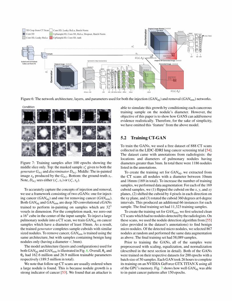
Figure 6: The network architecture, layers, and parameters used for both the injection (GANinj) and removal (GANrem) networks.
Figure 7: Training samples after 100 epochs showing themiddle slice only. Top: the masked sample x∗r given to both thegenerator Gin j and discriminator Din j. Middle: The in-paintedimage xg produced by the Gin j. Bottom: the ground-truth xr.Note, Din j sees either (x∗r , xr) or (x∗r , xg).
To accurately capture the concepts of injection and removal,we use a framework consisting of two cGANs: one for inject-ing cancer (GANinj) and one for removing cancer (GANrem).Both GANinj and GANrem are deep 3D convolutional cGANstrained to perform in-painting on samples which are 323
voxels in dimension. For the completion mask, we zero-outa 163 cube in the center of the input sample. To inject a largepulmonary nodule into a CT scan, we train GANinj on cancersamples which have a diameter of least 10mm. As a result,the trained generator completes sample cuboids with similarsized nodules. To remove cancer, GANrem is trained using thesame architecture, but with samples containing benign lungnodules only (having a diameter <3mm).
The model architecture (layers and configurations) used forboth GANinj and GANrem is illustrated in Fig. 6. Overall, θg andθd had 162.6 million and 26.9 million trainable parametersrespectively (189.5 million in total).
We note that follow up CT scans are usually ordered whena large nodule is found. This is because nodule growth is astrong indicator of cancer [53]. We found that an attacker is
able to simulate this growth by conditioning each canceroustraining sample on the nodule’s diameter. However, theobjective of this paper is to show how GANS can add/removeevidence realistically. Therefore, for the sake of simplicity,we have omitted this ‘feature’ from the above model.
5.2 Training CT-GAN
To train the GANs, we used a free dataset of 888 CT scanscollected in the LIDC-IDRI lung cancer screening trial [54].The dataset came with annotations from radiologists: thelocations and diameters of pulmonary nodules havingdiameters greater than 3mm. In total there were 1186 noduleslisted in the annotations.
To create the training set for GANinj, we extracted fromthe CT scans all nodules with a diameter between 10mmand 16mm (169 in total). To increase the number of trainingsamples, we performed data augmentation: For each of the 169cuboid samples, we (1) flipped the cuboid on the x, y, and xyplanes, (2) shifted the cuboid by 4 pixels in each direction onthe xy plane, and (3) rotated the cuboid 360 degrees at 6 degreeintervals. This produced an additional 66 instances for eachsample. The final training set had 11,323 training samples.
To create the training set for GANrem, we first selected cleanCT scans which had no nodules detected by the radiologists. Onthese scans, we used the nodule detection algorithm from [55](also provided in the dataset’s annotations) to find benignmicro nodules. Of the detected micro nodules, we selected 867nodules at random and performed the same data augmentationas above. The final training set had 58,089 samples.
Prior to training the GANs, all of the samples werepreprocessed with scaling, equalization, and normalization(described in the next section in detail). Both of the GANswere trained on their respective datasets for 200 epochs with abatch size of 50 samples. Each GAN took 26 hours to completeits training on an NVIDIA GeForce GTX TITAN X using allof the GPU’s memory. Fig. 7 shows how well GANinj was ableto in-paint cancer patterns after 150 epochs.
DICOMFILESS
MM
ORInjection:Find nodule or select random location
Removal:Find largest nodule
+=AWGNpaste
cut Equalize Normalize Mask CenterScale 1:1:1
Unequalize Unnormalize
read
write
readrea1
cccccccc2
3
q4 5
ask Ce6
7a
891011pp12
14
Touch-up
Preprocessing
Post-processingInject Cancer
Remove Cancer
Original ( ) Cropped & Scaled
Generated De-Norm/Equalized
Merge Weights: Final -pasted1:1:1
1.3 : 0.6 : 0.6
Rescaled ( )
32x32
Condition MaskNorm. & Equalized1:1
3 4,5 6
7b
7a 8,9
10O2 11 12
Merge ,13
RemFindFinddFind
dnodd
papaaa
dd
e
d
pp12
1133repeat?
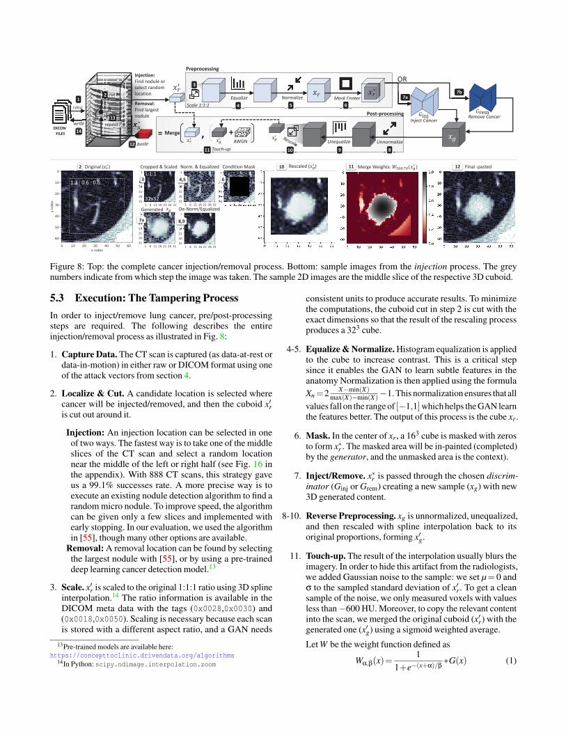
Figure 8: Top: the complete cancer injection/removal process. Bottom: sample images from the injection process. The greynumbers indicate from which step the image was taken. The sample 2D images are the middle slice of the respective 3D cuboid.
5.3 Execution: The Tampering ProcessIn order to inject/remove lung cancer, pre/post-processingsteps are required. The following describes the entireinjection/removal process as illustrated in Fig. 8:
1. Capture Data. The CT scan is captured (as data-at-rest ordata-in-motion) in either raw or DICOM format using oneof the attack vectors from section 4.
2. Localize & Cut. A candidate location is selected wherecancer will be injected/removed, and then the cuboid x′ris cut out around it.
Injection: An injection location can be selected in oneof two ways. The fastest way is to take one of the middleslices of the CT scan and select a random locationnear the middle of the left or right half (see Fig. 16 inthe appendix). With 888 CT scans, this strategy gaveus a 99.1% successes rate. A more precise way is toexecute an existing nodule detection algorithm to find arandom micro nodule. To improve speed, the algorithmcan be given only a few slices and implemented withearly stopping. In our evaluation, we used the algorithmin [55], though many other options are available.
Removal: A removal location can be found by selectingthe largest nodule with [55], or by using a pre-traineddeep learning cancer detection model.13
3. Scale. x′r is scaled to the original 1:1:1 ratio using 3D splineinterpolation.14 The ratio information is available in theDICOM meta data with the tags (0x0028,0x0030) and(0x0018,0x0050). Scaling is necessary because each scanis stored with a different aspect ratio, and a GAN needs
13Pre-trained models are available here:https://concepttoclinic.drivendata.org/algorithms
14In Python: scipy.ndimage.interpolation.zoom
consistent units to produce accurate results. To minimizethe computations, the cuboid cut in step 2 is cut with theexact dimensions so that the result of the rescaling processproduces a 323 cube.
4-5. Equalize & Normalize. Histogram equalization is appliedto the cube to increase contrast. This is a critical stepsince it enables the GAN to learn subtle features in theanatomy Normalization is then applied using the formulaXn=2 X−min(X)
max(X)−min(X)−1. This normalization ensures that allvalues fall on the range of [−1,1]which helps the GAN learnthe features better. The output of this process is the cube xr.
6. Mask. In the center of xr, a 163 cube is masked with zerosto form x∗r . The masked area will be in-painted (completed)by the generator, and the unmasked area is the context).
7. Inject/Remove. x∗r is passed through the chosen discrim-inator (Ginj or Grem) creating a new sample (xg) with new3D generated content.
8-10. Reverse Preprocessing. xg is unnormalized, unequalized,and then rescaled with spline interpolation back to itsoriginal proportions, forming x′g.
11. Touch-up. The result of the interpolation usually blurs theimagery. In order to hide this artifact from the radiologists,we added Gaussian noise to the sample: we set µ= 0 andσ to the sampled standard deviation of x′r. To get a cleansample of the noise, we only measured voxels with valuesless than−600 HU. Moreover, to copy the relevant contentinto the scan, we merged the original cuboid (x′r) with thegenerated one (x′g) using a sigmoid weighted average.
Let W be the weight function defined as
Wα,β(x)=1
1+e−(x+α)/β∗G(x) (1)
where parameter α is the HU threshold between wantedand unwanted tissue densities, and parameter β controlsthe smoothness of the cut edges. The function G(x) returnsa 0-1 normalized Gaussian kernel with the dimensions ofx. G(x) is used to decay the contribution of each voxel thefurther it is the cuboid’s center.
With W , we define the merging function asmergeα,β(x,y)=Wα,β(x)∗x+
(1−Wα,β(x)
)∗y (2)
where x is source (x′g) and y is the destination (x′r). Wefound that setting α = 500 and β = 70 worked best. Byapplying these touch-ups, the final cuboid x∗g is produced.
12. Paste. The cuboid x∗g is pasted back into the CT scan at theselected location. See Fig. 16 in the appendix for one sliceof a complete scan.
13. Repeat. If the attacker is removing cancer, then return tostep 2 until no more nodules with a diameter > 3mm arefound. If the attacker is injecting cancer, then (optionally)return to step 2 until four injections have been performed.The reason for this is because the risk of a patient beingdiagnosed with cancer is statistically greater in the presenceof exactly four solitary pulmonary nodules having adiameter >8mm [53].
14. Return Data. The scan is converted back into the originalformat (e.g. DICOM) and returned back to the source.
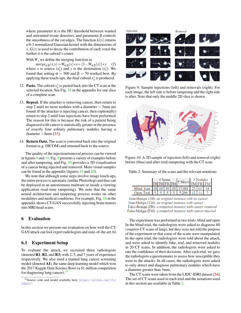
The quality of the injection/removal process can be viewedin figures 9 and 10. Fig. 9 presents a variety of examples beforeand after tampering, and Fig. 10 provides a 3D visualizationof a cancer being injected and removed. More visual samplescan be found in the appendix (figures 19 and 20).
We note that although some steps involve image touch-ups,the entire process is automatic (unlike Photoshop) and thus canbe deployed in an autonomous malware or inside a viewingapplication (real-time tampering). We note that the sameneural architecture and tampering process works on othermodalities and medical conditions. For example, Fig. 18 in theappendix shows CT-GAN successfully injecting brain tumorsinto MRI head scans.
6 Evaluation
In this section we present our evaluation on how well the CT-GAN attack can fool expert radiologists and state-of-the-art AI.
6.1 Experiment SetupTo evaluate the attack, we recruited three radiologists(denoted R1, R2, and R3) with 2, 5, and 7 years of experiencerespectively. We also used a trained lung cancer screeningmodel (denoted AI), the same deep learning model which wonthe 2017 Kaggle Data Science Bowl (a $1 million competitionfor diagnosing lung cancer).15
15Source code and model available here: https://github.com/lfz/DSB2017
Figure 9: Sample injections (left) and removals (right). Foreach image, the left side is before tampering and the right sideis after. Note that only the middle 2D slice is shown.
Figure 10: A 3D sample of injection (left) and removal (right)before (blue) and after (red) tampering with the CT scan.
Table 2: Summary of the scans and the relevant notations
The experiment was performed in two trials: blind and open.In the blind trial, the radiologists were asked to diagnose 80complete CT scans of lungs, but they were not told the purposeof the experiment or that some of the scans were manipulated.In the open trial, the radiologists were told about the attack,and were asked to identify fake, real, and removed nodulesin 20 CT scans. In addition, the radiologists were asked torate the confidence of their decisions. After each trial, we gavethe radiologists a questionnaire to assess how susceptible theywere to the attacks. In all cases, the radiologists were askedto only detect and diagnose pulmonary nodules which havea diameter greater than 3mm.
The CT scans were taken from the LIDC-IDRI dataset [54].The set of CT scans used in each trial and the notations usedin this section are available in Table 2.
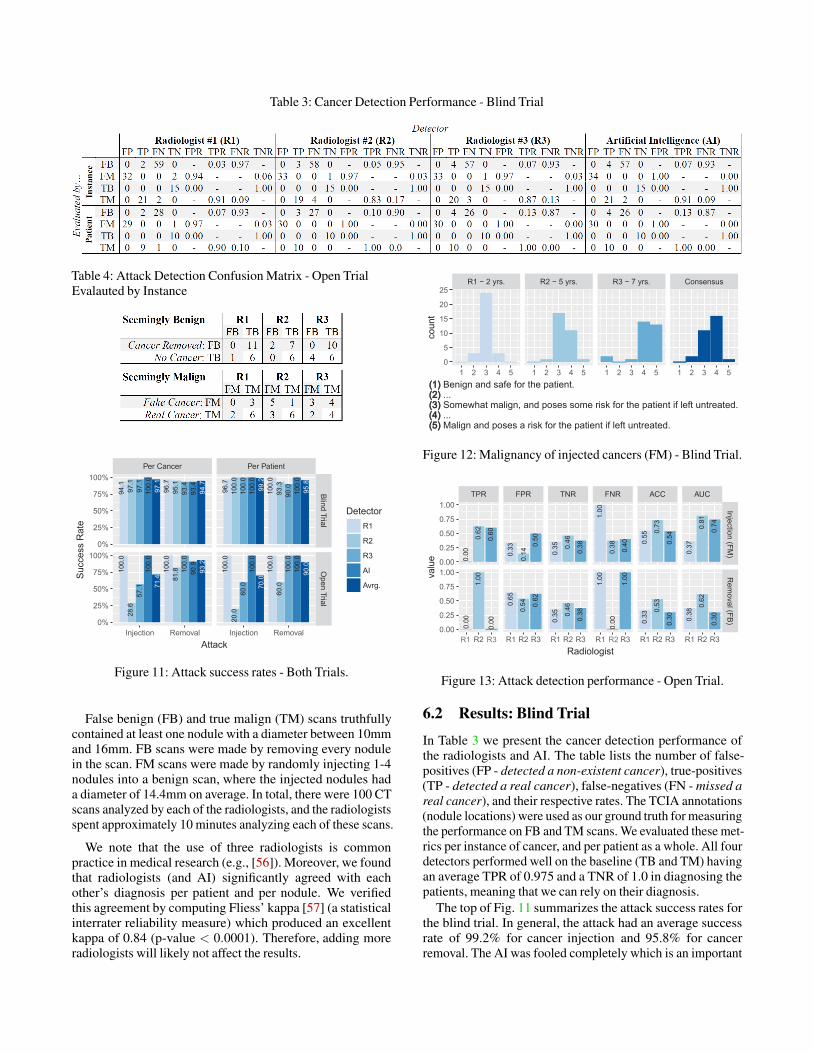
Table 3: Cancer Detection Performance - Blind Trial
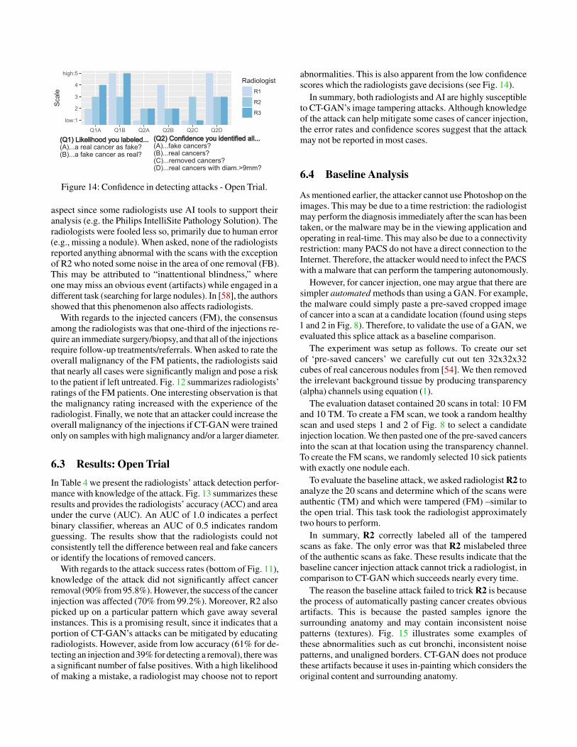
Table 4: Attack Detection Confusion Matrix - Open TrialEvalauted by Instance
97.1
100.
097
.197
.194
.1
94.7
93.4
93.4
95.1
96.7
71.4
100.
057
.128
.610
0.0
93.2
90.9
100.
081
.8100.
0
99.2
100.
010
0.0
100.
096
.7
95.8
100.
090
.093
.310
0.0
70.0
100.
060
.020
.010
0.0
90.0
100.
010
0.0
60.0
100.
0
Per Cancer Per Patient
Blind TrialO
pen Trial
Injection Removal Injection Removal
0%
25%
50%
75%
100%
0%
25%
50%
75%
100%
Attack
Succ
ess
Rat
e
DetectorR1
R2
R3
AI
Avrg.
Figure 11: Attack success rates - Both Trials.
False benign (FB) and true malign (TM) scans truthfullycontained at least one nodule with a diameter between 10mmand 16mm. FB scans were made by removing every nodulein the scan. FM scans were made by randomly injecting 1-4nodules into a benign scan, where the injected nodules hada diameter of 14.4mm on average. In total, there were 100 CTscans analyzed by each of the radiologists, and the radiologistsspent approximately 10 minutes analyzing each of these scans.
We note that the use of three radiologists is commonpractice in medical research (e.g., [56]). Moreover, we foundthat radiologists (and AI) significantly agreed with eachother’s diagnosis per patient and per nodule. We verifiedthis agreement by computing Fliess’ kappa [57] (a statisticalinterrater reliability measure) which produced an excellentkappa of 0.84 (p-value < 0.0001). Therefore, adding moreradiologists will likely not affect the results.
R1 − 2 yrs. R2 − 5 yrs. R3 − 7 yrs. Consensus
1 2 3 4 5 1 2 3 4 5 1 2 3 4 5 1 2 3 4 50
5
10
15
20
25
(1) Benign and safe for the patient.(2) ...(3) Somewhat malign, and poses some risk for the patient if left untreated.(4) ...(5) Malign and poses a risk for the patient if left untreated.
coun
t
Figure 12: Malignancy of injected cancers (FM) - Blind Trial.0.00
0.62
0.60
1.00
0.33
0.14
0.50
0.65
0.54 0.62
0.35 0.46
0.38
0.35 0.46
0.38
1.00
0.38 0.40
1.00
1.00
0.55
0.73
0.54
0.33
0.53
0.30
0.37
0.81
0.74
0.38
0.62
0.30
TPR FPR TNR FNR ACC AUC
InjectionRem
oval
R2 R1 R2 R3 R1 R2 R3 R1 R3 R1 R2 R3 R1 R2 R3
0.00
0.25
0.50
0.75
1.00
0.00
0.25
0.50
0.75
1.00
Radiologist
value
0.00
0.00
0.00
Figure 13: Attack detection performance - Open Trial.
6.2 Results: Blind Trial
In Table 3 we present the cancer detection performance ofthe radiologists and AI. The table lists the number of false-positives (FP - detected a non-existent cancer), true-positives(TP - detected a real cancer), false-negatives (FN - missed areal cancer), and their respective rates. The TCIA annotations(nodule locations) were used as our ground truth for measuringthe performance on FB and TM scans. We evaluated these met-rics per instance of cancer, and per patient as a whole. All fourdetectors performed well on the baseline (TB and TM) havingan average TPR of 0.975 and a TNR of 1.0 in diagnosing thepatients, meaning that we can rely on their diagnosis.
The top of Fig. 11 summarizes the attack success rates forthe blind trial. In general, the attack had an average successrate of 99.2% for cancer injection and 95.8% for cancerremoval. The AI was fooled completely which is an important
low:1
2
3
4
high:5
Q1A Q1B Q2A Q2B Q2C Q2D
(Q1) Likel hood you labeled...(A)...a real cancer as fake?(B)...a fake cancer as real?
(Q2) Confidence you identified all...(A)...fake cancers?(B)...real cancers?(C)...removed cancers?(D)...real cancers with diam.>9mm?
Scal
e
RadiologistR1
R2
R3
Figure 14: Confidence in detecting attacks - Open Trial.
aspect since some radiologists use AI tools to support theiranalysis (e.g. the Philips IntelliSite Pathology Solution). Theradiologists were fooled less so, primarily due to human error(e.g., missing a nodule). When asked, none of the radiologistsreported anything abnormal with the scans with the exceptionof R2 who noted some noise in the area of one removal (FB).This may be attributed to “inattentional blindness,” whereone may miss an obvious event (artifacts) while engaged in adifferent task (searching for large nodules). In [58], the authorsshowed that this phenomenon also affects radiologists.
With regards to the injected cancers (FM), the consensusamong the radiologists was that one-third of the injections re-quire an immediate surgery/biopsy, and that all of the injectionsrequire follow-up treatments/referrals. When asked to rate theoverall malignancy of the FM patients, the radiologists saidthat nearly all cases were significantly malign and pose a riskto the patient if left untreated. Fig. 12 summarizes radiologists’ratings of the FM patients. One interesting observation is thatthe malignancy rating increased with the experience of theradiologist. Finally, we note that an attacker could increase theoverall malignancy of the injections if CT-GAN were trainedonly on samples with high malignancy and/or a larger diameter.
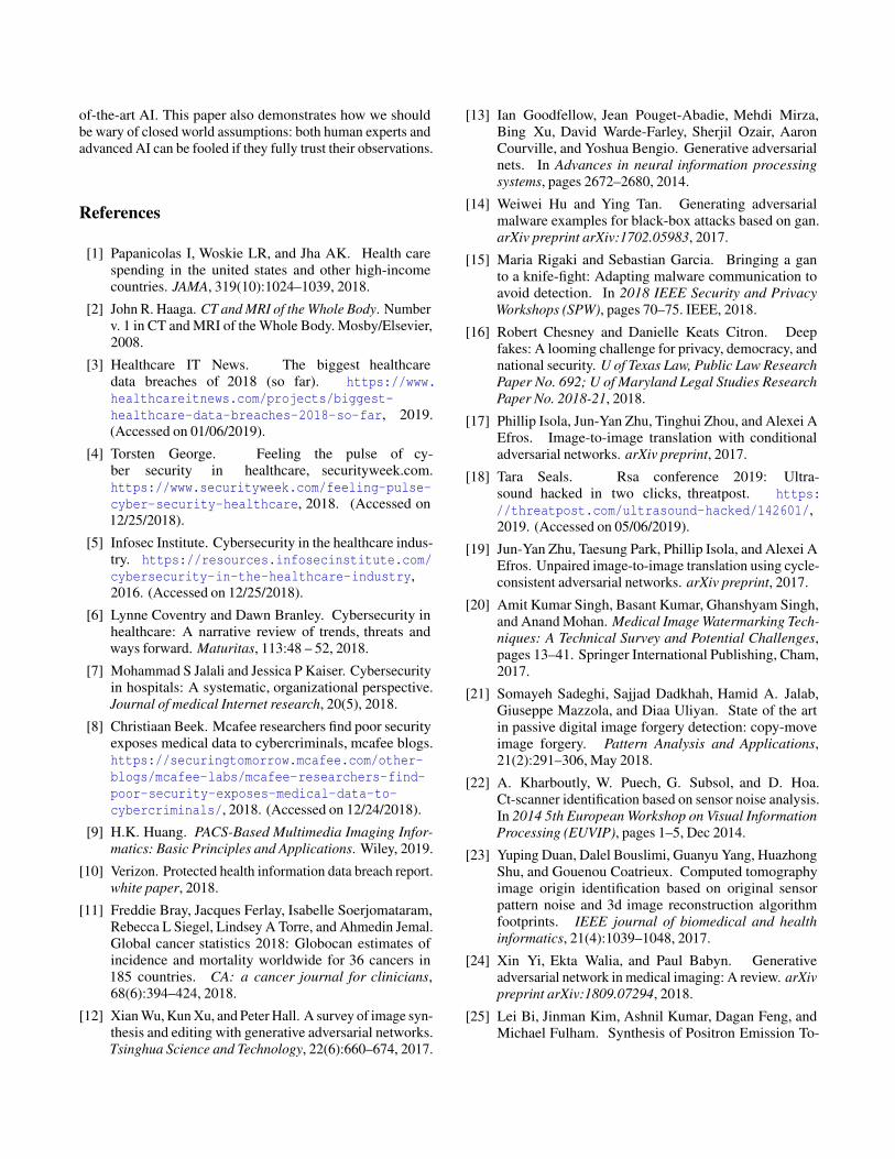
6.3 Results: Open TrialIn Table 4 we present the radiologists’ attack detection perfor-mance with knowledge of the attack. Fig. 13 summarizes theseresults and provides the radiologists’ accuracy (ACC) and areaunder the curve (AUC). An AUC of 1.0 indicates a perfectbinary classifier, whereas an AUC of 0.5 indicates randomguessing. The results show that the radiologists could notconsistently tell the difference between real and fake cancersor identify the locations of removed cancers.
With regards to the attack success rates (bottom of Fig. 11),knowledge of the attack did not significantly affect cancerremoval (90% from 95.8%). However, the success of the cancerinjection was affected (70% from 99.2%). Moreover, R2 alsopicked up on a particular pattern which gave away severalinstances. This is a promising result, since it indicates that aportion of CT-GAN’s attacks can be mitigated by educatingradiologists. However, aside from low accuracy (61% for de-tecting an injection and 39% for detecting a removal), there wasa significant number of false positives. With a high likelihoodof making a mistake, a radiologist may choose not to report
abnormalities. This is also apparent from the low confidencescores which the radiologists gave decisions (see Fig. 14).
In summary, both radiologists and AI are highly susceptibleto CT-GAN’s image tampering attacks. Although knowledgeof the attack can help mitigate some cases of cancer injection,the error rates and confidence scores suggest that the attackmay not be reported in most cases.
6.4 Baseline Analysis
As mentioned earlier, the attacker cannot use Photoshop on theimages. This may be due to a time restriction: the radiologistmay perform the diagnosis immediately after the scan has beentaken, or the malware may be in the viewing application andoperating in real-time. This may also be due to a connectivityrestriction: many PACS do not have a direct connection to theInternet. Therefore, the attacker would need to infect the PACSwith a malware that can perform the tampering autonomously.
However, for cancer injection, one may argue that there aresimpler automated methods than using a GAN. For example,the malware could simply paste a pre-saved cropped imageof cancer into a scan at a candidate location (found using steps1 and 2 in Fig. 8). Therefore, to validate the use of a GAN, weevaluated this splice attack as a baseline comparison.
The experiment was setup as follows. To create our setof ‘pre-saved cancers’ we carefully cut out ten 32x32x32cubes of real cancerous nodules from [54]. We then removedthe irrelevant background tissue by producing transparency(alpha) channels using equation (1).
The evaluation dataset contained 20 scans in total: 10 FMand 10 TM. To create a FM scan, we took a random healthyscan and used steps 1 and 2 of Fig. 8 to select a candidateinjection location. We then pasted one of the pre-saved cancersinto the scan at that location using the transparency channel.To create the FM scans, we randomly selected 10 sick patientswith exactly one nodule each.
To evaluate the baseline attack, we asked radiologist R2 toanalyze the 20 scans and determine which of the scans wereauthentic (TM) and which were tampered (FM) –similar tothe open trial. This task took the radiologist approximatelytwo hours to perform.
In summary, R2 correctly labeled all of the tamperedscans as fake. The only error was that R2 mislabeled threeof the authentic scans as fake. These results indicate that thebaseline cancer injection attack cannot trick a radiologist, incomparison to CT-GAN which succeeds nearly every time.
The reason the baseline attack failed to trick R2 is becausethe process of automatically pasting cancer creates obviousartifacts. This is because the pasted samples ignore thesurrounding anatomy and may contain inconsistent noisepatterns (textures). Fig. 15 illustrates some examples ofthese abnormalities such as cut bronchi, inconsistent noisepatterns, and unaligned borders. CT-GAN does not producethese artifacts because it uses in-painting which considers theoriginal content and surrounding anatomy.
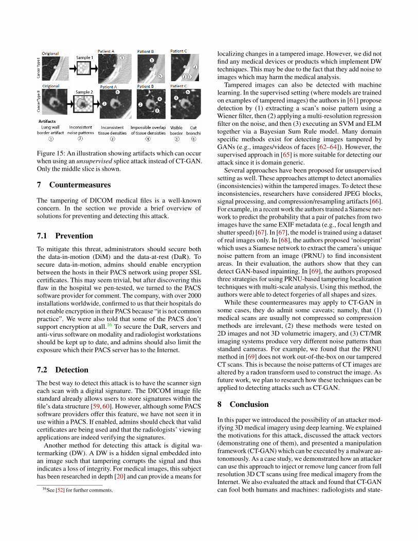
Figure 15: An illustration showing artifacts which can occurwhen using an unsupervised splice attack instead of CT-GAN.Only the middle slice is shown.
7 Countermeasures
The tampering of DICOM medical files is a well-knownconcern. In the section we provide a brief overview ofsolutions for preventing and detecting this attack.
7.1 PreventionTo mitigate this threat, administrators should secure boththe data-in-motion (DiM) and the data-at-rest (DaR). Tosecure data-in-motion, admins should enable encryptionbetween the hosts in their PACS network using proper SSLcertificates. This may seem trivial, but after discovering thisflaw in the hospital we pen-tested, we turned to the PACSsoftware provider for comment. The company, with over 2000installations worldwide, confirmed to us that their hospitals donot enable encryption in their PACS because “it is not commonpractice”. We were also told that some of the PACS don’tsupport encryption at all.16 To secure the DaR, servers andanti-virus software on modality and radiologist workstationsshould be kept up to date, and admins should also limit theexposure which their PACS server has to the Internet.
7.2 DetectionThe best way to detect this attack is to have the scanner signeach scan with a digital signature. The DICOM image filestandard already allows users to store signatures within thefile’s data structure [59, 60]. However, although some PACSsoftware providers offer this feature, we have not seen it inuse within a PACS. If enabled, admins should check that validcertificates are being used and that the radiologists’ viewingapplications are indeed verifying the signatures.
Another method for detecting this attack is digital wa-termarking (DW). A DW is a hidden signal embedded intoan image such that tampering corrupts the signal and thusindicates a loss of integrity. For medical images, this subjecthas been researched in depth [20] and can provide a means for
16See [52] for further comments.
localizing changes in a tampered image. However, we did notfind any medical devices or products which implement DWtechniques. This may be due to the fact that they add noise toimages which may harm the medical analysis.
Tampered images can also be detected with machinelearning. In the supervised setting (where models are trainedon examples of tampered images) the authors in [61] proposedetection by (1) extracting a scan’s noise pattern using aWiener filter, then (2) applying a multi-resolution regressionfilter on the noise, and then (3) executing an SVM and ELMtogether via a Bayesian Sum Rule model. Many domainspecific methods exist for detecting images tampered byGANs (e.g., images/videos of faces [62–64]). However, thesupervised approach in [65] is more suitable for detecting ourattack since it is domain generic.
Several approaches have been proposed for unsupervisedsetting as well. These approaches attempt to detect anomalies(inconsistencies) within the tampered images. To detect theseinconsistencies, researchers have considered JPEG blocks,signal processing, and compression/resampling artifacts [66].For example, in a recent work the authors trained a Siamese net-work to predict the probability that a pair of patches from twoimages have the same EXIF metadata (e.g., focal length andshutter speed) [67]. In [67], the model is trained using a datasetof real images only. In [68], the authors proposed ‘noiseprint’which uses a Siamese network to extract the camera’s uniquenoise pattern from an image (PRNU) to find inconsistentareas. In their evaluation, the authors show that they candetect GAN-based inpainting. In [69], the authors proposedthree strategies for using PRNU-based tampering localizationtechniques with multi-scale analysis. Using this method, theauthors were able to detect forgeries of all shapes and sizes.
While these countermeasures may apply to CT-GAN insome cases, they do admit some caveats; namely, that (1)medical scans are usually not compressed so compressionmethods are irrelevant, (2) these methods were tested on2D images and not 3D volumetric imagery, and (3) CT/MRimaging systems produce very different noise patterns thanstandard cameras. For example, we found that the PRNUmethod in [69] does not work out-of-the-box on our tamperedCT scans. This is because the noise patterns of CT images arealtered by a radon transform used to construct the image. Asfuture work, we plan to research how these techniques can beapplied to detecting attacks such as CT-GAN.
8 Conclusion
In this paper we introduced the possibility of an attacker mod-ifying 3D medical imagery using deep learning. We explainedthe motivations for this attack, discussed the attack vectors(demonstrating one of them), and presented a manipulationframework (CT-GAN) which can be executed by a malware au-tonomously. As a case study, we demonstrated how an attackercan use this approach to inject or remove lung cancer from fullresolution 3D CT scans using free medical imagery from theInternet. We also evaluated the attack and found that CT-GANcan fool both humans and machines: radiologists and state-
of-the-art AI. This paper also demonstrates how we shouldbe wary of closed world assumptions: both human experts andadvanced AI can be fooled if they fully trust their observations.
References
[1] Papanicolas I, Woskie LR, and Jha AK. Health carespending in the united states and other high-incomecountries. JAMA, 319(10):1024–1039, 2018.
[2] John R. Haaga. CT and MRI of the Whole Body. Numberv. 1 in CT and MRI of the Whole Body. Mosby/Elsevier,2008.
[3] Healthcare IT News. The biggest healthcaredata breaches of 2018 (so far). https://www.healthcareitnews.com/projects/biggest-healthcare-data-breaches-2018-so-far, 2019.(Accessed on 01/06/2019).
[4] Torsten George. Feeling the pulse of cy-ber security in healthcare, securityweek.com.https://www.securityweek.com/feeling-pulse-cyber-security-healthcare, 2018. (Accessed on12/25/2018).
[5] Infosec Institute. Cybersecurity in the healthcare indus-try. https://resources.infosecinstitute.com/cybersecurity-in-the-healthcare-industry,2016. (Accessed on 12/25/2018).
[6] Lynne Coventry and Dawn Branley. Cybersecurity inhealthcare: A narrative review of trends, threats andways forward. Maturitas, 113:48 – 52, 2018.
[7] Mohammad S Jalali and Jessica P Kaiser. Cybersecurityin hospitals: A systematic, organizational perspective.Journal of medical Internet research, 20(5), 2018.
[8] Christiaan Beek. Mcafee researchers find poor securityexposes medical data to cybercriminals, mcafee blogs.https://securingtomorrow.mcafee.com/other-blogs/mcafee-labs/mcafee-researchers-find-poor-security-exposes-medical-data-to-cybercriminals/, 2018. (Accessed on 12/24/2018).
[9] H.K. Huang. PACS-Based Multimedia Imaging Infor-matics: Basic Principles and Applications. Wiley, 2019.
[10] Verizon. Protected health information data breach report.white paper, 2018.
[11] Freddie Bray, Jacques Ferlay, Isabelle Soerjomataram,Rebecca L Siegel, Lindsey A Torre, and Ahmedin Jemal.Global cancer statistics 2018: Globocan estimates ofincidence and mortality worldwide for 36 cancers in185 countries. CA: a cancer journal for clinicians,68(6):394–424, 2018.
[12] Xian Wu, Kun Xu, and Peter Hall. A survey of image syn-thesis and editing with generative adversarial networks.Tsinghua Science and Technology, 22(6):660–674, 2017.
[13] Ian Goodfellow, Jean Pouget-Abadie, Mehdi Mirza,Bing Xu, David Warde-Farley, Sherjil Ozair, AaronCourville, and Yoshua Bengio. Generative adversarialnets. In Advances in neural information processingsystems, pages 2672–2680, 2014.
[14] Weiwei Hu and Ying Tan. Generating adversarialmalware examples for black-box attacks based on gan.arXiv preprint arXiv:1702.05983, 2017.
[15] Maria Rigaki and Sebastian Garcia. Bringing a ganto a knife-fight: Adapting malware communication toavoid detection. In 2018 IEEE Security and PrivacyWorkshops (SPW), pages 70–75. IEEE, 2018.
[16] Robert Chesney and Danielle Keats Citron. Deepfakes: A looming challenge for privacy, democracy, andnational security. U of Texas Law, Public Law ResearchPaper No. 692; U of Maryland Legal Studies ResearchPaper No. 2018-21, 2018.
[17] Phillip Isola, Jun-Yan Zhu, Tinghui Zhou, and Alexei AEfros. Image-to-image translation with conditionaladversarial networks. arXiv preprint, 2017.
[18] Tara Seals. Rsa conference 2019: Ultra-sound hacked in two clicks, threatpost. https://threatpost.com/ultrasound-hacked/142601/,2019. (Accessed on 05/06/2019).
[19] Jun-Yan Zhu, Taesung Park, Phillip Isola, and Alexei AEfros. Unpaired image-to-image translation using cycle-consistent adversarial networks. arXiv preprint, 2017.
[20] Amit Kumar Singh, Basant Kumar, Ghanshyam Singh,and Anand Mohan. Medical Image Watermarking Tech-niques: A Technical Survey and Potential Challenges,pages 13–41. Springer International Publishing, Cham,2017.
[21] Somayeh Sadeghi, Sajjad Dadkhah, Hamid A. Jalab,Giuseppe Mazzola, and Diaa Uliyan. State of the artin passive digital image forgery detection: copy-moveimage forgery. Pattern Analysis and Applications,21(2):291–306, May 2018.
[22] A. Kharboutly, W. Puech, G. Subsol, and D. Hoa.Ct-scanner identification based on sensor noise analysis.In 2014 5th European Workshop on Visual InformationProcessing (EUVIP), pages 1–5, Dec 2014.
[23] Yuping Duan, Dalel Bouslimi, Guanyu Yang, HuazhongShu, and Gouenou Coatrieux. Computed tomographyimage origin identification based on original sensorpattern noise and 3d image reconstruction algorithmfootprints. IEEE journal of biomedical and healthinformatics, 21(4):1039–1048, 2017.
[24] Xin Yi, Ekta Walia, and Paul Babyn. Generativeadversarial network in medical imaging: A review. arXivpreprint arXiv:1809.07294, 2018.
[25] Lei Bi, Jinman Kim, Ashnil Kumar, Dagan Feng, andMichael Fulham. Synthesis of Positron Emission To-
mography (PET) Images via Multi-channel GenerativeAdversarial Networks (GANs). pages 43–51. Springer,Cham, 2017.
[26] Avi Ben-Cohen, Eyal Klang, Stephen P. Raskin,Michal Marianne Amitai, and Hayit Greenspan. VirtualPET Images from CT Data Using Deep ConvolutionalNetworks: Initial Results. pages 49–57. Springer, Cham,2017.
[27] Avi Ben-Cohen, Eyal Klang, Stephen P. Raskin, ShellySoffer, Simona Ben-Haim, Eli Konen, Michal MarianneAmitai, and Hayit Greenspan. Cross-Modality Synthesisfrom CT to PET using FCN and GAN Networks forImproved Automated Lesion Detection. 2 2018.
[28] Qi Dou, Cheng Ouyang, Cheng Chen, Hao Chen, andPheng-Ann Heng. Unsupervised Cross-ModalityDomain Adaptation of ConvNets for Biomedical ImageSegmentations with Adversarial Loss. In Proceedingsof the Twenty-Seventh International Joint Conferenceon Artificial Intelligence, pages 691–697, California,7 2018. International Joint Conferences on ArtificialIntelligence Organization.
[29] Cheng-Bin Jin, Hakil Kim, Wonmo Jung, Seongsu Joo,Ensik Park, Ahn Young Saem, In Ho Han, Jae Il Lee, andXuenan Cui. Deep CT to MR Synthesis using Paired andUnpaired Data. 5 2018.
[30] Camilo Bermudez, Andrew J Plassard, Larry T Davis,Allen T Newton, Susan M Resnick, and Bennett ALandman. Learning implicit brain mri manifoldswith deep learning. In Medical Imaging 2018: ImageProcessing, volume 10574, page 105741L. InternationalSociety for Optics and Photonics, 2018.
[31] Maayan Frid-Adar, Idit Diamant, Eyal Klang, MichalAmitai, Jacob Goldberger, and Hayit Greenspan.GAN-based Synthetic Medical Image Augmentationfor increased CNN Performance in Liver LesionClassification. 3 2018.
[32] Jelmer M. Wolterink, Tim Leiner, and Ivana Isgum.Blood Vessel Geometry Synthesis using GenerativeAdversarial Networks. In 1st Conference on MedicalImaging with Deep Learning (MIDL 2018), Amsterdam,The Netherlands, The Netherlands, 2018.
[33] Christoph Baur, Shadi Albarqouni, and Nassir Navab.Melanogans: High resolution skin lesion synthesis withgans. arXiv preprint arXiv:1804.04338, 2018.
[34] Ali Madani, Mehdi Moradi, Alexandros Karargyris,and Tanveer Syeda-Mahmood. Chest x-ray generationand data augmentation for cardiovascular abnormalityclassification. In Medical Imaging 2018: ImageProcessing, volume 10574, page 105741M. InternationalSociety for Optics and Photonics, 2018.
[35] Maria JM Chuquicusma, Sarfaraz Hussein, Jeremy Burt,and Ulas Bagci. How to fool radiologists with generativeadversarial networks? a visual turing test for lung cancer
diagnosis. In Biomedical Imaging (ISBI 2018), 2018IEEE 15th International Symposium on, pages 240–244.IEEE, IEEE, 4 2018.
[36] W. Hruby. Digital (R)Evolution in Radiology. SpringerVienna, 2013.
[37] A. Peck. Clark’s Essential PACS, RIS and ImagingInformatics. Clark’s Companion Essential Guides. CRCPress, 2017.
[38] C. Carter and B. Veale. Digital Radiography and PACS.Elsevier Health Sciences, 2018.
[39] Bill Siwicki. Cloud-based pacs system cuts imaging costsby half for rural hospital | healthcare it news. https://www.healthcareitnews.com/news/cloud-based-pacs-system-cuts-imaging-costs-half-rural-hospital. (Accessed on 01/02/2019).
[40] Jennifer Bresnick. Picture archive communicationsystem use widespread in hospitals. https://healthitanalytics.com/news/picture-archive-communication-system-use-widespread-in-hospitals, 2016. (Accessed on 01/02/2019).
[41] Sébastien Jodogne, Claire Bernard, Magali Devillers,Eric Lenaerts, and Philippe Coucke. Orthanc-alightweight, restful dicom server for healthcare andmedical research. In Biomedical Imaging (ISBI), 2013IEEE 10th International Symposium on, pages 190–193.IEEE, 2013.
[42] Carlos Costa, Carlos Ferreira, Luís Bastião, Luís Ribeiro,Augusto Silva, and José Luís Oliveira. Dicoogle-an opensource peer-to-peer pacs. Journal of digital imaging,24(5):848–856, 2011.
[43] Ladi Adefala. Healthcare experiences twicethe number of cyber attacks as other industries.https://www.fortinet.com/blog/business-and-technology/healthcare-experiences-twice-the-number-of-cyber-attacks-as-othe.html,2018. (Accessed on 12/24/2018).
[44] Joram Borenstein Rebecca Weintraub. 11 thingsthe health care sector must do to improve cyberse-curity. https://hbr.org/2017/06/11-things-the-health-care-sector-must-do-to-improve-cybersecurity, 2017. (Accessed on 12/25/2018).
[45] Charlie Osborne. Us hospital pays $55,000to hackers after ransomware attack | zdnet.https://www.zdnet.com/article/us-hospital-pays-55000-to-ransomware-operators/, 2018.(Accessed on 01/04/2019).
[46] Joseph Muniz and Aamir Lakhani. Penetration testingwith raspberry pi. Packt Publishing Ltd, 2015.
[47] Mathy Vanhoef and Frank Piessens. Key reinstallation at-tacks: Forcing nonce reuse in wpa2. In Proceedings of the2017 ACM SIGSAC Conference on Computer and Com-munications Security, pages 1313–1328. ACM, 2017.
[48] Alfred NG. Security researchers find flaws inchips used in hospitals, factories and stores -cnet. https://www.cnet.com/news/security-researchers-find-flaws-in-chips-used-in-hospitals-factories-and-stores/, 2018. (Ac-cessed on 01/04/2019).
[49] Rebecca Myers Robin Henry and Jonathan Corke.Hospitals to struggle for days | news | the sunday times.https://www.thetimes.co.uk/article/nhs-cyberattack-bitcoin-wannacry-hospitals-to-struggle-for-days-k0nhk7p2b, 2017. (Accessedon 01/04/2019).
[50] DHS. Philips isite/intellispace pacs vulnerabili-ties (update a), ics-cert. https://ics-cert.us-cert.gov/advisories/ICSMA-18-088-01, 2018.(Accessed on 12/24/2018).
[51] John E Dunn. Imagine you’re having a ct scan andmalware alters the radiation levels – it’s doable •the register. https://www.theregister.co.uk/2018/04/11/hacking_medical_devices/, 2018.(Accessed on 01/04/2019).
[52] Kim Zetter. Hospital viruses: Fake cancerous nodesin ct scans, created by malware, trick radiologists.https://www.washingtonpost.com/technology/2019/04/03/hospital-viruses-fake-cancerous-nodes-ct-scans-created-by-malware-trick-radiologists/, April 2019. (Accessed on 05/07/2019).
[53] Heber MacMahon, David P Naidich, Jin Mo Goo,Kyung Soo Lee, Ann NC Leung, John R Mayo, Atul CMehta, Yoshiharu Ohno, Charles A Powell, MathiasProkop, et al. Guidelines for management of incidentalpulmonary nodules detected on ct images: from the fleis-chner society 2017. Radiology, 284(1):228–243, 2017.
[54] Samuel G Armato III, Geoffrey McLennan, Luc Bidaut,Michael F McNitt-Gray, Charles R Meyer, Anthony PReeves, Binsheng Zhao, Denise R Aberle, Claudia I Hen-schke, Eric A Hoffman, et al. The lung image databaseconsortium (lidc) and image database resource initiative(idri): a completed reference database of lung noduleson ct scans. Medical physics, 38(2):915–931, 2011.
[55] Keelin Murphy, Bram van Ginneken, Arnold MRSchilham, BJ De Hoop, HA Gietema, and MathiasProkop. A large-scale evaluation of automatic pulmonarynodule detection in chest ct using local image featuresand k-nearest-neighbour classification. Medical imageanalysis, 13(5):757–770, 2009.
[56] Andre Esteva, Brett Kuprel, Roberto A Novoa, Justin Ko,Susan M Swetter, Helen M Blau, and Sebastian Thrun.Dermatologist-level classification of skin cancer withdeep neural networks. Nature, 542(7639):115, 2017.
[57] Anthony J Conger. Integration and generalization ofkappas for multiple raters. Psychological Bulletin,88(2):322, 1980.
[58] Trafton Drew, Melissa L-H Võ, and Jeremy M Wolfe.The invisible gorilla strikes again: Sustained inatten-tional blindness in expert observers. Psychologicalscience, 24(9):1848–1853, 2013.
[59] F Cao, HK Huang, and XQ Zhou. Medical image securityin a hipaa mandated pacs environment. Computerizedmedical imaging and graphics, 27(2-3):185–196, 2003.
[60] NEMA. Digital imaging and communications inmedicine (dicom) digital signatures. ftp://medical.nema.org/medical/dicom/final/sup41_ft.pdf,2001. (Accessed on 2/11/19).
[61] Ahmed Ghoneim, Ghulam Muhammad, Syed UmarAmin, and Brij Gupta. Medical image forgery detectionfor smart healthcare. IEEE Communications Magazine,56(4):33–37, 2018.
[62] Andreas Rössler, Davide Cozzolino, Luisa Verdoliva,Christian Riess, Justus Thies, and Matthias Nießner.Faceforensics++: Learning to detect manipulated facialimages. arXiv preprint arXiv:1901.08971, 2019.
[63] Falko Matern, Christian Riess, and Marc Stamminger.Exploiting visual artifacts to expose deepfakes and facemanipulations. In 2019 IEEE Winter Applications ofComputer Vision Workshops (WACVW), pages 83–92.IEEE, 2019.
[64] Shahroz Tariq, Sangyup Lee, Hoyoung Kim, Youjin Shin,and Simon S Woo. Detecting both machine and humancreated fake face images in the wild. In Proceedings ofthe 2nd International Workshop on Multimedia Privacyand Security, pages 81–87. ACM, 2018.
[65] Davide Cozzolino, Justus Thies, Andreas Rössler,Christian Riess, Matthias Nießner, and Luisa Verdoliva.Forensictransfer: Weakly-supervised domain adaptationfor forgery detection. arXiv preprint arXiv:1812.02510,2018.
[66] Lilei Zheng, Ying Zhang, and Vrizlynn LL Thing.A survey on image tampering and its detection inreal-world photos. Journal of Visual Communicationand Image Representation, 58:380–399, 2019.
[67] Minyoung Huh, Andrew Liu, Andrew Owens, andAlexei A Efros. Fighting fake news: Image splicedetection via learned self-consistency. In Proceedings ofthe European Conference on Computer Vision (ECCV),pages 101–117, 2018.
[68] Davide Cozzolino and Luisa Verdoliva. Noiseprint: acnn-based camera model fingerprint. arXiv preprintarXiv:1808.08396, 2018.
[69] P. Korus and J. Huang. Multi-scale analysis strategiesin prnu-based tampering localization. IEEE Trans. onInformation Forensics & Security, 2017.
Appendix
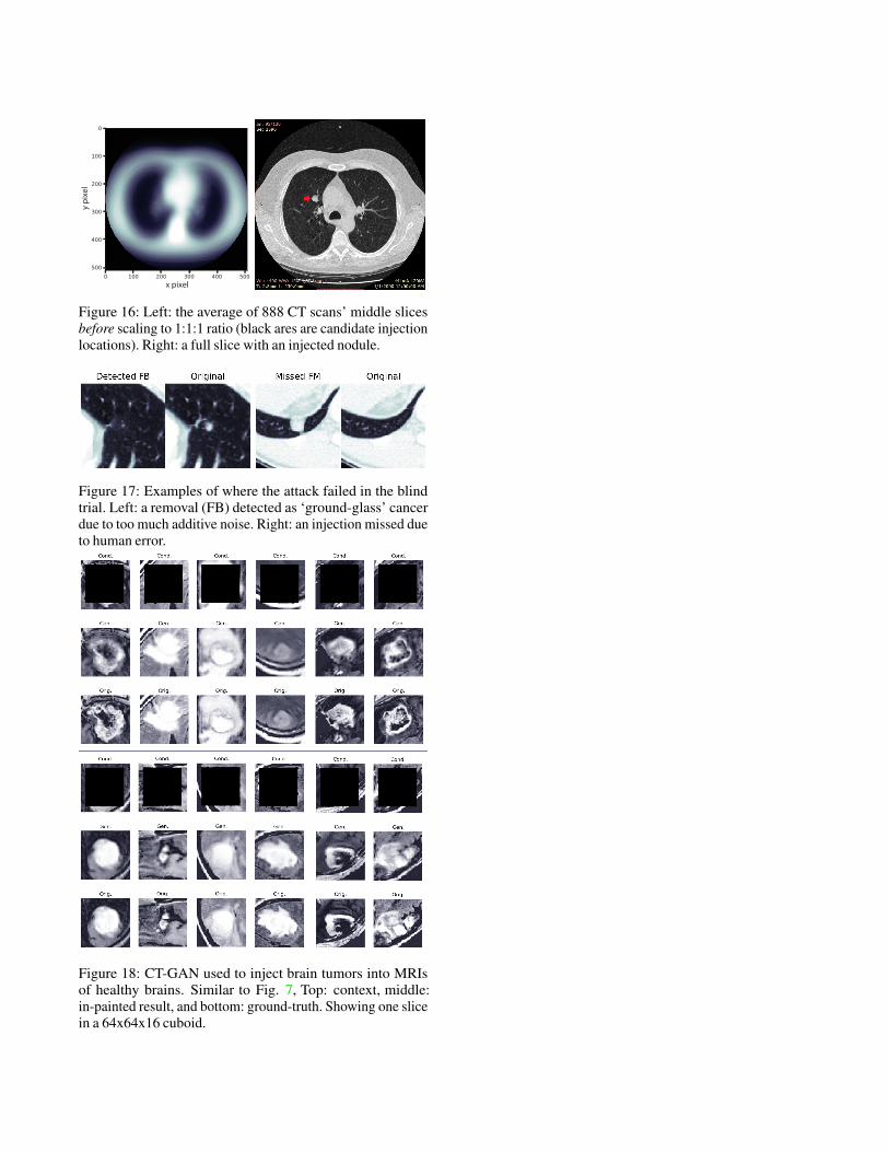
Figure 16: Left: the average of 888 CT scans’ middle slicesbefore scaling to 1:1:1 ratio (black ares are candidate injectionlocations). Right: a full slice with an injected nodule.
Figure 17: Examples of where the attack failed in the blindtrial. Left: a removal (FB) detected as ‘ground-glass’ cancerdue to too much additive noise. Right: an injection missed dueto human error.
Figure 18: CT-GAN used to inject brain tumors into MRIsof healthy brains. Similar to Fig. 7, Top: context, middle:in-painted result, and bottom: ground-truth. Showing one slicein a 64x64x16 cuboid.
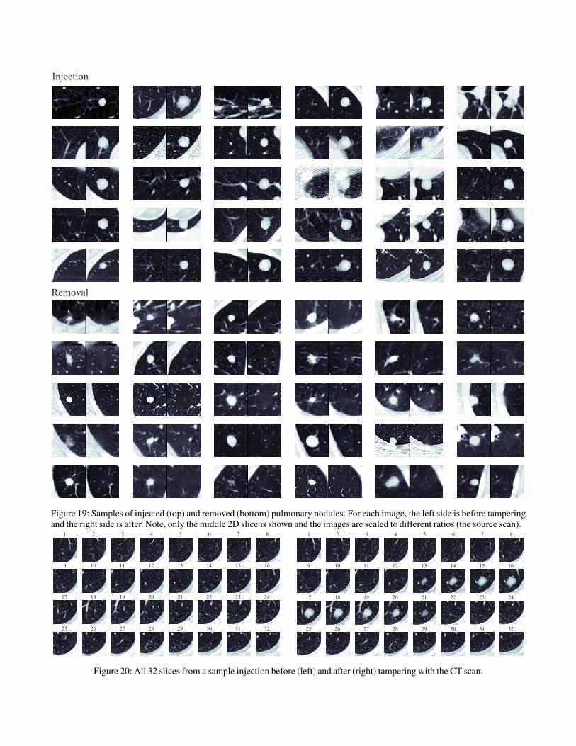
Figure 19: Samples of injected (top) and removed (bottom) pulmonary nodules. For each image, the left side is before tamperingand the right side is after. Note, only the middle 2D slice is shown and the images are scaled to different ratios (the source scan).
1 2 3 4 5 6 7 8
9 10 11 12 13 14 15 16
17 18 19 20 21 22 23 24
25 26 27 28 29 30 31 32
1 2 3 4 5 6 7 8
9 10 11 12 13 14 15 16
17 18 19 20 21 22 23 24
25 26 27 28 29 30 31 32
Figure 20: All 32 slices from a sample injection before (left) and after (right) tampering with the CT scan.