Embed Size (px)
Citation preview
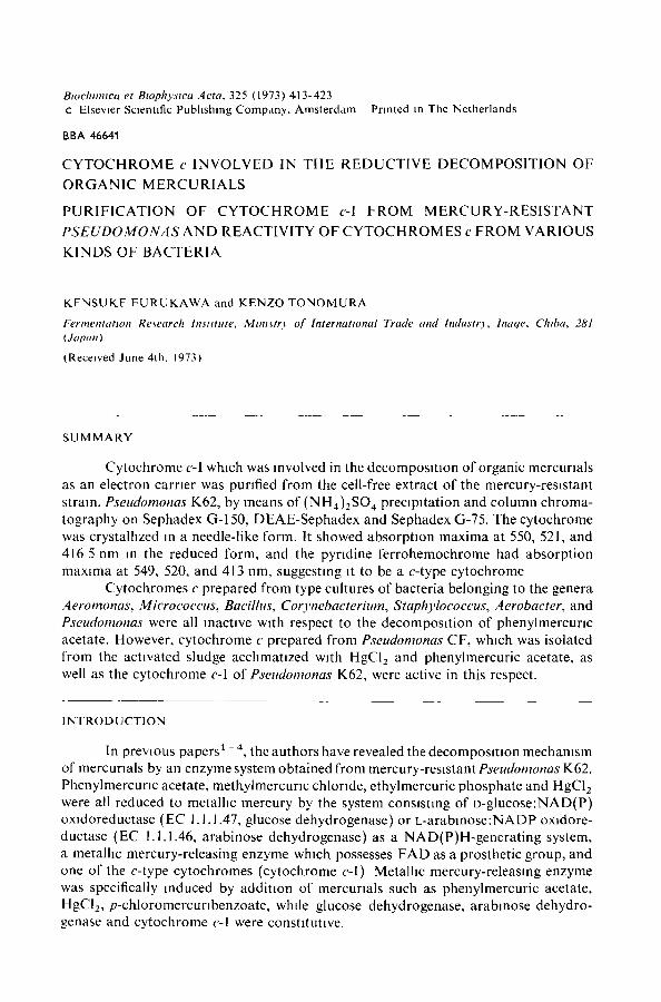
Biochmuca et Btophyslca Acta, 325 (1973) 413-423 c Elsevier ScJentlfic Pubhshmg Company. Amsterdam Printed m The Netherlands
BBA 4664"1
CYTOCHROME c INVOLVED IN THE REDUCTIVE DECOMPOSITION OF
O R G A N I C MERCURIALS
PURIFICATION OF CYTOCHROME c-I FROM MERCURY-RESISTANT
PSEUDOMONAS AND REACTIVITY OF CYTOCHROMES c FROM VARIOUS
KINDS OF BACTERIA
KENSUKE F U R U K A W A and K E N Z O T O N O M U R A
Fermentation Research Institute, Mmt~tr) of Internattonal Trade and lndustr3, lnaqe, Chtba, 281 (Japan)
(Received June 4th, 1973)
S U M M A R Y
Cytochrome c-1 which was revolved in the decomposition of organic mercurials as an electron carrier was purified from the cell-free extract of the mercury-resistant strata, Pseudomonas K62, by means of (N H 4 ) 2 S O ¢ precipitation and column chroma- tography on Sephadex G-150, DEAE-Sephadex and Sephadex G-75. The cytochrome was crystalhzed in a needle-like form. It showed absorption maxima at 550, 521, and 416 5 nm in the reduced form, and the pyrldine ferrohemochrome had absorption maxima at 549, 520, and 413 nm, suggesting it to be a c-type cytochrome
Cytochromes c prepared from type cultures of bacteria belonging to the genera Aeromonas, Micrococcus, Bacillus, Corvnebacterium, Staphylococcus, Aerobacter, and Pseudomonas were all reactive with respect to the decomposition of phenylmercurlc acetate. However, cytochrome c prepared from Pseudomonas CF, which was isolated from the activated sludge acchmatlzed with HgCI2 and phenylmercuric acetate, as well as the cytochrome c-I of Pseudomonas K62, were active in this respect.
INTRODUCTION
In previous papers ~ - ~, the authors have revealed the decomposmon mechanism of mercurials by an enzyme system obtained from mercury-resistant Pseudomonas K62. Phenylmercurlc acetate, methylmercurlc chloride, ethylmercuric phosphate and HgCI z were all reduced to metallic mercury by the system consisting of D-glucose:NAD(P) oxldoreductase (EC 1.1.1.47, glucose dehydrogenase) or L-arabmose:NADP oxldore- ductase (EC 1.1.1.46, arabinose dehydrogenase) as a NAD(P)H-generating system, a metallic mercury-releasing enzyme which possesses FAD as a prosthetic group, and one of the c-type cytochromes (cytochrome c-I) Metalhc mercury-releasing enzyme was specifically induced by addition of mercurials such as phenylmercuric acetate, HgCI2, p-chloromercurlbenzoate, while glucose dehydrogenase, arabmose dehydro- genase and cytochrome c-1 were constltUttve.
414 K. FURUKAWA, K. TONOMURA
Two kinds of cytochrome c (c-I and c-II) were found m the cell extract of th~s organism 2. Cytochrome c-I with a mol. wt of 26 000, was active m the decomposmon, while cytochrome c-II, with a mol. wt of 14 000, was inactive However, cytochrome c-I has not yet been purified thoroughly. The present paper describes the crystalliza- tion of cytochrome c-I and examination of some of its properties, and reactivity of cytochromes c prepared from various kinds of bacteria in the decomposition of phenyl- mercuric acetate. It was found that one of the c-type cytochromes from Pseudomonas CF which was newly isolated from activated sludge was able to couple m the reaction as well as the cytochrome c-I. The role of cytochrome e-I is discussed in comparison with ItS role in the respiratory chain
MATERIALS AND METHODS
Microorganisms and cultwatton A mercury-resistant strain, K62, of Pseudomonas was isolated from the soil of
a phenylmercuric acetate-producing factory previously 5. The composition of the culture medium (Medium A) for this organism was as follows' glucose, 2.5 g: KH 2- PO4, 0.2 g; KzHPO 4, 1.6 g; (NH4)2804, 1 g; MgSO 4 " H20, 0.2 g; CaCI2 • 2 H20, 0.02 g; NaCI, 0 1 g ; FeSO4' 7 H20, 0.01 g, NazMoO 4, 0.5 mg; MnSO 4, 0.5 mg; calcium pantothenate, 0.4 rag; Inositol, 0.2 mg; nicotinic acid, 0.4 mg; riboflavin, 0.4 mg, biotin, 2/~g; vitamin B12, 0.5 pg; phenylmercurlc acetate 30 mg; and distilled water, 1 1 (pH 7.5). Cultivation was carried out with shaking at 30 °C. Aeromonas hydrophila, Micrococcus luteus, Bacillus subtilis, Corynebacterium equi, Staphylococcus aureus, Aerobacter aerogenes, Pseudomonas fluorescens, P. aeruginosa, P. ovahs, P. riboflavina and P. mephitica var. lipolitiea were aerobically grown on a medium (Medium B) containing meat extract, 7 g; peptone, 10 g; NaCI, 3 g; FeSO 4 • 7 H 2 0 ,
0.02 g; and distilled water, I I (pH 7.0) at 30 °C. The CF strain of Pseudomonas was newly isolated from the activated sludge which was acclimatized with HgCI 2 and phenylmercuric acetate. The organism was aerobically grown on Medium B supple- mented with 20 mg phenylmercuric acetate.
Preparation of cell extract After cells were harvested and washed with 0.05 M phosphate buffer (pH 6 7),
they were disrupted by shaking with glass beads (0.10-0.11 mm diameter) using a Braun Cell Homogenizer. The homogenate was centrifuged at 15 000 × g for 30 mm. The supernatant fluid was dialyzed overnight against 0.05 M phosphate buffer (pH 6.7), and used as a crude extract.
Preparation of metalhc mercury-releasmg enzyme and arabinose dehydrogenase The crude extract of Pseudomonas K62 was fractlonated by precipitation w~th
(NH4)2SO 4 (20 to 70 of saturation), the precipitate was dissolved in 0.05 M phosphate buffer (pH 6.7) and dialyzed against 200 vol. of the same buffer. The dlalysate was then applied to a column (2.5 cm × 90 cm) of Sephadex G-150 which had been equili- brated with 0.05 M phosphate buffer (pH 6.7) and eluted with the same buffer at a flow rate of 10 m l / h Fractions containing metallic mercury-releasing enzyme, arabl- nose dehydrogenase, cytochrome e-I and cytochrome c-ll were pooled separately. Cytochrome c-I was further purified as described below
CYTOCHROME c AND ORGANIC MERCURIALS 415
Assay of metallic mercury-releasin9 enzyme and arabinose dehydrogenase Metallic mercury-releasing enzyme was assayed by measuring phenylmercunc
acetate decomposition in the following mixture" 5 • 10 -z M phosphate buffer (pH 5.8) 5 units of arabinose dehydrogenase, 6 . 1 0 - S M L-arabinose, 6. 1 0 - 5 M NADP, 1 " 1 0 - 7 M cytochrome c-I, 1 - 10 -6 M FAD, 5 • 1 0 - 4 M thloglycolate, 6 • 10 -5 M phenyl [2°3Hg]mercuric acetate and the enzyme. The reaction mixture was incubated m a L-form tube (15-cm long, 7-cm high, 2 5-cm diameter) with shaking at 30 °C. Metallic mercury formed from phenylmercurlc acetate was rapidly volatilized under th~s condition, and the radioactivity remaimng m the m~xture was assayed in a well- type scintillation counter. The actwity of arabinose dehydrogenase was measured by reading the increase of absorbance at 340 nm formed by N A D P H reduction. The reaction mixture was as follows: 5. 10 -2 M phosphate buffer (pH 8.5), 3 10 -3 M L-arabmose, 3 • 1 0 - 6 M NADP, and the enzyme.
Purification and crystalhzatlon oJ cytochrome c-1 The fraction containing cytochrome was separated by chromatography on
Sephadex G-150 and concentrated m a collodion bag (Sartorious Membrane Fdter Co. Ltd) The enzyme was then apphed to a column (2.5 c m × 2 8 c m ) of DEAE- Sephadex A-50 which had previously been equihbrated with 0.05 M phosphate buffer (pH 6.7), and eluted with a linear gradient of 0 to 0.5 M NaC1 at a flow rate of 10 ml/h. Fractions containing cytochrome c-I were concentrated m a collodion bag, and apphed to a column (2.5 cm ×40 cm) of Sephadex G-75 which had been equdlbrated with 0.05 M phosphate buffer (pH 6 7). The column was eluted with the same buffer. Crystallization of cytochrome c-I was carried out by adding finely powdered (NH4) z- SO 4 to the concentrated enzyme solution. The heine content of cytochrome c-I was estimated from the alkahne pyndme ferrohemochrome spectrum 6. After reduction with a few crystals of dith~onite, the absorbance was read at 549 nm Protein contents were estimated as mtrogen content by the micro-Kjeldahl method.
Preparation o/bacterial cytochrome c Cytochromes c of bacteria, exept for Pseudomonas K62, were separated f rom
each crude extract by chromatography using a column (2.5 cm ×90 cm) of Sephadex G-150. The cytochrome c fractions were pooled and stored at --20 °C until use.
Esttmation o/molecular weight of cytochrome c-I by gel filtratton A Sephadex G-75 column (1.5 cm ×70 cm) was prepared with 0.05 M phos-
phate buffer (pH 6.7)containing 0. I M NaCI. Ovalbumin ,ct-chymotrypsin and bovine cytochrome c were used as an internal standard on the column.
Spectrophotometric measurements A Hitachi electrop~totometer, Type 181, was used for the spectrophotometric
measurements.
Chemical Phenyl [2 O3Hg]mercuri c acetate was purchased from the Radiochemlcal Centre,
Amersham, England.
416 K F U R U K A W A , K, T O N O M U R A
RESULTS
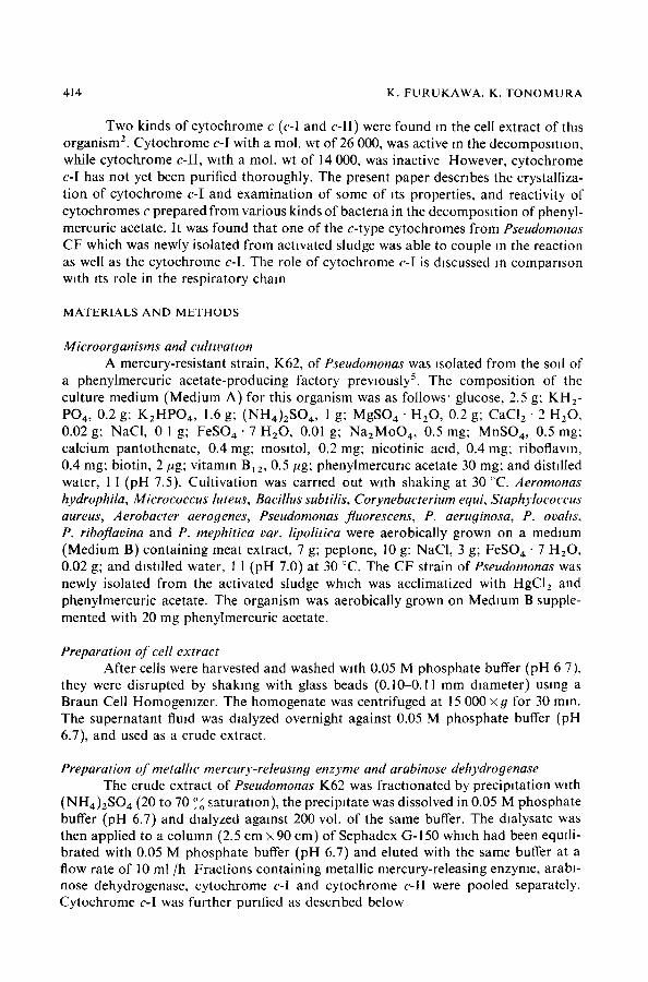
Purit~catton and crvstalhzatton oJ cvtochrome c-I Chromatography of the crude extract on Sephadex G-150 Js shown m Rg. 1,
mdlcatmg the eluuon of metalhc mercury-releasmg enzyme, arabmose dehydrogenase, cytochrome c-I and cytochrome c-ll. It has been reported in a previous paper z that only the eluted first cytochrome c was actwe for the decomposmon of mercurials as an electron carrier and that eluted secondly inactive The actwe cytochrome was
5 " I001
E 4 -_¢ 80 g
3-~6C
2~4C
I- 2C
[-__ ,10' MMR'Enz~iic -~ 8 ytc-I cyt cll 28O ~ i nrr , :
-~ 6
-uJ 41
I0 20 30 40 Fraction No (lOml)
03
o2~
Ol
Fig I Ch roma tog raphy of crude extract on Sephadex G-150, A fraction obtained from the crude extract by a m m o n i u m sulfate preclpJtaUon (0 2 0.7 sa tura t ion) **as applied to a Sephadex G-150 column (2 5 cm 90 cm), and eluted with 0 05 M phospha te buffer (pH 6 7) Metalhc mercury releasing enzyme ( M M R - E n z ) activity ( 0 - 0 ) and arablnose dehydrogenase ( A D H ) activity (O © ) ~as measured for each fraction. Protein ( ) was measured by reading absorbance at 280 nm. and cytochromes ( ) by reading at 415 nm
=833 ~n ,¢
~0~ 0
o]
i
cy t c-II
cyt c-[
I0 20 30 40 ,f~ Froction No ( lOml )
D3
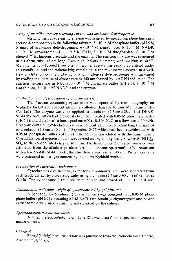
Fig. 2 C h r o m a t o g r a p h y of cy tochrome c-I and c-ll on DEAE-Sephadex Fract ions o f cy tochrome c-I and c-ll eluted f rom the Sephadex G-150 co lumn were pooled, concentrated and then apphed to a DEAE-Sephadex A-50 co lumn (2 5 cm 28 cml ~h lch had previously been equil ibrated with 0 05 M phospha te buffer (pH 6.7) A linear gradient (- - ) of 0 to 0 5 M NaCI in the same buffer ~a s used for elutlon o f cytochromes Protein ( ) was measured by reading absorbance at 280 nm, and cytochromes c (--- ) by absorbance at 415 nm.
C Y T O C H R O M E c A N D O R G A N I C M E R C U R I A L S 417
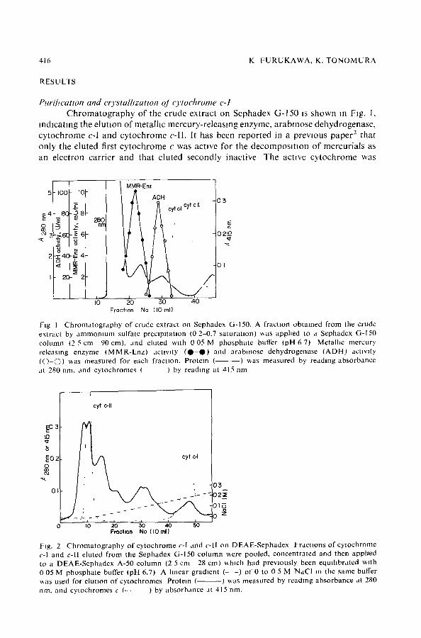
Fig 3. C r y s t a l h n e c y t o c h r o m e c-I ( 300)
designated as cytochrome c-I and mactwe cytochrome as cytochrome c-lI. Fractions of cytochrome c-! and c-ll, separated by column chromatography on Sephadex G-150, were further puNfied by DEAE-Sephadex A-50 chromatography (Fig 2). Cytochrome c-I was clearly separated from DEAE-Sephadex chromatography and was then sub- jected to column chromatography on Sephadex G-75. The mare fraction was collected and concentrated for crystalhzatlon. To the reddish solution thus obtained, finely powdered (NH4)2SO 4 was added gradually with gentle stirring until a faint turbidity appeared, and then the solution was kept standing for 3 days at 4 ~C. Cytochrome c-I was obtained in a crystalhne, needle-hke form as shown m Fig. 3. The results of a purification experiment are summarized m Table I.
T A B L E l
P U R I F I C A T I O N O F C Y T O C H R O M E c-I F R O M T H E C E L L E X T R A C T O F P S E U D O ~ I O N A S K62
Purthcatton step Total Total ab~orbance vol (ml)
280 nm 416 nm
C r u d e ex t r ac t 220 16 800 1750 0 104 (NH,D2SO.~ p r e c i p i t a t i o n 28 I I 500 1020 0 089 S e p h a d e x G-150 110 I 360 650 0 478 D E A E - S e p h a d e x A-50 70 66 206 3.20 S e p h a d e x G-75 20 42 150 3.57
/14 I 6 nm./A Z80 nm
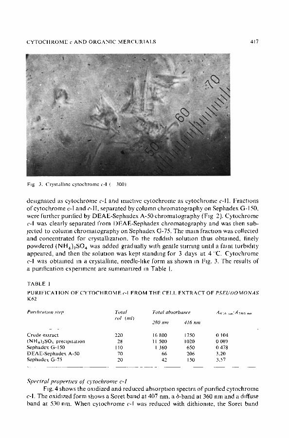
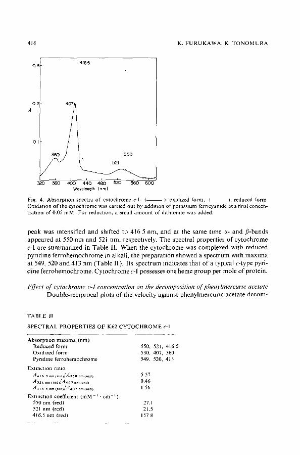
Spectral properttes o] O'tochrorne c-I Fig. 4 shows the oxidized and reduced absorptton spectra of purified cy tochrome
c-l. The oxidized form shows a Soret band at 407 nm, a 6-band at 360 nm and a diffuse band at 530 nm. When cytochrome c-I was reduced with dithiomte, the Soret band
418 K. F U R U K A W A , K T O N O M U R A
°3 I O2
' 4165
i
3~o 360 ~x) Wavelength ( nm }
550
521
440 400 520 560 60(
Fig. 4. Absorp t ion spectra of cytochrome c-l. ( ), oxidized form, ( ), reduced form Oxlda tmn of the cytochrome was carried out by a d d m o n of po tassmm ferncyamde at a final concen- t ra t ion of 0.05 mM For reducUon, a small amoun t of d l th lomte was added.
peak was intensified and shifted to 416 5 nm, and at the same time ~- and fl-bands appeared at 550 nm and 521 nm, respectively. The spectral properties of cytochrome c-I are summarized in Table II. When the cytochrome was complexed with reduced pyndme ferrohemochrome in alkali, the preparation showed a spectrum with maxima at 549, 520 and 413 nm (Table II). Its spectrum indicates that of a typical e-type pyri- dine ferrohemochrome. Cytochrome c-I possesses one heme group per mole of protein.
Effect of cvtochrome c-I concentration on the decomposition of phenylmercurtc acetate Double-reciprocal plots of the velocity against phenylmercunc acetate decom-
TABLE II
S P E C T R A L P R O P E R T I E S OF K62 C Y T O C H R O M E c-1
Absorp t ion max ima (nm) Reduced form Oxidized form PyrJdme fer rohemochrome
Extract ion ratio
A 4 1 6 5 nm ( r e d ) / A s s o nm (oxd) 5 57 A 5 2 1 nm (reaffA4o7 n,, (o~dj 0.46 A 4 1 6 5 am ( r e a ) / A 4 o 7 am (oxd) I 56
ExtmcUon coefficient ( m M - ~ • c m - t ) 550 nm (red) 27.1 521 nm (red) 21.5 416.5 nm (red) 157 8
550, 521, 416 5 530, 407, 360 549, 520, 413
C Y T O C H R O M E c A N D O R G A N I C M E R C U R I A L S 419
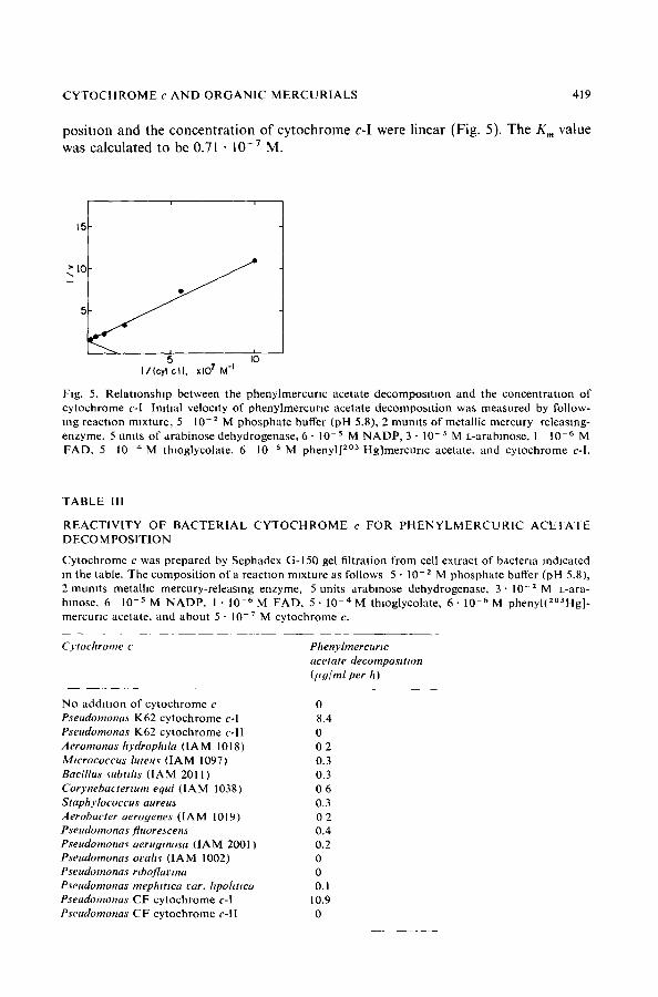
position and the concentration of cytochrome c-I were linear (Fig. 5). The K m value was calculated to be 0.71 • 1 0 - 7 M.
> IC e ~
I/lcytcl). xlO 7 M "l
Fig. 5. Rela t ionship between the phenylmercur lc acetate decomposi t ion and the concentra t ion of cytochrome c-I Ini t ial velocity of phenylmercunc acetate decomposi t ion was measured by follow- mg reaction mixture , 5 10 -2 M phosphate buffer (pH 5.8), 2 mumts of metall ic mercury releasing- enzyme, 5 umts of arabinose dehydrogenase, 6 - 10-s M N A D P , 3 - 10 -3 M L-arabmose, 1 10 -6 M FAD, 5 1 0 - 4 M thioglycolate, 6 10 -5 M phenyl[ 2°3 Hg]mercunc acetate, and cytochrome c-I.
T A B L E I l l
R E A C T I V I T Y OF B A C T E R I A L C Y T O C H R O M E c FOR P H E N Y L M E R C U R I C A C E T A T E DECO M POSITION
Cytochrome c was prepared by Sephadex G-150 gel f i l t rat ion from cell extract of bacteria indicated m the table. The composi t ion of a reaction mixture as follows 5 • 10 -2 M phosphate buffer (pH 5.8), 2 mumts metallic mercury-releasing enzyme, 5 units a rabmose dehydrogenase, 3 ' 10 -z M L-ara- hmose, 6 10 - s M NADP, 1 • 10 -6 M FAD, 5 • 10 -4 M thioglycolate, 6 • 10 -6 M phenyl(2°3Hg]- mercuric acetate, and about 5 - 1 0 - 7 M cytochrome c.
Cytochrome c Phenyhnercurtc acetate decompositton (l*Y/ml per h)
No addi t ion of cy tochrome c 0 Pseudomona~ K62 cytochrome c-I 8.4 Pseudomonas K62 cytochrome c- l l 0 Aeromonas hydrophtla ( IAM 1018) 0 2 Mwrococcus luteuv ( IAM 1097) 0.3 Bacillus ~ubtths ( IAM 2011) 0.3 Corynebactertum equi ( IAM 1038) 0 6 Staphylococcus aureus 0.3 Aerobacter aerogenes d A M 1019) 0 2 Pseadomonas fluore~cen~ 0.4 Pseudomonav aerugmosa ( IAM 2001 ) 0.2 Pseudomonas ovah~ ( IAM 1002) 0 Pseudomonas rtboflavma 0 Pseudomonas mephtttea ear. hpohtwa 0.1 Pseudomonas CF cytochrome c-I 10.9 Pseudomonas CF cytochrome c- l l 0
420 K. F U R U K A W A , K. T O N O M U R A
ReacttwO' o/c:vtoehromes c prepared from various kinds o/bacterta in the phenyhnereu- rtc aeetate decompositton
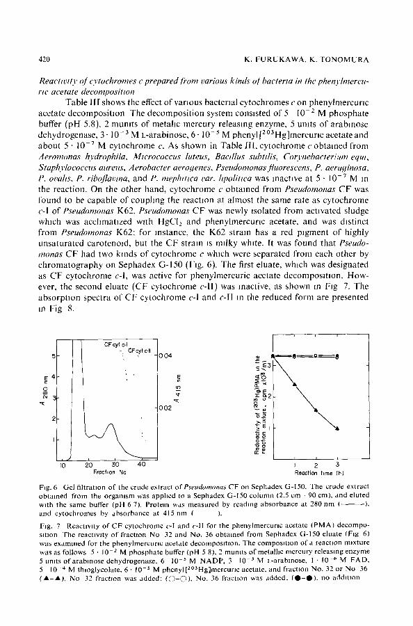
Table llI shows the effect of various bacterial cytochromes c on phenylmercurlc acetate decomposition The decomposition system consisted of 5 l0 -z M phosphate buffer (pH 5.8), 2 mumts of metahc mercury releasing enzyme, 5 umts of arabmose dehydrogenase, 3- 10-3 M L-arabinose, 6. 10-5 M phenyl [20aHg]mercurl c acetate and about 5 • 10 -v M cytochrome c. As shown in Table 111, cytochrome c obtained from Aeromonas hydrophila, Micrococcus luteus, Bactllus subtdis, Coo'nebacterium equt, Staphylocoectts attreus, Aerobacter aerogenes, Pseudomonas fluorescens, P. aeruginosa, P. ovahs, P. riboflavma, and P. mephmca var. hpoBttca was inactive at 5 • 10 -v M in the reaction. On the other hand, cytochrome c obtained from Pseudomonas CF was found to be capable of couphng the reaction at almost the same rate as cytochrome c-I of Pseudomonas K62. Pseudomonas CF was newly ~solated from activated sludge which was acchmatlzed w~th HgCI2 and phenylmercunc acetate, and was distinct from Pseudomonas K62: for instance, the K62 strain has a red pigment of highly unsaturated carotenoid, but the CF strain is milky white. It was found that Pseudo- monas CF had two kinds of cytochrome c which were separated from each other by chromatography on Sephadex G-150 (Fig. 6). The first eluate, which was designated as CF cytochrome c-l, was active for phenylmercuric acetate decomposmon. How- ever, the second eluate (CF cytochrome c-ll) was inactive, as shown m Fig 7. The absorption spectra of CF cytochrome c-I and c-II m the reduced form are presented m Fig 8.
i i
CFcyt c-I • -, CFcyt c.II
5 ~ 0 0 4 ~ I " f f i ~ O @ B
E 4 ' r ~ < S
~-~2 cJ roo
"~ 0 0 2 ~ "
:~__.
, I i i
I0 20 30 40 I 2 5 Frochon No Reocllon time (h)
Fig. 6 Ge l f i l t r a t ion o f the c rude ex t r ac t ofP~eudomonas C F on S e p h a d e x G-150. The c rude ex t rac t o b t a i n e d f rom the o r g a m s m was a p p h c d to a S e p h a d e x G-150 c o l u m n (2.5 cm - 90 cm) , and e lu ted wi th the same buffer (pH 6 7). P ro te in was m e a s u r e d by r ead ing a b s o r b a n c e at 280 n m ( ), and c y t o c h r o m e s by a b s o r b a n c e at 415 n m ( ).
Fig. 7 R e a c U w t y o f C F cy toch ro rne c-I and c - l l for the pheny lmercur~c ace ta te ( P M A ) d e c o m p o - s~tlon The reac t iv i ty o f fractzon N o 32 and No . 36 o b t a i n e d f r o m S e p h a d e x G-150 e lua te (F ig 6) was e x a m i n e d for the p h e n y l m e r c u r l c ace ta te decompos t t~on . The c o m p o s l U o n o f a reacUon m~xture was as fo l lows 5 • 10 -2 M p h o s p h a t e buffer (pH 5 8), 2 m u m t s o f me ta l l i c m e r c u r y r e l eas ing e n z y m e 5 umts o f a r a b m o s e d e h y d r o g e n a s e , 6 10 -5 M N A D P , 3 10 -3 M L-arabmose , 1 • 10 -~ M F A D , 5 10 -'~ M th log lyco la t e , 6 • 10 -5 M p h e n y l [ 2 ° a H g ] m e r c u r l c ace ta te , a n d f r ac t ion No. 32 or N o 36 ( A - A L No 32 f rac t ion was a d d e d : ( O 0 ) , No. 36 f r ac t ion was a d d e d , ( O - O ) , no a d d i t i o n
CYTOCHROME c AND ORGANIC MERCURIALS 421
i i I i
415
0 04
0 O2
55O
350 400 450 ~ 550' l , 600 Wovelength (nm)
Fig 8 Absorpt ion spectra o f CF cytochrome c-I and c-l l ( ), CF cytochrome c-I (reduced form), ( ), CF cytochrome c-II (reduced form) The reduction of cytochrome c was carried out by addit ion of a small amount o f dlthmomte.
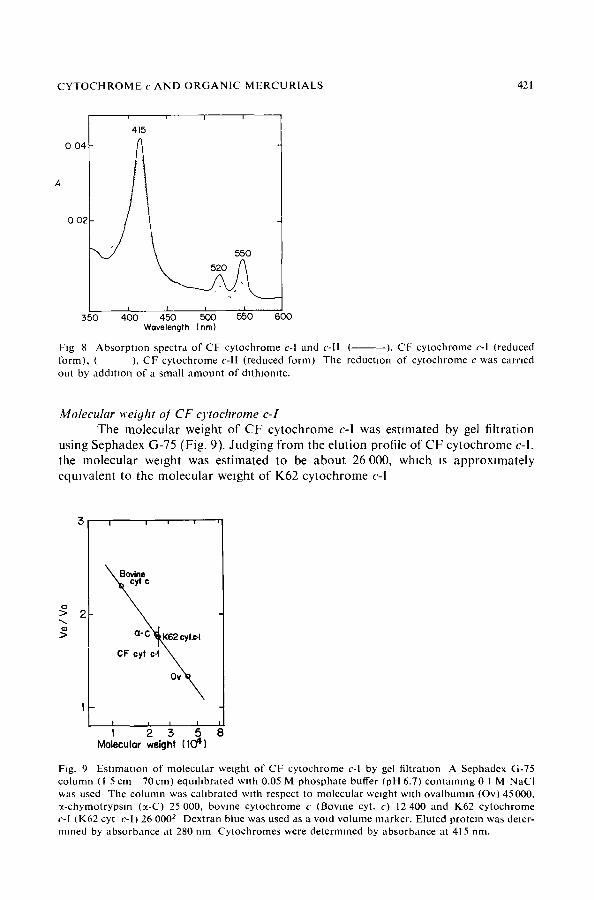
Molecular weiyht o! CF c)'tochrome c-I T h e m o l e c u l a r we igh t o f C F c y t o c h r o m e c-I was e s t i m a t e d by gel f i l t ra t ion
us ing S e p h a d e x G-75 (Fig. 9). J u d g i n g f r o m the e lu t ion prof i le o f C F c y t o c h r o m e c-l, the m o l e c u l a r we igh t was e s t i m a t e d to be a b o u t 26 000, w h i c h is a p p r o x i m a t e l y
e q u w a l e n t to the m o l e c u l a r we igh t o f K62 c y t o c h r o m e c-I
3 , , ,
~ c
a" C~K62 cyt.c-I
CF cylcq
ova,, I I / I
I 2 3 5 Molecular weight (IC7 ~1")
Fig. 9 Estimation of molecular weight of CF cytochrome c-I by gel filtration A Sephadex G-75 column (1 5 cm 70 cm) equdlbrated wtth 0.05 M phosphate buffer (pH 6.7) containing 0 1 M NaCI was used The column was cahbrated with respect to molecular weight with ovalbumm (Ov) 45000, ~-chymotrypsm (~-C) 25 000, bovine cytochrome c (Bovine cyt. c) 12 400 and K62 cytochrome c-I (K62 cyt c-l) 26 000 ~ Dextran blue was used as a void volume marker. Eluted protein was deter- mined by absorbance at 280 nm Cytochromes were determined by absorbance at 415 rim.
422 K FURUKAWA, K. TONOMURA
DISCUSSION
It is known that a c-type cytochrome is revolved in the reduction of nitrate, nitrite, sulfate and sulfite as an electron carrier in anaerobic respiration. Cytochrome c(551) acted as an electron carrier of the nitrite reductase system in Pseudomonas aerugmosa 7. lsh~moto et al. 8 and Postgate 9' 1 o suggested that cytochrome c3 was con- cerned in the sulfate reduction. However, it has not been reported so far that the c-type of cytochrome IS involved in the reduction of mercurials. In previous papers ~ 4, we described the mechanism of enzymatic decomposition of organomercurials to metalhc mercury in mercury-resistant Pseudomonas K62. The reductive reaction was considered to be as follows: NAD(P)H is generated by glucose dehydrogenase or arablnose dehydrogenase system, and the electrons are transferred to metalhc mercury-releasing enzyme, which has FAD as a prosthetic group, in the presence of cytochrome c-l. Organomercurlals would become a terminal acceptor of electrons, and then be reduced to metallic mercury by an cooperative action of metallic mercury-releasing enzyme and cytochrome c-l.
Cytochrome c-I was confirmed to be a c-type cytochrome. Yamanaka and Oku- nukl al indicated that there were two functionally distinct types of cytochrome c: those from plants and animals react with cytochrome oxldase derived from mammalian sources, and those from bacteria react w~th cytochromes oxidase derived from bacte- rial sources. However, bacterial cytochromes c obtained from type cultures belonging to the genera Aeromonas, Micrococcus, Bacillus, Corynebacterium, Staphylococcus, Aerobacter, and Pseudomonas were all reactive at the concentration tested m the phenyl- mercuric acetate decomposing system of Pseudomonas K62. On the other hand, cyto- chrome c obtained from Pseudomonas CF (CF cytochrome c-I) was capable of cou- phng m the reaction at almost the same rate as K62 cytochrome c-I Pseudomonas CF was isolated from the activated sludge acchmatized with HgCI 2 and phenylmercurlc acetate, and was quite different from Pseudomonas K62 with respect to taxonomic properties. It is interesting that the CF strain as well as the K62 strain had two kinds of cytochrome c which are different in molecular weight and function, CF cytochrome c-I and K62 cytochrome c-I had the same mol. wt of 26 000 and were active in phenyl- mercuric acetate decomposition. Furthermore, CF cytochrome c-II which had a low molecular we,ght was inactive in the reaction. Purification of CF cytochrome c-l and enzymatic decomposition of mercurials in CF strain are now in progress. It is likely that the decompos,tion mechanism found in the K62 strata wdl also exist in the CF strain.
Some cytochromes whose function was obscure were reported. Cytochrome c5 s6, a dlheme protein obtained from Pseudomonas aeruginosa, has a tool. wt of 77 200, but the function was not clear; it did not react with cytochrome oxidase of the orga- msm t2. The relationship between cytochrome c-I and cytochrome c-II, nor their involvement in the respiratory chain have as yet been disclosed. The formation of metallic mercury-releasing enzyme was specifically induced in the presence of mercuri- als, but the formation of cytochrome c-1 was not 4. Therefore, it may not be said that cytochrome c-I serves only for the mercurial reduction. It would be very interesting to know how widely cytochromes such as K62 cytochrome c-I or CF cytochrome c-I which are involved in the decomposition of organomercurlals are &stributed m other microorganisms and serve for the mercurial reduction.
C Y T O C H R O M E c A N D O R G A N I C M E R C U R I A L S 423
REFERENCES
1 Tonomura , K and Kanzakl, F (1969) Btochtm Btophys. Acta 184, 227-229 2 Furukawa, K. a n d T o n o m u r a , K (1971) Aortc Btol Chem 35,604 610 3 Furukawa, K. and Tonomura , K (1972) Agrtc. Btol. Chem 36, 217-226 4 Furukawa, K and Tonomura , K. (1972) Aqrte Btol. Chem. 36, 2441 2448 5 Tonomura , K , Maeda, K , Futal, F , NakagamJ, T. and Yamada, M (1968) Nature, 217, 644 646 6 Drucker, H , Troustl, E B, Campbell, L L , Barlow, G. H and Margohash, E. (1970) Bioche-
mtsto', 9, 1515 7 Yamanaka, T (1959) J Btochem. Tol~yo 46, 1289-1301 8 Ishlmoto, M , Koyama, J. and Nagal, Y. (1954) J Btochem Tohvo 41, 763-770 9 Postgate, J R (1954) Btochem. J. 56, xl-xn
10 Postgate, J P (1956) J. Gen Mteroblol. 15, 186-193 11 Yamanaka, T and Okunukl, K. (1968) m Structure attd Functton o/Cytochrome~ (Okunukl, K .
Kamen, M D and Sekuzu, I , eds), pp 390 403, Tokyo 12 Smgh, J. and Wharton, D C. (1973) Btoehtm, Btophy~ Acta 292, 391-401