Embed Size (px)
Citation preview
九州大学学術情報リポジトリKyushu University Institutional Repository
Cytochrome c Peroxidase from Phanerochaetechrysosporium
Nonaka,DaisukeLaboratory of Bioresources Chemistry, Division of Biomaterial Sciences, Department of Forestand Forest Products Sciences, Graduate School of Bioresource and Bioenvironmental Sciences,Kyushu University
Wariishi, Hiroyuki
http://hdl.handle.net/2324/4633
出版情報:九州大学大学院農学研究院紀要. 50 (1), pp.151-164, 2005-02-01. Faculty ofAgriculture, Kyushu Universityバージョン:published権利関係:
J. Fac. Agr.. Kyushu Univ., 50 (1), 151-164 (2005)
Cyiochrome c Peroxidase froln Phanerochaete chrysosporium
Daisuke NONAKA~ and Hiroyuki WARIISHI*
Laboratory of Bioresources Chemistry, Division of Biomaterial Science, Department of
Forest and Fo'rest Products Sciences, Faculty of Agriculture,
Kyushu University, Fukuoka 812-8581 , Japan
(Received November 5, 2004 aud accepted November 15, 2004)
Cytochrome c peroxidase from the white-rot basidiomycete Phanerochate chrysospori-
um (PcCcP) was investigated. A phylogenic analysis of PcCcP amino acid sequence showed
that PcCcP was closely related to cytochrome c peroxidase from Saccharomyces cerevisiae
(yeastCcP) and pea cytosolic ascorbate peroxidase (APX). Recombinant PcCcP was obtained
by expression in Eshcherichia coil and a heme incorporation into the apoenzymes. Spectral
charactersitics indicated that the heme iron of PcCcP was mainly 5-coordinated high spin
species. The absorption spectrum of PcCcP compound I and rapid-scan spectra of compound I
formation strongly suggested that PcCcP compound I was ferryoxy heme iron and protein cation radical, as observed in yeastCcP. Although several typical peroxidase substrates, small
organic or inorganic com~ounds, were not oxidized by PcCcP, ferrocytochrome c was effectively
oxidized. Both PcCcP and yeastCcP shared catalytic features. A homology modeling of PcCcP
and cytochrome c from P. chrysosporium (PcCc) strongly suggested the interaction between
PcCcP and PcCc.
INTRODUCTION Lignin is a heterogeneous, phenylpropanoid polymer that constitutes 20-300/0 of
woody 'plant cell walls (Sarkanen et al.; 1970). White-rot basidiomycetous fungi are pri-
marily responsible for initiating the depolymerization of lignin, which is a key step in ~he
earth's carbon cycle (Crawford, 1980; Gold et al., 1989; Kirk and Farrell, 1987; Tien,
1987). The best-studied lignin-degrading fungus, P/~a7~erocl~aete chrysosporium,
secretes two types of extracellular heme peroxidases, lignin peroxidase (LiP) and man-
ganese peroxidase (MnP), which are the major extracellular components of its ljgnin degradative system (Gold et pLl., 1989; and Kirk and Farrell, 1987) . Both enzyme~ were known to catalyze the hrst step of degradation of lignin polymers using hydrogen per-
oxide (Gold et al., 1989). . ' ' Since the whole genomic sequence of P. chrysosprium was ~tade open t9 public
(Mar~iriez et al., 2004), the survej for. all heme peroxidase genes in the P. ch~~sospo-
rium genome via BLAST homology search ~vas performed. Several LiP and ivlnP 'gene
fragments as well as a gene fragments showing a high similarity to cytochrome_ c per-oxidas~ from Saccharomyces cerevisiae (yea~tdcP) were found. ' ' '
YeastCcP (ferrocytochrome c/hydrogen p~roxide oxidoreductase; EC I .1 1 .1 .5) occurs
naturally in the mitochondrial intermembrane space and catalyzes the H20=-dependent
l Laboratory of Bioresources Chemistry, Division of Biomaterial Sciences, Department of_Forest and
Forest Products Sciences, Graduate School of Bioresource and Bioenvironmental Sciences, Kyushu
University ' * Corre~ponding author (E-inail: [email protected]:jp)
151
1 52 D. NONA1~4 an d H. WARIISHI
one-electron= oxidation of ferrous cytochrorne c (Cc'-'~) to ferric cytochrome c .(Cc=]+)
(Bosshad et al., 1990). Most striking difference between yeastCcP and common peroxi-
dases are found in their compound I structures (Sivaraja et al., 1989; Erman and Yonetani, 1975). Generally, a peroxidase catalytic cycle was described as below.
Restmg state (Fe]+) +H O - Compound I (Fe4+ =0, R e )
Compound I (Fe4+ = O, R e ) +SH - Compound H (Fe4+ = O) + S e
Compound H (Fe++ = O) + SH - Resting state (Fe='+) + S e + H20
In the first step of the peroxidase catalytic cycle, the resting state enzyme, which has
ferric heme iron, is oxidized by hydrogen peroxide to form compound I, where R is an
organic Inoiety. Then, compound I is reduced back to the resting enzyme via compound II
by subtracting two electrons from reducing substrates (SH). Usually, peroxidase com-
pound I was comprised of ferryloxy heme iron and porphyrin 7c cation radical. In yeastCcP compound I, ferryloxy heme iron is also formed as observed in most other per-
oxidases; however, cation radical is located at Trpl91 rather than porphyrin 7T cation
radical (Sivearaja et al., 1989) . In addition., interaction and electron transfer between
yeastCcP and cytochrome c have been well studied as a model system for two macro-
molecules (Bosshard et al., 1990; Erman and Vitello, 2002).
Consequently, yeastCcP has been paid much attention in respect to those unique
functions and catalyiic mechanisms. Since the forefront research techniques have been
applied to yeastCcP to reveal its characters, yeastCcP became the paradigm for all other
peroxidases.
In the present study, a structural characterization of cytochrome c peroxidase
(PcCcP) gene from P. chrysosporium as well as its cloning and heterologous expression
in Esc/berichia coli is reported. The recombinant protein exhibited the H202-dependent
activity of ferrous cytochrome c oxidation.
MATERIALS AND METHODS Chemicals
H20, (300/0 solution) and 2, 6-dimethoxyphenol (DMP) were obtained from Wako
Pure Chemicals Co. Ltd. H.O* stock solution (lO mM) was prepared daily and the concen-
tration was checked as previously described (Cotton and Dunford, 1973). DMP was recrystallized froin ethanoVhexane. All other- chemicals were of reagent grade. Deionized
water was obtained from Milli Q system (Millipore).
Cloning of PcCcP gene and construction of the expression vector P. chr~lsosporium cDNA was synthesized from totalRNA, whidh was isolated from
4-day-old stationary culture of P. chrysosporium, wlth M-MLV reverse transcriptase
(Takara) and oligo dT primers. Following primers were constructed according to BLAST
results using yeastCcP amino acid sequence against P. chrysosporium genome sequence;
CcP-S-51 (AGGGCTGCGGGCCTCCGTAA, 20mer), CcP-A-1448 (CGTATATGAG-CATCTATGACGCCTCGCA, 28mer). PcCcP fragment was synthesized by polymerase chain reaction, using CcP-S-51 and CcP-A-1448, and cloned into pGEM-T Easy Vector
Cytochrome c Peroxidase of P. chrysospol~um 153
(Promega) . Signal peptide prediction was achieved using SOSUI (http://sosui.proteome.
bio.tuat.ac.jp/sosuiframeOE.html) and SignalP (http://w~v.cbs.dtu.
dk/services/SignalP-2.0~ programs. For expression of PcCcP polypeptides in E. coli, a
mature PcCcP sequence with 74 base pairs truncated fragment was amplified using
following primers; PcCcP_exp_forw (CCCAAGCTTCATATGAGCGAAGCCGCGA~TCTG; 34mer) and PcCcP_exp_reve (CGGGATCCCTACGACGACTTCGCCTCCT; 28mer). The resulting fragment was digested with Nde I and Bam HI and cloned into pET-12a (+)
(Novagen). Resulting vector was named pET-tPcCcP.
Expression of PcCcP in Escherichia coli
E. coli BL21(DE3)pLysS (Novagen) transformed with pET-tPCCCP was grown in LB-broth supplemented wlth 100;hg/ml ampicillin and 34~hg/ml chloramphenicol at 37 'C.
An initial small-scale expression was conducted in 2 ml culture at 30'C or 37'C. At the
time of O.D.*,,,,, showing -0.6. 0.4mM isopropyl-thio-~-D-galactopyranoside (IPTG),
0.5 mM 5-aminolevulinic acid (ALA), and 0.2 mM ferrous sulfate was added to induce the
expression of active PcCcP. Incubation time was prolonged for another 6 to 2lh. The
cells were harvested by centrifugation at 9.000 rpm for 3 min and directly used for activity
assay.
Purification of PcCcP For a larger-scale preparation of PcCcP, the cells were grown in a 3-L jar-fermenter
wlthout the addition of ALA and ferrous sulfate; thus, apo-PcCcP was obtained. Arter
collection of the cells by centrifugation, they were resuspended in 50 mM Tris-C1, pH 8.0
containing 2 mM EDTA and stored at -80'C. Then, 2 mg/ml lysozyme, I mM phenyl-
methylsulfonylfluoride, 0.10/0 Triton X-100, I U DNase, and I U RNase were added and
incubated for I h at 30 'C with gentle stirring. The incorporation of heme into apo-protein
was achieved as reported for the yeastCcP expression system (Teske et al., 2000). A
concentrated hemin solution (final concentration of - 500~hM) was added into the lysed
cell suspention and incubated at 4 'C for at least 30min wlth gentle stirring. Then, the
solution was acidified at pH 5.0 with I M acetic acid, and incubated on ice for 30 min.
During this procedure, a large amount of contaminated proteins and excess heme were
precipitated. After centrifugation, the solution was diluted by 20 times with deionized
water and pH of the solution was adjusted to pH 6.0. The enzyme solution was loaded
onto DEAE Sepharose Fast Flow (Amersham Biosciences) equilibrated with 10 mM phos-
phate, pH 6.0. PcCcP was eluted with 500mM phosphate, pH 6.0. The fractions showlng
CcP activity were combined and dialysed against 10 mM phosphate, pH 6.0, for 16h. The
dialyzed sample was concentrated and final purification was achieved using Mono-Q
(Amersham Biosciences) with a multilinear gradient with 10mM phosphate, pH 6.0 and
1 M phophate, pH 6.0 at a flow rate of I mymin. The purified enzyme was dialyzed against
deionized water overnight and stored at 4 'C.
Cyiochrome c preparations Yeast is0-1 cytochrome c were purchased from Aldrich and used without further
purification. Ferrous cytochrome c (Cc'+) was prepared as previously described (Johjima
et al., 2002; Wariishi et al., 1994). Concentration'of Cc2+ was determined spectropho-
154 D. NONA1~4 aud H. WARIISHI
tometrically using a molar extinction coefficient at 550nm of 27.6mM-lcm~1 (Yonetani,
1965).
Spectroscopic Analysis Electronic absorption spectra were recorded using a Perkin Elmer Lambda 19
spectrophotometer at 25 'C.
Steady-state kinetic analysis Initial rates of substrate oxidation were spectrophotometrically measured at 25 'C.
All steady-state kinetic experiments were performed in 20mM phosphate, pH 6.0. The
rate of DMP oxidation (quinone dimmer formation) was determined from the increase in
absorbance at 469nm using Ac of 49.6mM-1cm~1 (Wariishi et al., 1992). The rate of fer-
rocyanide oxidation (ferricyanide formation) was determined using Ac4'_.'*' of I .02 mM-~cm~l
(Schellenberg and Hellerman., 1958) . The rate of ABTS oxidation (ABTS cation radical
formation) was determined using Ac+1* of 36.0mM-1cm~1 (Smith et al., 1990). All reac-
tions were initiated by adding 0.1 mM H202. Steady+state kinetic prameters were calcu-
lated from the Lineweaver-Burk prot. The reaction of PcCcP with Cc'-'+ and L-ascorbate
was determined at 550 nm and 290 nm, respectively.
Transient-state kinetic analysis
Kinetic measurements were conducted using a Photal RA 401S Rapid Reaction Analyzer (Otsuka Electronics Co. Ltd.) equipped with a 1-cm observation cell at 25.0~
0.1 'C. The formation rate of PcCcP compound I was determined at 417 nm, the maximum
absorption wavelength of compound I. One reservoir contained native PcCcP (ca. 2 71M)
and the other reservoir contained H202 (5-15llM) in 40 mM phosphate, pH 6.0. The pseu-
do-first order rate constants were determined by a non-1inear least-squares fit to expo-
nential traces.
Horuology modeling of PcCcP A homology model of the PcCcP tertiary structure was constructed with MOE
program (Chemical Computing Group Inc.), accorchng to manufacturer's instructions and
as previously reported (Rupasinghe et al., 2003) , using yeastCcP structure (PDB code:
2CYP) as a template structure. The truncated PcCcP amino acid sequence with a deletion of 82 amino acids in N-terminal and 10 amino acids in C-terminal sequences
from the original sequence was applied to homology model programs, since those sequences showed no homology to the mature yeastCcP sequence. After construction of
the initial homology model, further energy minimization was performed using the CHARMm22 force field (MacKerell et al., 1998) within the MOE distribution until the final
energy gradient became < 0.01 kcal/mol. A. A distance-dependent dielectric constant
was used in the calculations with a cutoff between 6.5 and 7 A.
RESULTS AND DISCUSSION
PcC6P amino acid sequence The fragment showed a high similarity to the yeastCcP sequence was amplified using
Cytochrome c Peroxidase of P. chrysosporium 155
primers designed from the P. chrysosporium genome sequence (Martinez et .al., 2004).
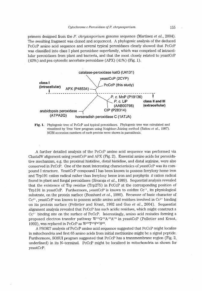
The resulting fragment was cloned and sequenced. A phylogenic analysis of the deduced
PcCcP amino acid sequence and several typical peroxidases clearly showed that PcCcP
was classified into class I plant peroxidase superfamily, which was comprised of intracel-
lular peroxidases from plant and bacteria, and that the most closely related to yeastCcP
(430/0) and pea cytosolic ascorbate peroxidase (APX) (410/0) (Fig. 1).
catalase-peroxidase katG (U41 31 )
yeastCcP (2CYP) class l
PcCcP (this study) (intracellular) APX (P48534)
_j______________________ ______ P. c. MnP~(~19136j ~ ~ ~ ~ ~1~ '
P. c. LiP class ll and lll
(AAB00798) (extracellular) CIP (P28314) arabidopsis peroxidase
(ATPA2Q) horseradish peroxidase C (1ATJA)
Fig. 1. Phylogenic tree of PcCcP and typical peroxidases. Phylogenic tree was calculated and
visualized by Tree View program using Neighbor-Joining method (Saitou et al., 1987).
NCBI accession numbers of each protein were shown in parenthesis.
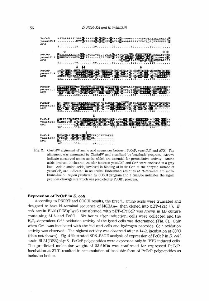
A further detailed analysis of the PcCcP amino acid sequence was performed via
ClustalW alignment using yeastCcP and APX (Fig. 2). Essential amino acids for peroxida-
tive mechanism, e.g. the proximal histidine, distal histidine, and distal arginine, were also
conserved in PcCcP. One of the most interesting characteristics of yeastCcP was its com-
pound I structure. YeastCcP compound I has been known to possess ferryloxy heme iron
and Trpl91 cation radical rather than ferryloxy heme iron and porphyrin lc cation radical
found in plant and fungal peroxidases (Sivaraja et aL, 1989) . Sequential analysis revealed
that the existence of Trp residue (Trp275) in PcCcP at the corresponding position of
Trpl91 in yeastCcP. Furthermore, yeastCcP is known to oxidize Cc'+, its physiologicai
substrate, on the protein surface (Bosshard et al., 1990). Becasuse of basic character of
Cc'+, yeastCcP was known to possess acidic amino acid residues involved in Cc'+ binding
on its protein surface (Pelletier and Kraut, 1992 and Guo et aL, 2004). Sequential
alignment analysis revealed that PcCcP has such acidic residues, which might construct a
Cc'+ binding site on the surface of PcCcP. Interestingly, amino acid resiudes forming a
proposed electron transfer pathway W**'*G**"'A*='=*A~*'+ in yeastCcP (Pelletier and Kraut,
1992) was replaced in PcCcP as W'~*T=~ 'F'~~S
A PSORT analysis of PcCcP amino acid sequence suggested that PcCcP might localize
in mitochondria and first 65 amino acids from initial methionine might be a signal peptide.
Furthermore, SOSUI program suggested that PcCcP has a transmembrane region (Fig. 2,
underlined) in its N-terminal. PcCcP might be localized in mitochondria as shown for
yeastCcP.
156 D. NONA1~4 an d H. W~~IISHI
PcCcP yeastCcP APX
PcCcP yeastCcP APX
PcCcP YeastCcP APX
MSFAALRASLRRA~APAPVA~R~s~IRS~V ~ ~1 ' ' ' ~LRRYETEPPPPPPPPKSHT ----------MTT VRLLPS G T HXR LYLF AAAAAAAAATFAYSQSQKRSSSSPG~
l"' "'10"' "'20"' "'30"' "'40"' "'50"' ** ~ ~~~~~~~SE~KSAGSAVRSGVQGAK~ATKFTP~~~~;~~~~~~~~~~~~~' AAi L :LREDDEY I N ~ ~ . , _ _ _ _ _ . MDKASDKER'Y ~ SNHGWNNWGKA LAS TTPLVHV SVEK~R
---------------------------------M K PTVSPD O AI A~CRKLRGFI 61""" 80"' "'90"' "'100"' "'110"'
, # , ~~~~~j:f;~~~~~~~~~~D N~;~~AP~~SEBK~~!l~il_b~~ K~~~~HV~RD~ YpG YE!~KK FNDPS. A QNG.FKF' j KE F t SKuEK~~I~
I!CHQA LABG N Dl~VR AEKK~:~i J
121"""I30"' "'140"' "'150"' "I60" "I70"
PcCcP yeastCcP APX
PcCcP yeastCcP APX
PcCcP yeastCcP APX
PcCcP yeastCcP APX
' ' ~~~~~i~G_ Y_AAQA~D , .QI. 'TPEDTT DN I D D 'YV TF ~~~~~~Y Q E ' KPEP E 181"""I90"' "'200"' "'210"' "'220"' "'230"'
, ~]~FD~;~~_ RC~~RAW~~~~~~~~~:~i~;~:~~[e'j{!~l~~~ibrIGLd;~:~:~~~~~1 TMS~~~~~~~lIEY~KL~l~~D K ~ ~~K~w
- ~ Q LNMN ) , L KT LKN GAAi NVET E YLN N D K E N ' SN'LI D Y~TE TG-------KA LS I QD T: AA KER F
241' ' ' ' ' '250""" '260""" '270""" "'290"' '280"" V
i~GPKQFEi~KGTRS ~~VjV~KS~EKW~A~D~j;Eil:ill~!r~~~j~~!~' Y ~;E~i;~~~~;~Ili~E~~~~!~' ~ ;i ~ I ANNEQW I - S~S . I S I"PKYL.S:~EE .N'QIK
---------E D LQ SIKA LT'SV~~P:1 EK A' ' 301"""310"' "'320"' "'330"' '*'340"' "'350"'
* EHQW~Ej T S EE~~:FEE~~~il~li~d~;!:~~AQD TKEAK S S
FPKD ' P-S ~A-----------361"""370"' "'380"'
Fig. 2. ClustalW alignment of amino acid sequences between PcCcP, yeastCcP and APX. The
alignment was generated by ClustalW and visualized by boxshade program. Arrows indicate conserved amino acids, which are essential for peroxidative activity. Amino
acids involved in electron transfer between yeastCcP and Cc2+ were enclosed in a gray
box. Acidic amino acids, involved in binding of basic Cc2+ at the enzyme surface of
yeastCcP, are indicated in asterisks. Underlined residues at N-terminal are mem-
brane-bound region predicted by SOSUI program and a triangle indicates the signal
peptides cleavage site which was predicted by PSORT program.
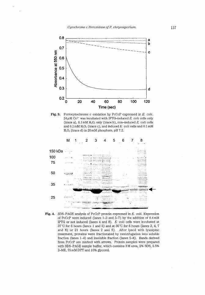
Expression of PcCcP in E. coli
According to PSORT and SOSUl results, the first 71 amino acids were truncated and
designed to have N-terminal sequence of MSEAA-, then cloned into pET-12a(+). E.
coli strain BL21(DE3)pLysS transformed with pET-tPCCCP was grown in LB culture
containing ALA and FeS0+. Six hours after induction, cells were collected and the
H O dependent Cc'+ oxidatlon actrnty of the lysed cells was determmed (Flg. 3). Only
when Cc'+ was incubated with the induced cells and hydrogen peroxide, Cc""+ oxidation
activity was observed. The highest activity was observed after a 14-h incubation at 30 'C
(data not shown). Fig. 4 illustrated SDS-PAGE analysis of expression of PcCcP in E. coli
strain BL21 (DE3)pLysS. PcCcP polypeptides were expressed only in IPTG induced cells.
The predicted molecular weight of 33.6kDa was confirmed for expressed PcCcP. Incubation at 37'C resulted in accumulation of insoluble form of PcCcP polypeptides as
inclusion bodies.
Cytochrome c Peroxidase ofP. chr~lsosporium 157
~
O LO LO
~ ,U
8 ~ S *Oco
n <
o. 8
0.7
0.6
0.5
0.4
0.3
0.2
a b
c
d
Fig. 3.
o 20 40 60 1 OO 1 20 80
Time (sec)
Ferrocytochrome c oxidation by PcCcP expressed in E. coli.
34~hM Cc2+ was incubated wlth IPTG-induced E. coli cells only
(trace a), 0.1 mM H202 only (trace b), non-induced E. coli cells
and 0.1 mM H.0= (trace c), and induced E. coli cells and 0.1 mM
H202'(trace d) in 20 mM phosphate, pH 7.2.
M 1 2 3 4 5 6 7 8 150 kDa
1 OO
75
・~
+・~'.~~~ ~~~ - ~~ ~F~i ~#~~
~ ~
~
50
35
*~* ~#~~f ~
t~~f~~~ S~~~~; ~~~~1* i~~f~! ~i
~f~~:~ ~~~~i+ ~;
~:" ~~~~f~!. *
* ' +(-- ・~~~'"~;~~ ~~##,~
25 ~~~~
Fig. 4. SDS-PAGE analysis of PcCcP protein expressed in E. coli. Expression
of PcCcP were induced (lanes 1-3 and 5-7) by the addition of 0.4mM
IPTG or not induced (lanes 4 and 8). E. coli cells were incubated at
37'C for 6 hours (1anes I and 5) and at 30'C for 6 hours (lanes 3, 4, 7
and 8) or 21 hours (lanes 2 and 6). After lysed with lysozyme treatment, proteins were fractionated by centrifugation into soluble
fraction (lanes 1~1) and insoluble fraction (1anes 5-8). Bands derived
from PcCcP are marked with arrows. Protein samples were prepared with SDS-PAGE sample buffer, which contains 3 M urea, 20/0 SDS, 2.50/0
2-ME, 75 mM DTT and 100/0 glycerol.
158 D. NONA1~4 aud H. WARIISHI
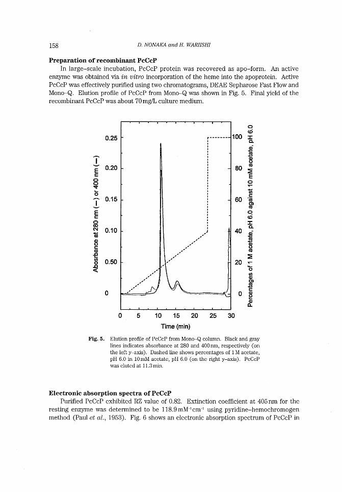
Preparation of recombinant PcCcP In large-scale incubation, PcCcP protein was recovered as apo-form. An active
enzyme was obtained via in vitro incorporation of the heme into the apoprotein. Active
PcCcP was effectively purified using two chromatograms, DEAE Sepharose Fast Flow and
Mono-Q. Elution profile of PcCcP from Mono-Q was shown in Fig. 5. Final yield of the
recombinant PcCcP was about 70 mg/L culture medium.
~ I
~ ~ o o It *o
~ l
~ ~ o co C~
~ ce
8=
~ coo
n <
0.25
0.20
0.15
0.10
0.50
o / ,
,
, , ,
(,
,,
,
If , ,, ,
1'
r--i
,
i
i
,
,
l
,
,
I
I
l
l
,
,
l
,
l
,
I
I
,
,
,
l
l
,
,
,
I
I
l
, I
J
o (c;
100 :E CL
~ CU
~ 8 cu
80 ~: E o .-
~ ~ 60 '~;
o) ,D
o. 'o
= a 40 o~ ~ cl'
~ ~
:~
20 -.* o (o
o o) CU ~c
08* ~
Fig. 5.
o 5 1 O 1 5 20 25 30 Time (min)
Elution profile of PcCcP from Mono-Q column. Black and gray
lrnes indicates absorbance at 280 and 400nm, respectively (on
the left y-axis). Dashed line shows percentages of I M acetate,
pH 6.0 in 10mM acetate, pH 6.0 (on the right y-axls). PcCcP was eluted at 1 1 .3 min.
Electronic absorption spectra of PcCcP Purified PcCcP exhibited RZ value of 0.82. Extinction coefficient at 405nm for the
resting enzyme was determined to be 1 18.9mM-1cm~* using pyridine-hemochromogen method (Paul et al., 1953) . Fig. 6 shows an electronic absorption spectrum of PcCcP in
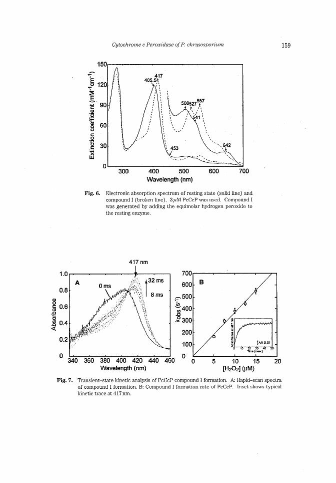
Cytochrome c Peroxidase of P. chr~lsosporium 159
1 50
~ -'E o
~. ~:
E ~. - 90 = o .5
~ (D 8 60 c o :i=
o 30 c ~~ UJ
1 20
o
'i I
i
t
l
,
t !
417 405 5t
,t ,,
t
, ~ , I t l I , 1
l 557 f
508527 I ,
J t i
l t * l] l l ,
' ~¥ ' 1 t I
, ¥' t l , f t
, t $ 1 l l
, I , I
t
, , l I I J
, I l t
l i I f ' ~ 1 , N ~l / t I t 1 I ¥ I
t ¥ 642 453 t .~ 1
L ¥ ¥ ¥
300 400 500 Wavelength (nm)
600 70 o
Fig. 6. Electronic absorption spectrum of resting state (solid line) and
compound I (broken line). 3~tM PcCcP was used. Compound I was generated by adding the equimolar hydrogen peroxide to
the resting enzyme.
417 nm
1 .o
0.8
8CCD 0.6
JD * o j~ 0.4
< 0.2
o
A
~ rr
o ms
¥
*'~rf'ff~_;:':::'~
!/ .. _~ f tlb" i'
f ti >, t!.~; ~ //^f "
,,. ,. ~.
f*.~~:, ~¥ .~
;'1L. t; .'LI'l,~ ='.
1,~~ t~ '4
.;~~~ t ~ t ?. a +*
! J~'~ ~~t t; * ~ iAI ~'
i 'f 1*F;
~l' ~
f32 ms
8 ms
j"ti
}~.
~~,
~*).
"'~*
~'~~t~
380 400 420 Wavelength (nm)
700
600
- 500 -b, ~co 400
~ o Qnn ~' .uu
200
1 oo
340 360 440 460 O o
B
E
N ~ 8 ~ 8 , <
tAA O.O1
Ttlle (rr~c]
Fig. 7.
5 10 15 [H202] (uM) 20
Transient-state kinetic analysis of PcCcP compound I formation. A: Rapid-scan spectra
of compound I formation. B: Compound I formation rate of PcCcP. Inset shows typical
kinetic trace at 417 nm.
160 D. NONA1~4 an d H. WARIISHI
20mM phosphate, pH 6.0. The absorption spectrum of resting enzyme showed the Soret
maximum at 405.5nm wlth a shoulder at -380nm, and visible peaks at 508nm, 541 nm and 642 nrn, strongly suggesting that the heme iron of recombinant PcCcP was five-coor-
dinated high spin. High numbers of aromatic amino acids (19 phenylalanines, 13 trypto-
phanes, and 12 tyrosines) in PcCcP is the reason for a low number of RZ value and could
also be one of the reasons that easier formation of inclusion bodies, compared to
yeastCcP.
PcCcP compound I was generated by adding the equimolar of hydrogen peroxide,
resulting in the Soret maximum at 417 nm. The absorption spectrum of PcCcP compound
I resembled that of yeastCcP compound I which has the Soret maximum at 419 run. Thus,
PcCcP compound I might contain the ferryloxy heme iron and an amino acid cation radi-
cal, probably at Trp275.
YeastCcP compound II was reported to be the mixture of ferryloxy iron and Trp cation radical; thus, its absorption spectrum is not distinguishable (Coulson et al., 1971).
PcCcP compund H was prepared by adding equimolar ferrocyanide to PcCcP compound I.
The resulting spectrum of PcCcP was very similar to that of the resting state (data not
shown), suggesting PcCcP compound H also possessed yeastCcP-like protein based cation radical, probably with less characteristics of ferryloxy feature of the heme.
Stopped-flow analysis of PcCcP cornpound I formation Further characterization of PcCcP compound I was conducted using a stopped-flow
rapid-scan analysis. Fig. 5A showed rapid-scan spectra of the reaction between the rest-
ing state enzyme and H202 from 8 ms to 32 ms after mixing. The isosbestic points at 345,
407, and 455nm clearly indicated that the reaction proceeded in a single step. Then, the
PcCcP compound I formation rate was determined by following the change in absorbance
at 417nm. The values of k.b, were obtained using exponential fit to the kinetic traces (Fig.
5B inset) . The first-order rate constant for PcCcP formation (kl) was evaluated to be
3.6X 10⁷M-Is~1 by plotting k.b, values against H202 concentrations (Fig. 5B). The inter-
section of the regression line of the plots at the origin indicated that the reaction between
PcCcP and H202 Was irreversible.
Cyiochrome c oxidation by PcCcP Since PcCcP amino acid sequence showed a high sirnilarity against both yeastCcP and
APX, as shown in Fig. 2, one-electron oxidation ability of purified PcCcP against
cytochrome c and L-ascorbate, which were physiological substrates for yeastCcP and
APX, respectively, were investigated. In the presence of H202, PcCcP effectively cat-
alyzed an one-electron oxidation of Cc'+ (Fig. 8). On the other hand, no significant
change was observed in the reaction between PcCcP and L-ascorbate (Fig. 8). These observations strongly suggested that L-ascorbate is not a substrate of PcCcP but Cc'+ is.
Substrate specificity of PcCcP Steady-state kinetic parameters for reactions between PcCcP and a series of typical
peroxidase substrates were determined (Table 1). PcCcP showed low activities against
DMP (phenolic substrate) , ABTS (typical anionic substrate) , ferrocyanide (inorganic sub-
strate) . Furthermore, veratryl alcohol and manganese (H) , which are physiological sub-
Cytochrome c Peroxidase ofP. chrysosporium 161
O O C CD !:1*
O Q,
~ <
1 .O
0.8
0.6
0.4
0.2
o
80 c:' to
e 8 O. ~O
300 Ascorbate at 2eo nm
oo
::.:~~~
;:;~
1 O 20 eo 40 Time (seo)
/~,/;.~~~~/r~~~_ . '
~r'$~~'i
//11 ' /~¥L1' / / " _ ;¥~1
/ /': ・;(.: ~~
lltl'/ '- ~
31 uM ycc(Il) l¥ at 5CO nm ' I l l
,t l t ,~l
50 60 ' f ¥It , l l .. ll
l!11 ,, IL ~i .. ~]
l~: / ~ ':~~
t ': ' ¥" 1 ,' S fii/:' :!/" ~ ,.
r¥'~'::~ ~ ':"' ;::~¥'~~~!} i.~~:*-
~.::
O sec
300 sec
~F 1~: ~~: ~t・・.~~~_~
¥t_;・ *:~!*1:¥s. _ ., -.-:. -¥,=*
:L '-:~ ,L " ~.~~
500 540 560 520 Wavelength (nm)
580
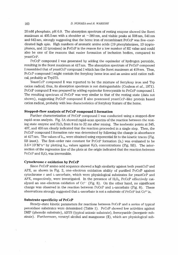
Fig. 8. Time course of the reaction between PcCcP and Cc2+. 3llhM Cc""+ was incubated wlth
0.85 nM PcCcP and 50~tM H20, in 20mM phosphate, pH 6.0. Each spectrum was mea-sured every 3 minutes after addition of H_.02. Inset shows kinetic traces of reactions
between PcCcP and Cc2+ or L-ascorbate.
Table 1. Steady-state kinetic parameters of various substrates oxidation
catalyzed by PcCcP.
Km (mM)
k***
(mmoVmin/mmol)
k** JKm
(mM-Imin~1)
DMP ABTS
Ferrocyanide
17
0.41
68
56
390
213800
3.3
951
3144
strates for LiP and MnP were not reacted with PcCcP.
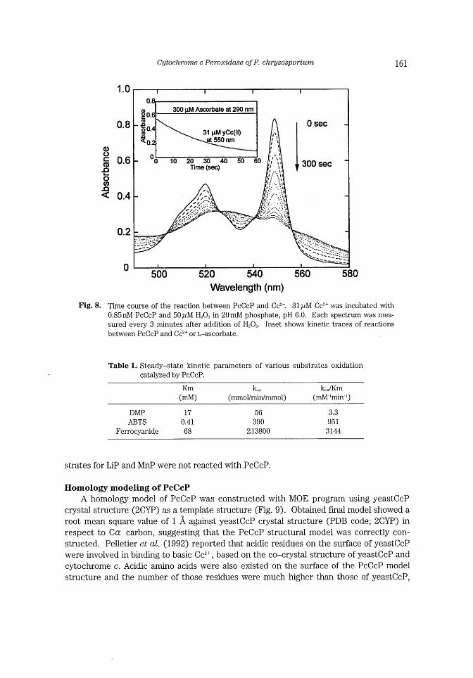
Holnology Inodeling of PcCcP A homology model of PcCcP was constructed with MOE program using yeastCcP
crystal structure (2CYP) as a template structure (Fig. 9). Obtained final model showed a
root mean square value of I A against yeastCcP crystal structure (PDB code; 2CYP) in
respect to Ca carbon, suggesting that the PcCcP structural model was correctly con-
structed. Pelletier et al. (1992) reported that acidic residues on the surface of yeastCcP
were involved in binding to basic Cc2+, based on the co-crystal structure of yeastCcP and
cyiochrome c. Acidic amino acids were also existed on the surface of the PcCcP model
structure and the number of those residues were much higher than those of yeastCcP,
162 D. NONA1~4 aud H. WARIISHI
A y
~f'~
i ~ ;'~/'L;j~¥::;~~f~~
' ' ~~ '~ I~'~/ ~
'""~~~'
PcCcP
J '~... '= s~'~:~:~'-"~~j'~:--/~
, "s~~~ -
yeastCcP
Fig. 9.
~
'____~;~~it~:~')~
/ _ ~~' -
tii
~i' r-'
*~; Pcccp
r'~~~~'!~'~/¥
~~~T
~~:S~~~i~:~~/!~~~~ii~;~L~"~~~~;;f! ~"
~~~~~1;~/cS~;?'~~1~:
i~~~ ~ #~' ~ ;1~T~1~*~~~~~~i~ ~~~
APX
;,?
Homology model of PcCcP. Panel A: Acidic amino acids on the protein
surface of PcCcP and yeastCcP. Acidic arnino acids (glutamate or aspar-
tate) are indicated in dark color. The proposed electron transfer site in
yeastCcP (Pelltier and Kraut, 1992) or the corresponding residues in
PcCcP were enclosed in circles. Panel B: Comparison of heme pocket
architecture of PcCcP and APX. Heme was emphasized in gray.
indicating surface of PcCcP was much more acidic than that of yeastCcP (Fig. 9A) . The
model structure for cytochrome c of P. chr~lsosporium (PcCc) , which was annotated
from P. chr~lsosporium genome sequence by a BLAST search using cytochrome c sequence of S. cerevisiae was constructed. The surface of PcCc was basic with cal-
culated pl of 9.6, which was comparable to pl of 1 1 for yeast is0-1 cytochrome c. Because
of lower pl for PcCc, strongly acidic surface characteristics might be required for PcCcP.
APX shows no activity against Cc2+despite of its sequence similarity against yeastCcP.
The most preferred substrate of APX is L-ascorbate, its physiological substrate. Crystal
structure of ascorbate peroxidase-ascorbate complex (Sharp et al., 2003) showed that
cationic amino acid residues, Lys 30 and Arg 172, were mostly contributed to the binding
Cytochrome c Peroxidase of P. chrysosporium 163
of L-ascorbate, an anionic substrate. These two cationic amino acids were substituted to
Gly and Asn in yeastCcP and Gly and Trp in PcCcP, respectively (Fig. 2). These struc-
tural characteristics are in good accordance with the observation that PcCcP exhibited
the oxidation activity towards Cc'+ but not to L-ascorbate (Fig. 8) .
PcCcP showed only a weak activity towards typical peroxidase substrates (Table 1).
Furthermore, PcCcP showed no activity against manganese (H) or veratryl alcohol, which
were physiological substrates for MnP and LiP, respectively (Gold et al., 1989). These
observations clearly indicated that substrate specificity of PcCcP was strictly restricted to
Cc'+ and, furthermore, PcCcP seemed not to be involved in neither L-ascorbate nor lignin
metabolism. Substrate specificity of PcCcP was further evidenced by the homology
model of PcCcP. Fig. 9B showed comparison of heme pocket entrance between PcCcP
and APX. Narrower heme pocket of PcCcP, compared to that of APX, might prevent the
interaction of reducing substrates with the heme of PcCcP.
A physiological function of yeastCcP was not completely understood. Especially, a
physiological meaning of Cc2+ oxidation by yeastCcP is still unclear. However, the induc-
tion of yeastCcP mRNA by oxidative stress was reported (Kwon et al., 2003) and this
enzyme might be involved in anitioxidative system in mitochondria. Proteomic analysis
surveying the conditions to optimize PcCcP production is now underway. Our recent pro-
teomic data indicated the up-regulation of PcCcP production under 1000/0 oxygen atmosphere (data not shown). PcCcP may be involved in antioxidative system.
CONCLUSION P. chr~lsospoium genome sequence data enabled us to survey various enzymes. This
is the first time to exhibit that cytochrome c peroxidase was active and functional in
basidiomycetes. Recombinant PcCcP was successfully obtained via the E. coli expression
system. The structural characteristics were suggested to be very similar to yeastCcP.
Spectral, kinetic, and sequential analyses of PcCcP revealed that recombinant PcCcP
catalyzes the H202-dependent Cc"-+ oxidation via a typical peroxidase catalytic cycle.
REFERENCES Bosshard, H. R H Anm and T Yonetam 1990 Yeast cytochrome c peroxidase In "Peroxldases m
Chemistry and Biology" (Everse, J., Everse, K. E., and Grisham, M. B., Eds.), Vol. ll, CRC Press, Boca
Raton, FL, pp. 51-84
Cotton, M. L. and H. B. Dunford 1973 Studies on horseradish peroxidase XI: On the nature of com-pounds I and II as determined from the kinetics of the oxidation of ferrocyanide. Can. J. Chem., 51:
582-587 Coulson, A. F. W., J. E. Erman and T. Yonetani 1971 Studies on cytochrome c peroxidase. XVII.
Stoichiometry and mechanism of the reaction of compound ES wlth donors. J. l~iol. Chem., 246:
9 1 7-924
Crawford, R. L. 1980 Lignin 1~iodegradati07~ aud Transformation,Wliey, New York
Erman. J. E. and L. B. Vitello 2002 Yeast cytochrome c peroxidase: mechanistic studies via protein
engineering. Biochim. ~iophys. Acta., 1597: 193-220
Erman, J. E. and T. Yonetani 1975 A kinetic study of the endogenous reduction of the oxidized sites in
the primary cytochrome c peroxidase-hydrogen peroxide compound. Biochim. Biophys. Acta., 393:
350-357 Gold, M. H., H. Wariishi and K. Valli 1989 Whitaker, J. R. and P. Sonnet (Eds.),Proceedings ofthe AC~
164 D. NONA1~4 an d H. W~RIISHI
Symposium Series 389 on l~iocatalysis in Agricultural 1~iotechnology, American Chemical Society, Washington, DC, pp. 127-140
Guo, M., B. Bhaskar, H. Li, T. P. Barrows and T. L. Poulos 2004 Crystal structure and characterization of
a cytochrome c peroxidase-cytochrome c site-specific cross-link. Proc. Natl. Acad. Sci. U. S. A.,
lO1: 5940-5945 Johjima, T., H. Wariishi and H. Tanaka 2002 Veratryl alcohol binding sites of lignin peroxidase from
Phanerochaete chrysosporium. J. Mol. Cat. 1~: Enzym., 17: 49-57
Kirk, T. K. and R. L. Farrell 1987 Enzymatic "combustion": the microbial degradation of lignin. An?zu.
Rev. Microbiol., 41: 465-505
Kwon, M.. S. Chong, S. Han and K. Kim 2003 Oxidative stresses elevate the expression of cytochrome c
peroxidase in Saccharomyces cerevisiae. i~iochim. Biophys. Acta., 1623: 1-5
MacKerell. A. D., Jr., D. Bashford, R. L. Bellott, R. L. Dunbrack, Jr., J. D. Evan~eck, M. J. Field, S. Fischer,
J. Gao, H. Guo, S. Ha, D. Joseph-McCarthy, L. Kuchnir, K. Kuczera, F. T. K. Lau, C. Mattos, S.
Michnick, T. Ngo, D. T. Nguyen, B. Prodhom, W. E. Reiher, HI, B. Roux, M. Schlenkrich, J. C. Smith,
R. Stote, J. Straub, M. Watanabe, J. Wiorkiewicz-Kuczera, D. Yin and M. Karplus 1998 All-Atom
Empirical Potential for Molecular Modeling and Dynamics Studies of Proteins. J. Phys. Chem. B.,
102: 3586-3616 Martinez, D., L. F. Larrondo, N. Putnam, M. D. Gelpke, K. Huang, J. Chapman, K. G. Helfenbein, P.
Ramaiya, J. C. Detter, F. Larimer, P. M. Coutinho, B. Henrissat, R. Berka, D. Cullen and D. Rokhsar
2004 Genome sequence of the lignocellulose degrading fungus Phanerochaete chrysosporium strain
RP78. Nat. Biotechnol., 22: 695-700 Paul, K. G., H. Theorell and A. Akeson 1953 The molar light absorption of pyridine ferroprotoporphyrin
(pyridine haemochromogen). Acttb Chem. Sca?7;d., 7: 1284-1287
Pelletier, H. and J. Kraut 1992 Crystal Structure of a Complex Between Electron Transfer Partners,
Cyiochrome c Peroxidase and Cyiochrome c. Science, 258: 1748-1755
Rupasinghe, S., J. Baudry and M. A. Schuler 2003 Conunon active site architecture and binding strategy
of four phenylpropanoid P450s from Arabidopsis thaliana as revealed by molecular modeling. Protein Eng., 16: 721-731
Saitou, N. and M. Nei 1987 The neighbor-joining method: a new method for reconstructing phylogenetic
trees. Mol. Biol. Evol., 4: 406~25
Sarkanen. K. V. and C. H. Ludwlg 1970 Lignins: Ocurrence. Formation. Structure a7td Reaclions,
Wliey, New York Schellenber~, K. A. and L. Hellerman 1958 Oxidation of reduced diphosphopyridine nucleotide. J. Biol.
Chem., 231: 547-556
Sharp, K. H., M. Mewies, P. C. Moody and E. L. Raveh 2003 Crystal structure of the ascorbate peroxidase-ascorbate complex. Nat. Struct. ~iol., 4: 303-307
Sivaraja, M., D. B. Goodin, M. Smith and B. M. Hoffman 1989 Identification by ENDOR of Trpl91 as the
free-radical site in cytochrome c peroxidase compound ES. Science, 245: 738-740
Smith, A. T., N. Santama, S. Dacey, M. Edwards, R. C. Bray, R. N. Thorneley and J. F. Burkp 1990
Expression of a synthetic gene for horseradish peroxidase C in Escherichia coli and folding and
activation of the recombinant enzyme with Ca;+ and heme. J. Biol. Chem., 265: 13335-13343
Teske, J. G., M. I. Savenkova, J. M. Mauro, J. E. Erman and J. D. Satterlee 2000 Yeast cytochrome c
peroxidase expression in Escherichia coli and rapid isolation of various highly pure holoenzymes.
Protein Expr. Purif., 19: 139-147
Tien, M. 1987 Properties of ligninase from Phanerochaete chrysosporium and their possible applications. CRC Crit. Rev. Microbiol., 15: 141-168
Wariishi, H., K. Valli and M. H. Gold 1992 ManganeseCll) oxidation by manganese peroxidase from the
basidiomycete Phanerochaete chrysosporium. Kinetic mechanism and role of chelators. J. Biol.
Chem., 267: 23688-23695
Wariishi, H., D. Sheng and M. H. Gold 1994 Oxidation of ferrocytochrome c by lignin peroxidase
Biochemistry, 33: 5545-5552
Yonetani. T. 1965 Studies on cytochrome c peroxidase. ll. Stoichiometry between enzyme, H*O,, and
ferrocyiochrome c and enzymic determination of extinction coefficients of cytochrome c. J. Biol.
Chem., 240: 4509~1514 Yonetai, T. 1976 The Enzymes,3rd ed., P. D. Boyer, ed.,Academic,NewYork, Vol. 13, pp. 345-361