Embed Size (px)
Citation preview
LETTER TO THE EDITOR Korean J Intern Med 2012;27:470-473http://dx.doi.org/10.3904/kjim.2012.27.4.470
pISSN 1226-3303 eISSN 2005-6648http://www.kjim.org
Cytomegalovirus Induced Interstitial Nephritis and
Ureteral Stenosis in Renal Transplant Recipient
To the Editor,Cytomegalovirus (CMV) infections occur in up to
60% of kidney transplant recipients, with most cases
developing between the second and 6 months after
transplantation [1]. The clinical manifestations of CMV
infection vary widely. Asymptomatic viremia is the
most common symptom, although the infection can oc-
casionally invade various organs such as the lung, or
the gastrointestinal tract [2]. Likewise, in the urogenital
tract, CMV can induce ureteral stenosis by direct inva-
sion of ureter tissue [3]. Involvement of grafted kidneys
by CMV occurs frequently. In previous reports, CMV
genomes were documented by in situ hybridization
techniques in more than 40% of routine graft biopsies
[3]. However, significant pathologic changes and al-
lograft dysfunction induced by CMV invasion are rarely
observed. Indeed, CMV inclusions in tissue have been
reported in less than 1% of renal biopsies when exam-
ined by routine light microscopy. Therefore, an alterna-
tive method to detect CMV involvement in graft kidneys
is required.
In a previous report, pathological CMV nephritis was
successfully diagnosed by immunohistochemistry (IHC)
and tissue polymerase chain reaction (PCR) [3]. In this
regard, the present case illustrates manifestation of
CMV nephritis both by ureteral stenosis and CMV inter-
stitial nephritis, diagnosed by IHC stain for CMV. The
patient was successfully treated with ganciclovir and
immunosuppression reduction. We present this case re-
port with a review of the current literature.
Our patient was a 48-year-old Korean man who suf-
fered from end-stage renal failure attributed to hy-
pertension who had been on continuous ambulatory
peritoneal dialysis for 2 years. He had received a living
donor renal transplant in January 2010, but the specific
record of the donor was not available because the proce-
dure was performed overseas in China. A week after the
transplant, when he returned to Korea and visited the
hospital, he was put on a dual therapy with tacrolimus
and prednisolone with a serum creatinine of 3.1 mg/dL.
Tacrolimus was titrated to 4 mg twice a day, mycophe-
nolate mophetil was continued 750 mg twice a day, and
prednisolone was added at 25 mg twice a day. Labora-
tory investigations revealed the following values: white
blood cells, 4,840/mm3 with 94.5% polymorphonuclear
cells, 1.4% lymphocytes, 4.1% monocytes; hemoglobin
10 g/dL; hematocrit, 29.7%; platelet count, 151,000/
mm3; creatinine, 3.1 mg/dL; total bilirubin, 0.45 mg/
dL; AST, 8 U/L; ALT, 9 U/L; albumin, 3.1 g/dL; random
blood sugar, 133 mg/dL; C-reactive protein, 0.16 mg/dL;
and urinary microscopic hematuria.
A Doppler ultrasound of the grafted kidney revealed
hydronephrosis without visible obstructive lesions and
favorable renal perfusion. The patient underwent per-
cutaneous nephrostomy and showed contrast leakage
from the ureterovesical anastomosis. Subsequently, an
anterograde double J (DJ) catheter was inserted with
a ureteral stent via the right hypogastric posterolateral
approach. A Doppler ultrasound performed after the
catheter insertion showed complete regression of hydro-
nephrosis of the graft kidney; however, renal function
was only partially recovered and the patient’s creatinine
level hovered around 2.2 mg/dL 2 weeks after catheter
insertion.
To evaluate the cause of the incomplete recovery of
allograft function, a renal biopsy was performed that
revealed calcineurin inhibitor toxicity with tubulopathy.
Glomerular capillary intracytoplasmic CMV inclusions
Copyright © 2012 The Korean Association of Internal MedicineThis is an Open Access article distributed under the terms of the Creative Commons Attribution Non-Commercial License (http://creativecommons.org/licenses/by-nc/3.0/) which permits unrestricted non-commercial use, distribution, and reproduction in any medium, provided the original work is properly cited.
Received : March 21, 2012Revised : May 9, 2012Accepted : July 16, 2012
Correspondence to Bum Soon Choi, M.D. Department of Internal Medicine, Seoul St. Mary’s Hospital, The Catholic University of Korea College of Medicine, 222 Banpo-daero, Seocho-gu, Seoul 137-701, KoreaTel: 82-2-2258-7570, Fax: 82-2-599-3589, E-mail: [email protected]
Bae SH, et al. CMV induced interstitial nephritis 471
http://dx.doi.org/10.3904/kjim.2012.27.4.470 http://www.kjim.org
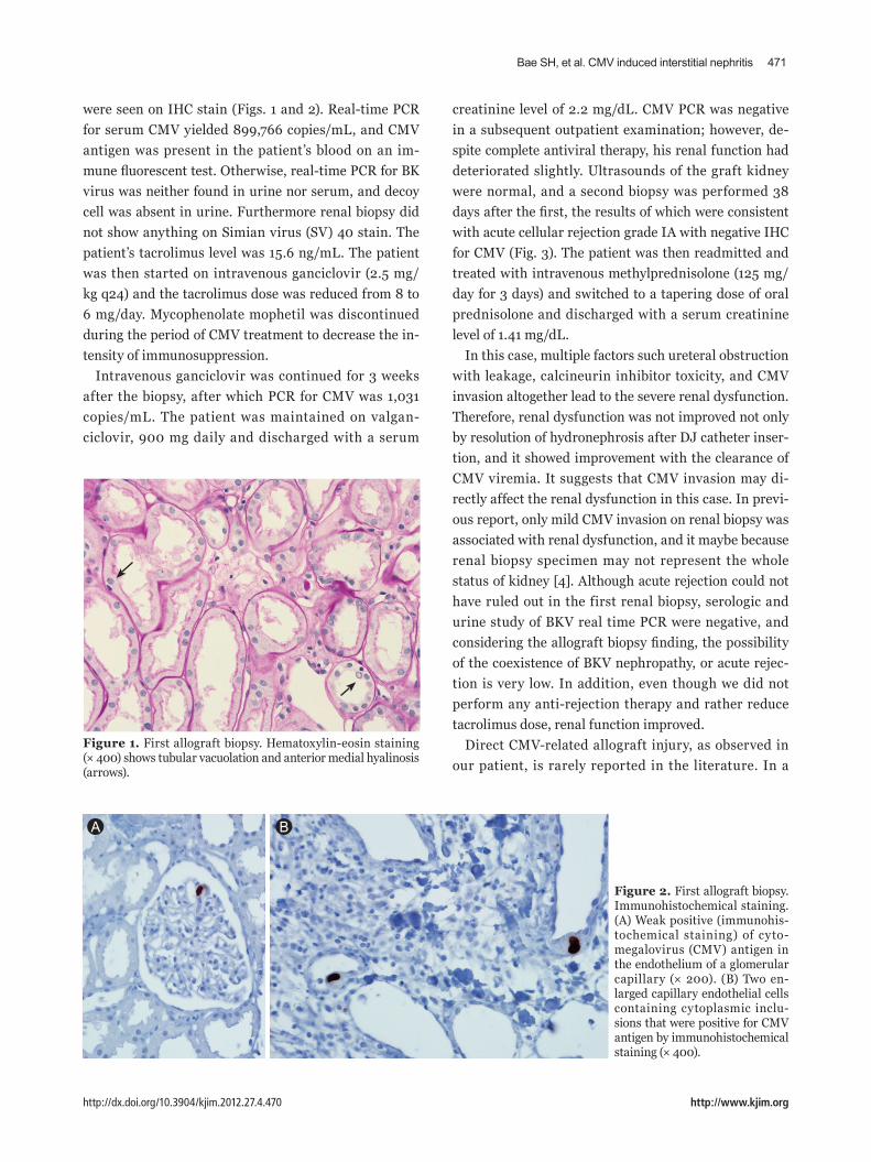
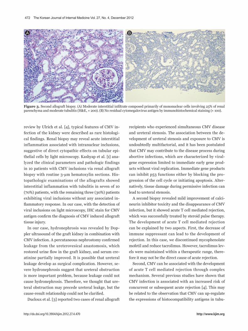
were seen on IHC stain (Figs. 1 and 2). Real-time PCR
for serum CMV yielded 899,766 copies/mL, and CMV
antigen was present in the patient’s blood on an im-
mune fluorescent test. Otherwise, real-time PCR for BK
virus was neither found in urine nor serum, and decoy
cell was absent in urine. Furthermore renal biopsy did
not show anything on Simian virus (SV) 40 stain. The
patient’s tacrolimus level was 15.6 ng/mL. The patient
was then started on intravenous ganciclovir (2.5 mg/
kg q24) and the tacrolimus dose was reduced from 8 to
6 mg/day. Mycophenolate mophetil was discontinued
during the period of CMV treatment to decrease the in-
tensity of immunosuppression.
Intravenous ganciclovir was continued for 3 weeks
after the biopsy, after which PCR for CMV was 1,031
copies/mL. The patient was maintained on valgan-
ciclovir, 900 mg daily and discharged with a serum
creatinine level of 2.2 mg/dL. CMV PCR was negative
in a subsequent outpatient examination; however, de-
spite complete antiviral therapy, his renal function had
deteriorated slightly. Ultrasounds of the graft kidney
were normal, and a second biopsy was performed 38
days after the first, the results of which were consistent
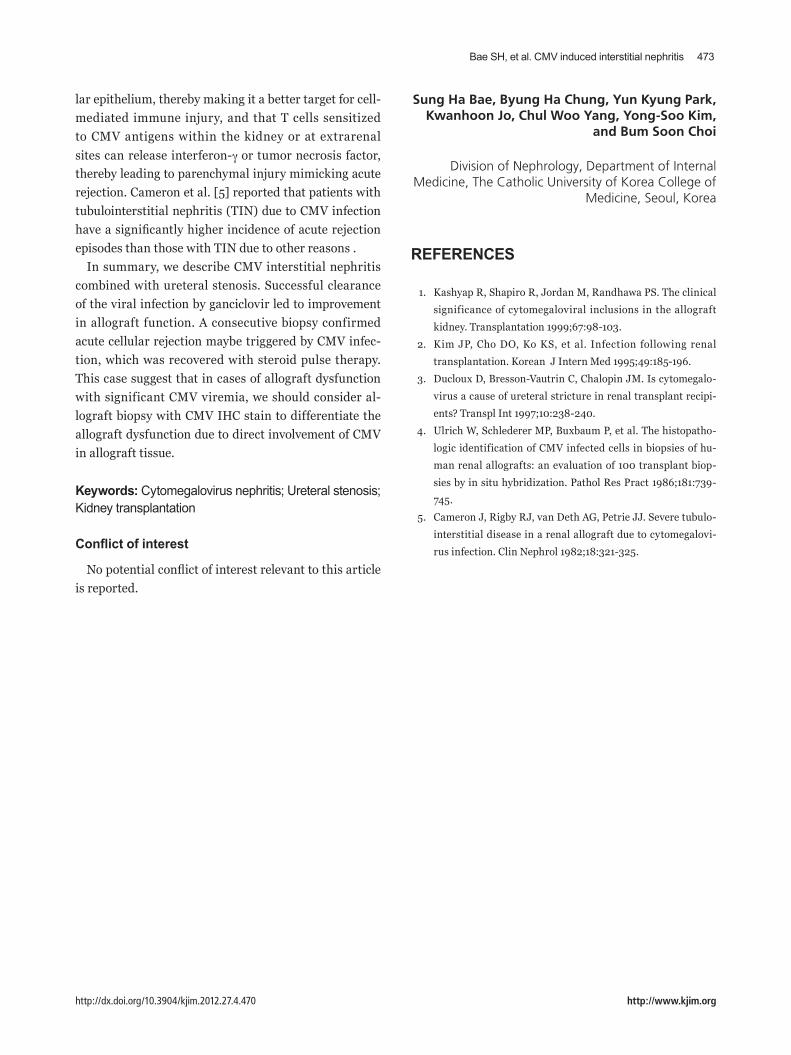
with acute cellular rejection grade IA with negative IHC
for CMV (Fig. 3). The patient was then readmitted and
treated with intravenous methylprednisolone (125 mg/
day for 3 days) and switched to a tapering dose of oral
prednisolone and discharged with a serum creatinine
level of 1.41 mg/dL.
In this case, multiple factors such ureteral obstruction
with leakage, calcineurin inhibitor toxicity, and CMV
invasion altogether lead to the severe renal dysfunction.
Therefore, renal dysfunction was not improved not only
by resolution of hydronephrosis after DJ catheter inser-
tion, and it showed improvement with the clearance of
CMV viremia. It suggests that CMV invasion may di-
rectly affect the renal dysfunction in this case. In previ-
ous report, only mild CMV invasion on renal biopsy was
associated with renal dysfunction, and it maybe because
renal biopsy specimen may not represent the whole
status of kidney [4]. Although acute rejection could not
have ruled out in the first renal biopsy, serologic and
urine study of BKV real time PCR were negative, and
considering the allograft biopsy finding, the possibility
of the coexistence of BKV nephropathy, or acute rejec-
tion is very low. In addition, even though we did not
perform any anti-rejection therapy and rather reduce
tacrolimus dose, renal function improved.
Direct CMV-related allograft injury, as observed in
our patient, is rarely reported in the literature. In a
Figure 1. First allograft biopsy. Hematoxylin-eosin staining(× 400) shows tubular vacuolation and anterior medial hyalinosis (arrows).
Figure 2. First allograft biopsy. Immunohistochemical staining. (A) Weak positive (immunohis-tochemical staining) of cyto-megalovirus (CMV) antigen in the endothelium of a glomerular capillary (× 200). (B) Two en-larged capillary endothelial cells containing cytoplasmic inclu-sions that were positive for CMV antigen by immunohistochemical staining (× 400).
A B
472 The Korean Journal of Internal Medicine Vol. 27, No. 4, December 2012
http://dx.doi.org/10.3904/kjim.2012.27.4.470 http://www.kjim.org
review by Ulrich et al. [4], typical features of CMV in-
fection of the kidney were described as rare histologi-
cal findings. Renal biopsy may reveal acute interstitial
inflammation associated with intranuclear inclusions,
suggestive of direct cytopathic effects on tubular epi-
thelial cells by light microscopy. Kashyap et al. [1] ana-
lyzed the clinical parameters and pathologic findings
in 10 patients with CMV inclusions via renal allograft
biopsy with routine 3-μm hematoxylin sections. His-
topathologic examinations of the allografts showed
interstitial inflammation with tubulitis in seven of 10
(70%) patients, with the remaining three (30%) patients
exhibiting viral inclusions without any associated in-
flammatory response. In our case, with the detection of
viral inclusions on light microscopy, IHC stain for CMV
antigen confirm the diagnosis of CMV induced allograft
tissue injury.
In our case, hydronephrosis was revealed by Dop-
pler ultrasound of the graft kidney in combination with
CMV infection. A percutaneous nephrostomy confirmed
leakage from the ureterovesical anastomosis, which
restored urine flow in the graft kidney, and serum cre-
atinine partially improved. It is possible that ureteral
leakage develop as surgical complication. However, se-
vere hydronephrosis suggest that ureteral obstruction
is more important problem, because leakage could not
cause hydronephrosis. Therefore, we thought that ure-
teral obstruction may precede ureteral leakge, but the
cause-result relationship could not be clarified.
Ducloux et al. [3] reported two cases of renal allograft
recipients who experienced simultaneous CMV disease
and ureteral stenosis. The association between the de-
velopment of ureteral stenosis and exposure to CMV is
undoubtedly multifactorial, and it has been postulated
that CMV may contribute to the disease process during
abortive infections, which are characterized by viral-
gene expression limited to immediate early gene prod-
ucts without viral replication. Immediate gene products
can inhibit p53 functions either by blocking the pro-
gression of the cell cycle or initiating apoptosis. Alter-
natively, tissue damage during permissive infection can
lead to ureteral stenosis.
A second biopsy revealed mild improvement of calci-
neurin inhibitor toxicity and the disappearance of CMV
infection, but it showed acute T cell mediated rejection,
which was successfully treated by steroid pulse therapy.
The development of acute T cell mediated rejection
can be explained by two aspects. First, the decrease of
immune suppressant can lead to the development of
rejection. In this case, we discontinued mycophenolate
mofetil and reduce tacrolimus. However, tacrolimus lev-
els were maintained within a therapeutic range, there-
fore it may not be the direct cause of acute rejection.
Second, CMV can be associated with the development
of acute T cell mediated rejection through complex
mechanism. Several previous studies have shown that
CMV infection is associated with an increased risk of
concurrent or subsequent acute rejection [4]. This may
be related to the observation that CMV can up-regulate
the expressions of histocompatibility antigens in tubu-
Figure 3. Second allograft biopsy. (A) Moderate interstitial infiltrate composed primarily of mononuclear cells involving 25% of renal parenchyma and moderate tubulitis (H&E, × 200). (B) No residual cytomegalovirus antigen by immunohistochemical staining (× 100).
A B
Bae SH, et al. CMV induced interstitial nephritis 473
http://dx.doi.org/10.3904/kjim.2012.27.4.470 http://www.kjim.org
lar epithelium, thereby making it a better target for cell-
mediated immune injury, and that T cells sensitized
to CMV antigens within the kidney or at extrarenal
sites can release interferon-γ or tumor necrosis factor,
thereby leading to parenchymal injury mimicking acute
rejection. Cameron et al. [5] reported that patients with
tubulointerstitial nephritis (TIN) due to CMV infection
have a significantly higher incidence of acute rejection
episodes than those with TIN due to other reasons .
In summary, we describe CMV interstitial nephritis
combined with ureteral stenosis. Successful clearance
of the viral infection by ganciclovir led to improvement
in allograft function. A consecutive biopsy confirmed
acute cellular rejection maybe triggered by CMV infec-
tion, which was recovered with steroid pulse therapy.
This case suggest that in cases of allograft dysfunction
with significant CMV viremia, we should consider al-
lograft biopsy with CMV IHC stain to differentiate the
allograft dysfunction due to direct involvement of CMV
in allograft tissue.
Keywords: Cytomegalovirus nephritis; Ureteral stenosis; Kidney transplantation
Conflict of interest
No potential conflict of interest relevant to this article
is reported.
Sung Ha Bae, Byung Ha Chung, Yun Kyung Park, Kwanhoon Jo, Chul Woo Yang, Yong-Soo Kim,
and Bum Soon Choi
Division of Nephrology, Department of Internal Medicine, The Catholic University of Korea College of
Medicine, Seoul, Korea
REFERENCES
1. Kashyap R, Shapiro R, Jordan M, Randhawa PS. The clinical
significance of cytomegaloviral inclusions in the allograft
kidney. Transplantation 1999;67:98-103.
2. Kim JP, Cho DO, Ko KS, et al. Infection following renal
transplantation. Korean J Intern Med 1995;49:185-196.
3. Ducloux D, Bresson-Vautrin C, Chalopin JM. Is cytomegalo-
virus a cause of ureteral stricture in renal transplant recipi-
ents? Transpl Int 1997;10:238-240.
4. Ulrich W, Schlederer MP, Buxbaum P, et al. The histopatho-
logic identification of CMV infected cells in biopsies of hu-
man renal allografts: an evaluation of 100 transplant biop-
sies by in situ hybridization. Pathol Res Pract 1986;181:739-
745.
5. Cameron J, Rigby RJ, van Deth AG, Petrie JJ. Severe tubulo-
interstitial disease in a renal allograft due to cytomegalovi-
rus infection. Clin Nephrol 1982;18:321-325.