Embed Size (px)
Citation preview

Cytoplasmic Organelles
• Plastids• Plant organelle that may take many forms. • Examples include chloroplast, leukoplasts
(which store food), & chromoplasts (which store pigment molecules).


Cytoplasmic Organelles
• Cytoskeleton • (It has replaced the idea of “protoplasm”) • 2 main components:
• (1) Microtubules—support cell shape and help organelles move through the cell.
• (2) Microfiliments—function in movement of both the cytoplasm and the cell.

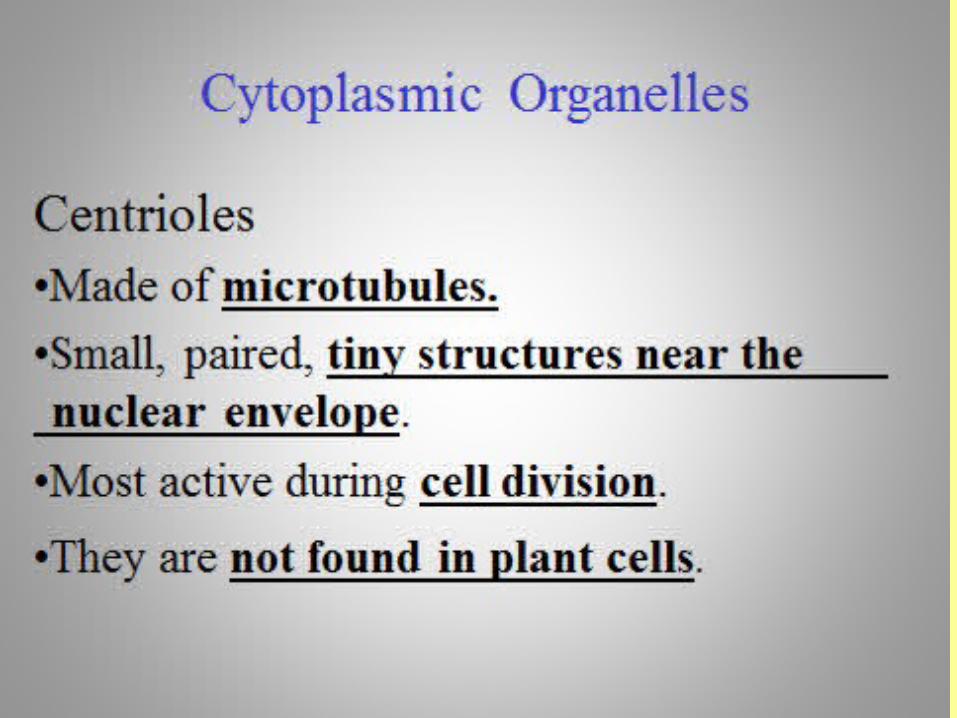
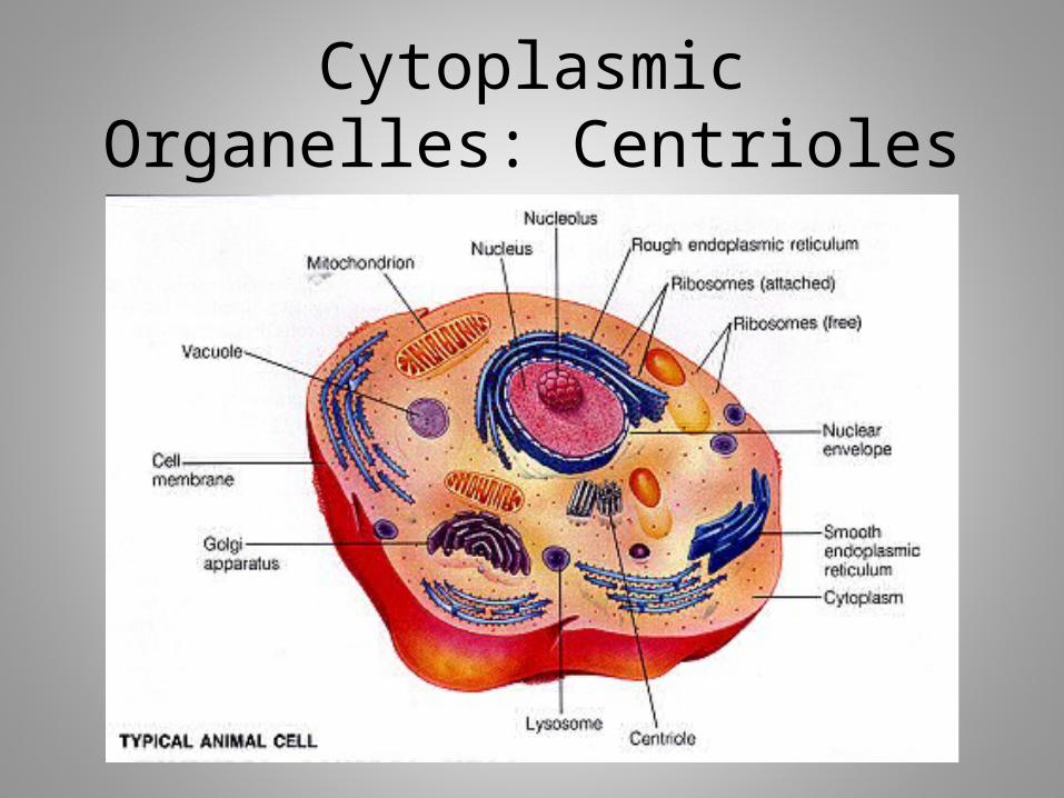
Cytoplasmic Organelles: Centrioles
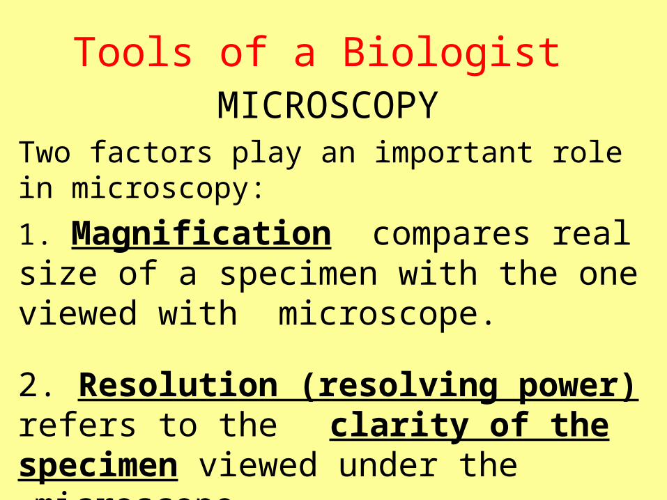
Tools of a BiologistMICROSCOPY
Two factors play an important role in microscopy:
1. Magnification compares real size of a specimen with the one viewed with microscope.
2. Resolution (resolving power) refers to the clarity of the specimen viewed under the
microscope.

Tools of a BiologistCOMPOUND LIGHT MICROSCOPE
•The most commonly used microscope.
• Cells and small organisms can be observed while they are still alive.
•Light microscopes are limited to about
1000 times magnification due to the limit of resolution.
Tools of a Biologist
COMPOUND LIGHT MICROSCOPE
•Limit of resolution is the point of magnification beyond which images
become blurry and lose detail.
•This occurs because light passing through a lens is scattered, making it hard to form a clear image.

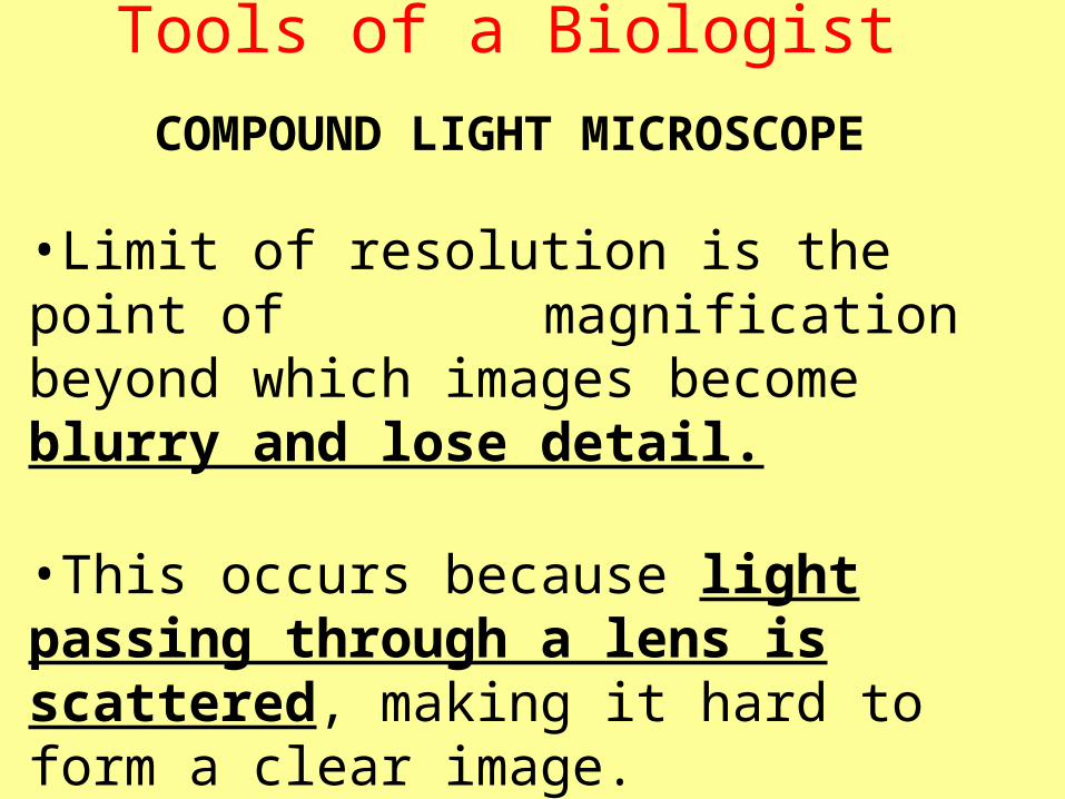
Tools of a BiologistCOMPOUND LIGHT MICROSCOPE
•Limit of resolution is the point of magnification beyond which images become blurry and lose detail.
•This occurs because light passing through a lens is scattered, making it hard to form a clear image.
•For standard light microscopes, the limit of resolution is about 0.2 micrometers.

Tools of a BiologistELECTRON MICROSCOPES
•These microscopes use a beam of electrons and magnets instead of light and lenses.
• They have a shorter wave length (0.2 nanometers) than a light microscope.
•Because of the high-energy particles involved, these microscopes cannot view
living specimens.

Tools of a BiologistTransmission Electron Microscope (TEM)
TEM transmits a beam of electrons through a very thinly sliced specimen.
TEM can magnify objects up to 1,000,000 times.
The electron beam can also be used to expose photographic film to produce a permanent image.

Tools of a BiologistScanning Electron Microscope (SEM)
• SEM get their name from a pencil like beam of electrons that scans back and forth
across the surface of a specimen.
•Electrons that bounce off the specimen are picked up by detectors that provide
information to form a three-dimensional picture.
•SEM can magnify objects up to 300,000 times.

Tools of a BiologistPROBE MICROSCOPES
• A new class of microscopes developed in the 1980s.
•They do not use lenses to produce images.
• These instruments trace the surfaces of a sample with a fine tip known as a probe.
•They are called scanning probe microscopes.
Tools of a BiologistPROBE MICROSCOPES
•Scanning probe microscopes have even made it possible to observe single atoms.
• Unlike electron microscopes, scanning probe microscopes do not require that specimens be placed in a vacuum.