Embed Size (px)
Citation preview
JOURNAL OF VIROLOGY, Nov. 2010, p. 11440–11447 Vol. 84, No. 210022-538X/10/$12.00 doi:10.1128/JVI.01030-10Copyright © 2010, American Society for Microbiology. All Rights Reserved.
Cytotoxicity of ORF3 Proteins from a Nonpathogenic and aPathogenic Porcine Circovirus�
Mark Chaiyakul,1,2 Karolynn Hsu,1 Rkia Dardari,1 Frank Marshall,1,3 and Markus Czub1,2*Department of Production Animal Health, Faculty of Veterinary Medicine,1 and Department of Microbiology and
Infectious Diseases, Faculty of Medicine,2 University of Calgary, Calgary, Alberta, Canada, andMarshall Swine Health Services, Camrose, Alberta, Canada3
Received 12 May 2010/Accepted 21 August 2010
Porcine circovirus type 2 (PCV2) infection is associated with significant and serious swine diseases world-wide, while PCV1 appears to be a nonpathogenic virus. Previous studies demonstrated that the ORF3 proteinof PCV2 (PCV2ORF3) was involved in PCV2 pathogenesis via its proapoptotic capability (J. Liu, I. Chen, Q.Du, H. Chua, and J. Kwang, J. Virol. 80:5065-5073, 2006). If PCV2ORF3-induced apoptosis is a determinantof virulence, PCV1ORF3 is hypothesized to lack this ability. The properties of PCV1 and PCV2 ORF3,expressed as fusion proteins to an enhanced green fluorescent protein (eGFP), were characterized with regardto their ability to cause cellular morphological changes, detachment, death, and apoptosis. PCV1ORF3significantly induced more apoptotic cell death and was toxic to more different cell types than PCV2ORF3 was.PCV1ORF3-associated cell death was caspase dependent. PCV1ORF3 also induced poly(ADP-ribose) poly-merase 1 (PARP) cleavage; however, whether PARP was involved in cell death requires further studies.Truncation of PCV1 and elongation of PCV2 ORF3 proteins revealed that the first 104 amino acids contain adomain capable of inducing cell death, whereas the C terminus of PCV1ORF3 contains a domain possiblyresponsible for enhancing cell death. These results suggest that the pathogenicity of PCV2 for pigs is either notdetermined or not solely determined by the ORF3 protein.
Lymphocyte depletion and the presence of porcine circovi-rus type 2 (PCV2) genome and antigens (4, 6, 21) are hall-marks of PCV-associated disease (PCVAD), a wasting andimmunosuppressive ailment of postweaned pigs. Despite twodecades of research, little is known about the molecular patho-genesis of PCVAD.
PCV2 is the smallest known autonomous vertebrate viruscontaining a 1.7-kb single-stranded, ambisense DNA genome(32). The virus has two major open reading frames (ORFs)that encode the replication proteins (Rep and Rep�) involvedin the initiation of virus replication (17) and a capsid protein(Cap), forming the capsid of the virion (32). A third ORFencodes an ORF3 protein that has been characterized as aninducer of apoptosis (14). Abrogation of ORF3 expressionattenuated PCV2 pathogenesis in BALB/c mice (13) and spe-cific-pathogen-free piglets (9). This led to the hypothesis thatORF3 is involved in PCV2 pathogenesis by inducing apoptosisin infected lymphocytes, leading to lymphocyte depletion andultimately immunosuppression (13, 25). The closely related,yet nonpathogenic porcine circovirus type 1 (PCV1) also has athird open reading frame, but the properties of the ORF3protein of PCV1 (PCV1ORF3) have not been characterized.Analysis of over 250 PCV2 variants and 30 PCV1 variantsshows a consistent single-nucleotide substitution in the ORF3coding sequence of PCV2 (PCV2ORF3), resulting in a stopcodon and a protein that is half the size of PCV1ORF3(PCV2ORF3 is made up of 104 amino acids [aa] compared to
PCV1ORF3, which is made up of 206 aa). A comparison be-tween PCV1 and PCV2 ORF3 translated regions reveals only60% amino acid sequence identity (5), making ORF3 the mostvariable protein among the three identified major proteins ofPCV. If ORF3 is a determinant of virulence of PCV2 via itsapoptotic capability, PCV1ORF3 is hypothesized to lack theability to induce apoptotic cell death. This report demonstratesthe differences in cytotoxic properties between PCV1ORF3and PCV2ORF3. Interestingly, PCV1ORF3 appeared to bemore cytotoxic than PCV2ORF3, activating a caspase-depen-dent apoptotic pathway and potentially a caspase-independent,poly(ADP-ribose) polymerase 1 (PARP) cleavage pathway.Further analysis of truncated PCV1ORF3 and elongatedPCV2ORF3 showed that different ORF3 proteins had similarpatterns of cytotoxicity, although full-length PCV1ORF3 wasthe most potent inducer of cell death.
MATERIALS AND METHODS
Generation of recombinant eukaryotic expression vectors. Coding sequencesof the ORF3 genes were PCR amplified from synthetic PCV1 and PCV2 ge-nomes (DNA 2.0, Menlo Park, CA) by using primers. For PCV1ORF3, primers5�-ATTCTCGAGCCATGATATCCATCCCACCACT-3� (forward) (nucleotidepositions 658 to 639) and 5�-AATGGATCCTCAGTGAAAATGCCAAGCAAG-3� (reverse) (nucleotide positions 38 to 58) were used. For PCV2ORF3, prim-ers 5�-ATTCTCGAGCCATGGTAACCATCCCACCACTT-3� (forward) (nu-cleotide positions 671 to 651) and 5�-AATGGATCCTTACTTATTGAATGTGGAGC-3� (reverse) (nucleotide positions 357 to 376) were used. PCR wasperformed with iProof high-fidelity DNA polymerase (Bio-Rad, Mississauga,Ontario, Canada) in a GeneAmp PCR system 9700 (PE Applied Biosystems,Carlsbad, CA). PCR consisted of the following: a predenaturation step (30 s at98°C); (ii) 36 cycles, where 1 cycle consisted of a denaturation step (10 s at 98°C),an annealing step (30 s at 60°C), and an extension step (60 s at 72°C); and (iii)a final extension step (10 min at 72°C). PCR products of expected size werepurified using a QIAquick gel extraction kit (Qiagen, Mississauga, Ontario,Canada). The BamHI/XhoI fragments of ORF3 were cloned into the correspond-
* Corresponding author. Mailing address: Faculty of VeterinaryMedicine, University of Calgary, 3330 Hospital Drive NW, Calgary,Alberta T2N 4N1, Canada. Phone: (403) 220-4744. Fax: (403) 210-7882. E-mail: [email protected].
� Published ahead of print on 1 September 2010.
11440
on January 28, 2019 by guesthttp://jvi.asm
.org/D
ownloaded from
ing sites of the eukaryotic expression plasmid peGFP-C1 (Clontech, MountainView, CA) generating N-terminal enhanced green fluorescent protein (eGFP)fusion proteins under the control of a human cytomegalovirus promoter.
To generate a truncated PCV1ORF3 (eGFP-PCV1ORF3:Y105*) that resem-bles the analogous PCV2ORF3, a change of tyrosine residue (Y) to a stop codon(*) at amino acid position 105 was required. To generate an elongatedPCV2ORF3 (eGFP-PCV2ORF3:*105Y), a substitution of a stop codon (*) to atyrosine residue (Y) at amino acid position 105 was achieved through a two-stepprocedure. ORF3 genes were first amplified from the viral genomes by usingprimers. To create truncated PCV1ORF3, primers 5�-ATTCTCGAGCCATGATATCCATCCCACCACT-3� (forward) (nucleotide positions 658 to 639) and5�-AATGGATCCTTACTTATCGAGTGTGGAGC-3� (reverse) (nucleotidepositions 344 to 363) were used. To create elongated PCV2ORF3, primers5�-ATTCTCGAGCCATGGTAACCATCCCACCACTT-3� (forward) (nucleo-tide positions 671 to 651) and 5�-AATGGATCCTCACCCAGCAAGAAGAATGG-3� (reverse) (nucleotide positions 54 to 70) were used. PCR was performedwith 1� PCR Taq plus master mix (Applied Biological Materials Inc., Richmond,British Columbia, Canada) and consisted of a predenaturation step (10 min at94°C), followed by 35 cycles, with 1 cycle consisting of denaturation (60 s at94°C), annealing (60 s at 60°C), and extension at (60 s at 72°C), and a finalextension step (10 min at 72°C). The BamHI/XhoI fragments of ORF3 werecloned into peGFP-C1 in a manner similar to that described previously for theauthentic ORF3 genes. To create an elongated PCV2ORF3, one base mutationwas introduced using a QuikChange site-directed mutagenesis kit (Stratagene,La Jolla, CA) with 50 ng of DNA template from the first step and 150 ng of thefollowing mutagenesis primers: 5�-CTGCAGTAAAGAAGGCAACATACTGATTGAGTGTGGAGCTC-3� (forward) (nucleotide positions 338 to 378) and5�-GAGCTCCACACTCAATCAGTATGTTGCCTTCTTTACTGCAG-3� (re-verse) (nucleotide positions 378 to 338). PCR was performed with 1� Taq DNApolymerase (Fermentas, Burlington, Ontario, Canada) and consisted of a pre-denaturation step (2 min at 95°C), followed by 12 cycles, with 1 cycle consistingof denaturation (1 min at 95°C), annealing (1 min at 50°C), and extension (6 minat 68°C), and a final extension step at 68°C for 8 min. All ORF3 sequences wereverified by sequencing (Eurofins MWG Operon, Huntsville, AL).
Cells. Human embryonic kidney epithelial 293T and carcinomic human alve-olar basal epithelial lung A549 cell lines were maintained in Dulbecco’s modifiedEagle medium (Sigma-Aldrich, Oakville, Ontario, Canada) with 10% fetal bo-vine serum, 2% penicillin-streptomycin solution, and 4% 200 mM L-glutamine at37°C and 5% CO2. PCV-free porcine kidney epithelial PK-15 cell line andprimary pig kidney epithelial cells (PPKCs) were maintained in Eagle’s minimumessential medium (Sigma-Aldrich) with 10% fetal bovine serum, 2% penicillin-streptomycin solution, and 4% 200 mM L-glutamine. PPKCs derived from thekidney of a healthy pig free of PCV1 and PCV2 were used starting from passages4 to 16.
Transfection. The wells of a 12-well plate (3.8 cm2 per well) were coated withpoly-D-lysine (Sigma-Aldrich) and cells were seeded 1 day prior to transfection ingrowth medium without antibiotics (1 ml per well in a 12-well plate). When thecells were 70 to 80% confluent, 2 �g of DNA was transfected into the cells ineach well with Lipofectamine 2000 (Invitrogen, Burlington, Ontario, Canada).One day after transfection, 1 ml of new growth medium without antibiotics wasadded to each well that was to be analyzed 48 h after transfection.
Western blot analysis. Whole-cell lysates were resolved by 13% sodium do-decyl sulfate-polyacrylamide gel electrophoresis (SDS-PAGE) and blotted ontopolyvinylidene fluoride (PVDF) membranes (GE Healthcare, Baie d’Urfe, Que-bec, Canada) via a semidry transfer method (Bio-Rad). To prevent nonspecificbinding, the membranes were blocked in phosphate-buffered saline containingTween 20 (PBST) at pH 7.4 (140 mM NaCl, 2.7 mM KCl, 6.5 mM Na2HPO4, 1.5mM KH2PO4, 0.1% Tween 20) with 5% skim milk for 1 h at room temperatureor overnight at 4°C. They were then incubated with primary antibody for 1 h atroom temperature or overnight at 4°C, washed three times with PBST for 5 mineach time, incubated with secondary antibody conjugated to horseradish perox-idase for 1 h at room temperature, and washed again three times with PBSTbefore incubation with an enhanced chemiluminescent substrate reagent mix(GE Healthcare) at room temperature for 5 min. Membranes were analyzed witha VersaDoc 5000 MP imaging system (Bio-Rad) and Quantity One software(Bio-Rad). Primary antibodies included mouse anti-GFP (sc-9996; Santa CruzBiotechnology, Santa Cruz, CA), rabbit anti-PARP (P7605; Sigma-Aldrich),rabbit anti-mitogen-activated protein kinase (anti-MAPK) (M7927; Sigma-Al-drich), and rabbit anti-�-actin (G046; Applied Biological Materials Inc.).
Cell detachment assay. Detached cells were collected from the supernatantand one washing (1 ml) of adherent cells. The pellet of detached cells wasresuspended in 50 �l of 1� PBS. After one wash, adherent cells were trypsinizedwith 200 �l of 0.25% trypsin-EDTA solution (Sigma-Aldrich) and resuspended
in 500 �l of growth medium to make a total volume of 700 �l. Detached andadherent cells were counted using a hemocytometer to determine the percentageof detached cells.
Cell viability assay. All cells, including both detached and adherent cells, werecollected, centrifuged at 2,000 rpm for 3 min, washed twice with PBS, andresuspended in PBS at 1 � 106 cells/ml. For each treatment, 100 �l of cellsuspension was stained with 5 �l of propidium iodide (PI) (BD Biosciences,Mississauga, Ontario, Canada) for 5 to 10 min at room temperature. Fourhundred microliters of PBS was added to each tube prior to fluorescence-activated cell sorting (FACS) analysis.
Phosphatidylserine (PS) externalization detection assay. All cells were col-lected by centrifugation, washed twice with PBS, and resuspended in annexinV-binding buffer at 1 � 106 cells/ml. Each sample was stained with 1 �l of7-amino-actinomycin D (7-AAD) (Sigma-Aldrich) and 5 �l of annexin V-phy-coerythrin (AV-PE) (BD Biosciences) for 15 min at room temperature in thedark. Subsequently, 400 �l of 1� annexin V-binding buffer was added to eachtube prior to FACS analysis. Etoposide (Sigma-Aldrich), an apoptosis-inducingchemical, was used as a positive control in this assay.
Cell cycle analysis. All cells were collected by centrifugation, washed twicewith PBS, and fixed in ice-cold 70% ethanol for at least 1 h at 4°C. Fixed cellswere washed twice with PBS, resuspended in 100 �l of 50-�g/ml PI solution(Invitrogen) containing 5 �l of 10 �g/ml RNase A (Invitrogen) working solutionthat had been boiled for 5 min, and incubated at 37°C for 45 min prior to FACSanalysis.
PARP cleavage detection assay. Whole-cell lysates were subjected to Westernblot analysis, using rabbit anti-PARP antibody (Sigma-Aldrich).
Pancaspase inhibitor assay. At the time of transfection, cells were incubatedwith a pancaspase inhibitor, benzyloxycarbonyl-L-aspart-1-yl-[(2,6-dichloroben-zoyl)oxy]methane (Z-Asp-CH2-DCB; Santa Cruz Biotechnology), to differentfinal concentrations not exceeding 100 �M. Cell death (PI staining), apoptosis(AV-PE/7-AAD staining), cell cycle (PI staining), and PARP cleavage wereassessed.
Statistical analysis. All statistical analyses were performed using MINITABsoftware (Minitab Inc., State College, PA). One-way analysis of variance(ANOVA) and Tukey’s multiple-comparison tests were performed when as-sumptions of normality and equal variance were met; otherwise, a nonparamet-ric, Kruskal-Wallis pairwise multiple-comparison (Dunn’s) test was conducted. AP value of �0.05 was considered significant. At least two independent trials wereconducted for each experiment.
Nucleotide sequence accession numbers. ORF3 coding regions from PCV1(GenBank accession number AY184287) and PCV2 (GenBank accession num-bers EF394779, AY094619, and AY847748) were used in this study.
RESULTS AND DISCUSSION
ORF3-induced cellular detachment of human embryonickidney 293T cells. Previous studies indicated that PCV2ORF3is involved in apoptotic death of porcine and simian cells (14)and PCV2-associated pathology in pigs (9). Here, we hypoth-esized that if PCV2ORF3 is a major determinant for the vir-ulence of PCV2, the analogous protein derived from the non-pathogenic PCV1 would not be able to kill cells.Upon transfection with an expression plasmid for eGFP-PCV1ORF3, human embryonic kidney 293T cells displayedmorphological changes with a shrunken, grape-like appearance(Fig. 1A). Quantitative analysis of detached and adherent 293Tcell numbers showed that cellular detachment was significantlyhigher in eGFP-PCV1ORF3-transfected cells compared toeGFP-PCV2ORF3- and eGFP-transfected cells (P � 0.05)(Fig. 1B). Etoposide, an apoptosis-inducing substance, likewiseinduced significant cellular detachment (P � 0.05). No signif-icant difference in cellular detachment was observed betweeneGFP-PCV2ORF3- and eGFP-transfected cell populations.PCVORF3 coding sequences cloned into a retroviral expres-sion vector led to similar results (data not shown). Immunoblotanalysis revealed that eGFP-PCV1ORF3 was predominantlyseen in the detached, but not the adherent cell fraction (Fig.
VOL. 84, 2010 CYTOTOXICITY OF ORF3 PROTEINS FROM PORCINE CIRCOVIRUS 11441
on January 28, 2019 by guesthttp://jvi.asm
.org/D
ownloaded from
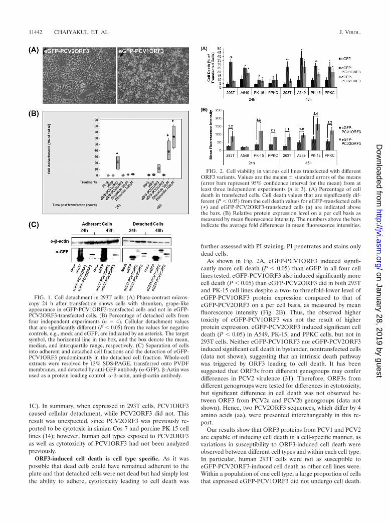
1C). In summary, when expressed in 293T cells, PCV1ORF3caused cellular detachment, while PCV2ORF3 did not. Thisresult was unexpected, since PCV2ORF3 was previously re-ported to be cytotoxic in simian Cos-7 and porcine PK-15 celllines (14); however, human cell types exposed to PCV2ORF3as well as cytotoxicity of PCV1ORF3 had not been analyzedpreviously.
ORF3-induced cell death is cell type specific. As it waspossible that dead cells could have remained adherent to theplate and that detached cells were not dead but had simply lostthe ability to adhere, cytotoxicity leading to cell death was
further assessed with PI staining. PI penetrates and stains onlydead cells.
As shown in Fig. 2A, eGFP-PCV1ORF3 induced signifi-cantly more cell death (P � 0.05) than eGFP in all four celllines tested. eGFP-PCV1ORF3 also induced significantly morecell death (P � 0.05) than eGFP-PCV2ORF3 did in both 293Tand PK-15 cell lines despite a two- to threefold-lower level ofeGFP-PCV1ORF3 protein expression compared to that ofeGFP-PCV2ORF3 on a per cell basis, as measured by meanfluorescence intensity (Fig. 2B). Thus, the observed highertoxicity of eGFP-PCV1ORF3 was not the result of higherprotein expression. eGFP-PCV2ORF3 induced significant celldeath (P � 0.05) in A549, PK-15, and PPKC cells, but not in293T cells. Neither eGFP-PCV1ORF3 nor eGFP-PCV2ORF3induced significant cell death in bystander, nontransfected cells(data not shown), suggesting that an intrinsic death pathwaywas triggered by ORF3 leading to cell death. It has beensuggested that ORF3s from different genogroups may conferdifferences in PCV2 virulence (31). Therefore, ORF3s fromdifferent genogroups were tested for differences in cytotoxicity,but significant difference in cell death was not observed be-tween ORF3 from PCV2a and PCV2b genogroups (data notshown). Hence, two PCV2ORF3 sequences, which differ by 4amino acids (aa), were presented interchangeably in this re-port.
Our results show that ORF3 proteins from PCV1 and PCV2are capable of inducing cell death in a cell-specific manner, asvariations in susceptibility to ORF3-induced cell death wereobserved between different cell types and within each cell type.In particular, human 293T cells were not as susceptible toeGFP-PCV2ORF3-induced cell death as other cell lines were.Within a population of one cell type, a large proportion of cellsthat expressed eGFP-PCV1ORF3 did not undergo cell death.
FIG. 1. Cell detachment in 293T cells. (A) Phase-contrast micros-copy 24 h after transfection shows cells with shrunken, grape-likeappearance in eGFP-PCV1ORF3-transfected cells and not in eGFP-PCV2ORF3-transfected cells. (B) Percentage of detached cells fromfour independent experiments (n � 4). Cellular detachment valuesthat are significantly different (P � 0.05) from the values for negativecontrols, e.g., mock and eGFP, are indicated by an asterisk. The targetsymbol, the horizontal line in the box, and the box denote the mean,median, and interquartile range, respectively. (C) Separation of cellsinto adherent and detached cell fractions and the detection of eGFP-PCV1ORF3 predominantly in the detached cell fraction. Whole-cellextracts were resolved by 13% SDS-PAGE, transferred onto PVDFmembranes, and detected by anti-GFP antibody (�-GFP). �-Actin wasused as a protein loading control. �-�-actin, anti-�-actin antibody.
FIG. 2. Cell viability in various cell lines transfected with differentORF3 variants. Values are the means � standard errors of the means(error bars represent 95% confidence interval for the mean) from atleast three independent experiments (n � 3). (A) Percentage of celldeath in transfected cells. Cell death values that are significantly dif-ferent (P � 0.05) from the cell death values for eGFP-transfected cells(*) and eGFP-PCV2ORF3-transfected cells (a) are indicated abovethe bars. (B) Relative protein expression level on a per cell basis asmeasured by mean fluorescence intensity. The numbers above the barsindicate the average fold differences in mean fluorescence intensities.
11442 CHAIYAKUL ET AL. J. VIROL.
on January 28, 2019 by guesthttp://jvi.asm
.org/D
ownloaded from
Similarly, it has been reported that cells from different originsvary in their tolerance and responses to apoptosis-inducingagents, such as lithium chloride (15, 35). Therefore, it is pos-sible that ORF3 is proapoptotic but that it is the state of thecellular host that determines the final outcome of virus-cellinteractions.
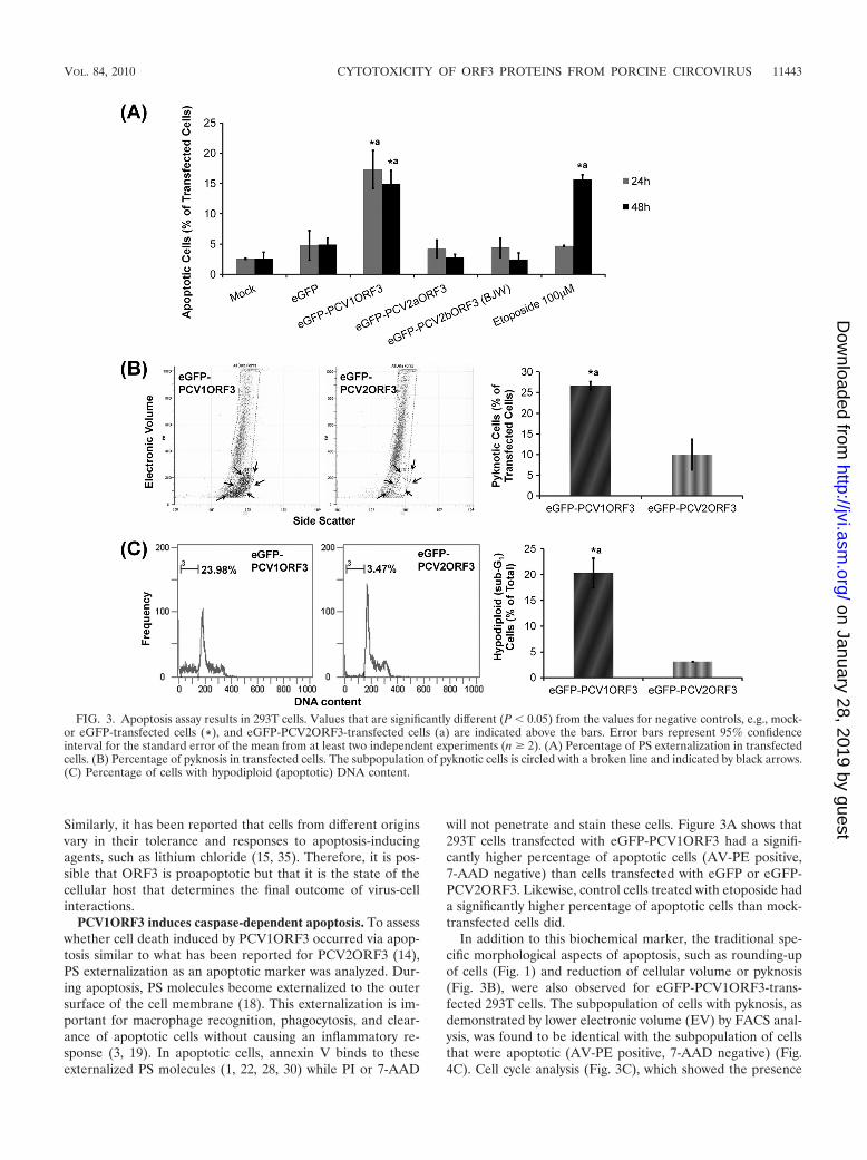
PCV1ORF3 induces caspase-dependent apoptosis. To assesswhether cell death induced by PCV1ORF3 occurred via apop-tosis similar to what has been reported for PCV2ORF3 (14),PS externalization as an apoptotic marker was analyzed. Dur-ing apoptosis, PS molecules become externalized to the outersurface of the cell membrane (18). This externalization is im-portant for macrophage recognition, phagocytosis, and clear-ance of apoptotic cells without causing an inflammatory re-sponse (3, 19). In apoptotic cells, annexin V binds to theseexternalized PS molecules (1, 22, 28, 30) while PI or 7-AAD
will not penetrate and stain these cells. Figure 3A shows that293T cells transfected with eGFP-PCV1ORF3 had a signifi-cantly higher percentage of apoptotic cells (AV-PE positive,7-AAD negative) than cells transfected with eGFP or eGFP-PCV2ORF3. Likewise, control cells treated with etoposide hada significantly higher percentage of apoptotic cells than mock-transfected cells did.
In addition to this biochemical marker, the traditional spe-cific morphological aspects of apoptosis, such as rounding-upof cells (Fig. 1) and reduction of cellular volume or pyknosis(Fig. 3B), were also observed for eGFP-PCV1ORF3-trans-fected 293T cells. The subpopulation of cells with pyknosis, asdemonstrated by lower electronic volume (EV) by FACS anal-ysis, was found to be identical with the subpopulation of cellsthat were apoptotic (AV-PE positive, 7-AAD negative) (Fig.4C). Cell cycle analysis (Fig. 3C), which showed the presence
FIG. 3. Apoptosis assay results in 293T cells. Values that are significantly different (P � 0.05) from the values for negative controls, e.g., mock-or eGFP-transfected cells (*), and eGFP-PCV2ORF3-transfected cells (a) are indicated above the bars. Error bars represent 95% confidenceinterval for the standard error of the mean from at least two independent experiments (n � 2). (A) Percentage of PS externalization in transfectedcells. (B) Percentage of pyknosis in transfected cells. The subpopulation of pyknotic cells is circled with a broken line and indicated by black arrows.(C) Percentage of cells with hypodiploid (apoptotic) DNA content.
VOL. 84, 2010 CYTOTOXICITY OF ORF3 PROTEINS FROM PORCINE CIRCOVIRUS 11443
on January 28, 2019 by guesthttp://jvi.asm
.org/D
ownloaded from
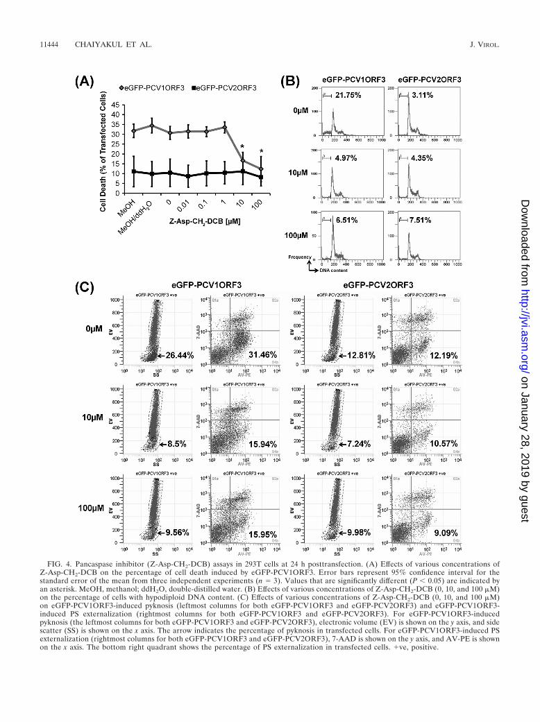
FIG. 4. Pancaspase inhibitor (Z-Asp-CH2-DCB) assays in 293T cells at 24 h posttransfection. (A) Effects of various concentrations ofZ-Asp-CH2-DCB on the percentage of cell death induced by eGFP-PCV1ORF3. Error bars represent 95% confidence interval for thestandard error of the mean from three independent experiments (n � 3). Values that are significantly different (P � 0.05) are indicated byan asterisk. MeOH, methanol; ddH2O, double-distilled water. (B) Effects of various concentrations of Z-Asp-CH2-DCB (0, 10, and 100 �M)on the percentage of cells with hypodiploid DNA content. (C) Effects of various concentrations of Z-Asp-CH2-DCB (0, 10, and 100 �M)on eGFP-PCV1ORF3-induced pyknosis (leftmost columns for both eGFP-PCV1ORF3 and eGFP-PCV2ORF3) and eGFP-PCV1ORF3-induced PS externalization (rightmost columns for both eGFP-PCV1ORF3 and eGFP-PCV2ORF3). For eGFP-PCV1ORF3-inducedpyknosis (the leftmost columns for both eGFP-PCV1ORF3 and eGFP-PCV2ORF3), electronic volume (EV) is shown on the y axis, and sidescatter (SS) is shown on the x axis. The arrow indicates the percentage of pyknosis in transfected cells. For eGFP-PCV1ORF3-induced PSexternalization (rightmost columns for both eGFP-PCV1ORF3 and eGFP-PCV2ORF3), 7-AAD is shown on the y axis, and AV-PE is shownon the x axis. The bottom right quadrant shows the percentage of PS externalization in transfected cells. ve, positive.
11444 CHAIYAKUL ET AL. J. VIROL.
on January 28, 2019 by guesthttp://jvi.asm
.org/D
ownloaded from

of hypodiploid (sub-G1) cells with lower DNA content ineGFP-PCV1ORF3-transfected population, further confirmedthe apoptotic capability of PCV1ORF3. eGFP-PCV1ORF3-induced apoptotic cell death was inhibitable by Z-Asp-CH2-DCB, as increasing concentrations (10 �M and 100 �M) of thisbroad-spectrum caspase inhibitor significantly reduced the per-centage of eGFP-PCV1ORF3-mediated apoptotic cell death(P � 0.05) (Fig. 4A) by diminishing the subpopulation of cellswith hypodiploid DNA content (Fig. 4B), pyknosis, and PSexternalization (Fig. 4C). Caution was taken when using theterm caspase dependent, because although Z-Asp-CH2-DCBis supposed to specifically inhibit caspases, a family of cysteineproteases, it has been shown that another broad-spectrumcaspase inhibitor, Z-VAD-FMK (benzyloxycarbonyl-Val-Ala-DL-Asp conjugated to fluoromethylketone), can also inhibitcalpains and cathepsins (24), especially at high concentration(10 �M), and can associate with targets that are not part ofa caspase-dependent pathway by binding to other cysteine pro-teases and proteins (12). Since only a 10 �M concentration ofthe pancaspase inhibitor was used to inhibit PCV1ORF3-me-diated apoptotic cell death, it should be safe to assume thatPCV1ORF3-mediated apoptotic cell death is caspase depen-dent. Future experiments using small interfering RNA(siRNA) to knock down different caspases will confirm theinvolvement of specific caspases.
These results demonstrate that PCV1ORF3 induced apop-totic cell death via a caspase-dependent pathway, withPCV1ORF3 being a more potent inducer of cell death thanPCV2ORF3. Yet, only PCV2, not PCV1, leads to lymphocytedepletion and disease in pigs. Similarly, PCV1 persistently in-fects PK-15 cells without any obvious cytopathic effects (7, 32,33), whereas PCV2 infection appears to kill PK-15 cells. AsORF3 protein expression has not been reported over thecourse of PCV1 infection, either in vitro or in vivo, it is possiblethat the expression of PCV1ORF3 is tightly regulated or evennot occurring in vitro and conceivably in vivo as well. Perhapsthe lack of ORF3 expression in PCV1 infection in pigs inca-pacitates the virus from causing PCVAD. Alternatively, ifPCV1ORF3 were expressed in vivo, the ability to induce apop-tosis to a greater extent could render PCV1 nonpathogenic,because PCV1-infected cells would be recognized, phagocy-tosed, and cleared by the immune cells without an activation ofinflammatory responses.
PCV1ORF3 induces PARP cleavage. Activated caspases arecapable of cleaving and thereby inactivating PARP molecules,a DNA repair enzyme, leading to apoptosis (2, 20, 27, 29). Tofurther elucidate the mechanism of apoptosis induced byPCV1ORF3, we analyzed PARP cleavage. As expected, eGFP-PCV2ORF3 did not induce PARP cleavage (Fig. 5A), becauseit did not cause significant apoptotic cell death in 293T cells(Fig. 1 to 3). This was further confirmed when a pancaspaseinhibitor did not influence the level of PARP cleavage ineGFP-PCV2ORF3-transfected cells (Fig. 5B). However, im-munoblot analysis (Fig. 5A) showed a strong signal for thecleaved 27-kDa PARP fragment in eGFP-PCV1ORF3-trans-fected cells and in control cells treated with etoposide. Inter-estingly, eGFP-PCV1ORF3-mediated PARP cleavage was notreduced in the presence of 10 �M Z-Asp-CH2-DCB but wasreduced in the presence of a higher concentration (100 �M) ofthis pancaspase inhibitor (Fig. 5B). As indicated previously, it
is possible that the pancaspase inhibitor can be caspase non-specific when used at a high concentration (12, 24). PARPcleavage may occur independently of cell death, and multiplepathways might be activated by PCV1ORF3. Transforminggrowth factor �1, a cytokine that plays a role in many physio-logical processes, such as cell proliferation, differentiation,apoptosis, and growth inhibition, has also been shown to in-duce caspase-dependent apoptosis and caspase-independentPARP cleavage (34), similar to the data on PCV1ORF3 pre-sented here. Taken together, these results show thatPCV1ORF3 activates PARP cleavage that might be dissoci-ated from a caspase-dependent apoptotic cell death pathway.
Effects of modified PCVORF3 proteins. Comparison ofPCV1 and PCV2 ORF3 sequences revealed a consistent single-nucleotide substitution resulting in a PCV2ORF3 protein thatis truncated compared to PCV1ORF3. The stop codon is con-served in most published PCV2 genomes, making it likely thatit preserves virus survival of PCV2 in pigs. The truncatedPCV2ORF3 could either be associated with an unknown gainof function or with an attenuation of an otherwise even moredetrimental PCV2 virus, i.e., a PCV2 with an elongated ORF3.To test the latter hypothesis, we generated truncatedPCV1ORF3 (eGFP-PCV1ORF3:Y105*) and elongatedPCV2ORF3 (eGFP-PCV2ORF3:*105Y). Figure 6A illustratesthat eGFP-PCV1ORF3:Y105* induced significantly less celldeath than eGFP-PCV1ORF3 did, despite two- to ninefold-higher levels of expression (Fig. 6C). eGFP-PCV2ORF3:*105Y induced the same level of cell death as eGFP-PCV2ORF3 did (Fig. 6B), despite two- to fivefold-lower levelsof expression (Fig. 6D). The finding that the elongated ORF3
FIG. 5. PARP cleavage assay in 293T cells. (A) Detection of thecleaved 27-kDa PARP fragment in etoposide-treated and eGFP-PCV1ORF3-transfected cells. (B) Effects of various micromolar con-centrations of Z-Asp-CH2-DCB on eGFP-PCV1ORF3-mediatedPARP cleavage at 24 h posttransfection. Whole-cell extracts wereresolved by 13% SDS-PAGE, transferred onto PVDF membranes, anddetected by anti-PARP antibody (�-PARP). Either �-actin or MAPKwas used as a protein loading control.
VOL. 84, 2010 CYTOTOXICITY OF ORF3 PROTEINS FROM PORCINE CIRCOVIRUS 11445
on January 28, 2019 by guesthttp://jvi.asm
.org/D
ownloaded from
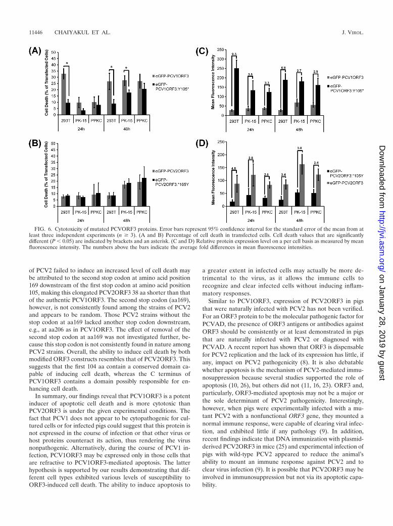
of PCV2 failed to induce an increased level of cell death maybe attributed to the second stop codon at amino acid position169 downstream of the first stop codon at amino acid position105, making this elongated PCV2ORF3 38 aa shorter than thatof the authentic PCV1ORF3. The second stop codon (aa169),however, is not consistently found among the strains of PCV2and appears to be random. Those PCV2 strains without thestop codon at aa169 lacked another stop codon downstream,e.g., at aa206 as in PCV1ORF3. The effect of removal of thesecond stop codon at aa169 was not investigated further, be-cause this stop codon is not consistently found in nature amongPCV2 strains. Overall, the ability to induce cell death by bothmodified ORF3 constructs resembles that of PCV2ORF3. Thissuggests that the first 104 aa contain a conserved domain ca-pable of inducing cell death, whereas the C terminus ofPCV1ORF3 contains a domain possibly responsible for en-hancing cell death.
In summary, our findings reveal that PCV1ORF3 is a potentinducer of apoptotic cell death and is more cytotoxic thanPCV2ORF3 is under the given experimental conditions. Thefact that PCV1 does not appear to be cytopathogenic for cul-tured cells or for infected pigs could suggest that this protein isnot expressed in the course of infection or that other virus orhost proteins counteract its action, thus rendering the virusnonpathogenic. Alternatively, during the course of PCV1 in-fection, PCV1ORF3 may be expressed only in those cells thatare refractive to PCV1ORF3-mediated apoptosis. The latterhypothesis is supported by our results demonstrating that dif-ferent cell types exhibited various levels of susceptibility toORF3-induced cell death. The ability to induce apoptosis to
a greater extent in infected cells may actually be more de-trimental to the virus, as it allows the immune cells torecognize and clear infected cells without inducing inflam-matory responses.
Similar to PCV1ORF3, expression of PCV2ORF3 in pigsthat were naturally infected with PCV2 has not been verified.For an ORF3 protein to be the molecular pathogenic factor forPCVAD, the presence of ORF3 antigens or antibodies againstORF3 should be consistently or at least demonstrated in pigsthat are naturally infected with PCV2 or diagnosed withPCVAD. A recent report has shown that ORF3 is dispensablefor PCV2 replication and the lack of its expression has little, ifany, impact on PCV2 pathogenicity (8). It is also debatablewhether apoptosis is the mechanism of PCV2-mediated immu-nosuppression because several studies supported the role ofapoptosis (10, 26), but others did not (11, 16, 23). ORF3 and,particularly, ORF3-mediated apoptosis may not be a major orthe sole determinant of PCV2 pathogenicity. Interestingly,however, when pigs were experimentally infected with a mu-tant PCV2 with a nonfunctional ORF3 gene, they mounted anormal immune response, were capable of clearing viral infec-tion, and exhibited little if any pathology (9). In addition,recent findings indicate that DNA immunization with plasmid-derived PCV2ORF3 in mice (25) and experimental infection ofpigs with wild-type PCV2 appeared to reduce the animal’sability to mount an immune response against PCV2 and toclear virus infection (9). It is possible that PCV2ORF3 may beinvolved in immunosuppression but not via its apoptotic capa-bility.
FIG. 6. Cytotoxicity of mutated PCVORF3 proteins. Error bars represent 95% confidence interval for the standard error of the mean from atleast three independent experiments (n � 3). (A and B) Percentage of cell death in transfected cells. Cell death values that are significantlydifferent (P � 0.05) are indicated by brackets and an asterisk. (C and D) Relative protein expression level on a per cell basis as measured by meanfluorescence intensity. The numbers above the bars indicate the average fold differences in mean fluorescence intensities.
11446 CHAIYAKUL ET AL. J. VIROL.
on January 28, 2019 by guesthttp://jvi.asm
.org/D
ownloaded from
ACKNOWLEDGMENTS
We thank Avril Hatherell, Sampson Law, Sandi Nishikawa, G. vanMarle, F. Jirik, and R. Yates and the DNA and Flow Cytometry CoreFacilities at the University of Calgary for training and technical sup-port.
This work was supported by the Natural Sciences and EngineeringResearch Council (NSERC), Alberta Agriculture Consortium, andUniversity of Calgary Faculties of Medicine and Veterinary Medicine.
REFERENCES
1. Andree, H. A., C. P. Reutelingsperger, R. Hauptmann, H. C. Hemker, W. T.Hermens, and G. M. Willems. 1990. Binding of vascular anticoagulant alpha(VAC alpha) to planar phospholipid bilayers. J. Biol. Chem. 265:4923–4928.
2. Boulares, A. H., A. G. Yakovlev, V. Ivanova, B. A. Stoica, G. Wang, S. Iyer,and M. Smulson. 1999. Role of poly(ADP-ribose) polymerase (PARP)cleavage in apoptosis. Caspase 3-resistant PARP mutant increases rates ofapoptosis in transfected cells. J. Biol. Chem. 274:22932–22940.
3. Duvall, E., A. H. Wyllie, and R. G. Morris. 1985. Macrophage recognition ofcells undergoing programmed cell death (apoptosis). Immunology 56:351–358.
4. Ellis, J., L. Hassard, E. Clark, J. Harding, G. Allan, P. Willson, J. Strokappe,K. Martin, F. McNeilly, B. Meehan, D. Todd, and D. Haines. 1998. Isolationof circovirus from lesions of pigs with postweaning multisystemic wastingsyndrome. Can. Vet. J. 39:44–51.
5. Finsterbusch, T., and A. Mankertz. 2009. Porcine circoviruses–small butpowerful. Virus Res. 143:177–183.
6. Harding, J. C., C. D. Baker, A. Tumber, K. A. McIntosh, S. E. Parker, D. M.Middleton, J. E. Hill, J. A. Ellis, and S. Krakowka. 2008. Porcine circovirus-2DNA concentration distinguishes wasting from nonwasting pigs and is cor-related with lesion distribution, severity, and nucleocapsid staining intensity.J. Vet. Diagn. Invest. 20:274–282.
7. Hattermann, K., C. Roedner, C. Schmitt, T. Finsterbusch, T. Steinfeldt, andA. Mankertz. 2004. Infection studies on human cell lines with porcinecircovirus type 1 and porcine circovirus type 2. Xenotransplantation 11:284–294.
8. Juhan, N. M., T. LeRoith, T. Opriessnig, and X. J. Meng. 2010. The openreading frame 3 (ORF3) of porcine circovirus type 2 (PCV2) is dispensablefor virus infection but evidence of reduced pathogenicity is limited in pigsinfected by an ORF3-null PCV2 mutant. Virus Res. 147:60–66.
9. Karuppannan, A. K., M. H. Jong, S. H. Lee, Y. Zhu, M. Selvaraj, J. Lau, Q.Jia, and J. Kwang. 2009. Attenuation of porcine circovirus 2 in SPF pigletsby abrogation of ORF3 function. Virology 383:338–347.
10. Kiupel, M., G. W. Stevenson, E. J. Galbreath, A. North, H. HogenEsch, andS. K. Mittal. 2005. Porcine circovirus type 2 (PCV2) causes apoptosis inexperimentally inoculated BALB/c mice. BMC Vet. Res. 1:7.
11. Krakowka, S., J. Ellis, F. McNeilly, B. Meehan, M. Oglesbee, S. Alldinger,and G. Allan. 2004. Features of cell degeneration and death in hepatic failureand systemic lymphoid depletion characteristic of porcine circovirus-2-asso-ciated postweaning multisystemic wasting disease. Vet. Pathol. 41:471–481.
12. Kroemer, G., L. Galluzzi, P. Vandenabeele, J. Abrams, E. S. Alnemri, E. H.Baehrecke, M. V. Blagosklonny, W. S. El-Deiry, P. Golstein, D. R. Green, M.Hengartner, R. A. Knight, S. Kumar, S. A. Lipton, W. Malorni, G. Nunez,M. E. Peter, J. Tschopp, J. Yuan, M. Piacentini, B. Zhivotovsky, and G.Melino. 2009. Classification of cell death: recommendations of the Nomen-clature Committee on Cell Death 2009. Cell Death Differ. 16:3–11.
13. Liu, J., I. Chen, Q. Du, H. Chua, and J. Kwang. 2006. The ORF3 protein ofporcine circovirus type 2 is involved in viral pathogenesis in vivo. J. Virol.80:5065–5073.
14. Liu, J., I. Chen, and J. Kwang. 2005. Characterization of a previously un-identified viral protein in porcine circovirus type 2-infected cells and its rolein virus-induced apoptosis. J. Virol. 79:8262–8274.
15. Lucas, K. C., D. A. Hart, and R. W. Becker. 2010. Porcine proximal tubularcells (LLC-PK1) are able to tolerate high levels of lithium chloride in vitro:assessment of the influence of 1–20 mM LiCl on cell death and alterations incell biology and biochemistry. Cell Biol. Int. 34:225–233.
16. Mandrioli, L., G. Sarli, S. Panarese, S. Baldoni, and P. S. Marcato. 2004.
Apoptosis and proliferative activity in lymph node reaction in postweaningmultisystemic wasting syndrome (PMWS). Vet. Immunol. Immunopathol.97:25–37.
17. Mankertz, A., J. Mankertz, K. Wolf, and H. J. Buhk. 1998. Identification ofa protein essential for replication of porcine circovirus. J. Gen. Virol. 79:381–384.
18. Martin, S. J., C. P. Reutelingsperger, A. J. McGahon, J. A. Rader, R. C. vanSchie, D. M. LaFace, and D. R. Green. 1995. Early redistribution of plasmamembrane phosphatidylserine is a general feature of apoptosis regardless ofthe initiating stimulus: inhibition by overexpression of Bcl-2 and Abl. J. Exp.Med. 182:1545–1556.
19. Morris, R. G., A. D. Hargreaves, E. Duvall, and A. H. Wyllie. 1984. Hor-mone-induced cell death. 2. Surface changes in thymocytes undergoingapoptosis. Am. J. Pathol. 115:426–436.
20. Nicholson, D. W., A. Ali, N. A. Thornberry, J. P. Vaillancourt, C. K. Ding, M.Gallant, Y. Gareau, P. R. Griffin, M. Labelle, Y. A. Lazebnik, N. A. Munday,S. M. Raju, M. E. Smulson, T.-T. Yamin, V. L. Yu, and D. K. Miller. 1995.Identification and inhibition of the ICE/CED-3 protease necessary for mam-malian apoptosis. Nature 376:37–43.
21. Opriessnig, T., X. J. Meng, and P. G. Halbur. 2007. Porcine circovirus type2 associated disease: update on current terminology, clinical manifestations,pathogenesis, diagnosis, and intervention strategies. J. Vet. Diagn. Invest.19:591–615.
22. Raynal, P., and H. B. Pollard. 1994. Annexins: the problem of assessing thebiological role for a gene family of multifunctional calcium- and phospho-lipid-binding proteins. Biochim. Biophys. Acta 1197:63–93.
23. Resendes, A. R., N. Majo, J. Segales, E. Mateu, M. Calsamiglia, and M.Domingo. 2004. Apoptosis in lymphoid organs of pigs naturally infected byporcine circovirus type 2. J. Gen. Virol. 85:2837–2844.
24. Schotte, P., W. Declercq, S. Van Huffel, P. Vandenabeele, and R. Beyaert.1999. Non-specific effects of methyl ketone peptide inhibitors of caspases.FEBS Lett. 442:117–121.
25. Shen, H. G., J. Y. Zhou, X. Zhang, Z. Y. Huang, J. L. He, and Y. Yan. 2009.Interference of porcine circovirus type 2 ORF2 immunogenicity by ORF1and ORF3 mixed DNA immunizations in mice. Virology 393:104–111.
26. Shibahara, T., K. Sato, Y. Ishikawa, and K. Kadota. 2000. Porcine circovirusinduces B lymphocyte depletion in pigs with wasting disease syndrome. J.Vet. Med. Sci. 62:1125–1131.
27. Simbulan-Rosenthal, C. M., D. S. Rosenthal, S. Iyer, H. Boulares, and M. E.Smulson. 1999. Involvement of PARP and poly(ADP-ribosyl)ation in theearly stages of apoptosis and DNA replication. Mol. Cell. Biochem. 193:137–148.
28. Tait, J. F., D. Gibson, and K. Fujikawa. 1989. Phospholipid binding prop-erties of human placental anticoagulant protein-I, a member of the lipocor-tin family. J. Biol. Chem. 264:7944–7949.
29. Tewari, M., L. T. Quan, K. O’Rourke, S. Desnoyers, Z. Zeng, D. R. Beidler,G. G. Poirier, G. S. Salvesen, and V. M. Dixit. 1995. Yama/CPP32 beta, amammalian homolog of CED-3, is a CrmA-inhibitable protease that cleavesthe death substrate poly(ADP-ribose) polymerase. Cell 81:801–809.
30. Thiagarajan, P., and J. F. Tait. 1990. Binding of annexin V/placental anti-coagulant protein I to platelets. Evidence for phosphatidylserine exposure inthe procoagulant response of activated platelets. J. Biol. Chem. 265:17420–17423.
31. Timmusk, S., P. Wallgren, I. M. Brunborg, F. H. Wikstrom, G. Allan, B.Meehan, M. McMenamy, F. McNeilly, L. Fuxler, K. Belak, D. Podersoo, T.Saar, M. Berg, and C. Fossum. 2008. Phylogenetic analysis of porcine cir-covirus type 2 (PCV2) pre- and post-epizootic postweaning multisystemicwasting syndrome (PMWS). Virus Genes 36:509–520.
32. Tischer, I., H. Gelderblom, W. Vettermann, and M. A. Koch. 1982. A verysmall porcine virus with circular single-stranded DNA. Nature 295:64–66.
33. Tischer, I., R. Rasch, and G. Tochtermann. 1974. Characterization of pa-povavirus-and picornavirus-like particles in permanent pig kidney cell lines.Zentralbl. Bakteriol. Orig. A 226:153–167.
34. Yang, Y., S. Zhao, and J. Song. 2004. Caspase-dependent apoptosis and-independent poly(ADP-ribose) polymerase cleavage induced by transform-ing growth factor beta1. Int. J. Biochem. Cell Biol. 36:223–234.
35. Zhang, W. V., M. Jullig, A. R. Connolly, and N. S. Stott. 2005. Early generesponse in lithium chloride induced apoptosis. Apoptosis 10:75–90.
VOL. 84, 2010 CYTOTOXICITY OF ORF3 PROTEINS FROM PORCINE CIRCOVIRUS 11447
on January 28, 2019 by guesthttp://jvi.asm
.org/D
ownloaded from