Embed Size (px)
Citation preview
DENTISTRY TODAY...
The journal is indexed with ‘Indian Science Abstract’ (ISA)(Published by National Science Library), www.ebscohost.com, www.indianjournals.com
The journal is printed on ACID FREE paper.
JADCH is available (full text) online:Website- www.adc.org.in/html/viewJournal.php
This journal is an official publication of Ahmedabad Dental Collegeand Hospital, published bi-annually in the month of March andSeptember.
ISSN 0976-2256E-ISSN:2249-6653
A
Pediatric dentistry provides primary care and comprehensive dental speciality treatments for infants, children, adolescents and individuals with special health care needs.
The successful practice of pediatric dentistry is nit merely the completion of any operative procedure but also ensuring a positive dental outcome for the future oral health behaviour of the individual and their family. To this end, an understanding of child development- physical, cognitive and psychosocial - is paramount. Traditionally, dental caries has been regarded as a static phenomenon, eventulation in loss of tooth structure while the basis for treatment and management of this ubiquitious disease has essentially been mechanical. However, with current developments in new dental materials, techniques and preventive strategies, a more precise understanding and appreciation of the nature of the caries process is obtained. The newer concept of using micro-invasive resin infiltrants in enamel and early dentine lesion is promoted. There have been advances in four key areas related to pediatric dentistry ;(1) caries detection tools, (2) early interventions to arrest disease progression, (3) caries risk assesement tools, (4) trends in pediatric procedures and dental materials. It is the only specialization in dentistry that is age defined and not specific to any treatment modality. Hence, with the adoption of newer material and techniques working on a child becomes one of the most satisfying experience in all dental practice.
Editor - in - ChiefDr. Darshana Shah
Co - EditorDr. Rupal Vaidya
Editorial Board:Dr. Mihir ShahDr. Vijay BhaskarDr. Monali ChalishazarDr. A. R. ChaudharyDr. Neha VyasDr. Sonali MahadeviaDr. Shraddha ChokshiDr. Bhavin DudhiaDr. M GaneshDr. Mahadev DesaiDr. Darshit DalalDr. Harsh Shah
JADCH
EDITORIAL
FROM THE EDITOR'S DESK .....................................................................................................................................................01
DARSHANA SHAH
REVIEW ARTICLES
1) MALODOR AND PERIODONTITIS: CASUAL OR CAUSAL? ........................................................................................02
SHILPI SHAH*, TEJAL SHETH**, MIHIR SHAH***, SANDIP LADANI****, PRAGNESH SOLANKI*****
ORIGINAL ARTICLES
2) DENTAL FLUOROSIS – A RETROSPECTIVE STUDY IN GANDHINAGAR DISTRICT.................................................10
PURV PATEL*, BHAVIN DUDHIA**, A. R. CHAUDHARY***, PARUL BHATIA****, YESHA JANI*****,
PURVA BUTALA******
3) KNOWLEDGE, ATTITUDE AND PRACTICE REGARDING ORAL CANCER AND SCREENING PROCEDURES
AMONG PRIMARY HEALTH CARE AND COMMUNITY HEALTHCARE WORKERS OF WAGHODIA, GUJARAT.......20
BAFNA HARSHAL P *, AJITHKRISHNAN CG**, KALANTHARAKATH THANVEER***, RICKY PAL SING*,
HEMAL PATEL****
4) EVALUATION OF SHEAR BOND STRENGTH USING THREE DIFFERENT TYPES OF ADHESIVE PRIMERS
UNDER NON-CONTAMINATED AND CONTAMINATED CONDITIONS – AN IN VITRO STUDY..................................26
NIRAV M. PATEL*, C.R. NAIK**, RAVI GUPTA***
5) AN IN-VIVO EVALUATION OF THE EFFECT OF CHEWING CORIANDER SEEDS ON SALIVARY PH. .....................31
RICKY PAL S *, AJITHKRISHNAN CG **, THANVEER K***, HARSHAL B*, HEMAL P****
CASE REPORT
6) GIANT CELL FIBROMA...................................................................................................................................................35
MANISH LIMBACHIYA*, BRIJESH PATEL*, MINAL BAKSHI*, RINA DAVE*
7) DIRECTING THE FORCE VECTOR DIRECTLY: USE OF MICRO-IMPLANTS FOR CORRECTION OF
GUMMY SMILES.............................................................................................................................................................38
HARESH AHIR*, SONALI MAHADEVIA**, KRISHNAMURTHY***, TOSHIF KUMAR****, RINKALKUMAR SHAH*****
8) ROOT PIECES ON DENTAL RADIOGRAPHS: INCIDENTAL FINDINGS WITH DIFFERENTIAL DIAGNOSIS.............43
DINESH TRIVEDI*, BHAVIN DUDHIA**, RUTU JANI***, YESHA JANI****, SANYAL SHAH*****
9) PLEOMORPHIC ADENOMA ...........................................................................................................................................48
NEHA VYAS*, SACHIN DALAL**, SAURABH JAIN***, MEGHA VYAS****
ContentsContents
JADCH
Subscription:Rate per issue: Rs. 400/-, for one year: Rs. 750/-, for three years: Rs. 2,000/-Contact: Ahmedabad Dental College & Hospital Vivekanand Society, Bhadaj-Ranchhod Pura Road, Santej, Post: Rancharda, Ta: Kalol, Dist: Gandhinagar, Gujarat, India.
B
Dear friends,
The current method of dental and medical training is dependent on professors, Obsolete Text books and opinion of the seniors who are often dogmatic and unresponsiveto new ideas.
such a method is insufficient to carry on lifelong clinical practice in a very comptent manner.
the younger doctors are computer savvy, inquisitive and want to know more. We in india are known for hardwork, logical reasoning and cultural strengths.
We have to incorporate this evidence based education into our mainstream dental education if we are going to maintain and provide the personnel for the whole global village.
the critical feature of "Evidence-Based Dentistry is that dentists, when faced with any problem in the clinical context of a patient, should be able to: perform a literature search; identify the evidence available pertaining tio the clinical condition; critically evaluate it and determine the "Best evidence" to diagnose /treat/manage the patient.
The crux of the matter in this cycle is the ability of the dentist to search and retrieve the literature in the shortest possible time in an efficient manner and apply it in practice
in the "global scenario", the term EBD became more widely used in the early 1990s, and was later formally defined by Sackett in 1996.
The first National Workshop on Evidence-Base Dentistry in India was held at College of Dental Sciences, Davangere between the 8th and 11th March 2001.
With over 85 delegates registered from all over India, it was perhaps the largest ever dedicated workshop on evidence-based dentistry in the world.
This is the beacon of what th e future holds for Evidence based dentistry in India.
1The Journal of Ahmedabad Dental College and Hospital; 5 (1), March 2014 - August 2014
Dr. Darshana ShahEditor JADCHEditorial Office:Prof. & Head Dept. of ProsthodonticsAhmedabad Dental College & Hospital,Dist.: Gandhinagar, Gujarat.Email: [email protected]
Malodor and periodontitis: casual or causal?
DEPARTMENT OF PERIODONTICS AND ORAL IMPLANTOLOGY, AHMEDABAD DENTAL COLLEGE AND HOSPITAL, GANDHINAGAR, GUJARAT, INDIA.
The Journal of Ahmedabad Dental College and Hospital; 5 (1), March 2014 - August 2014 2
Review Article
* Reader, ** Senior Lecturer, ***Professor and Head, ****Senior Lecturer, *****Tutor
INTRODUCTION:
Halitosis is a medical term first coined by Listerine
Company in 1921, used to describe “unpleasant,
offensive, stale or foul smelling breath emitted from
the mouth regardless of whether the odorous
substances in the breath originate from oral or extra-
oral sources.”
This condition is commonly responsible for social
embarrassment, emotional and psychological
distress leading to a lack of self-esteem, self-image
and self-confidence. Furthermore it may signal the
presence of disease.
Oral halitosis or oral malodor is the term, especially
used to describe bad breath with an origin within the
oral cavity. In fact most adult subjects have socially
unacceptable bad breath when waking up in the
morning. This problem is transitory and attributed
to physiologic causes such as reduced salivary flow
during sleep. Although these transitory problems
are easily controlled, persistent bad breath may be
indicative of either oral disease (i.e. Periodontal
disease, the presence of bacterial reservoir in
SHILPI SHAH*, TEJAL SHETH**, MIHIR SHAH***, SANDIP LADANI****, PRAGNESH SOLANKI*****
ABSTRACT
Halitosis can be a crippling social problem.However, in the last 5 to 6 years, it has come to the forefront of public and dental professional awareness.The mouth is home to hundreds of bacterial species that produce several fetid substances as a result of protein degradation. Volatile sulfur compound (VSC)-producing bacteriacolonizing the lingual dorsum, gingival pockets, and tonsillar crypts have recently been implicated in the generation of halitosis. Understanding causes, assessment, and treatment of oral malodor can help dental professionals find ways to decrease its prevalence and increase their patients' well-being.This article reviews the etiology and various connections among periodontal pathogenic microorganisms, periodontal disease and oral malodor from a periodontal perspective.
Keywords: halitosis, volatile sulphur compounds, gingivitis, periodontitis, role of microflora in malodor, co-relation between halitosis and periodontitis
Received: 02-12-2013; Review Completed: 15-02-2014; Accepted: 26-02-2014
mouth) or indicative of systemic disease (i.e. Hiatus
hernia, hepatic cirrhosis, renal failure, diabetes
mellitus etc.).
ETIOLOGY
The etiology of oral malodor is multifactorial. In the
presence of adequate substrate with appropriate
conditions, a sequence of events leads to the release
into the oral cavity of pungent gases that pollute
exhaled air and are perceived as bad breath.
Research has identified several microorganisms
that produce these offensive odors and provided a
fair explanation of the conditions necessary for their
production. In addition to the presence of certain
types of bacteria, the type and amount of substrate,
and oxygen and pHlevels influence the occurrence
and severity of oral malodor.
Certain chemical end-products of bacterial
putrefaction known as volatile sulfur compounds
(VSCs) are foul smelling and have been found to be 1-4
the primary culprit of engendering oral malodor.
Nonsulfur-containing compounds such as
cadaverine, putrescine, indole and skatole5,6 have
ADDRESS FOR AUTHOR CORROSPONDENCE : DR. SHILPI SHAH, Tel: +91 9374407773
also been implicated in the foul smell of oral
malodor, but their contribution is thought to be
limited.7 VSCs such as hydrogen sulfide, methyl
mercaptan and dimethyl disulfide make up more
than 90 percent of the putrid odors from the mouth.8
Two of these VSCs, hydrogen sulfide and methyl
mercaptan, account for approximately 90 percent of
the total VSCs identified with putrid odors from the 9mouth.
Source of VSC production in periodontitis
patientsAnaerobic bacteria, oxygen depletion, alkaline pH
and sulfur-containing substrates are some of the
requirements for oral malodor to occur .The
bacteria that produce these VSCs can be found by
evaluating biofilm and scraped specimens from the
lingual dorsum, gingival pockets, and tonsillar
crypts.7 (fig. 1)VSCs are produced by gram-
negative anaerobic bacteria that live on the lingual
dorsum.8 These bacteria can thrive on the tongue
because food debris accumulates rapidly on the
tongue's large surface area and papillae.Periodontal
pathogens have been positively correlated with oral
malodor.1-4,10 Several periodontal pathogens
i n c l u d i n g T r e p o n e m a d e n t i c o l a ,
Porphymonasgingivalis and Bacteroidesforsythus
have been identified, with BANA hydrolysis, on the
posterior tongue, contributing to oral malodor.11
Additional periodontal pathogens, including
F u s o b a c t e r i u m n u c l e a t u m a n d
Bacteroidesmelanogenicus have been identified as
VSC formers.8 These microorganisms produce
copious amounts of hydrogen sulfide, methyl
mercaptan and dimethyl disulfide.However, other
compounds in mouth air may also be offensive such
as diamines (e.g. Putrescine, cadaverine), indole,
skatole and butyric or propionic acid. Most of these
compounds results from proteolytic degradation by
oral microorganisms of peptides present in saliva,
shed epithelium, food debris, gingival crevicular
fluid, interdental plaque, post-nasal drip and blood.Breath malodor, a significant social and/or
psychological handicap, may be caused by several
intra- and extraoral factors.
Intraoral causes:1. Tongue coating ( primary source of malodor)2. Dentition
a. Carious lesionb. Food impactionc. Extraction wounds filled with blood clots
3. Periodontal infections like Pockets, ANUG,
Purulent discharge from gums4. Xerostomia
Extra oral Causes:Halitosis of the upper respiratory tract: Chronic
sinusitis, Nasal obstruction, nasopharyngeal
abscess, Carcinoma of larynxHalitosis of the lower respiratory tract:
Bronchitis, Bronchiectasis, Pneumonia, Carcinoma
of the lungsCauses of blood borne halitosis:Systemic diseases: Hepatic failure/ Liver cirrhosis,
U r e a m i a / K i d n e y f a i l u r e , D i a b e t i c
ketoacidosis/Diabetes mellitusM e t a b o l i c d i s o r d e r s : I s o l a t e d
PersistantHypermethioninemia, Fish odor
syndrome, Medication: Disulfiram, Dimethyl sulphoxide,
Cysteamine, Food: garlic, onion, alcohol, tobacco
ASSOCIATION BETWEEN HALITOSIS AND
PERIODONTAL DISEASE
Periodontal disease result from the combination of
many factors present in vivo. These processes
include chronic activation of immune system,
alteration in connective tissue metabolism
production of proteinases and cytokines, direct
destruction of host tissue by bacterial enzymes, and
virulence factors and a multitude of other
mechanisms. One of these volatile sulfur
compounds (VSCs) not only be associated with oral
3
SHILPI SHAH Malodor and periodontitis: casual or causal? et. al. :
The Journal of Ahmedabad Dental College and Hospital; 5 (1), March 2014 - August 2014
malodor but probably contribute to the etiology of
both gingivitis and periodontitis. Different lines of
evidence have demonstrated this association
between halitosis and periodontal disease.
An increase in production of VSCs from
periodontal pockets provides a plausible
explanation for the intensification of oral malodor
observed in pa t ients wi th per iodonta l
disease.Several studies suggest that periodontitis
increases the severity of oral malodor.7One
possible explanation is the increased amount of
substrate available to be metabolized. In patients
with periodontitis, more sulfur-containing protein
substrate is available through increased exfoliation
of epithelial cells and crevicular effusion of
leukocytes.Yaegaki and Sanada found that bleeding
on probing and periodontal pocket depth positively
correlated with the production of VSCs.4
In contrast to the view that periodontal disease
contributes to oral malodor, Bosy and colleagues
found that oral hygiene levels and not periodontal
pockets were more indicat ive of ora l
malodor,11which supports the concept that oral
malodor may be an independent entity.
Certainly, some gram-negative anaerobic bacteria,
which are not known to be periodontal pathogens
( F u s o b a c t e r i u m p o l y m o r p h u m ,
Veillonellaalcalescens, Bacteroidesfundiliformis
and Klebsiellapneumoniae) have been identified
with oral malodor.12,13 The bacteria contributing
to oral malodor in healthy individuals are most
commonly located on the posterior dorsal tongue
surface as opposed to in periodontal locations.6
As currently understood, periodontal disease
progression consists of a shift in the bacterial plaque
from a gram-positive aerobic flora to a gram-
negative anaerobic and motile flora. Some studies
suggest that the production of VSCs by these
microorganisms may contribute to the progression
of periodontal disease via breakdown of the oral
mucosa leading to bacterial invasion.
This finding suggests that the VSCs of oral malodor
could contribute in the pathogenesis of
periodontitis.
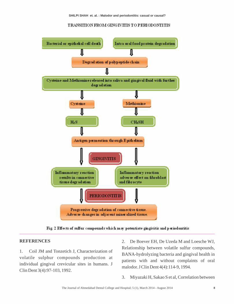
T R A N S I T I O N F R O M H E A LT H T O
GINGIVITIS:-
Gingivitis is characterized by an immune response
to antigens in bacterial plaque as well as by
alteration in connective tissue(Fig 2).One of the
earliest events associated with disease is enhanced
permeability of the lining epithelium with the
gingival sulcus. VSCs are potentially capable of
altering permeability of the gingival tissues,
including inflammatory response and modulating
functions of gingival fibroblast. Rizzo (1970)
indicated that a facilitating agent is required to
allow lipolysaccharides (LPS) to penetrate healthy
gingival epithelium and subsequently initiate an
inflammatory response.14 Studies demonstrated
that thiol participate in early stages of the
inflammatory response and may be important
initiator of gingivitis. Gingivitis results from the
induction of an immune response and may be
accompanied by alteration in fibroblast function.
Methyl mercaptan (CH SH) has been shown to 3
induce secretion of interleukin-1β (IL-1β) from
mononuclear cells. Methyl mercaptan has also been
shown to act synergistically with both LPS and IL-
1β to increase secretion of prostaglandin E and 2
collagenase, important mediators of inflammation 15and tissue destruction.
VSCs have direct effect on the formation of
extracellular matrix by human gingival fibroblast.
In addition they lower total protein production by
4
SHILPI SHAH Malodor and periodontitis: casual or causal? et. al. :
The Journal of Ahmedabad Dental College and Hospital; 5 (1), March 2014 - August 2014
these cells.
The effect of methyl mercaptan (CH SH) on 3
collagen metabolism is a reflection of both
decreased synthesis and increased degradation of
protein. This increased degradation is likely to be
associated with inhibition of procollagen peptidase
enzymes which are essential for procollagen
processing and for cross linking to form mature
collagen fibrils.
TRANSITION FROM GINGIVITIS TO
PERIODONTITIS:In the change from gingivitis to periodontitis, there
is a continuation of all the events in the oral
malodor and gingivitis section as well as a new
group of events that occur in the development of
periodontitis (Fig.2). Periodontitis results from
destruction of both the soft and hard tissue
structures which support teeth. The transition from
gingivitis to periodontitis is mainly an anatomical
difference in which the disease progresses into the
underlying bone.Since the periodontal ligament
cells are associated with the formation and
maintenance of the mineralized supporting
structures, effects of thiols on these ligament cells
are particularly relevant. Increase in probing
pocket depth and bleeding on probing with increase
in methyl mercaptan in these pocket are also
relevant. The effects resulting from exposure to
CH3SH become increasingly important in
periodontitis.1
Yaegaki&Sanada showed a correlation between
increases in CH SH/H S concentration ratio and 3 2
10increases in periodontal pocket depth.
Studies have shown that PDL cells exposed to
methyl mercaptan (CH3SH) alter their intracellular
pHand become more acidic. In addition, they
exhibit decreased motility, lowered protein
synthesis, and alteration in collagen metabolism.
These changes are predominantly determined to the
ability of these cells to maintain or regenerate
mineralized tissues. In addition there is substantial
reduction in amount of type III collagens.16 This
observation is significant since periodontally
involved tissue are known to exhibit substantial
losses of type III collagens which decrease from 20-
30% to 4% of total collagen.17 Fibronectin in PDL
cell are also affected by VSCs.
Severity of Periodontitis
In periodontitis, different studies have shown a
correlation between VSC concentration in mouth 3,6,10air and increased pocket depth. However De
Boever (1996) found that tongue odor was
negatively correlated with probing depth
suggesting an inverse relationship between malodor
and periodontal parameters.2 Similarly Bosy et al.
(1994) did not find a relationship between
periodontal disease and the prevalence or severity 11
of halitosis.
Correlation between the presence of a
p a t h o g e n i c m i c r o f l o r a i n t h e
subgingivalmicrobiota & Halitosis
In 1994,Bosy et al.founda moderately strong
correlation between the BANA (Benzoyl- DL-
arginine-2-Napthylamide) scores with floss odor
based on trypsin like activity detected by the BANA
test . They also found that 87.5% of tooth sites were
BANA positive as compared with 74.5% of tooth 11
sites positive in healthy individuals.
A significant correlation has been found between
the presence of motile organisms and P.
intermediaon the tongue dorsum in individual with
periodontitis as opposed to periodontally-healthy
subjects. This indicates that the tongue may act as a
reservoir for some periodontopathogens that
contribute to oral malodor.
SHILPI SHAH Malodor and periodontitis: casual or causal? et. al. :
5The Journal of Ahmedabad Dental College and Hospital; 5 (1), March 2014 - August 2014
Tonzetichfound the pathogenic, proteolytic strains
of Bacteroidesmelanogenicus produced more VSCs
t h a n n o n - p r o t e o l y t i c
s t r a i n s . 1 8 T r e p o n o m a d e n t i c o l a ,
P o r p h y r o m o n a s g i n g i v a l i s ,
Porphyromonasendodontalis, Prevotellaintermedia
and Bacteroidesloescheii produced significantly
higher amount of sulfides than other bacteria.19
Other bacterial species recovered from periodontal
pocke t s such a s En t e robac t e r i a eceae ,
Bacteroidesforsythus, Centipedaperiodontii,
E i k e n e l l a c o r r o d e n s ,
Fusobacteriumperiodonticumetc. also had high
capability to generate VSCs in vitro19, 20.
FUTURE PERSPECTIVE:VSC in periodontal pockets might be used as a
predictor of periodontal disease. Many authors have
proposed to utilize hydrogen sulfide, one of
causative periodontal pathogens' products as an
indicator for disease severity. The value of this
hypothesis remains to be elucidated. It is also
interesting to determine the degree of contribution
by pocket VSC to whole mouth odor. For this study,
we have to measure VSC level in periodontal
pockets quantitatively. Then multifactor analysis
including pocket VSC level and odor level of the
tongue and the other parts of oral cavity will provide
us useful information in management of patients
with oral malodor.
CONCLUSION:An estimated 80 percent to 90 percent of all bad
breath odors originate from the mouth and are
caused by bacteria. The accumulation of plaque and
debris and the stagnation of saliva occur most
commonly in areas where tooth and tissue
crevices,posterior dorsum tongue, interdental
spaces and subgingival areas lend themselves to
stagnant microenvironments.
Although oral malodor is probably not caused by
periodontal disease; there is ample evidence to
suggest that periodontal disease increases the
severity of oral malodor. Periodontitis worsens the
severity of oral malodor by providing additional
sites of VSC production (interdental and
subgingival), an increased availability of sulfur-
containing substrate (exfoliated epithelial cells and
leukocytes) and an increased rate of methionine
metabolism (precursor to methyl mercaptan).
Periodontitis contributes to an increased tongue
coating with higher VSC production. There is
evidence to suggest that VSCs, i.e., oral malodor,
may contribute to the progression and pathogenesis
of periodontal disease via increased mucosal
permeability.
This article outlines the efficacy of volatile sulphur
compounds in causing malodor and showing the
casual and causal association among periodontal
pathogenic microorganisms, periodontal disease
and oral malodor which has been strongly
implicated but not proved.
ACKNOWLEDGEMENT: NILCONFLICT OF INTEREST: NIL
6
SHILPI SHAH Malodor and periodontitis: casual or causal? et. al. :
The Journal of Ahmedabad Dental College and Hospital; 5 (1), March 2014 - August 2014
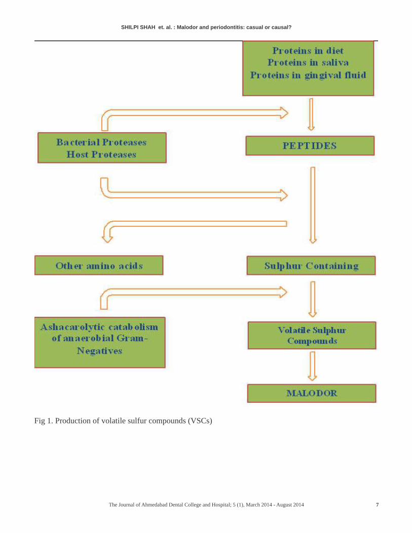
Fig 1. Production of volatile sulfur compounds (VSCs)
7
SHILPI SHAH Malodor and periodontitis: casual or causal? et. al. :
The Journal of Ahmedabad Dental College and Hospital; 5 (1), March 2014 - August 2014
REFERENCES
1. Coil JM and Tonzetich J, Characterization of
volatile sulphur compounds production at
individual gingival crevicular sites in humans. J
Clin Dent 3(4):97-103, 1992.
2. De Boever EH, De Uzeda M and Loesche WJ,
Relationship between volatile sulfur compounds,
BANA-hydrolyzing bacteria and gingival health in
patients with and without complaints of oral
malodor. J Clin Dent 4(4):114-9, 1994.
3. Miyazaki H, Sakao S et al, Correlation between
8
SHILPI SHAH Malodor and periodontitis: casual or causal? et. al. :
The Journal of Ahmedabad Dental College and Hospital; 5 (1), March 2014 - August 2014
volatile sulphur compounds and certain oral health
measurements in the general population.
JPeriodontol 66(8):679-84, 1995.
4. Yaegaki K and Sanada K, Volatile sulfur
compounds in mouth air from clinically healthy
subjects and patients with periodontal disease. J
periodontal Res 27(4 Pt 1):233-8, 1992.
5. Goldberg S, Kozlovsky A et al, Cadaverine a
putative component of oral malodor. J Dent Res
73(6):1168-72, 1994.
6. Rosenberg M, Clinical assessment of bad
breath: current concepts. J Am Dent Assoc,
127(4):475-82, 1996.
7. Tonzetich J. Production and origin of oral
malodor: a review of mechanisms and methods of
analysis. J Periodontol 48(1):13-20, 1977.
8. McNamara TF, Alexander JF and Lee M, The
role of microorganisms in the production of oral
malodor. Oral Surg Oral Med Oral Pathol 34(1):41-
8, 1972.
9. Schmidt NF, Missan SR and Tarbet WJ, The
correlation between organoleptic mouth-odor
ratings and levels of volatile sulfur compounds.
Oral Surg Oral Med Oral Pathol 45(4):560-7, 1978.
10. Yaegaki K and Sanada K, Biochemical and
clinical factors influencing oral malodor in
periodontal patients. J Periodontol 63(9):783-9,
1992.
11. Bosy A, Kulkarni GV, et al., Relationship of
oral malodor to periodontitis: evidence of
independence in discrete subpopulations. J
Periodontol 65(1):37-46, 1994
12. McNamara TF, Alexander JF and Lee M, The
role of microorganisms in the production of oral
malodor. Oral Surg Oral Med Oral Pathol 34(1):41-
8, 1972.
13. Solis-Gaffar MC, Fischer TJ and Gaffar A,
Instrumental evaluation of odor produced by
specific oral microorganisms. J SocCosmetChem
30:241-7, 1979.
14. Rizzo A. Histologic and immunologic
evaluation of antigen penetration into oral tissues
after topical application. J Periodontol 1970;
41:210-212.
15. Ratkay LG, Waterfeild JD, Tonzetich J.
Stimulation of enzyme and cytokine production
by methyl mercaptan in human gingival fibroblast
and monocyte cell culture. Arch Oral Biol
1995;40:337-344.
16. Lancero H, Niu JJ, Johnson PW. Exposure of
periodontal ligament cells to methyl mercaptan
reduces intracellular pH and inhibits cell migration.
J Dent Res 1996; 75:1994-2002.
17. Narayanan AS, Page RC. Biochemical
characterization of collagen synthesized by
fibroblast derived from normal and diseased human
gingiva. J BiolChem 1977; 251:5464-5469.
18. Tonzetich J and McBride BC, Characterization
of volatile sulfur production by pathogenic and non-
pathogenic strains of bacteroides. Arch Oral Biol
26:963-9, 1981.
19. Persson S, Edlund MB, Claesson R, Carlsson J.
The formation of hydrogen sulphide and methyl
m e r c a p t a n b y o r a l b a c t e r i a . O r a l
MicrobiolImmunol 1990; 5: 195-201.
20. Goldberg S, Cardash H, Browning H III, Sahly
H, Rosenberg M. Isolation of Enterobacteriaecea
from the mouth and potential association with
malodor. J Dent Res 1997; 76: 1770-1775.
9
SHILPI SHAH Malodor and periodontitis: casual or causal? et. al. :
The Journal of Ahmedabad Dental College and Hospital; 5 (1), March 2014 - August 2014
DENTAL FLUOROSIS – A RETROSPECTIVE STUDY IN GANDHINAGAR DISTRICT
PURV PATEL*, BHAVIN DUDHIA**, A. R. CHAUDHARY***, PARUL BHATIA****, YESHA JANI*****, PURVA BUTALA******
ADDRESS FOR AUTHOR CORROSPONDENCE : DR. , PHONE: 94272 19470PURV PATEL
10
Original Article
AHMEDABAD DENTAL COLLEGE & HOSPITAL, BHADAJ-RANCHHODPURA ROAD, TA:- KALOL DIST:-GANDHINAGAR.
*SENIOR LECTURER, ** PROFESSOR & HEAD, ***PROFESSOR, ****PROFESSSOR, *****SENIOR LECTURER, ******SENIOR LECTURER
ORAL MEDICINE, DIAGNOSIS & RADIOLOGY DEPARTMENT, AHMEDABAD DENTAL COLLEGE & HOSPITAL, GUJARAT.
ABSTRACT
IntroductionDental Fluorosis is an irreversible but preventable disease commonly caused by excessive intake of fluoride during critical period of teeth development. Dental fluorosis is endemic in many areas of Indian subcontinent including many districts in the North Central part of Gujarat state.
Aims & ObjectivesThe study was undertaken to correlate the severity of dental fluorosis with variables like age, gender, drinking water fluoride levels and dental caries.
Materials & MethodsA total of 53 patients (30 males and 23 females) affected with dental fluorosis were selected for the study. The subjects were assessed for their age range, severity of the dental fluorosis (based on Dean's Fluorosis Index) and their drinking water fluoride levels (determined by Ion Selective Electrode method). The subjects were also assessed for their dental caries prevalence using the DMFT index.
ResultsMajority of the subjects belonged to third and fourth decades of life followed by the second decade. The highest number of subjects manifested moderate degree of dental fluorosis followed by severe degree and mild degrees respectively. There was a positive correlation of the severity of dental fluorosis with the level of fluoride in the drinking water as well as the DMFT index of the subjects.
ConclusionA dentist often plays a prime role in detection of dental fluorosis, and hence in identification of the areas with higher water fluoride levels. This necessitates the dentists to be familiar with the clinical presentation of dental fluorosis as well as with areas affected by endemic dental fluorosis.Received: 08-10-2013; Review Completed: 12-12-2013; Accepted: 15-01-2014
INTRODUCTION
Fluorosis is described as a state of toxicity of
the trace element called fluorine within an [1]
organism. In 1901, Dr. Frederick McKay of
Colorado (USA) accidentally discovered that many
patients had apparently permanent stain on their
teeth which was often referred to as COLORADO [2]STAIN. “Shoe Leather Survey” of Trendley Dean
(1931) lead to the establishment that concentration
of fluoride in drinking water was directly correlated
with the severity of fluorosed and mottled enamel. [2,3]
However, the United States Food and Drug
Administration (1973) has listed Fluoride as an
essential nutrient for human health. The report of
WHO expert committee includes Fluoride in its list
of 14 trace elements which are physiologically
essential for normal growth and development of [2,4]
human beings.
Dental Fluorosis develops due to chronic
and excessive use of fluoride compounds, most
common causative factor being use of drinking
water with higher levels of fluoride, especially
during first 6 years of age when teeth are [3,5-12]
developing. Dental Fluorosis is more
The Journal of Ahmedabad Dental College and Hospital; 5 (1), March 2014 - August 2014

PURV PATEL Dental Fluorosis – A Retrospective Study in Gandhinagar District et. al. :
commonly observed in patients obtaining drinking
water from tube wells, bore wells or hand pumps. [13,14]
Diet, seafood and tea intake does not influence [3]
prevalence of dental fluorosis.
There is no reported significant difference
in prevalence and severity of dental fluorosis [3,14]
related to age and gender. However, certain
studies report it to be least common within first
decade due to higher number of primary teeth and
only a few erupted permanent teeth. It is reportedly
more common in 12 – 14 years of age as maximum [1,5,12,15] permanent teeth have erupted by this age.
Some studies report greater prevalence of fluorosis [5,9,12]
among males than females. One possible
explanation might be that men drink more water
than women to compensate for fluid loss during [16]
field work. This could also be due to greater [5] number of male population in a particular area.
Fluorosis is reported to be more severe in maxillary [14]
teeth than in homologous mandibular teeth. This
was probably related to unrecognized trauma in the
maxillary teeth or other local, unspecified types of [17]
insult during tooth development. Dental
Fluorosis is observed in both primary and
permanent dentition. Primary tooth fluorosis is less
common and usually less severe than in permanent
teeth, as explained by the fact that very high fluoride
levels (> 10 ppm) are required in drinking water for
it to cross placental barrier and affect primary
dentition as most primary teeth develop during [4,8,10,18]
intrauterine life.
Dental fluorosis results in a variety of [19]
pathological changes in the structure of the teeth.
It is characterized by occasional opaque, lusterless
white spots in the enamel which constitute
questionable degree of fluorosis (based on Dean's
Fluorosis index). When the white flecks cover less
than 25% of tooth surfaces, it is said to be very mild
degree fluorosis. When the white flecks cover more
than 25% but less than 50% of tooth surfaces, it is
said to have mild degree fluorosis. Cloudy striations
or 'snow capping' white flecks covering more than
50% of tooth surfaces or presence of brown or
yellow stains constitute moderate degree fluorosis.
Porous and weak, pitted or hypoplastic tooth with
loss of its general form constitutes severe degree [4,6,11,15,19,20] fluorosis. The fluorotic changes showed
high degree of bilateral symmetry in the buccal [14]
surfaces of homologous pairs of teeth.
Contradictory reports are available in the
literature regarding the correlation of the severity of
Dental Fluorosis and the DMFT Index. Some
authors have reported that as the severity of dental
fluorosis increased, the DMFT increased upto the
level of mild fluorosis and then decreased as the
severity increased from moderate to severe [5] fluorosis. Others have reported that when
fluorosis level increases upto moderate, the DMFT
value correspondingly decreases. But as the
fluorosis level increases beyond moderate, the
DMFT rate increases in cases of severe fluorosis
due to pitting of enamel surface which promotes the [9]
accumulation of microbial plaque.
The optimal concentration of fluoride for
drinking water is that level which offers minimal
risk of dental fluorosis while providing significant [21]
protection against caries. The Environmental
Protection Agency has recommended optimum [13,15,22]water fluoride level from 0.7 to 1.0 ppm. The
World Health Organization has set a maximum
concentration of 1.5 ppm fluoride in drinking water [21] to avoid dental fluorosis. In the Indian context,
even in regions with water fluoride concentration as
low as 0.5 ppm in drinking water, mild forms of [21,22] dental fluorosis have been reported. Dental
Fluorosis is endemic in 15 states of India, including
The Journal of Ahmedabad Dental College and Hospital; 5 (1), March 2014 - August 2014 11
[3-6,14,23] Gujarat. Fluorosis is endemic in almost all the
[14] districts of Gujarat. Fluorosis mostly affects
people in rural India who have no access to safe
drinking water and drink water drawn from wells or [1, 4] hand pumps.
There is a positive association between the
mean annual temperature of a place and total [9,14]
fluoride intake of people residing at that place.
There are reports of fluorosis even at places with
low water fluoride levels. This could be explained
by A. K. Susheela's explanation, that in an endemic
fluorosis area, a great amount of fluoride is
incorporated into food materials and ingested into
the body. Also, with high temperature of the place [14] necessitates greater intake of water. The
occurrence of fluorosis can vary widely among
different locations having almost the same Fluoride
concentrations in drinking water and can be affected
by a number of other factors such as nutritional
status, climate, individual susceptibility and
biological response, duration of fluoride exposure,
dissolved salts in drinking water, and most [5,11,12]importantly the frequency of fluoride intake.
A stepwise increase in severity of fluorosis
has been reported with rise in level of fluoride in [3,19] water consumed. The proposed possible
mechanisms for such severe manifestations in high
water fluoride content areas include: (1) High
atmospheric temperatures during summer months
(2) Hard physical labour activity (3) Poor nutrition,
deficient in calories and also vitamin C (4)
Continued exposure to fluoride (5) Impaired renal
function (6) Abnormal concentration of certain [4,14]trace elements.
Some standard methods for assay of
fluoride in the scientific literature include Ion
Se l ec t ive e l ec t rode , Co lo r ime t ry, Ion [24] Chromatography and Electrometry. Out of these,
Ion Selective Electrode is found to be accurate upto [13] about 98%. Dental Fluorosis is irreversible, but
preventable by appropriate and timely intervention. [4,25]
There are several basic types of water
purification systems, e.g., reverse osmosis,
distillation, filtration, oxidation, disinfection,
cation exchange softening, anion exchange, [13] activated carbon, etc. Reverse osmosis water
purification systems remove 90 to 95% of the [13,20]
fluoride content in water.
A variety of treatment modalities are
available for Dental Fluorosis, with cost being a
major limitation. In – Office bleaching is the most
commonly used method for the removal of stains.
Moreover, depending upon the clinical condition, a
synergistic approach of combining bleaching with
other modalities such as micro abrasion and
fabrication of veneers can help in gaining an [26]
excellent clinical outcome. A minimal invasive
technique combining the triad of micro reduction,
micro abrasion and conventional vital bleaching
allows good esthetics and a possible cost reduction [16]for treating mild to moderate fluorosis.
The purpose of this study is to evaluate the
correlation between high ground water fluoride
content & severity as well as extent of Dental
Fluorosis in rural areas in and around Gandhinagar
district of Gujarat state.
PURV PATEL Dental Fluorosis – A Retrospective Study in Gandhinagar District et. al. :
The Journal of Ahmedabad Dental College and Hospital; 5 (1), March 2014 - August 2014 12
PURV PATEL Dental Fluorosis – A Retrospective Study in Gandhinagar District et. al. :
AIMS AND OBJECTIVESØ To estimate the occurrence of Dental Fluorosis
in patients coming to the Out Patient Department (OPD) of Ahmedabad Dental College and Hospital
Ø To evaluate the association of Dental Fluorosis with Age and Sex
Ø To estimate water fluoride level and correlate it with the degree of Dental Fluorosis
Ø To evaluate different degrees of Dental Fluorosis in terms of type and severity
Ø To estimate the incidence and severity of dental caries in patients having Dental Fluorosis
MATERIALS AND METHODS
Ø The study was conducted on 53 patients from amongst all the patients coming to the Out Patient Department (OPD) of the Oral Medicine and Radiology Department of Ahmedabad Dental College and Hospital during the period of June 2009 to September 2010
Ø Inclusion Criteria• Patients having chalky white spots or brown
staining or structural abnormalities of teeth• Patients who have lived in the same place
where they were born and have procured drinking water from the same source throughout their life
Ø Exclusion Criteria• Patients with history of being treated by
long term antibiotic medication in early childhood or whose mother has been treated by such medication during pregnancy
• Patients who had migrated to some other place after birth or who were not permanent residents of any one place since birth
• Patients who have obtained drinking water from more than one source since birth
• Patients having some severe systemic disease or condition
Ø The severity of Dental Fluorosis was estimated based on Dean's Fluorosis Index.
Ø Patients selected for the study were evaluated and examined thoroughly and the findings were recorded in a Proforma prepared specially for
the study
Ø The drinking water samples were procured from patients and sent to laboratory for estimation of fluoride content in ppm (parts per million) based on Ion Selective Electrode Method.
COLLECTION OF WATER SAMPLE
Ø The ground water pumped from the borewell into overhead water tanks comprised the source of water for samples collected in the study
Ø The water samples were collected from overhead water tanks after obtaining permission from the respective Gram Panchayats of villages under study.
Ø The water sample collected from a single place was equally divided into two unused plastic containers and precoded by the investigator and then submitted to the laboratory technician on the same day. Thus, the laboratory technician was kept unaware of the place to which the water sample belonged, to eliminate any potential bias.
Ø The same procedure was repeated for all places under study.
Ø Two samples were thus submitted from each place to the technician in order to test the same sample twice and hence eliminate major errors. The mean of two samples was taken as final reading. If the difference between the two readings was greater than 0.5 ppm, the sample was discarded; a new sample was obtained from the same place and submitted for water fluoride estimation.
Ø From the ppm content of water samples procured, a chart of fluoride content of different areas of Gandhinagar district was prepared.
RESULTS
A total of 53 patients with Fluorosis were selected for the study from the patients coming to the Out Patient Department (OPD) at Oral Medicine, Diagnosis & Radiology Department
The Journal of Ahmedabad Dental College and Hospital; 5 (1), March 2014 - August 2014 13
14
of Ahmedabad Dental College & Hospital.
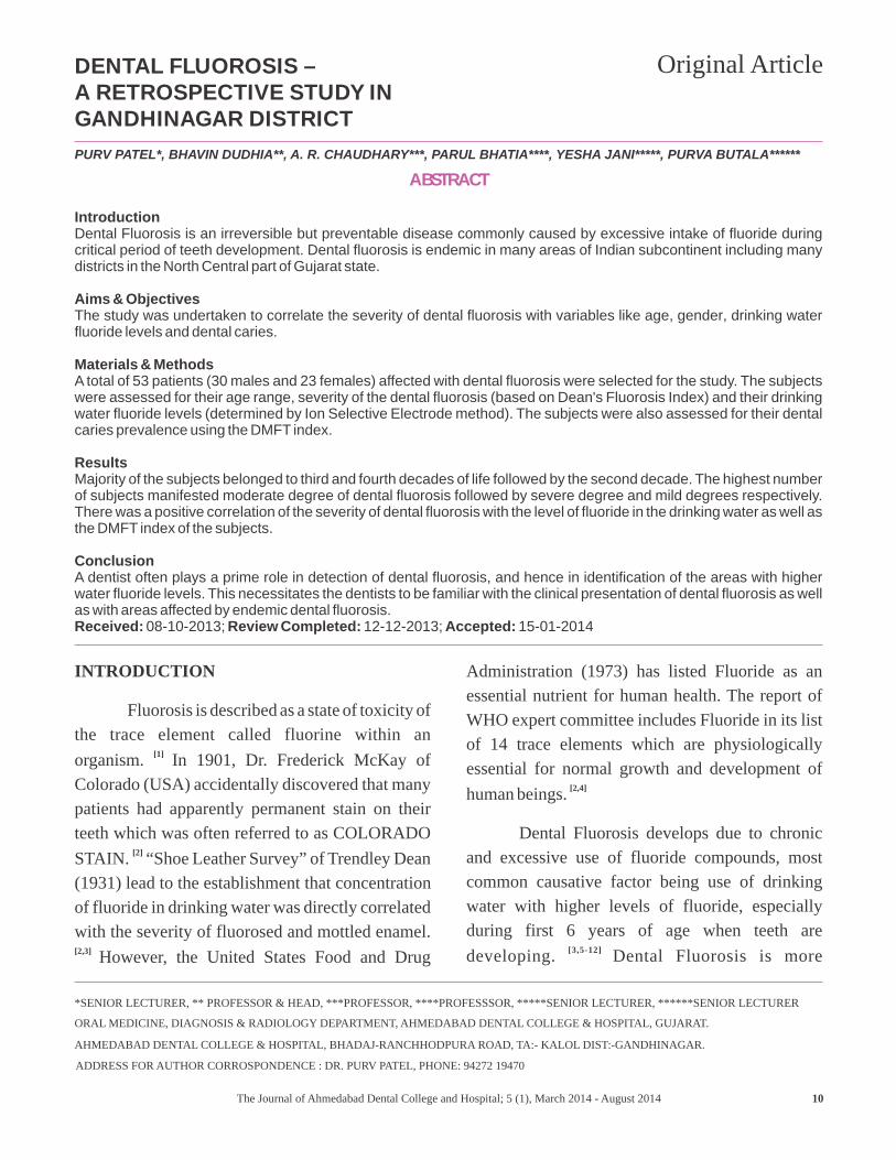
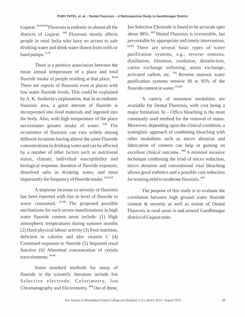
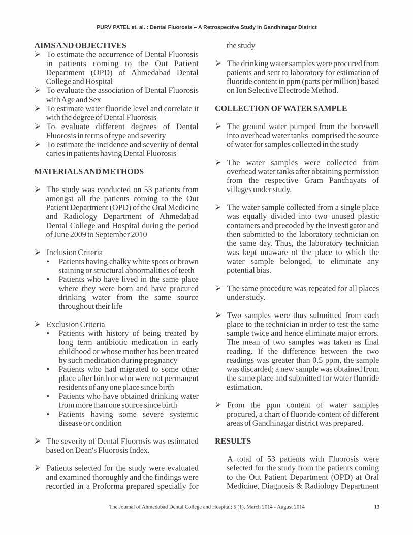
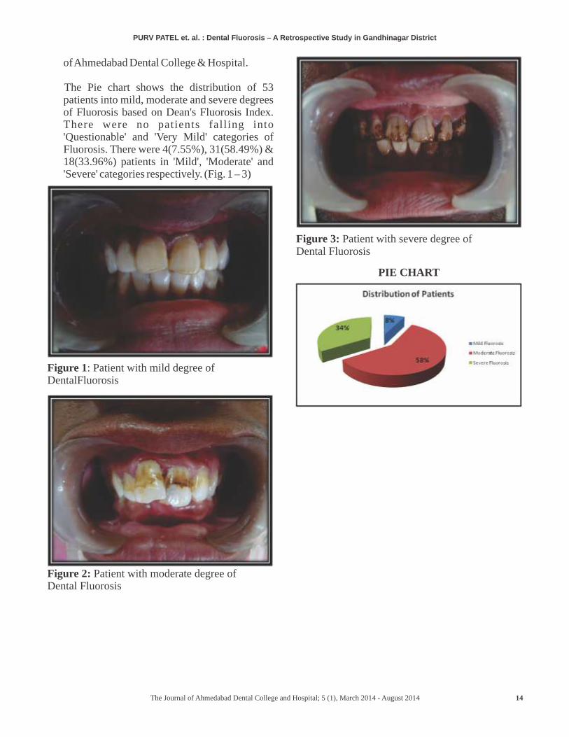
The Pie chart shows the distribution of 53 patients into mild, moderate and severe degrees of Fluorosis based on Dean's Fluorosis Index. There were no patients falling into 'Questionable' and 'Very Mild' categories of Fluorosis. There were 4(7.55%), 31(58.49%) & 18(33.96%) patients in 'Mild', 'Moderate' and 'Severe' categories respectively. (Fig. 1 – 3)
Figure 1: Patient with mild degree of DentalFluorosis
Figure 2: Patient with moderate degree of Dental Fluorosis
Figure 3: Patient with severe degree of Dental Fluorosis
PIE CHART
PURV PATEL Dental Fluorosis – A Retrospective Study in Gandhinagar District et. al. :
The Journal of Ahmedabad Dental College and Hospital; 5 (1), March 2014 - August 2014
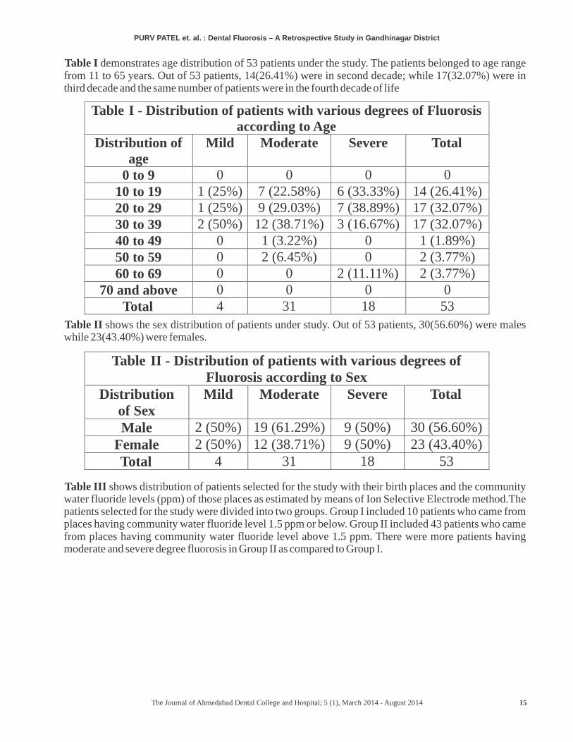
Table I demonstrates age distribution of 53 patients under the study. The patients belonged to age range from 11 to 65 years. Out of 53 patients, 14(26.41%) were in second decade; while 17(32.07%) were in third decade and the same number of patients were in the fourth decade of life
Table II shows the sex distribution of patients under study. Out of 53 patients, 30(56.60%) were males while 23(43.40%) were females.
Table III shows distribution of patients selected for the study with their birth places and the community water fluoride levels (ppm) of those places as estimated by means of Ion Selective Electrode method.The patients selected for the study were divided into two groups. Group I included 10 patients who came from places having community water fluoride level 1.5 ppm or below. Group II included 43 patients who came from places having community water fluoride level above 1.5 ppm. There were more patients having moderate and severe degree fluorosis in Group II as compared to Group I.
Table I
-
Distribution of patients with various degrees of Fluorosis according to Age
Distribution of age
Mild
Moderate Severe Total
0 to 9
0
0
0 010 to 19
1 (25%)
7 (22.58%) 6 (33.33%) 14 (26.41%)20 to 29
1 (25%)
9 (29.03%) 7 (38.89%) 17 (32.07%)30 to 39 2 (50%) 12 (38.71%) 3 (16.67%) 17 (32.07%)40 to 49 0 1 (3.22%) 0 1 (1.89%)50 to 59 0 2 (6.45%) 0 2 (3.77%)60 to 69 0 0 2 (11.11%) 2 (3.77%)
70 and above 0 0 0 0Total 4 31 18 53
Table II - Distribution of patients with various degrees of Fluorosis according to Sex
Distribution of Sex
Mild Moderate Severe Total
Male 2 (50%) 19 (61.29%) 9 (50%) 30 (56.60%)Female 2 (50%) 12 (38.71%) 9 (50%) 23 (43.40%)Total 4 31 18 53
15
PURV PATEL Dental Fluorosis – A Retrospective Study in Gandhinagar District et. al. :
The Journal of Ahmedabad Dental College and Hospital; 5 (1), March 2014 - August 2014
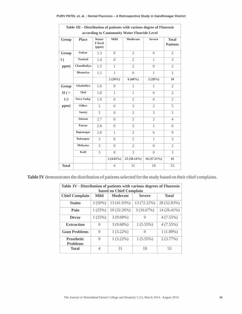
Table III – Distribution of patients with various degree of Fluorosis
according to Community Water Fluoride Level
Group Place Water F level (ppm)
Mild Moderate Severe Total
Patients
Group
I (≤ 1.5
ppm)
Vadsar
1.3
0
2
0
2
Nasmed
1.4
0
2
1
3
Chandlodiya
1.5
1
2 0
3
Bhamriya 1.5 1 0 1 2
2 (20%)
6 (60%)
2 (20%) 10
Group
II ( >
1.5
ppm)
Ghatlodiya
1.6
0
1
1
2
Thol
1.8
1
1
0
2
Nava Vadaj
1.9
0
2
0
2
Odhav
2
0
3
2
5
Santej
2
0
2
3
5
Julasan
2.7
0
2
2
4
Pansar
2.8
0
5
1
6
Bapunagar
2.8
1
2
6
9
Rakanpur 3 0 2 1 3
Mehsana 3 0 2 0 2
Kadi 3 0 3 0 3
2 (4.65%) 25 (58.14%) 16 (37.21%) 43
Total 4 31 18 53
PURV PATEL Dental Fluorosis – A Retrospective Study in Gandhinagar District et. al. :
Table IV demonstrates the distribution of patients selected for the study based on their chief complains.
Table IV - Distribution of patients with various degrees of Fluorosis based on Chief Complain
Chief Complain
Mild
Moderate
Severe
Total
Stains
2 (50%)
13 (41.93%)
13 (72.22%) 28 (52.83%)
Pain
1 (25%)
10 (32.26%)
3 (16.67%)
14 (26.41%)
Decay
1 (25%)
3 (9.68%)
0
4 (7.55%)
Extraction 0 3 (9.68%) 1 (5.55%) 4 (7.55%)
Gum Problems 0 1 (3.22%) 0 1 (1.89%)
Prosthetic Problems
0 1 (3.22%) 1 (5.55%) 2 (3.77%)
Total 4 31 18 53
The Journal of Ahmedabad Dental College and Hospital; 5 (1), March 2014 - August 2014 16
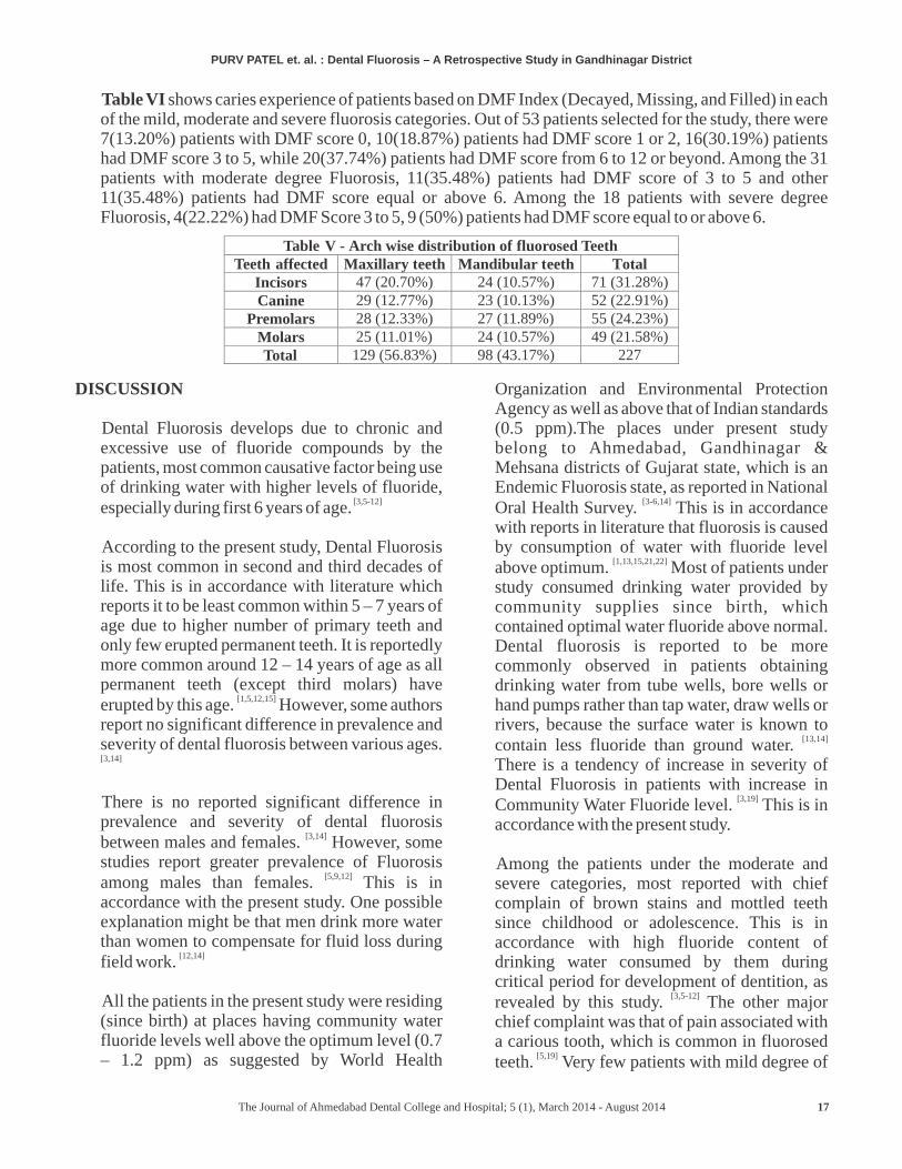
Table VI shows caries experience of patients based on DMF Index (Decayed, Missing, and Filled) in each of the mild, moderate and severe fluorosis categories. Out of 53 patients selected for the study, there were 7(13.20%) patients with DMF score 0, 10(18.87%) patients had DMF score 1 or 2, 16(30.19%) patients had DMF score 3 to 5, while 20(37.74%) patients had DMF score from 6 to 12 or beyond. Among the 31 patients with moderate degree Fluorosis, 11(35.48%) patients had DMF score of 3 to 5 and other 11(35.48%) patients had DMF score equal or above 6. Among the 18 patients with severe degree Fluorosis, 4(22.22%) had DMF Score 3 to 5, 9 (50%) patients had DMF score equal to or above 6.
Table V - Arch wise distribution of fluorosed TeethTeeth affected Maxillary teeth Mandibular teeth Total
Incisors 47 (20.70%) 24 (10.57%) 71 (31.28%)Canine 29 (12.77%) 23 (10.13%) 52 (22.91%)
Premolars 28 (12.33%) 27 (11.89%) 55 (24.23%)Molars 25 (11.01%) 24 (10.57%) 49 (21.58%)Total 129 (56.83%) 98 (43.17%) 227
PURV PATEL Dental Fluorosis – A Retrospective Study in Gandhinagar District et. al. :
DISCUSSION
Dental Fluorosis develops due to chronic and excessive use of fluoride compounds by the patients, most common causative factor being use of drinking water with higher levels of fluoride,
[3,5-12]especially during first 6 years of age.
According to the present study, Dental Fluorosis is most common in second and third decades of life. This is in accordance with literature which reports it to be least common within 5 – 7 years of age due to higher number of primary teeth and only few erupted permanent teeth. It is reportedly more common around 12 – 14 years of age as all permanent teeth (except third molars) have
[1,5,12,15]erupted by this age. However, some authors report no significant difference in prevalence and severity of dental fluorosis between various ages. [3,14]
There is no reported significant difference in prevalence and severity of dental fluorosis
[3,14]between males and females. However, some studies report greater prevalence of Fluorosis
[5,9,12]among males than females. This is in accordance with the present study. One possible explanation might be that men drink more water than women to compensate for fluid loss during
[12,14]field work.
All the patients in the present study were residing (since birth) at places having community water fluoride levels well above the optimum level (0.7 – 1.2 ppm) as suggested by World Health
Organization and Environmental Protection Agency as well as above that of Indian standards (0.5 ppm).The places under present study belong to Ahmedabad, Gandhinagar & Mehsana districts of Gujarat state, which is an Endemic Fluorosis state, as reported in National
[3-6,14]Oral Health Survey. This is in accordance with reports in literature that fluorosis is caused by consumption of water with fluoride level
[1,13,15,21,22]above optimum. Most of patients under study consumed drinking water provided by community supplies since birth, which contained optimal water fluoride above normal. Dental fluorosis is reported to be more commonly observed in patients obtaining drinking water from tube wells, bore wells or hand pumps rather than tap water, draw wells or rivers, because the surface water is known to
[13,14]contain less fluoride than ground water. There is a tendency of increase in severity of Dental Fluorosis in patients with increase in
[3,19]Community Water Fluoride level. This is in accordance with the present study.
Among the patients under the moderate and severe categories, most reported with chief complain of brown stains and mottled teeth since childhood or adolescence. This is in accordance with high fluoride content of drinking water consumed by them during critical period for development of dentition, as
[3,5-12]revealed by this study. The other major chief complaint was that of pain associated with a carious tooth, which is common in fluorosed
[5,19]teeth. Very few patients with mild degree of
The Journal of Ahmedabad Dental College and Hospital; 5 (1), March 2014 - August 2014 17
Fluorosis presented for the study, which reflects the fact that Fluorosis of a moderate degree is much more noticeable due to presence of brown stains than mild degree of Fluorosis which often goes unnoticed.
Dental Fluorosis is found to be more severe in maxillary teeth than in homologous mandibular teeth, with maxillary incisors as most commonly involved teeth group, in the present study. This is in accordance with the reports in
[14]literature. This was probably related to unrecognized trauma in the maxillary teeth or other local, unspecified types of insult during
[17]tooth development.
There is an increase in prevalence of dental caries with increase in severity of Dental Fluorosis in the present study. This is in accordance with studies reporting elevated caries levels associated with the brittleness of
[3]moderately and severely mottled teeth. However, some authors have reported that when Fluorosis severity increased from normal to moderate, the DMFT value correspondingly decreased, while as the fluorosis level increased beyond moderate, the DMFT rate increased. The DMFT increased in cases of severe Fluorosis due to pitting of enamel surface which promoted the accumulation of microbial plaque. [9] While, some authors have reported that as the severity of Dental Fluorosis increased the DMFT increased upto the level of mild Fluorosis and then decreased as the severity
[5]increased from moderate to severe Fluorosis.
In the present study, there are more patients with greater DMFT scores among the patients with moderate and severe degree of Fluorosis as compared to those with mild degree of Fluorosis.
SUMMARY AND CONCLUSION
• Majority of patients came from Ahmedabad, Gandhinagar & Mehsana districts of Gujarat state, which are areas of Endemic Fluorosis according to National Oral Health Survey
• Majority of patients affected with Dental Fluorosis belonged to second, third and fourth decades of life.
• There was a slight male predominance among the affected patients.
• Amongst the patients with Dental Fluorosis, a step wise increase in severity of fluorosis was noted with increase in drinking water fluoride content (estimated by Ion Selective Electrode Method).
• Majority of patients fell into moderate to severe degree Fluorosis categories of Dean's Fluorosis Index
• There was an increase in Caries prevalence with corresponding increase in severity of Dental Fluorosis.
• Among the treatment modalities evaluated, Bleaching was found to give esthetically acceptable results in patients with brown stains while veneers were satisfactory as treatment modality for hypoplastic enamel with surface irregularities in patients with severe degree of Fluorosis.
REFERENCES
1. Alien. Fluorosis – Causes, Symptoms, and Treatment. Content Corner.com 2008.
2. Amrit T, Ved J. Fluorides and Dental Caries – A Compendium. Publication of the Journal of Indian Dental Association 1986.
3. Jagan Kumar B, Clement R, Aswath N. Prevalence of dental fluorosis and associated risk factors in 11 – 15 year old school children of Kanyakumari District, Tamilnadu, India: A Cross Sectional survey. Indian J Dent Res 2008; 19(4): 297 – 303.
4. Vineet D, Maheep B. Physiology and toxicity of fluoride. Indian J Dent Res 2009; 20(3): 350 – 5.
5. Tuli A, Rehani U, Aggrawal A. Caries experience evidenced in children having dental fluorosis. Int J Clinical Ped Dent 2009; 2(2): 25 – 31.
6. Bronckers A, Lyaruu D, DenBesten P. The Impact of Fluoride on Ameloblasts and the mechanisms of Enamel Fluorosis. J Dent Res 2009; 88(10): 877 – 93.
7. Fatemeh V, Anne M, Paula J. Sources of dietary fluoride Intake in 6-7 year old english children
PURV PATEL Dental Fluorosis – A Retrospective Study in Gandhinagar District et. al. :
The Journal of Ahmedabad Dental College and Hospital; 5 (1), March 2014 - August 2014 18
receiving optimally, sub-optimally, and non-fluoridated water. Journal Public Health Dent 2006; 66(4): 227 – 34.
8. Teresa A, Steven M, John J, Barbara B, Julie M, Phyllis J. Associations between intakes of fluoride from beverages during infancy and dental fluorosis of primary teeth. Journal of the American College of Nutrition 2004; 23(2): 108 – 16.
9. Ramezani G, Valaei N, Eikani H. Prevalence of DMFT and Fluorosis in the students of Dayer City (Iran). J Indian Soc Pedod Prevent Dent 2004; 22(2): 49 – 53.
10. Jian R, Asgeir R, Anne S, RuiZhe H, ZhiLun W, Kjell B. Dental fluorosis in children in areas with fluoride polluted air, high fluoride water, and low fluoride water as well as low fluoride air: A study of deciduous and permanent teeth in the Shaanxi p rov ince , China . Acta Odontologica Scandinavica 2007; 65: 65 – 71.
11. Ana A, Carlo E, Juan F, Gerardo M, Mirna M, Sayde O. Dental fluorosis in cohorts born before, during, and after the national salt fluoridation program in a community in Mexico. Acta Odontologica Scandinavica 2006; 64: 209 – 13.
12. Choubisa S. Endemic fluorosis in Southern Rajasthan, India. Fluoride 2001; 34(1): 61 – 70.
13. Prabhakar A, Raju O, Kurthukoti A, Vishwas T. The effect of water purification systems on fluoride content of drinking water. J Indian Soc Pedod Prevent Dent 2008; 26(1): 6 – 11.
14. Sudhir K, Prashant G, Subba Reddy V, Mohandas U, Chandu G. Prevalence and severity of dental fluorosis among 13 to 15 year old school children of an area known for endemic fluorosis: Nalgonda district of Andhra Pradesh. J Indian Soc Pedod Prevent Dent 2009; 27(4): 190 – 6.
15. Lia Silva C, Efigenia F, Leila N, Lucia M, Edson P. Beliefs and attitudes about endemic dental fluorosis among adolescents in rural Brazil. Rev Saude Publica 2010; 44(2): 261 – 6.
16. Harikumar V, Arun A. Management of mild to moderate fluorosis with a combined chemomechanical approach. Annals and Essences of Dentistry 2010; 2(3): 73 – 6.
17. Vera S, Dorte H, Carolina T, Thais M, Sven P. Prevalence and distribution of demarcated opacities and their sequelae in permanent 1st molars and incisors in 7 to 13 year old Brazilian children. Acta Odontologica Scandinavica 2009; 67: 170 – 5.
18. Dhar V, Jain A, Van Dyke T, Kohli A. Prevalence of gingival diseases, malocclusion and fluorosis in school going children of rural areas in Udaipur district. J Indian Soc Pedod Prevent Dent 2007; 25(2): 103 – 5.
19. Susheela A, Bhatnagar M, Gnanasundaram N, Saraswathy T. Structural aberrations in fluorosed human teeth: Biochemical and scanning electron microscopic studies. Current Science 1999; 77: 1677 – 81.
20. Pediatric Dentistry – Special Supplemental Issue. Access, January 2000.
21. Parkar S, Ajithkrishnan C. Estimation of fluoride concentration in Community water supply & packaged drinking water sold in Vadodara City – A Comparative Study. J Indian Assoc Public Health Dent 2010(15): 105 – 9.
22. Martin S Spiller. Fluoride. Doctor Spiller.com 2000.
23. Bali R, Mathura V, Stalwart P, Canaan H. National Oral Health Survey and Fluoride Mapping 2002 – 2003, Gujarat. Dental Council of India, New Delhi, 2004.
24. Kolashi J, Dastjerdi M. Assay of fluoride levels in drinking water. Ann Saudi Med 2005; 25(2): 175.
25. Wolfgang A, Anabel H, Julia H, Zeno G, Jolan B, Peter G. Effect of pH of amine fluoride c o n t a i n i n g t o o t h p a s t e s o n e n a m e l remineralization in vitro. BMC Oral Health 2007; 7: 14.
26. Thosre D, Mulay S. Smile enhancement the conservative way: Tooth whitening procedures. J Conserv Dent 2009; 12(4): 164 – 8.
PURV PATEL Dental Fluorosis – A Retrospective Study in Gandhinagar District et. al. :
The Journal of Ahmedabad Dental College and Hospital; 5 (1), March 2014 - August 2014 19
KNOWLEDGE, ATTITUDE AND PRACTICE REGARDING ORAL CANCER AND SCREENING PROCEDURES AMONG PRIMARY HEALTH CARE AND COMMUNITY HEALTHCARE WORKERS OF WAGHODIA, GUJARATBAFNA HARSHAL P *, AJITHKRISHNAN CG**, KALANTHARAKATH THANVEER***, RICKY PAL SING*, HEMAL PATEL****
ABSTRACT
Background: India has the highest rate of oral cancer in world and there is a 60% rise in past three decades. Mortality rate for oral cancer is higher in population with poor access to oral health care. With 72% of Indians residing in rural areas they have a better access to community health care providers (CCPs) and primary health care providers (PCPs) who, could play a major role in oral cancer prevention and reduce death rate.
Objectives: The study aimed at evaluating knowledge, attitude and practice regarding oral cancer and screening practices among CCP's and PCP's by using pretested questionnaire.
Material and Methods: A cross sectional questionnaire study was conducted among all CCP's and PCP's of Waghodia, Vadodara. A self designed, modified close ended and pre-piloted questionnaire was used for recording data. Mean and percentage were used for statistical analysis.
Results: 78.24% believed that prevention and early detection was important, while 53% had actually referred cases in past one year. All believed tobacco to be a risk factor for oral cancer but only 49% answered for other factors. 91.37% were ready to be a part of continuing education (CE).
Conclusion: The participants had deficient knowledge about the risk factors for oral cancer and were ready to participate in CE.
Key-words: Oral Cancer, Health Care Workers, Gujarat
Received: 12-11-2013; Review Completed: 22-01-2014; Accepted: 13-02-2014
ADDRESS FOR AUTHOR CORROSPONDENCE : DR. , PHONE: 8469048732BAFNA HARSHAL P
20
Original Article
*P.G. STUDENT, **PROF. & HEAD, ***PROFFESOR, ****SR.LEC, DEPT.
DEPT. OF PUBLIC HEALTH DENTISTRY, K.M. SHAH DENTAL COLLEGE
INTRODUCTION:
The World Health Organization reports a
worldwide death toll from tobacco use to be four
million per year. The death toll is expected to rise to [1]
ten million per year by 2020's or early 2030's.
Tobacco is thought to be one of the most important
etiological factors in development of oral cancer.
India has the highest rate of oral cancer in the world
with a 60% increase in past three decades and no
improvement in the mortality rate has been seen [2]
during this period.
Oral cancer is a disease with known high risk factors
and an asymptomatic phase with identifiable
clinical features. The mortality rate for oral cancer
is higher in population who traditionally experience [3]poor access to the oral health care system. Also
unfortunately, for a large segment of high-risk
individuals, access to dental care is limited where
dental surgeons can help to identify these
individuals and provide necessary treatment. About
those residing in rural areas, they have a better
access to non dental CCPs and PCPs. Therefore,
CCPs and PCPs could play a major role in
prevention of oral cancer and reducing death toll
owing to oral cancer. One way to achieve these
goals is to develop a network of dental and other
healthcare providers to promote the early screening
The Journal of Ahmedabad Dental College and Hospital; 5 (1), March 2014 - August 2014
of oral soft tissue lesions that may be pre-cancerous [4]or cancerous.
India has a population of 1.21billion and with 72% [5] [6]
of the Indian population residing in rural areas, ,
the government has implemented various rural
health strategies and national programmes through
Primary and community health care centres,
National Cancer Control Programme (NCCP) being
one of them. Lack of such a network with regards to
oral health care practices in India and unavailability
of oral health professionals to provide on site basic
oral health services discourages further the [7]
integration of oral health services in these centres.
Thus it becomes necessary for these health care
workers to play a role in oral cancer prevention and
therefore the present study was proposed to assess
the knowledge of oral cancer, attitude towards it
prevention and screening practice of oral cancer
among CCPs and PCPs Waghodia Taluka,
Vadodara, Gujarat.
MATERIAL AND METHODS:
Ethical approval was obtained from the Institutional
Ehics committee. Permissions were obtained from
the Chief District Health Officer (CDHO) of
Vadodara district and the Block Health Officer of
Waghodia Taluka to conduct the survey at the
primary and community health care centres of
Waghodia taluka. The purpose and procedure of the
study was informed to each participant and
participant information sheet was provided to them.
Also informed consent was obtained from each
participant who was willing to participate in the
study.
A pilot study was conducted in the beginning for
testing of validity and reliability of the developed
questionnaire among four Experts and four
respondents. The medical officers were considered
as experts while the other staff members were
BAFNA HARSHAL KAP regarding Oral Cancer among PCP's and CCP's et. al. :
21
considered as respondents and four from each were
chosen randomly by lottery method as participants
for pilot study. The validity of questionnaire using
Concurrent Validity method was 90.3. The
reliability results obtained by test – retest was
89.92% which showed a high agreement.The sampling frame included all Medical officers,
staff nurses, health workers, health educator, and
health assistants working at these centre's during the
study period i.e. from June 2012 to August 2012
which constituted a total of 85 subjects. The
individuals who were not willing to participate in
the study or were absent at the centre during three
consecutive visits were excluded from the study.
For each health care centre the questionnaire was
administered on first day of visit and recollected on
the second day by the principal investigator himself.
The participants who were absent on the first day of
visit were contacted over the phone and a later date
was fixed with them for the questionnaire fill up. All
the absent individuals responded in the first call.
Data was collected with the help of a self designed,
modified close ended & pre-tested questionnaire
used in English and Gujarati language. The Gujarati
version was validated by back translation method.
The questionnaire consisted of six, four and five
questions pertaining to knowledge of Oral cancer,
screening practice of Oral cancer and attitude
towards Oral cancer prevention respectively.
The data was analyzed using Descriptive statistical
analysis i.e. percentage in Microsoft Excel 2007
spreadsheet.
The Journal of Ahmedabad Dental College and Hospital; 5 (1), March 2014 - August 2014
RESULTS:
As seen in Table 1 participants had a good level of
knowledge regarding oral cancer prevalence, its
ability to metastasize and its life threatening
potential.
Table 1: Table showing knowledge of participants
regarding oral cancer
Table 2 shows that participants had good level of
knowledge about different methods for detection of
oral cancer but only less than half had actually
referred patients for suspicious oral cancer cases in
past 12 months. A larger no agreed that health care
providers needed more education about oral cancer
screening.
Table 2: Table representing knowledge of oral
cancer screening among the participants
The participants had a positive attitude towards
prevention of oral cancer development, while a
larger number believed that its early detection could
help in successful treatment of the patient (Table 3).
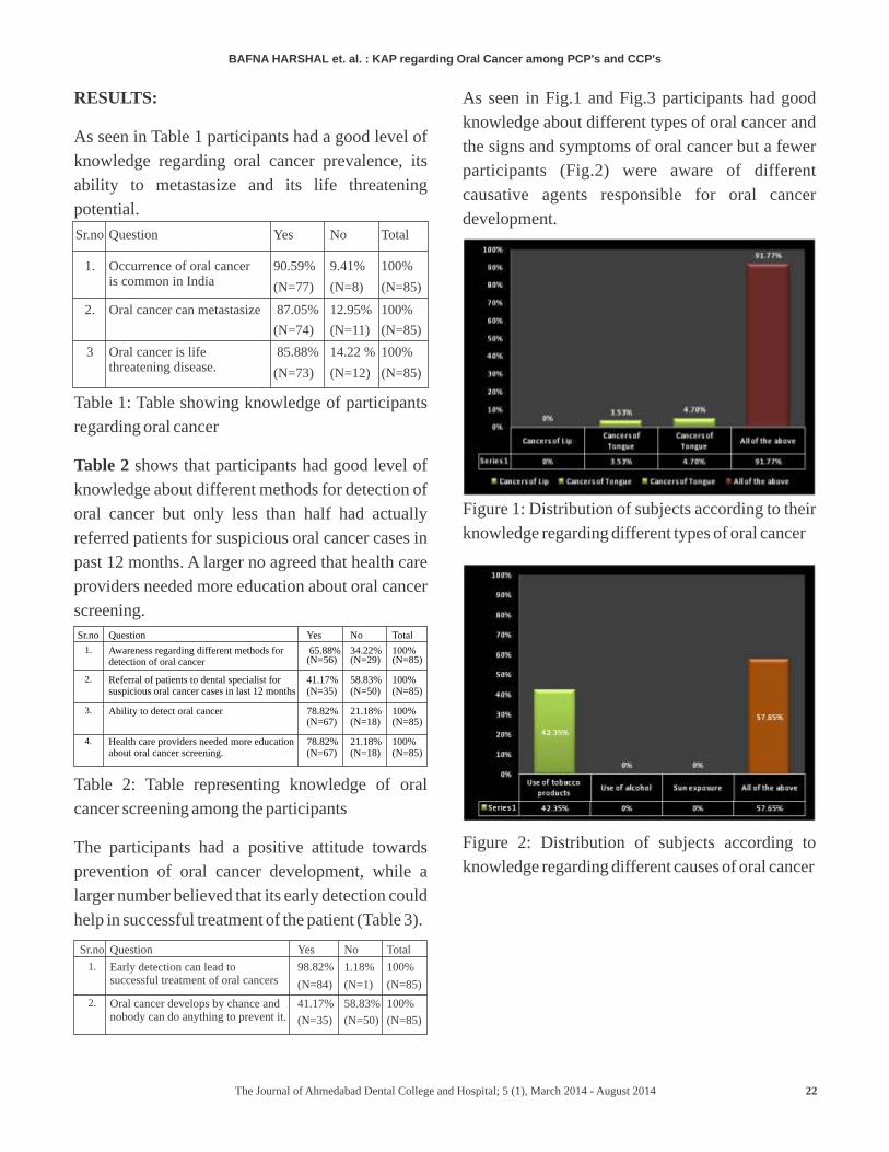
As seen in Fig.1 and Fig.3 participants had good
knowledge about different types of oral cancer and
the signs and symptoms of oral cancer but a fewer
participants (Fig.2) were aware of different
causative agents responsible for oral cancer
development.
Figure 1: Distribution of subjects according to their
knowledge regarding different types of oral cancer
Figure 2: Distribution of subjects according to
knowledge regarding different causes of oral cancer
Sr.no Question Yes No Total
1.
Occurrence of oral cancer is common in India
90.59%
(N=77)
9.41%
(N=8)
100%
(N=85)
2.
Oral cancer can metastasize
87.05%
(N=74)
12.95%
(N=11)
100%
(N=85)
3 Oral cancer is life threatening disease.
85.88%
(N=73)
14.22 %
(N=12)
100%
(N=85)
Sr.no Question Yes No Total
1. Awareness regarding different methods for detection of oral cancer
65.88%
(N=56)
34.22% (N=29)
100% (N=85)
2. Referral of patients to dental specialist for suspicious oral cancer cases in last 12 months
41.17% (N=35)
58.83% (N=50)
100% (N=85)
3. Ability to detect oral cancer 78.82% (N=67)
21.18% (N=18)
100% (N=85)
4. Health care providers needed more education about oral cancer screening.
78.82% (N=67)
21.18%(N=18)
100% (N=85)
Sr.no Question Yes No Total
1. Early detection can lead to successful treatment of oral cancers
98.82%
(N=84)
1.18%
(N=1)
100%
(N=85)
2.
Oral cancer develops by chance and nobody can do anything to prevent it.
41.17%
(N=35)
58.83%
(N=50)
100%
(N=85)
BAFNA HARSHAL KAP regarding Oral Cancer among PCP's and CCP's et. al. :
22The Journal of Ahmedabad Dental College and Hospital; 5 (1), March 2014 - August 2014
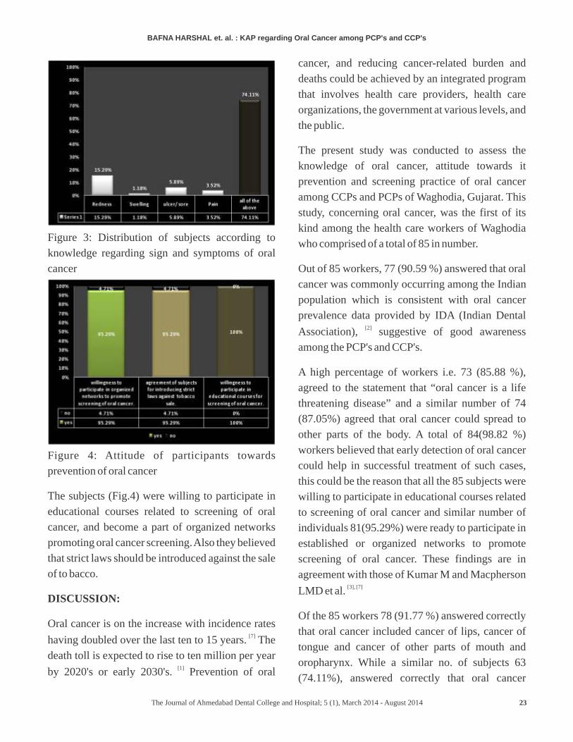
Figure 3: Distribution of subjects according to
knowledge regarding sign and symptoms of oral
cancer
Figure 4: Attitude of participants towards
prevention of oral cancer
The subjects (Fig.4) were willing to participate in
educational courses related to screening of oral
cancer, and become a part of organized networks
promoting oral cancer screening. Also they believed
that strict laws should be introduced against the sale
of to bacco.
DISCUSSION:
Oral cancer is on the increase with incidence rates [7]having doubled over the last ten to 15 years. The
death toll is expected to rise to ten million per year [1]
by 2020's or early 2030's. Prevention of oral
cancer, and reducing cancer-related burden and
deaths could be achieved by an integrated program
that involves health care providers, health care
organizations, the government at various levels, and
the public.
The present study was conducted to assess the
knowledge of oral cancer, attitude towards it
prevention and screening practice of oral cancer
among CCPs and PCPs of Waghodia, Gujarat. This
study, concerning oral cancer, was the first of its
kind among the health care workers of Waghodia
who comprised of a total of 85 in number.
Out of 85 workers, 77 (90.59 %) answered that oral
cancer was commonly occurring among the Indian
population which is consistent with oral cancer
prevalence data provided by IDA (Indian Dental [2]
Association), suggestive of good awareness
among the PCP's and CCP's.
A high percentage of workers i.e. 73 (85.88 %),
agreed to the statement that “oral cancer is a life
threatening disease” and a similar number of 74
(87.05%) agreed that oral cancer could spread to
other parts of the body. A total of 84(98.82 %)
workers believed that early detection of oral cancer
could help in successful treatment of such cases,
this could be the reason that all the 85 subjects were
willing to participate in educational courses related
to screening of oral cancer and similar number of
individuals 81(95.29%) were ready to participate in
established or organized networks to promote
screening of oral cancer. These findings are in
agreement with those of Kumar M and Macpherson [3], [7]
LMD et al.
Of the 85 workers 78 (91.77 %) answered correctly
that oral cancer included cancer of lips, cancer of
tongue and cancer of other parts of mouth and
oropharynx. While a similar no. of subjects 63
(74.11%), answered correctly that oral cancer
BAFNA HARSHAL KAP regarding Oral Cancer among PCP's and CCP's et. al. :
23The Journal of Ahmedabad Dental College and Hospital; 5 (1), March 2014 - August 2014