Embed Size (px)
Citation preview
Data Acquisition Working Group
Tom ChenevertPaul Kinahan
Yantian Zhang, NCI liaison
QIN annual meeting April 3-4, 2014
Charter
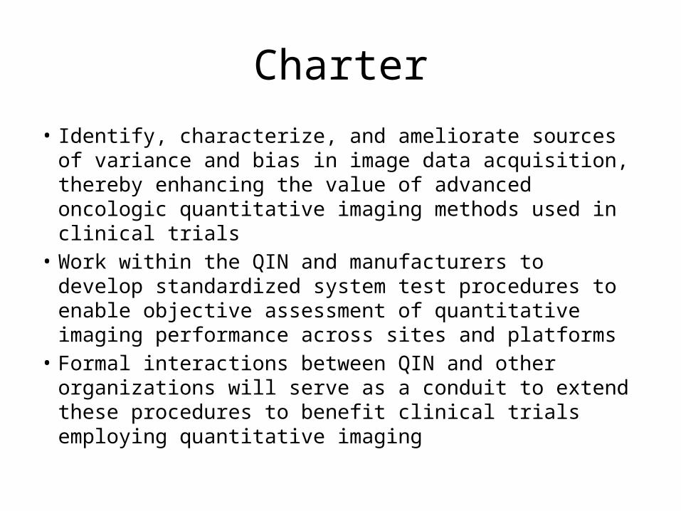
• Identify, characterize, and ameliorate sources of variance and bias in image data acquisition, thereby enhancing the value of advanced oncologic quantitative imaging methods used in clinical trials
• Work within the QIN and manufacturers to develop standardized system test procedures to enable objective assessment of quantitative imaging performance across sites and platforms
• Formal interactions between QIN and other organizations will serve as a conduit to extend these procedures to benefit clinical trials employing quantitative imaging
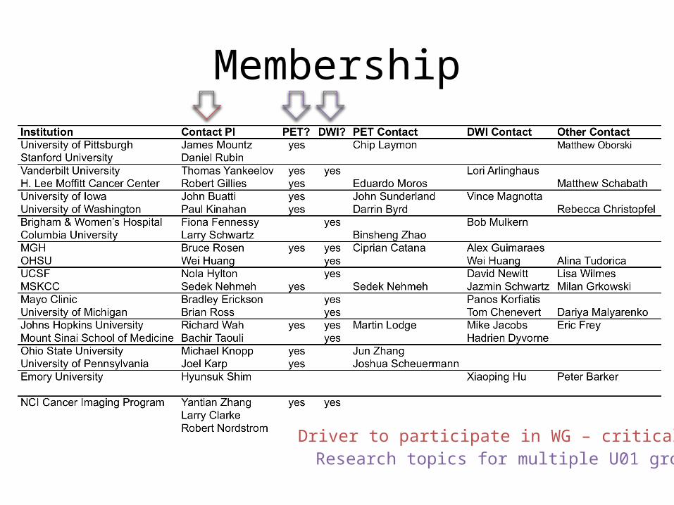
Membership
Driver to participate in WG – critical stepResearch topics for multiple U01 groups
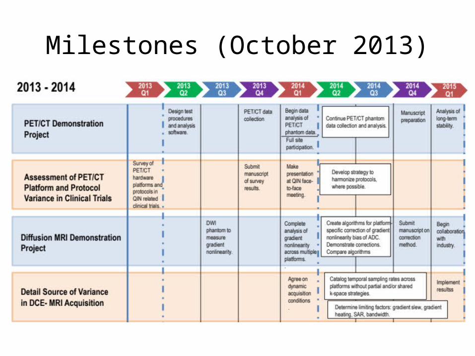
Milestones (October 2013)

Main goals
• PET/CT Demonstration Project– Multicenter data acquisition /processing survey– Longitudinal multicenter scanner calibration and
stability• MRI-DWI Demonstration Project
– Gradient Nonlinearity Bias in Multi-center Trials

Incoming co-chairs
• John Sunderland, PhD - University of Iowa• Bachir Taouli, MD - Mt Sinai School of
Medicine

PET/CT Demonstration Project
• Data acquisition /processing survey• Longitudinal survey of multicenter scanner
calibration and stability
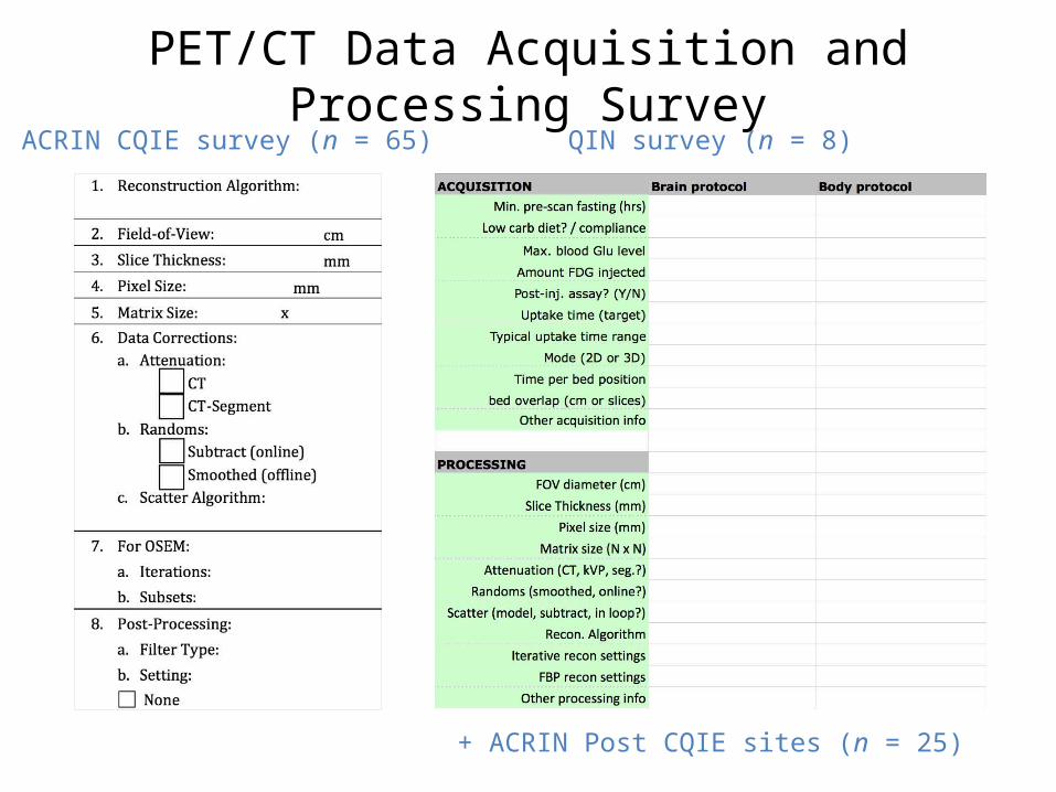
PET/CT Data Acquisition and Processing SurveyACRIN CQIE survey (n = 65) QIN survey (n = 8)
+ ACRIN Post CQIE sites (n = 25)

Longitudinal survey of multicenter scanner calibration and stability
Paul Kinahan, Darrin Byrd, Rebecca Christopfel, John Sunderland, Martin Lodge, Chip Laymon, Jun Zhang, Joshua Scheurmann, Cipriana Catana, Eduardo Moros, Sedek Nehmeh
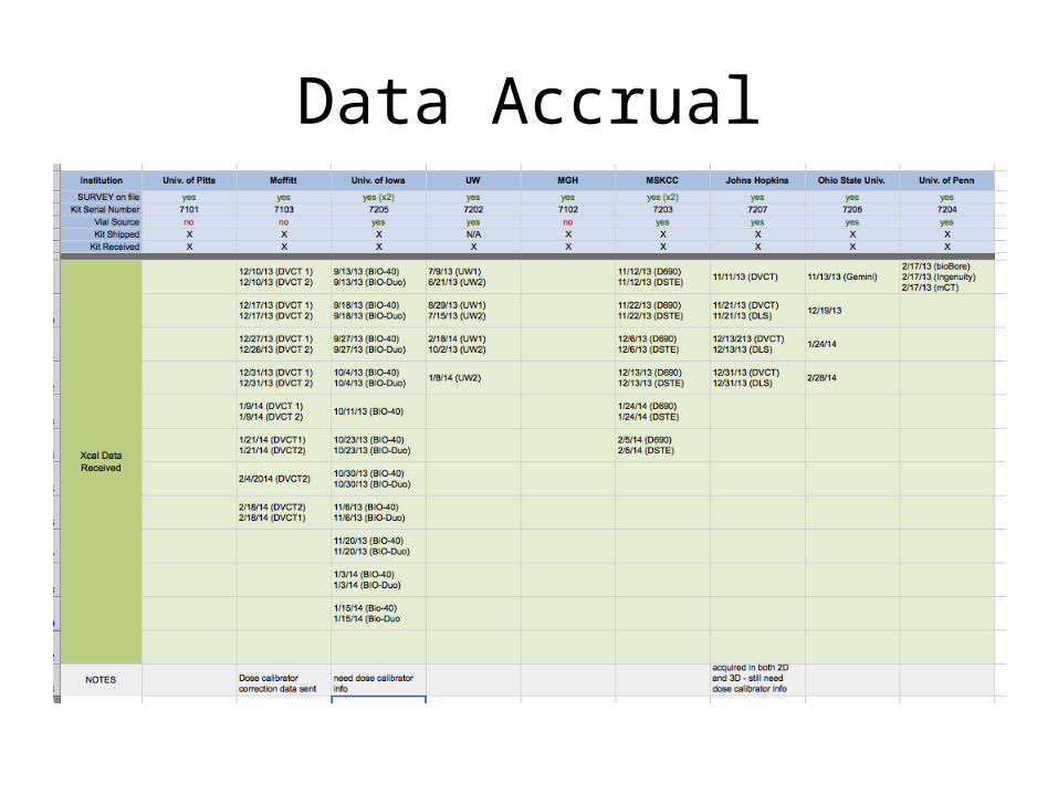
Data Accrual

Early results
QIN DAWG Demonstration Project: Gradient Nonlinearity Bias in Multi-center Trials
Dariya Malyarenko1, David Newitt2, Alina Tudorica3, Robert Mulkern4, Karl G. Helmer5, Michael A. Jacobs6, Lori Arlinghaus7, Thomas Yankeelov7, Fiona Fennessy4, Wei Huang3, Nola Hylton2, and Thomas L. Chenevert1
1University of Michigan Radiology, 2University of California San Francisco Radiology and Biomedical Imaging, 3Oregon Health and Science University, 4Dana Faber Harvard Cancer Center, 5Massachusetts General Hospital, 6John Hopkins University School of Medicine, 7Vanderbilt University Institute of Imaging Science
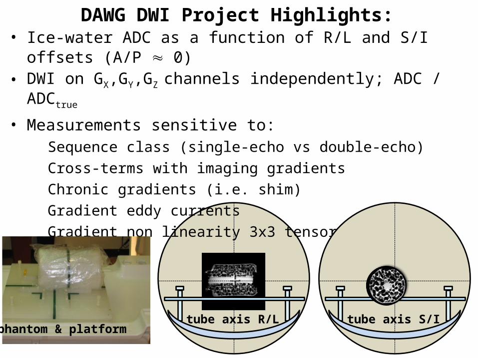
DAWG DWI Project Highlights:• Ice-water ADC as a function of R/L and S/I offsets (A/P 0)• DWI on GX,GY,GZ channels independently; ADC / ADCtrue
• Measurements sensitive to: Sequence class (single-echo vs double-echo) Cross-terms with imaging gradients Chronic gradients (i.e. shim) Gradient eddy currents Gradient non linearity 3x3 tensor, L
tube axis R/Lphantom & platform
tube axis S/I

Method:
Isotropic ADC phantom: DWIx,y,z acquisition
kth LAB-DWI ADC describes of
kth gradient-channel (Gk) S
I offs
ets
(+/-1
50m
m)
ADC measured from ROId=10mm
RL offsets (+/-150mm)
ADC bias for individual gradient-channels
DWI axes = (GX, GY, GZ)
ADCice-water = 1.1.10-3mm2/s
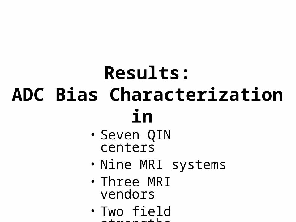
Results:ADC Bias Characterization in
• Seven QIN centers• Nine MRI systems• Three MRI vendors• Two field strengths
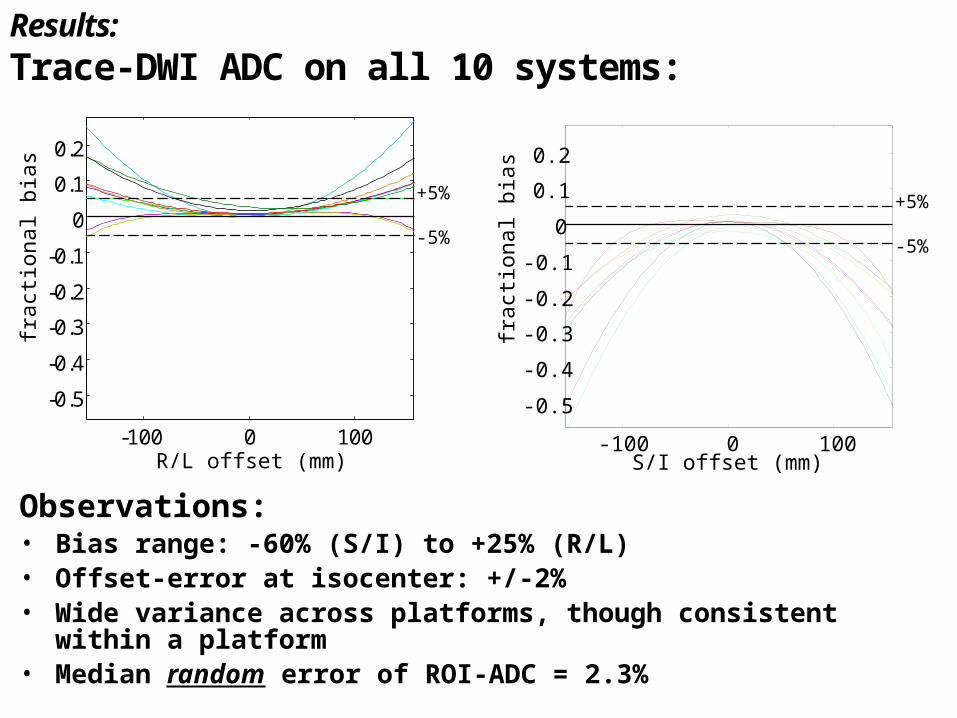
Results: Trace-DWI ADC on all 10 systems:
-100 0 100
-0.5
-0.4
-0.3
-0.2
-0.1
0
0.1
0.2
+5%
-5%
R/L offset (mm)
frac
tiona
l bia
s
S/I offset (mm)
frac
tiona
l bia
s
+5%
-5%
-100 0 100
-0.5
-0.4
-0.3
-0.2
-0.1
0
0.1
0.2
Observations:• Bias range: -60% (S/I) to +25% (R/L)• Offset-error at isocenter: +/-2%• Wide variance across platforms, though consistent within a platform • Median random error of ROI-ADC = 2.3%

Analysis of Results:
Gradient-bias contributors: Manifestation on ADC:
bkgnd gradients i.e. shim
eddy currents
imaginggradients
gradient (L)nonlinearity
nonuniform ADC
imaging channel ADC “shift”
uncertainty and asymmetry
bias asymmetry at +/- offset
co-reg. to b=0DWI-GX
AP “
shift
”im
age
shift
sc
ale
& s
hear
-5%
+5%
ADC-GX
SI, mm
co-reg.
SI, mm
-5%
+5%
GX-channel(SAG)
-5%
+5%
RL, mm
GZ-channel(AX)
RL, mm
GX GY -5%
+5%
SI, mm
AP/R
L
SI
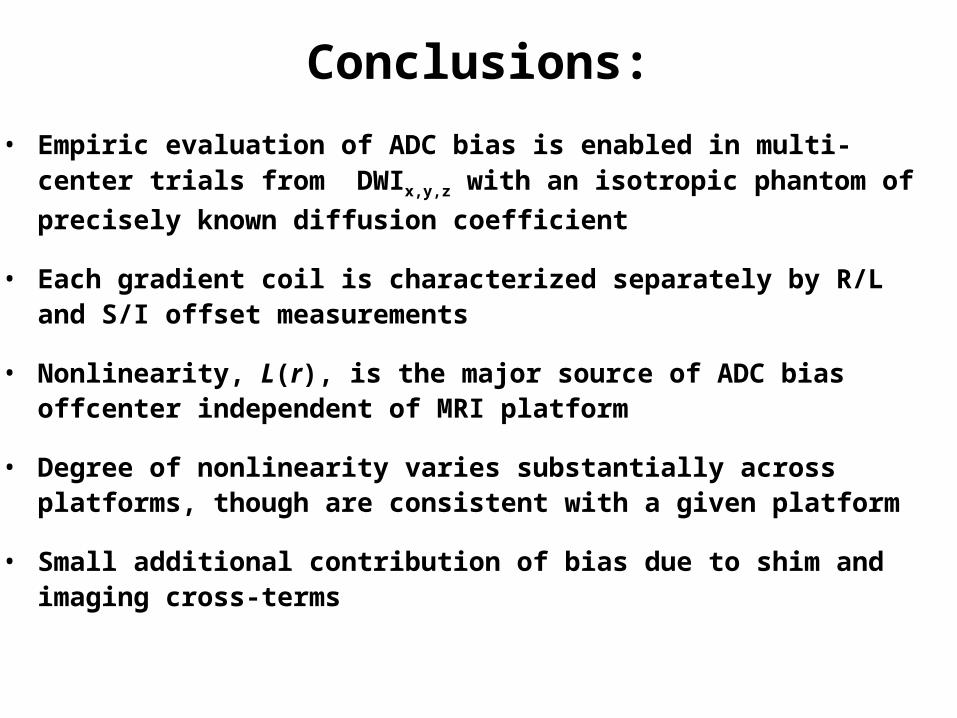
Conclusions:
• Empiric evaluation of ADC bias is enabled in multi-center trials from DWIx,y,z with an isotropic phantom of precisely known diffusion coefficient
• Each gradient coil is characterized separately by R/L and S/I offset measurements
• Nonlinearity, L(r), is the major source of ADC bias offcenter independent of MRI platform
• Degree of nonlinearity varies substantially across platforms, though are consistent with a given platform
• Small additional contribution of bias due to shim and imaging cross-terms


![deteRMinACiÓn deL HinCHAMiento LineAL en CoRteS de ... · En otro artículo publicado por Chenevert en 1970 “Shale control with balanced-activity oil-continuos muds"[8], el autor](https://img.pdfslide.net/doc/110x75/5e1398fd96243f60b966595e/determinacin-del-hinchamiento-lineal-en-cortes-de-en-otro-artculo-publicado.jpg)