Embed Size (px)
Citation preview

STS General Thoracic Data Specifications v5.21.1
P a g e 1 | 207
Data Collection Variables Updated 9/30/2021
Important Information for ALL SITES!
General Thoracic Surgery Database Homepage
STS National Database Webinars
Data Manager Education
Data Collection Resources (version specific abstraction documents)
Ask an Abstraction Question
STS National Database News - Publication for STS Data Managers
Advances in Quality & Outcomes: A Data Managers Meeting
Public Reporting
Contact Information ------------------------------------------------------------------------------------------------------------------------------------------------------ Introduction --------------------------------------------------------------------------------------------------------------------- ---------------------------------------------
This manual is intended to clarify field definition and intent. This document contains the most up to date instructions for v. 5.21.1 data abstraction. Do not refer to old manuals or other data definitions. Please review this document prior to submitting clinical questions. FAQs will be added to the document in red to provide additional examples and clarification. Comments in green are provided for clarification within prior updates. Please do not print this document since it will change frequently. Using the web version will ensure that you have the most up to date information. Occasionally there may be changes or important information that will be highlighted here and will be also included in STS Database Newsletters. Unless otherwise indicated, Data Managers are not required to go back in past records to update them based on new updated FAQ’s and Updates to the Training Manual. Data Mangers should move forward with new updated FAQ’s and Updates to the Training Manual as they abstract records. In the event of an audit, records will be audited based on the Training Manual at the time of the OR date. Use the Ctrl + F function to search for a number or term of interest. Bookmarks will be added each month with updates.
------------------------------------------------------------------------------------------------------------------------------------------------------
General Information: Procedure Inclusion – The STS General Thoracic Registry version 5.21.1 requires submission of all lung resections for primary lung cancer and all esophageal resections for primary esophageal cancer. Lung and esophageal resections for primary cancer are analyzed including national outcomes for benchmarking, risk adjusted outcomes, and star rating. Participants in the General Thoracic Registry may choose to submit Thymus/Mediastinal Mass

STS General Thoracic Data Specifications v5.21.1
P a g e 2 | 207
Resection, Tracheal Resection, and Hiatal Hernia/GERD cases. These case types are optional modules for submission to the registry and benchmark data will be available in the national report if submitted. All other case types are not required for collection or submission. They will not be available in the national report if submitted. Case Examples – Case #1: Patient has nodule on CT scan and is also worked up with a PET scan. Surgeon thinks it could be cancer, so lung resection is completed, and path comes back as lung cancer. This case is required for the registry. Enter this case as a lobectomy for primary lung cancer including the clinical and path staging. Case #2: Same as above but path comes back as hamartoma. This case is not required for submission to the registry because resection ultimately was not for primary lung cancer. This case will not be analyzed if submitted. Case #3 Patient worked up for presumed lung cancer and taken to OR for planned wedge resection followed by lobectomy if frozen section shows cancer. Frozen section comes back as granuloma, so surgery ends at wedge resection. This case is not required for submission because resection ultimately was not for lung cancer. This case would not be analyzed if submitted. Case #4 Patient presents to hospital with pneumonia. CT shows necrotic fluid suspicious for lung abscess in LLL. Patient taken to OR to drain effusion and wedge resection of abscess. Completion lobectomy was then undertaken because the lung was not salvageable. There was never suspected cancer. This case is not required for submission because resection ultimately was not for lung cancer. This case would not be analyzed if submitted. Case #5 Patient with history of breast cancer and previous mets to the lung removed via wedge and presents now with a new nodule. Surgeon assumes it’s another met. Taken to OR for therapeutic wedge resection. Final pathology returns as early-stage primary lung cancer. This case is required for the registry. Enter this case as a wedge resection for primary lung cancer including the clinical and pathological staging. This case will be analyzed. Case #6 Patient presents with empyema and undergoes decortication. This case is not required for submission to the registry because it is not a lung resection for primary lung cancer. This case will not be analyzed if submitted. Case #7 Patient presents with mediastinal mass. Ultimately undergoes thymectomy. Pathology returns with stage II thymoma. This case is an optional submission using the Thymus/Mediastinal Mass module. This case will be analyzed if submitted. Data Collection Forms – The General Thoracic Surgery Database requires a separate data collection form for every OR/procedural area visit for major general thoracic procedure(s). If there are additional questions about when a second DCF should be filled out for a new admission requiring an GTSD procedure, then send in a FAQ. Training Manual Updates – Training Manual updates will occur monthly and will be posted on the STS Website. When abstracting data, use the Training Manual updates at the time of the OR date. For example, if the OR date is November 15th, the abstractor should use the updates available in the November Training Manual. Importing Data from Other Data Sources - Although the data many participants are entering into their STS certified software may be gathered from another electronic data system at their site (such as an EMR), it is strictly

STS General Thoracic Data Specifications v5.21.1
P a g e 3 | 207
against STS policy for vendors to provide the users with the means to import this data automatically. It is not practical for the STS to certify the mapping of data from each site’s EMR to the STS data specifications, which would be required to ensure the integrity of the overall STS database. There is only one exception to this policy:
• Demographic data fields can be imported from an Admission/Discharge/Transfer (ADT) system. See Software Specifications for detailed information.
No and Unknown Questions - When a history and physical or a consultation exists in the medical record and the values are not specifically addressed in the documentation, code no. For example, if there is no mention of a history of cancer, then code No to history of cancer. Unknown should be coded in the circumstance where no clinical documentation exists, and the patient cannot give history and in certain situations for example when you know the patient has a history of cancer, but you do not know if it is within 5 years. These certain circumstances are field specific and will be addressed in the TM. If the patient is alone, intubated, and unable to give history; use the information from the patient’s family if they become available. No and Not Documented Questions - When a history and physical or a consultation exists in the medical record and the values are not specifically addressed in the documentation, code no. For example, if there is no documentation of 10% of body weight in the last three months, then code No. Not Documented should be coded in the circumstance where you have clinical documentation such as serial weights, however weight loss is not addressed in the H&P.
Text Fields – For fields where there is no option to choose yes/no/not documented/unknown, leave the field blank if you do not have an answer. For example, for Total number of Lymph Nodes sampled/harvested, if you do not have the total number, leave the field blank. Values Outside an Acceptable Range - When entering values into the DCF, if the values are outside of the maximum or minimum allowable range (specified as the low or high values in the Data Specifications) for the field an illegal value message will appear in the vendor tool. In this situation, enter the highest / lowest allowable value for that field. For example, the patient is 111 years old. The maximum allowable value for age is 1 to 110 per the Data Specifications Manual. In this situation code 110. Before using the highest or lowest allowable values, please verify the unit for the value is correct.

STS General Thoracic Data Specifications v5.21.1
P a g e 4 | 207
30 Day Mortality / 30 Day Readmission – Data Managers need to keep some type of log to include verification source, date assessed, status of mortality, and readmit. For example, this can be done on an excel spreadsheet or a document attached to DCF such as John Doe - Surgery 1/1/20 - Discharge 1/5/20 - checked with MD office and checked Medical Record on 2/6/20 and alive with no infection or readmit.
Case Inclusion for Star Ratings:

STS General Thoracic Data Specifications v5.21.1
P a g e 5 | 207
----------------------------------------------------------------------------------------------------------------------------------------------------------------- Administrative -------------------------------------------------------------------------------------------------------------------------------------- SeqNo: 10 Long Name: Operations Table Record Identifier Short Name: RecordID Format: Text Definition: An arbitrary, unique value generated by the software that permanently identifies each record in the participant's database (note that unlike the PatID value, this does not identify the individual patient). The value of the identifier is a combination of a code assigned to the software developer by the STS, and a value generated by the software to create a unique value. Once assigned to a record, this value can never be changed or reused. The data warehouse will use this value to communicate issues about individual records with the participant. It may also be used by the data warehouse to link this record to other clinical data. Intent/Clarification: SeqNo: 20 Long Name: Procedures Table Record Identifier Short Name: RecordID Format: Text

STS General Thoracic Data Specifications v5.21.1
P a g e 6 | 207
Definition: This field is the foreign key that links this record with the associated records in the "Operations" table. Intent/Clarification: SeqNo: 30 Long Name: Software Vendor's Identification Short Name: VendorID Format: Text Definition: Software vendor's identification assigned by the STS. Intent/Clarification: Name must match what is listed as the Active vendor for your Participant ID in the database. Any mismatch will cause your data file submission not to process. SeqNo: 50 Long Name: Version of STS Data Specification Short Name: DataVrsn Format: Text Definition: Version number of the STS Data Specifications/Dictionary, to which the record conforms. The value will identify which fields should have data, and what are the valid data values for those fields. It must be the version implemented in the software at the time the record was created. The value must be entered into the record automatically by the software. Intent/Clarification: Data version must be appropriate for the procedure date listed in the record. Valid date ranges can be found in the current Software Specifications. Any mismatch will cause your data file submission not to process. ----------------------------------------------------------------------------------------------------------------------------------------------------------------- Demographics -------------------------------------------------------------------------------------------------------------------------------------- SeqNo: 60 Text Long Name: Participant ID Short Name: ParticID Format: Definition: Participant ID is a unique number assigned to each database Participant by the STS. A database Participant is defined as one entity that signs a Participation Agreement with the STS, submits one data file to the harvest, and gets back one report on their data. The ParticipantID must be entered into each record. Intent/Clarification: The participant ID must be entered into each record. Each participant's data if submitted to harvest must be in one data file. If one participant keeps their data in more than one file (e.g., at two sites), then the participant must combine them back into one file for harvest submission.

STS General Thoracic Data Specifications v5.21.1
P a g e 7 | 207
If two or more participants share a single purchased software, and enter cases into one database, then the data must be extracted into two different files, one for each participant ID, with each record having the correct participant ID number. SeqNo: 70 Long Name: Demographics Table Data Version Short Name: DemogDataVrsn Format: Text Definition: Version number of the STS Data Specifications/Dictionary, to which the Demographics record conforms. The value will identify which fields should have data, and what are the valid data for those fields. It must be the version implemented in the software at the time the record was created. The value must be entered into the record automatically by the software. Note that the data version of the demographics record does not necessarily need to match the data version of all the associated operation records for that patient. This is because new data versions might be implemented in the software and used for the creation of operation records after a demographics record has been created for a patient. Intent/Clarification: SeqNo: 80 Long Name: Demographics Table Patient Identifier Short Name: PatID Format: Text Definition: An arbitrary value that uniquely and permanently identifies each patient. The value of the identifier is a combination of a code assigned to the software developer by the STS, and a value generated by the software to create a unique value. The value in this field cannot be a value that would identify the patient outside of the database (Such as Medical Record Number or Social Security Number). Once a value has been assigned to a patient, it can never be changed or reused. This field is the primary key that links this record with the associated records in the "Operations" table. Intent/Clarification: SeqNo: 90 Long Name: Operations Table Patient Identifier Short Name: PatID Format: Text Definition: The foreign key that links this record with the associated record in the "Demographics" table. Intent/Clarification: SeqNo: 100 Long Name: Medical Record # Short Name: MedRecN Format: Text Definition: Indicate the patient's medical record number at the hospital where surgery occurred. This field should be collected in compliance with state/local privacy laws.

STS General Thoracic Data Specifications v5.21.1
P a g e 8 | 207
Intent/Clarification: This field is not required for record inclusion. SeqNo: 110 Long Name: Patient's First Name Short Name: PatFName Format: Text Definition: Indicate the patient's first name documented in the medical record. This field should be collected in compliance with state/local privacy laws. Intent/Clarification: This field is not required for record inclusion. SeqNo: 120 Long Name: Patient Middle Name Short Name: PatMName Format: Text Definition: Indicate the patient's middle name as documented in the medical record. Leave "blank" if no middle name. This field should be collected in compliance with state/local privacy laws. Intent/Clarification: This field is not required for record inclusion. SeqNo: 130 Long Name: Patient's Last Name Short Name: PatLName Format: Text Definition: Indicate the patient's last name documented in the medical record. This field should be collected in compliance with state/local privacy laws. Intent/Clarification: This field is not required for record inclusion. SeqNo: 140 Long Name: Social Security Number Known Short Name: SSNKnown Format: Text (categorical values specified by STS) Definition: Indicate if the social security number or national identifier is known. Harvest Codes: Code: Value: 1 Yes 2 No 3 Patient Refused Intent/Clarification: ‘Patient Refused’ means the patient did not wish to share the information. ‘No’ means the information was not available, or the participant site did not wish to provide.

STS General Thoracic Data Specifications v5.21.1
P a g e 9 | 207
Do not use the Medicare number as the Social Security number. If you do not have the entire social security number, then code Seq 140 as No. SeqNo: 150 Long Name: Social Security Number Short Name: SSN Format: Text Definition: Indicate the patient’s Social Security Number (SSN). Although this is the Social Security Number in the USA, other countries may have a different National Patient Identifier Number. For example, in Canada, this would be the Social Insurance Number. This field should be collected in compliance with state/local privacy laws. ParentLongName: Social Security Number Known ParentShortName: SSNKnown ParentValue: 1 ParentHarvestCodes: = "Yes" Intent/Clarification: If SSNKnown is answered ‘Yes,’ then a response is expected in this field. Please provide the patient’s entire Social Security Number or, for sites not located in the United States, the corresponding National Patient Identifier. SeqNo: 160 Long Name: Permanent Street Address Short Name: PatAddr Format: Text Definition: Indicate the patients permanent street address at the time of admission. Intent/Clarification: If the medical record does not document if the address is permanent or not, then use the street address at which the patient resides at time of admission. If patient is homeless, enter "Homeless". A post office box may be used if no other address is available. If the patient has a northern and a southern address, choose the address where they spend most of their time. The intent is to identify patients who travel outside their local area for treatment. CMS is tracking disparities in health care delivery and looking at underserved areas. This also assists with long term follow up locally. SeqNo: 170 Long Name: Patients Permanent City Short Name: PatCity Format: Text Definition: Indicate the patients permanent city. Intent/Clarification: If the medical record does not document whether the city is permanent or not, then use the city in which the patient resides at time of admission. SeqNo: 180 Long Name: Patients Permanent Region Short Name: PatRegion Format: Text Definition: Indicate the patient’s permanent region (i.e., state or province) in which the patient resides at the time of admission.

STS General Thoracic Data Specifications v5.21.1
P a g e 10 | 207
Intent/Clarification: If the medical record does not document if the region is permanent or not, then use the region of the country (i.e., state or province) in which the patient resides at time of admission. SeqNo: 190 Long Name: Country Short Name: PatientCountry Format: Text (categorical values specified by STS) Definition: Indicate the patient country of residence at the time of admission. Harvest Codes: Code: Value: 237 United States of America 1 Afghanistan 11 Argentina 14 Australia 17 Bahamas 25 Bermuda 31 Brazil 40 Canada 46 China 53 Costa Rica 88 Greece 92 Guam 93 Guatemala 105 India 109 Ireland 111 Israel 112 Italy 113 Jamaica 114 Japan 116 Jordan 143 Mexico 166 State of Palestine 173 Peru 176 Poland 178 Puerto Rico 184 Russian Federation 196 Saudi Arabia 300 Scotland 201 Singapore 215 Switzerland 225 Trinidad and Tobago 227 Turkey 231 Uganda 233 United Arab Emirates 234 United Kingdom of Great Britain And Northern Ireland 235 United Republic of Tanzania 236 United States Minor Outlying Islands 238 United States Virgin Islands 242 Venezuela (Bolivarian Republic Of) 246 Yemen 2 Åland Island

STS General Thoracic Data Specifications v5.21.1
P a g e 11 | 207
999 Other Intent/Clarification: Harvest codes can change between versions. Please use the most current list of harvest codes for the version you are abstracting. The list above references harvest codes for v5.21 – OR dates starting July 1, 2021. SeqNo: 200 Long Name: Postal Code Short Name: PostalCode Format: Text Definition: Indicate the ZIP Code of the patient's residence. Outside the USA, this data may be known by other names such as Postal Code (needing 6 characters). Software should allow sites to collect at least up to 10 characters to allow for Zip+4 values. This field should be collected in compliance with state/local privacy laws. Intent/Clarification: Document the zip code of the patient permanent address. If the medical record does not document whether the zip code is permanent or not, then use the zip code in which the patient resides at time of admission. SeqNo: 210 Long Name: Patient Participating In STS-Related Clinical Trial Short Name: ClinTrial Format: Text (categorical values specified by STS) Definition: Indicate which, if any, STS-related clinical trial in which the patient is participating. The STS will assign a code to each clinical trial as they begin collecting data. Harvest Codes: Code: Value: 1 None 2 Trial 1 3 Trial 2 4 Trial 3 5 Trial 4 6 Trial 5 7 Trial 6 Intent/Clarification: A list of trials will be posted as they are started. Instructions will be provided for each trial SeqNo: 220 Long Name: Patient Participating In STS-Related Clinical Trial - Patient ID Short Name: ClinTrialPatID Format: Text Definition: Indicate the patient identifier used to identify the patient in the clinical trial. ParentLongName: Patient Participating In STS-Related Clinical Trial ParentShortName: ClinTrial ParentValue: <>1 And Is Not Missing ParentHarvestCodes: Is Not "None" And Is Not Missing Intent/Clarification: Instructions will be provided for each trial.
STS General Thoracic Data Specifications v5.21.1
P a g e 12 | 207
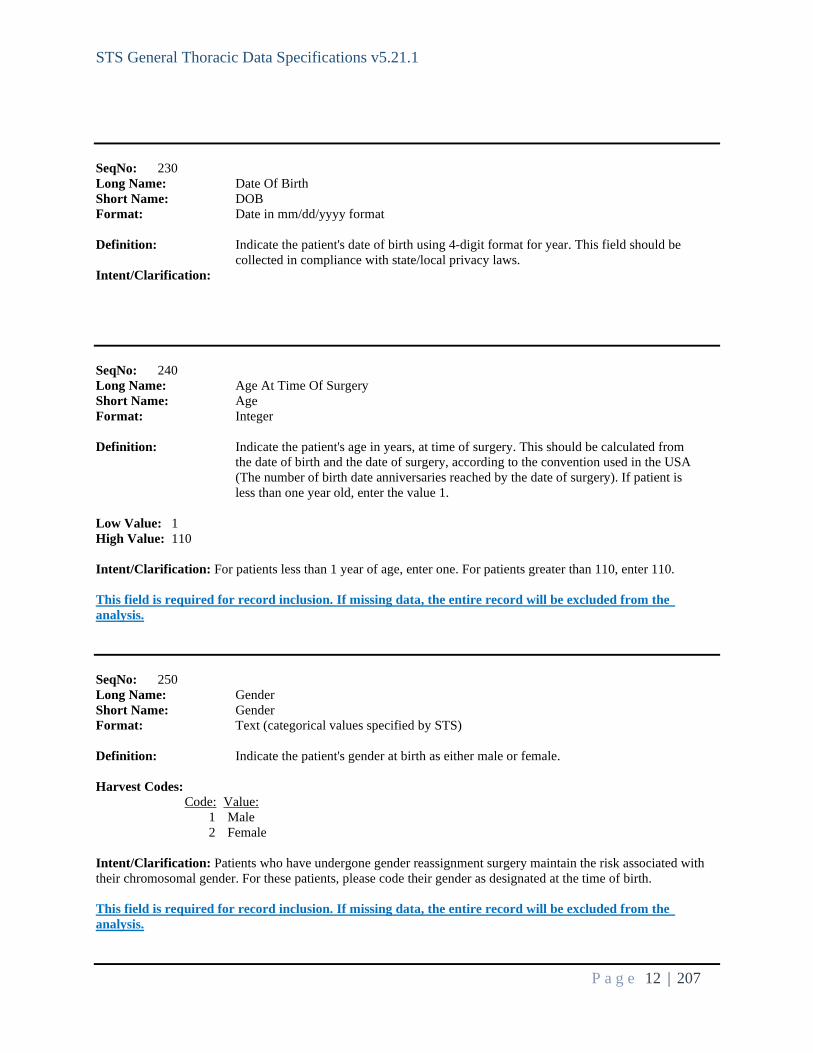
SeqNo: 230 Long Name: Date Of Birth Short Name: DOB Format: Date in mm/dd/yyyy format Definition: Indicate the patient's date of birth using 4-digit format for year. This field should be collected in compliance with state/local privacy laws. Intent/Clarification: SeqNo: 240 Long Name: Age At Time Of Surgery Short Name: Age Format: Integer Definition: Indicate the patient's age in years, at time of surgery. This should be calculated from the date of birth and the date of surgery, according to the convention used in the USA (The number of birth date anniversaries reached by the date of surgery). If patient is less than one year old, enter the value 1. Low Value: 1 High Value: 110 Intent/Clarification: For patients less than 1 year of age, enter one. For patients greater than 110, enter 110. This field is required for record inclusion. If missing data, the entire record will be excluded from the analysis. SeqNo: 250 Long Name: Gender Short Name: Gender Format: Text (categorical values specified by STS) Definition: Indicate the patient's gender at birth as either male or female. Harvest Codes: Code: Value: 1 Male 2 Female Intent/Clarification: Patients who have undergone gender reassignment surgery maintain the risk associated with their chromosomal gender. For these patients, please code their gender as designated at the time of birth. This field is required for record inclusion. If missing data, the entire record will be excluded from the analysis.

STS General Thoracic Data Specifications v5.21.1
P a g e 13 | 207
SeqNo: 260 Long Name: Race Documented Short Name: RaceDocumented Format: Text (categorical values specified by STS) Definition: Indicate whether race is documented. Harvest Codes: Code: Value: 1 Yes 2 No 3 Patient declined to disclose Intent/Clarification: Race should be self-reported by the patient/family. Do not assign race or make assumptions if race is not documented.
• Yes = race is documented • No = race is not documented • Patient Declined to Disclose = patient declined to provide race
It has been reported that some EHR’s report multi-race if the patient reports being of more than one race, instead of listing each race separately. For this scenario, code ‘no’ to race documented and work within your facility to accurately code race within your EHR’s. It is important to accurately capture race, as it is used in the risk modeling. This field is required for record inclusion. If missing data, the entire record will be excluded from the analysis. SeqNo: 270 Long Name: Race - Multi-Select Short Name: RaceMulti Format: Multi-Select Definition: Indicate the patient's race(s) selecting all that apply. ParentLongName: Race Documented ParentShortName: RaceDocumented ParentValue: 1 ParentHarvestCodes: = "Yes" Harvest Codes: Code: Value: 1 White/Caucasian 2 Black/African American 3 Asian 4 American Indian/Alaskan Native 5 Native Hawaiian/Pacific Islander 6 Other Intent/Clarification: The Census Bureau collects race data in accordance with guidelines provided by the U.S. Office of Management and Budget, these data are based on self-identification. The racial categories included in the census form generally reflect a social definition of race recognized in this country and are not an attempt to define race biologically, anthropologically, or genetically. In addition, it is recognized that categories of the race item include racial and national origin or socio-cultural groups. People may choose to report more than one race to

STS General Thoracic Data Specifications v5.21.1
P a g e 14 | 207
indicate their racial mixture, such as American Indian and White. People who identify their origin (ETHNICITY) as Hispanic, Latino or Spanish may be of any race. If a race is not specified for Hispanic/Latino patients, code race as ‘Pt declined to disclose/NA’ This field is required for record inclusion. If missing data, the entire record will be excluded from the analysis. General Information: Race Description White - "White" refers to a person having origins in any of the original peoples of Europe, the Middle East, or North Africa. It includes people who indicated their race(s) as "White" or reported entries such as Irish, German, Italian, Lebanese, Arab, Moroccan, or Caucasian. [The 2010 Census Redistricting Data (Public Law 94-171) Summary File] Black / African American - "Black or African American" refers to a person having origins in any of the Black racial groups of Africa. It includes people who indicated their race(s) as "Black, African Am., or Negro" or reported entries such as African American, Kenyan, Nigerian, or Haitian. This includes a person having origins in any of the black racial groups of Africa. Terms such as “Haitian” or “Negro” can be used in addition to “Black or African American.” [The 2010 Census Redistricting Data (Public Law 94-171) Summary File]. Definition source: Standards for Maintaining, Collecting and Presenting Federal Data on Race and Ethnicity: The minimum categories for data on race and ethnicity for Federal statistics, program administrative reporting and civil rights compliance reporting. Asian - "Asian" refers to a person having origins in any of the original peoples of the Far East, Southeast Asia, or the Indian subcontinent, including, for example, Cambodia, China, India, Japan, Korea, Malaysia, Pakistan, the Philippine Islands, Thailand, and Vietnam. It includes people who indicated their race(s) as "Asian" or reported entries such as "Asian Indian", "Chinese", "Filipino", "Korean", "Japanese", "Vietnamese", and "Other Asian" or provided other detailed Asian responses. [The 2010 Census Redistricting Data (Public Law 94-171) Summary File]. Definition source: Standards for Maintaining, Collecting and Presenting Federal Data on Race and Ethnicity: The minimum categories for data on race and ethnicity for Federal statistics, program administrative reporting and civil rights compliance reporting. American Indian / Alaskan - "American Indian or Alaska Native" refers to a person having origins in any of the original peoples of North and South America (including Central America) and who maintains tribal affiliation or community attachment. This category includes people who indicated their race(s) as "American Indian or Alaska Native" or reported their enrolled or principle tribe, such as Navajo, Blackfeet, Inupiat, Yup’ik, or Central American Indian groups or South American Indian groups. This includes all in North American native peoples such as American Indian/Alaskan Native, Inuit. [The 2010 Census Redistricting Data (Public Law 94-171) Summary File] Hawaiian / Pacific Islander - "Native Hawaiian or Other Pacific Islander" refers to a person having origins in any of the original peoples of Hawaii, Guam, Samoa, or other Pacific Islands. It includes people who indicated their race(s) as "Pacific Islander" or reported entries such as "Native Hawaiian", "Guamanian or Chamorro", "Samoan", and "Other Pacific Islander" or provided other detailed Pacific Islander responses. [The 2010 Census Redistricting Data (Public Law 94-171) Summary File]. Definition source: Standards for Maintaining, Collecting and Presenting Federal Data on Race and Ethnicity. The minimum categories for data on race and ethnicity for Federal statistics, program administrative reporting and civil rights compliance reporting. Other - "Some Other Race" includes all other responses not included in the White, Black or African American, American Indian or Alaska Native, Asian, and Native Hawaiian or Other Pacific Islander race categories described above. [The 2010 Census Redistricting Data (Public Law 94-171) Summary File] SeqNo: 340

STS General Thoracic Data Specifications v5.21.1
P a g e 15 | 207
Long Name: Hispanic Or Latino Ethnicity Short Name: Ethnicity Format: Text (categorical values specified by STS) Definition: Indicate if the patient is of Hispanic or Latino ethnicity as determined by the patient / family. Hispanic or Latino ethnicity includes patient report of Cuban, Mexican, Puerto Rican, South or Central American, or other Spanish culture or origin, regardless of race. Harvest Codes: Code: Value: 1 Yes 2 No 3 Not documented Intent/Clarification: People who identify their origin as Hispanic, Latino or Spanish may be of any race. Do not make assumptions about ethnicity if it is not documented in the medical record. ------------------------------------------------------------------------------------------------------------------------------------------------------------------------------------ Admission ------------------------------------------------------------------------------------------------------------------------------------------------------------------------------------ SeqNo: 350 Long Name: Admission Status Short Name: AdmissionStat Format: Text (categorical values specified by STS) Definition: Indicate whether the procedure was an Inpatient or Outpatient / Observation procedure. Harvest Codes: Code: Value: 1 Inpatient 2 Outpatient/Observation Intent/Clarification: This field is required for Record Inclusion. If missing data, the entire record will be excluded from the analysis. Outpatient/Observation should be selected if the operation was performed as an ambulatory procedure or if it included a period of overnight observation. For patients who enter the hospital as an outpatient or observation status and later change to inpatient status, the admit status should be captured as an inpatient. To further clarify, if at any time during the hospitalization the patient is considered inpatient status, then inpatient would be coded. This field is required for record inclusion. If missing data, the entire record will be excluded from the analysis. SeqNo: 360 Long Name: Admission Date Short Name: AdmitDt Format: Date in mm/dd/yyyy format Definition: Indicate the date of admission. For those patients who originally enter the hospital in an out-patient capacity, the admit date is the date the patient's status changes to in- patient.
STS General Thoracic Data Specifications v5.21.1
P a g e 16 | 207
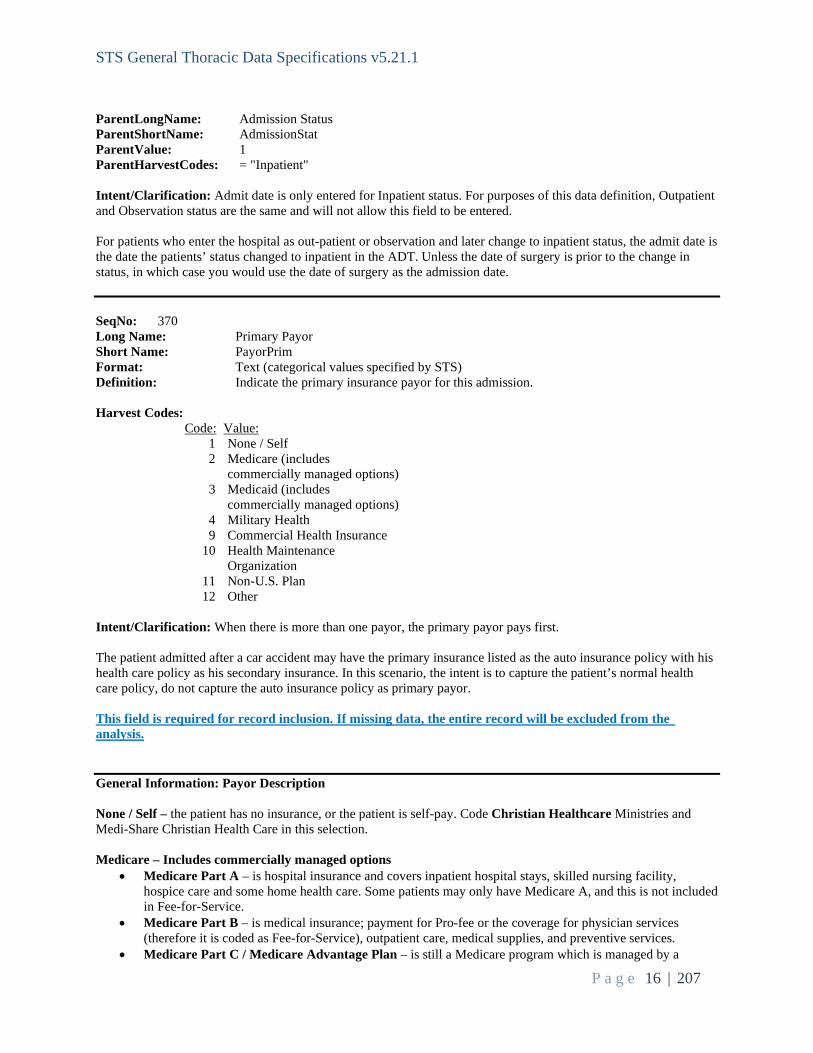
ParentLongName: Admission Status ParentShortName: AdmissionStat ParentValue: 1 ParentHarvestCodes: = "Inpatient" Intent/Clarification: Admit date is only entered for Inpatient status. For purposes of this data definition, Outpatient and Observation status are the same and will not allow this field to be entered. For patients who enter the hospital as out-patient or observation and later change to inpatient status, the admit date is the date the patients’ status changed to inpatient in the ADT. Unless the date of surgery is prior to the change in status, in which case you would use the date of surgery as the admission date. SeqNo: 370 Long Name: Primary Payor Short Name: PayorPrim Format: Text (categorical values specified by STS) Definition: Indicate the primary insurance payor for this admission. Harvest Codes: Code: Value: 1 None / Self 2 Medicare (includes commercially managed options) 3 Medicaid (includes commercially managed options) 4 Military Health 9 Commercial Health Insurance 10 Health Maintenance Organization 11 Non-U.S. Plan 12 Other Intent/Clarification: When there is more than one payor, the primary payor pays first. The patient admitted after a car accident may have the primary insurance listed as the auto insurance policy with his health care policy as his secondary insurance. In this scenario, the intent is to capture the patient’s normal health care policy, do not capture the auto insurance policy as primary payor. This field is required for record inclusion. If missing data, the entire record will be excluded from the analysis. General Information: Payor Description None / Self – the patient has no insurance, or the patient is self-pay. Code Christian Healthcare Ministries and Medi-Share Christian Health Care in this selection. Medicare – Includes commercially managed options
• Medicare Part A – is hospital insurance and covers inpatient hospital stays, skilled nursing facility, hospice care and some home health care. Some patients may only have Medicare A, and this is not included in Fee-for-Service.
• Medicare Part B – is medical insurance; payment for Pro-fee or the coverage for physician services (therefore it is coded as Fee-for-Service), outpatient care, medical supplies, and preventive services.
• Medicare Part C / Medicare Advantage Plan – is still a Medicare program which is managed by a

STS General Thoracic Data Specifications v5.21.1
P a g e 17 | 207
commercial insurance company. It is not the same as supplemental insurance. Medicare Advantage Plan covers most Medicare benefits and usually require patients to see specific providers in their network. All Medicare Advantage/ Managed Care plans (i.e., Humana HMO Medicare) are captured in the payor category as Medicare only. • For example, if the patient has BlueCross Advantage, code as primary payor Medicare, there is no
secondary payor in this scenario. • Medicare Part D is prescription drug coverage. Medicare Part D is optional, and it’s available only through
private insurance companies that contract with Medicare (Medicare Advantage or Managed Care plans). Medicare Supplement plans are not part of Medicare – this is a separate private health insurance plan that is bought by the subscriber in addition to Medicare.
Click here for more information on Medicare Plans. Medicaid - Medicaid in the United States is a federal and state program that helps with medical costs for some people with limited income and resources. Medicaid also offers benefits not normally covered by Medicare, including nursing home care and personal care services. All Medicaid Commercial / Managed Care plans (i.e., Humana Medicare, Star Molina Medicaid) are captured in the payor category as Medicaid only.
Commercial Health Insurance - Commercial health insurance is health insurance provided and administered by non-governmental entities. It covers medical expenses and disability income for the insured. Commercial insurance includes Medicare Supplement plans such as Medigap or AARP etc. It is a private insurance policy that can help pay for some of the health care cost Medicare doesn’t cover, such as co-payments, coinsurance, and deductibles. This is not part of Medicare – this is a separate private health insurance plan. Point-of-service plan (POS) and Preferred Provider Organization (PPO) plans not associated with Medicare Advantage plans will be captured here. Health Maintenance Organization (HMO) - An HMO gives you access to certain doctors and hospitals within its network. A network is made up of providers that have agreed to lower their rates for plan members and meet quality standards. But unlike PPO plans, care under an HMO plan is covered only if you see a provider within that HMO’s network. There are few opportunities to see a non-network provider. There are also typically more restrictions for coverage than other plans, such as allowing only a certain number of visits, tests, or treatments. Military – US Military provides insurance. Typically reported as VA insurance or Tricare. Non-U.S. Plan – Insurance covered by a non-U.S. source. Other – All other insurance not listed in the above selections such as Indian Health Services, Correctional Facility, State Specific plans, other government insurance, charitable care, or foundation funding. SeqNo: 380 Long Name: Commercially Managed Medicare Plan - Primary Short Name: ComMngMedPlnPrim Format: Text (categorical values specified by STS) Definition: Indicate whether the patient's primary payor is a commercially managed Medicare plan. ParentLongName: Primary Payor ParentShortName: PayorPrim ParentValue: 2 ParentHarvestCodes: = "Medicare (includes commercially managed options)" Harvest Codes: Code: Value: 1 Yes 2 No

STS General Thoracic Data Specifications v5.21.1
P a g e 18 | 207
Intent/Clarification: Commercially managed Medicare plans are also referred to as Medicare Advantage plans. This is Medicare Part C. Medicare Part C (aka Advantage Plan) – is still a Medicare program which is managed by a commercial insurance company. It is not the same as supplemental insurance. Medicare Advantage Plan covers most Medicare benefits and usually require patients to see specific providers in their network. All Medicare Advantage/ Managed Care plans (i.e., Humana HMO Medicare) are captured in the payor category as Medicare only.
• For example, if the patient has BlueCross Advantage, code as primary payor Medicare, there is no secondary payor in this scenario, and code Commercially Managed Medicare Plan as ‘Yes’
Click here for more information on Medicare Plans. SeqNo: 390 Long Name: HICN / MBI Known - Primary Short Name: HICNMBIKnown Format: Text (categorical values specified by STS) Definition: Indicate whether patient's HICN or MBI is known for primary ParentLongName: Commercially Managed Medicare Plan - Primary ParentShortName: ComMngMedPlnPrim ParentValue: 2 ParentHarvestCodes: = "No" Harvest Codes: Code: Value: 1 Yes 2 No Intent/Clarification: HICN numbers are made up of a nine-byte social security number plus a one to two-character Beneficiary Identification Code. A HICN number is not the same as a member number and is only associated with traditional Medicare. With the risk of identity theft becoming more and more prevalent CMS launched the Social Security Number Removal Initiative (SSNRI) years ago to remove the social security number from Medicare beneficiary identifiers. Beginning in 2018 the Medicare HICN number will be replaced with a new identifier called a Medicare Beneficiary Identifier (MBI). The MBI numbers will be eleven bytes in length, randomly generated, and will derive no components from a beneficiary’s identification. Here is an example of an MBI number: 1EG4-TE5-MK73. Patients with Medicare Advantage will not have a HICN/MBI. SeqNo: 400 Long Name: HICN / MBI Number Primary Short Name: HICNMBI Format: Text Definition: Indicate the HICN or MBI number for primary coverage. ParentLongName: HICN / MBI Known - Primary ParentShortName: HICNMBIKnown ParentValue: 1 ParentHarvestCodes: = "Yes" Intent/Clarification: SeqNo: 410

STS General Thoracic Data Specifications v5.21.1
P a g e 19 | 207
Long Name: Primary Payor Medicare Fee For Service Short Name: PrimMCareFFS Format: Text (categorical values specified by STS) Definition: Indicate whether the patient is covered by Medicare Fee For Service (Part B). ParentLongName: Primary Payor ParentShortName: PayorPrim ParentValue: 2 ParentHarvestCodes: = "Medicare (includes commercially managed options)" Harvest Codes: Code: Value: 1 Yes 2 No Intent/Clarification: Medicare Part B – is payment for Professional-fee or the coverage for physician services therefore it is coded as Fee-for-Service (FFS). This field is for traditional Medicare plans that pay via FFS and is often referred to as Medicare Part B. Medicare Replacement (Medicare Advantage) and Managed Care plans that pay via PFFS (Private-Fee-for-Service) are not captured as Medicare FFS. SeqNo: 420 Long Name: Secondary (Supplemental) Payor Short Name: PayorSecond Format: Text (categorical values specified by STS) Definition: Indicate which if any secondary insurance payor was used for this admission. ParentLongName: Primary Payor ParentShortName: PayorPrim ParentValue: <>1 And Is Not Missing ParentHarvestCodes: Is Not "None / Self" And Is Not Missing Harvest Codes: Code: Value: 1 None / Self 2 Medicare (includes commercially managed options) 3 Medicaid (includes commercially managed options) 4 Military Health 9 Commercial Health Insurance 10 Health Maintenance Organization 11 Non-U.S. Plan 12 Other Intent/Clarification: When there is more than one payor, the secondary payor pays after the primary payor. SeqNo: 430 Long Name: Commercially Managed Medicare Plan Secondary Short Name: ComMngMedPlnSec Format: Text (categorical values specified by STS) Definition: Indicate whether the patient's secondary payor is a commercially managed Medicare

STS General Thoracic Data Specifications v5.21.1
P a g e 20 | 207
plan. ParentLongName: Secondary (Supplemental) Payor ParentShortName: PayorSecond ParentValue: 2 ParentHarvestCodes: = "Medicare (includes commercially managed options)" Harvest Codes: Code: Value: 1 Yes 2 No Intent/Clarification: Commercially managed Medicare plans are also referred to as Medicare Advantage plans. This is Medicare Part C. Medicare Part C (aka Advantage Plan) – is still a Medicare program which is managed by a commercial insurance company. It is not the same as supplemental insurance. Medicare Advantage Plan covers most Medicare benefits and usually require patients to see specific providers in their network. All Medicare Advantage/ Managed Care plans (i.e., Humana HMO Medicare) are captured in the payor category as Medicare only.
• For example, if the patient has BlueCross Advantage, code as primary payor Medicare, there is no secondary payor in this scenario, and code Commercially Managed Medicare Plan as ‘Yes’
Click here for more information on Medicare Plans. SeqNo: 440 Long Name: HICN / MBI Known - Secondary Short Name: HICNMBIKnownSec Format: Text (categorical values specified by STS) Definition: Indicate whether patient's HICN or MBI is known for secondary. ParentLongName: Commercially Managed Medicare Plan Secondary ParentShortName: ComMngMedPlnSec ParentValue: 2 ParentHarvestCodes: = "No" Harvest Codes: Code: Value: 1 Yes 2 No Intent/Clarification: HICN numbers are made up of a nine-byte social security number plus a one to two-character Beneficiary Identification Code. A HICN number is not the same as a member number and is only associated with traditional Medicare. With the risk of identity theft becoming more and more prevalent CMS launched the Social Security Number Removal Initiative (SSNRI) years ago to remove the social security number from Medicare beneficiary identifiers. Beginning in 2018 the Medicare HICN number will be replaced with a new identifier called a Medicare Beneficiary Identifier (MBI). The MBI numbers will be eleven bytes in length, randomly generated, and will derive no components from a beneficiary’s identification. Here is an example of an MBI number: 1EG4-TE5-MK73. SeqNo: 450 Long Name: HICN / MBI Number - Secondary Short Name: HICNMBINumberSec Format: Text Definition: Indicate patient's HICN or MBI number for secondary.

STS General Thoracic Data Specifications v5.21.1
P a g e 21 | 207
ParentLongName: HICN / MBI Known - Secondary ParentShortName: HICNMBIKnownSec ParentValue: 1 ParentHarvestCodes: = "Yes" Intent/Clarification: SeqNo: 460 Long Name: Secondary Payor Medicare Fee For Service Short Name: SecondMCareFFS Format: Text (categorical values specified by STS) Definition: Indicate whether the patient is covered by Medicare Fee For Service (Part B). ParentLongName: Secondary (Supplemental) Payor ParentShortName: PayorSecond ParentValue: 2 ParentHarvestCodes: = "Medicare (includes commercially managed options)" Harvest Codes: Code: Value: 1 Yes 2 No Intent/Clarification: Medicare Part B – is payment for Professional-fee or the coverage for physician services therefore it is coded as Fee-for-Service (FFS). This field is for traditional Medicare plans that pay via FFS and is often referred to as Medicare Part B. Medicare Replacement (Medicare Advantage) and Managed Care plans that pay via PFFS (Private-Fee-for-Service) are not captured as Medicare FFS. SeqNo: 470 Long Name: Surgeon's Name Short Name: Surgeon Format: Text (categorical values specified by User) Definition: Indicate the name of the surgeon responsible for the patient's care. Intent/Clarification: If two surgeons participate in the procedure and both surgeons participate in the Database, the surgeon to list for this field is the physician under whom the patient is admitted or the physician responsible for the care of the patient. If this is not evident from the EHR, communication with the involved physicians is necessary. SeqNo: 480 Long Name: Surgeon's National Provider Identifier Short Name: SurgNPI Format: Text (categorical values specified by User) Definition: Indicate the individual-level National Provider Identifier of the surgeon performing the procedure. For Non-US surgeons a unique identifier will be assigned by STS. Intent/Clarification: Field must be populated. Missing or inaccurate data will cause your data file submission not to process. It is crucial to enter the correct surgeon identifier since it may impact public reporting and physician quality reporting. This link provides an NPI search – https://nppes.cms.hhs.gov/#/

STS General Thoracic Data Specifications v5.21.1
P a g e 22 | 207
This field is required for record inclusion. If missing data, the entire record will be excluded from the analysis. SeqNo: 490 Long Name: Taxpayer Identification Number Short Name: TIN Format: Text (categorical values specified by User) Definition: Indicate the Taxpayer Identification Number for the Taxpayer holder of record for the Surgeon's National Provider Identifier that performed the procedure. This may be an individual TIN or a group TIN depending on billing. This information is used for MIPS reporting. This field will be blank for Non-US participants. Intent/Clarification: This field is required for record inclusion. If missing data, the entire record will be excluded from the analysis. SeqNo: 500 Long Name: Hospital Name Short Name: HospName Format: Text (categorical values specified by User) Definition: Indicate the full name of the facility where the procedure was performed. Values should be full, official hospital name with no abbreviations or variations in spelling for a single hospital. Values should also be in mixed-case. Intent/Clarification: User maintains list of valid values. New values are made available through a utility that is separate from entering a data record. Update Hospital Name Information (with STS) Here SeqNo: 510 Long Name: Hospital Region Short Name: HospStat Format: Text Definition: Indicate the region of the country (i.e., state or province) in which the hospital is located. ParentLongName: Hospital Name ParentShortName: HospName ParentValue: Is Not Missing ParentHarvestCodes: Is Not Missing Intent/Clarification: SeqNo: 520 Long Name: Hospital Postal Code Short Name: HospZIP Format: Text

STS General Thoracic Data Specifications v5.21.1
P a g e 23 | 207
Definition: Indicate the ZIP Code of the hospital. Outside the USA, this data may be known by other names such as "Postal Code". Software should allow sites to collect up to 10 characters to allow for Zip+4 values. ParentLongName: Hospital Name ParentShortName: HospName ParentValue: Is Not Missing ParentHarvestCodes: Is Not Missing Intent/Clarification: __________________________________________________________________________ SeqNo: 530 Long Name: Hospital National Provider Identifier Short Name: HospNPI Format: Text (categorical values specified by User) Definition: Indicate the hospital's National Provider Identifier (NPI). This number, assigned by the Center for Medicare and Medicaid Services (CMS), is used to uniquely identify facilities for Medicare billing purposes. Non-US participants will have a unique hospital ID number assigned by STS. Intent/Clarification: STS maintains a list of Hospital NPIs associated with Participation Agreements. Data files that include other hospitals cannot be processed. This is different from the Surgeon NPI. https://nppes.cms.hhs.gov/NPPES/NPIRegistryHome.do. If the field is missing or incorrect, the file will not be processed. If the hospital NPI is changed (e.g., thru mergers/acquisitions) it is crucial that STS be notified as soon as possible. This will ensure records are handled appropriately at harvest. Update Hospital and Surgeon information here ------------------------------------------------------------------------------------------------------------------------------------------------------------------------------------------- Pre-Operative Evaluation ------------------------------------------------------------------------------------------------------------------------------------------------------------------------------------------- SeqNo: 540 Long Name: Height In Centimeters Short Name: HeightCm Format: Real Definition: Indicate the height of the patient in centimeters. Low Value: 20.00 High Value: 251.00 Intent/Clarification: Used to calculate BSA (body surface area) and is a field for risk calculation. 1 inch = 2.54 cm. For patients who have had lower extremity amputations, code the patient’s original height. Time frame: Capture height closest to time of OR for index procedure.
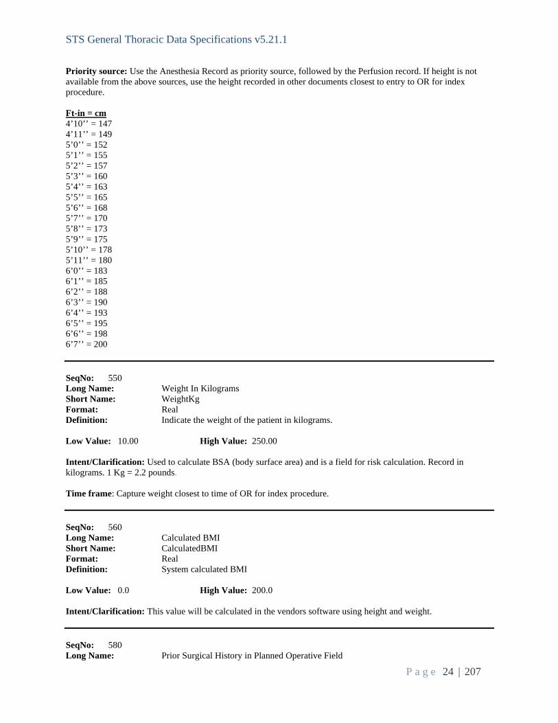
STS General Thoracic Data Specifications v5.21.1
P a g e 24 | 207
Priority source: Use the Anesthesia Record as priority source, followed by the Perfusion record. If height is not available from the above sources, use the height recorded in other documents closest to entry to OR for index procedure. Ft-in = cm 4’10’’ = 147 4’11’’ = 149 5’0’’ = 152 5’1’’ = 155 5’2’’ = 157 5’3’’ = 160 5’4’’ = 163 5’5’’ = 165 5’6’’ = 168 5’7’’ = 170 5’8’’ = 173 5’9’’ = 175 5’10’’ = 178 5’11’’ = 180 6’0’’ = 183 6’1’’ = 185 6’2’’ = 188 6’3’’ = 190 6’4’’ = 193 6’5’’ = 195 6’6’’ = 198 6’7’’ = 200 SeqNo: 550 Long Name: Weight In Kilograms Short Name: WeightKg Format: Real Definition: Indicate the weight of the patient in kilograms. Low Value: 10.00 High Value: 250.00 Intent/Clarification: Used to calculate BSA (body surface area) and is a field for risk calculation. Record in kilograms. 1 Kg = 2.2 pounds. Time frame: Capture weight closest to time of OR for index procedure. SeqNo: 560 Long Name: Calculated BMI Short Name: CalculatedBMI Format: Real Definition: System calculated BMI Low Value: 0.0 High Value: 200.0 Intent/Clarification: This value will be calculated in the vendors software using height and weight. SeqNo: 580 Long Name: Prior Surgical History in Planned Operative Field

STS General Thoracic Data Specifications v5.21.1
P a g e 25 | 207
Short Name: Reop Format: Text (categorical values specified by STS) Definition: Indicate whether this is a cardiac or thoracic re-operation that affects this operative field (i.e., patient has had a previous surgical procedure in the same cavity or organ). The current surgery must be in the same operative field that was previously entered. Harvest Codes: Code: Value: 1 Yes 2 No Intent/Clarification: The intent of this field is to capture if the patient had a previous procedure within the same anatomical space as the current procedure. Access through the same incision is not a requirement. For example:
1. Patient had a previous right middle lobe wedge and is returning for a right lobectomy. This is considered the same operative field (pleural space) and should be coded as ‘Yes.’
2. Patient had a previous coronary artery bypass and is returning for an esophagectomy. This is not considered the same operative field because the heart lies in the pericardial cavity and the esophagus lies in the superior mediastinum. This example should be coded as ‘No.’
Aug 2021: Only capture prior surgical procedures within the same anatomical space – not percutaneous procedures such as chest tubes, thoracentesis, paracentesis etc.
Reference: https://courses.lumenlearning.com/suny-ap1/chapter/anatomical-terminology/ SeqNo: 590 Long Name: History of Cardiopulmonary Disease Short Name: HistCarPulDis

STS General Thoracic Data Specifications v5.21.1
P a g e 26 | 207
Database Table Name: Operations Data Source: User Format: Multi-Select Definition: Indicate the patient history of cardiopulmonary disease. Select all that apply or 'none'. Harvest Codes: Code: Value: 1 None 2 Hypertension 3 Coronary Artery Disease (CAD) 4 Atrial Fibrillation within the last year; with or without treatment 5 Pulmonary Hypertension 6 Congestive Heart Failure (CHF) 7 Myocardial Infarction 8 Aortic Valve Disease 9 Mitral Valve Disease 10 Tricuspid Valve Disease 11 Pulmonic Valve Disease 12 Interstitial Fibrosis/ Interstitial Lung Disease Intent/Clarification: This field is required for record inclusion. If missing data, the entire record will be excluded from the analysis. 1. None:
a. Code none if the patient does have any of the following cardiovascular diseases
2. Hypertension (HTN): a. Indicate if the patient has or had a diagnosis of hypertension defined by any 1 of the following:
i. Hypertension diagnosis treated with medication, diet, and/or exercise. ii. Has undergone pharmacological therapy for treatment of hypertension, including patients who
are normotensive. b. Capturing hypertension as a risk factor must be based on Provider documentation of hypertension in
the medical record c. Please verify diagnosis with provider if there is conflicting information in the patients’ chart. If
conflicting information persists, then do not code. d. https://www.ahajournals.org/doi/10.1161/HYP.0000000000000065 e. Time frame: Onset can occur anytime between birth and entry to OR for index procedure.
3. Coronary Artery Disease (CAD):
a. Coronary artery disease is a type of atherosclerosis in which plaque builds up inside the arteries that carry blood to the heart. As the artery walls thicken, the passageway for blood narrows. Sometimes platelets gather at the narrowing, forming a clot that decreases or prevents blood flow to the region of the heart supplied by the artery.
b. Capture this for patients who have any one of the following: i. Documented blockage ≥ 50% of one or more coronary arteries
ii. Documentation of CAD in H&P iii. Documentation of angina, myocardial infarction (MI), Coronary artery bypass graft (CABG),
Percutaneous Coronary Intervention (PCI), angioplasty (balloon), coronary atherectomy, coronary artery stenting, or sudden cardiac death with no known cause may be included.

STS General Thoracic Data Specifications v5.21.1
P a g e 27 | 207
c. Medication without documentation of CAD is not sufficient to code CAD d. Capturing as a risk factor must be based on Provider documentation of CAD in the medical record. e. Do not make assumptions of disease presence without provider documentation. f. Time frame: Capture any occurrence between birth and entry to OR for index procedure.
4. Atrial Fibrillation within the last year; with or without treatment:
a. Atrial fibrillation (also called A Fib) is an irregular heartbeat (arrhythmia) that can lead to blood clots, stroke, heart failure and other heart-related complications.
b. This data element is intended to capture A Fib. Documentation of A Fib with Aflutter is captured. Aflutter alone is not captured.
c. Capturing as a risk factor must be based on Provider documentation of Atrial Fibrillation or Atrial Fibrillation/Atrial Flutter in the medical record.
d. Include patients with persistent or paroxysmal atrial fibrillation if present within the last year per providers documentation.
e. An EKG does not have to be present within the medical record. f. Time Frame: Up to one year prior to OR Entry for index procedure.
5. Pulmonary Hypertension (PH, PAH, PHT, PHTN)
a. High blood pressure in the arteries that supply the lungs is called pulmonary hypertension (PH, PAH, PHT, PHTN). The blood vessels that supply the lungs constrict and their walls thicken, so they cannot carry as much blood. It is identified as:
b. Capturing as a risk factor must be based on Provider diagnosis of Pulmonary Hypertension (PH, PAH, PHT, PHTN)
c. Do not make a diagnosis based on test results. The documentation must be documented by a provider. d. Time frame: Capture any occurrence between birth and entry to OR for index procedure.
6. Congestive Heart Failure (CHF)
a. Heart failure is described as unusual dyspnea on light exertion, recurrent dyspnea occurring in the supine position, fluid retention; or the description of rales, jugular venous distension, pulmonary edema on physical exam, or pulmonary edema on chest x‐ray presumed to be cardiac dysfunction. A low ejection fraction alone, without clinical evidence of heart failure does not qualify as heart failure. An elevated BNP without other supporting documentation should not be coded as CHF.
b. NYHA Class documentation alone cannot be used for diagnosis for heart failure, you must have physician documentation that states heart failure. There needs to be preoperative documentation in the chart that the patient has been in or was in a state of heart failure.
c. Do not code heart failure for a diagnosis of Cardiomyopathy. A diagnosis of heart failure must be documented in the medical record to code heart failure. Cardiomyopathy may or may not be associated with a heart failure diagnosis.
d. Capturing as a risk factor must be based on Provider documentation of Congestive Heart Failure (CHF)
e. Patients who had CHF in a previously transplanted heart are not considered to still be diseased. Do not code for these patients unless a diagnosis of CHF is present with their current heart.
f. Time frame: Capture any occurrence between birth and entry to OR for index procedure.
7. Myocardial Infarction (MI) a. Indicate if the patient has a history of a myocardial infarction b . Do not code slight troponin increase and no EKG changes alone as MI without confirmation in the
medical record by a physician or physician extender. c . Do not use phrases such as “cannot rule out”, “suggestive”, “probable”, “cannot exclude”, etc. to code
MI. d. Capturing as a risk factor must be based on Provider documentation of Myocardial Infarction (MI) e. Time frame: Capture any occurrence between birth and entry to OR for index procedure.
8. Aortic Valve Disease
a. Indicate if the patient has had or has the presence of dysfunction of the aortic valve, identified as: i. Moderate or severe (2+) aortic valve insufficiency
ii. Moderate, or severe (2+) aortic valve stenosis

STS General Thoracic Data Specifications v5.21.1
P a g e 28 | 207
b. Calcification alone is not sufficient to code disease. There must be documentation of stenosis or insufficiency.
c. Regurgitation or Prolapse alone is not sufficient to code disease. There must be documentation of stenosis or insufficiency.
d. Excludes surgically corrected valvular disease e. Capturing as a risk factor must be based on Provider documentation of Aortic Valve disease f. Time Frame: Up to six months prior to OR Entry for index procedure.
9. Mitral Valve Disease
a. Indicate if the patient has had or has the presence of dysfunction the mitral valve, identified as: i. Moderate or severe (2+) mitral valve insufficiency
ii. Moderate, or severe (2+) mitral valve stenosis b. Excludes surgically corrected valvular disease c. Calcification alone is not sufficient to code disease. There must be documentation of stenosis or
insufficiency. d. Regurgitation or Prolapse alone is not sufficient to code disease. There must be documentation of
stenosis or insufficiency. e. Capturing as a risk factor must be based on Provider documentation of Mitral Valve disease f. Time Frame: Up to six months prior to OR Entry for index procedure.
10. Tricuspid Valve Disease
a. Indicate if the patient has had or has the presence of dysfunction the tricuspid valve, identified as: i. Moderate or severe (2+) tricuspid valve insufficiency
ii. Moderate, or severe (2+) tricuspid valve stenosis b. Excludes surgically corrected valvular disease c. Calcification alone is not sufficient to code disease. There must be documentation of stenosis or
insufficiency. d. Capturing as a risk factor must be based on Provider documentation of Tricuspid Valve disease e. Time Frame: Up to six months prior to OR Entry for index procedure.
11. Pulmonic Valve Disease
a. Indicate if the patient has had or has the presence of dysfunction the pulmonic valve, identified as: i. Moderate or severe (2+) pulmonic valve insufficiency
ii. Moderate, or severe (2+) pulmonic valve stenosis b. Excludes surgically corrected valvular disease c. Calcification alone is not sufficient to code disease. There must be documentation of stenosis or
insufficiency. d. Regurgitation or Prolapse alone is not sufficient to code disease. There must be documentation of
stenosis or insufficiency. e. Capturing as a risk factor must be based on Provider documentation of Pulmonic Valve disease f. Time Frame: Up to six months prior to OR Entry for index procedure.
12. Interstitial Fibrosis/ Interstitial Lung Disease
a. Interstitial lung disease (ILD) refers to a group of lung diseases affecting the interstitium (the tissue and space around the air sacs of the lungs). It involves alveolar epithelium, pulmonary capillary endothelium, basement membrane, peri-vascular and peri-lymphatic tissues.
b. This is not the same as chronic lung disease (CLD) or black lung disease c. Patients who had ILD in previously transplanted lungs are not considered to still be diseased. Do not
code for these patients unless it is presented with their current lungs. d. Capturing as a risk factor must be based on Provider diagnosis of Interstitial Fibrosis/ILD e. Time frame: Capture any occurrence between birth and entry to OR for index procedure.
SeqNo: 600 Long Name: Preoperative Ejection Fraction Short Name: EF

STS General Thoracic Data Specifications v5.21.1
P a g e 29 | 207
Database Table Name: Operations Data Source: User Format: Real Definition: Indicate the percentage of the blood emptied from the left ventricle at the end of the contraction. Use the most recent determination prior to the surgical intervention documented on a diagnostic report. Enter a percentage in the range of 1 - 99. If a qualitative description is reported, code the mean value for that range, i.e., normal (50-70%) is coded as 60%. If no diagnostic report is in the medical record, a value documented in the medical record is acceptable. Low Value: 1.0 High Value: 99.0 ParentLongName: History of Cardiopulmonary Disease ParentShortName: HistCarPulDis ParentValue: contains (6) ParentHarvestCodes: Contains ("Congestive Heart Failure (CHF)") Intent/Clarification: Ejection fraction (EF) indicates the efficiency of the left ventricle (ability to pump blood sufficiently to the rest of the body). It compares the amount of blood in the left ventricle at the end of systole (when the ventricle is fuller) to the end of diastole (after the ventricle contracted and should be less full). Issues effecting the left ventricles pumping ability include preload (the amount of blood deposited into the ventricle prior to diastole), afterload (amount of pressure the ventricle must pump against typically high because of elevated systemic venous pressure), ventricular hypertrophy (the enlargement of the ventricle which results in stretching of the ventricle causing decreased contractility and is a usually a result of congestive heart failure), and valvular insufficiency. Ejection fraction is typically reported in a percentage (1-99%) or described with words. Time Frame: It is preferred to use the result within the last 6 months, closest and prior to OR Entry for index procedure. If results are not available within the last 6 months, then results can be used from within the last 12 months. If there is no documented EF withing the past 12 months, leave blank. If a percentage range is reported, report a whole number using the “mean”. For example, a range of 55-60 is coded as 58%. For echo reports that have a descriptive term such as normal documented in the impression / conclusion / summary for EF and the measurement portion of the report says 60-70% by visual estimate, use the numerical values first and capture as 65%. Use descriptive terms when you have no numerical values. If only a descriptive term is reported, code as below:
• Hyperdynamic: code 71% • Normal: code 60% • Mild dysfunction: code 45% • Moderate dysfunction: code 35% • Severe dysfunction: code 29% ACCF/AHA 2013
If the EF closest to surgery is on a Nuclear stress test with a post-stress and rest EF documented, use the rest EF. SeqNo: 610 Long Name: History of Vascular Disease Short Name: HistVasDis Format: Multi-Select Definition: Indicate the patients’ history of vascular disease. Harvest Codes: Code: Value:

STS General Thoracic Data Specifications v5.21.1
P a g e 30 | 207
1 None 2 Major Aortic or Peripheral Vascular Disease (PVD) 3 Deep Vein Thrombosis/Pulmonary Embolism (DVT/PE) 4 Transient Ischemic Attack (TIA) 5 Cerebrovascular Accident (CVA) Intent/Clarification: This field is required for record inclusion. If missing data, the entire record will be excluded from the analysis. Time frame – capture any occurrence between birth and entry to OR for index procedure
1. None
2. Major Aortic or Peripheral Vascular Disease (MVD/PVD) a. Examples include AAA repair or stent; amputation for arterial insufficiency, aorto-iliac occlusive
disease reconstruction, peripheral vascular bypass surgery, angioplasty or stent, renal artery atherosclerosis, aortic aneurysm, aortic dissection, aortic enlargement, collagen vascular disease
b. Patients with documentation of a major vascular disease (MVD) or peripheral vascular disease (PVD) but not having had surgery and/or not receiving medical treatment should also be captured as having MVD/PVD
c. Capturing as a risk factor must be based on Provider documentation of MVD/PVD
3. Deep Vein Thrombosis/Pulmonary Embolism (DVT/PE) a. DVT occurs when a blood clot forms in one or more of the deep veins in the body, usually the
legs. Pulmonary embolism is a clot located in one of the pulmonary arteries in the lungs. In most cases, the clot(s) have traveled to the lungs from the legs or other parts of the body
b. Capturing as a risk factor must be based on Provider documentation of DVT/PE
4. Transient Ischemic Attack (TIA) a. Transient ischemic attack (TIA) is defined as a transient episode of focal neurological dysfunction
caused by brain, spinal cord, or retinal ischemia, without acute infarction, where the neurological dysfunction resolves within 24 hours
b. Capturing as a risk factor must be based on Provider documentation of TIA
5. Cerebrovascular Accident (CVA)/Stroke a. Stroke is an acute episode of focal or global neurological dysfunction caused by brain, spinal cord,
or retinal vascular injury because of hemorrhage or infarction, where the neurological dysfunction lasts for greater than 24 hours.
b. Include any confirmed neurological deficit of abrupt onset caused by a disturbance in cerebral blood supply that did not resolve within 24 hours of the event. The physical deficit can be in the form of extremity weakness, facial asymmetry, language (speech and/or cognitive thinking) impairment. The intent is to differentiate between neurological events that resolve within 24 hours and those that don’t.
c. Code for CVA if the patient has no history of stroke and no symptoms but imaging study results show an infarct (old/chronic or new) or cerebral septic emboli.
d. Not all subarachnoid hemorrhages (SAH) will create a stroke. There must be some form of deficit (symptoms lasting > 24 hr.) documented in the chart to code SAH as a CVA.
e. Do not code if any neurologic dysfunction occurred or was suspected, did not resolve in 24 hours, and could not be confirmed or when there is conflicting information in the medical record and/or
STS General Thoracic Data Specifications v5.21.1
P a g e 31 | 207
with the patient/family and/or patient/family unable to provide history. f. Capturing as a risk factor must be based on Provider documentation of CVA
SeqNo: 620 Long Name: Permanent Neurologic Impairment Short Name: PNI Format: Text (categorical values specified by STS) Definition: Indicate if the patient has any permanent neurological impairments. ParentLongName: History of Vascular Disease ParentShortName: HistVasDis ParentValue: contains(5) ParentHarvestCodes: Contains ("Cerebrovascular Accident (CVA)") Harvest Codes: Code: Value: 1 Yes 2 No Intent/Clarification: Code ‘yes’ for patients who had permanent neurological impairment following a cerebral vascular accident. SeqNo: 630 Long Name: History of Endocrine GI Renal Disease Short Name: HistEndoGiRenDis Format: Multi-Select Definition: Indicate the patient's history of endocrine, gastrointestinal, and/or renal disease. Select all that apply or 'none'. Harvest Codes: Code: Value: 1 None 2 Diabetes 3 Liver Dysfunction 4 Dialysis Intent/Clarification: This field is required for record inclusion. If missing data, the entire record will be excluded from the analysis.
1. None 2. Diabetes
a. Indicate if the patient has a history of diabetes mellitus diagnosed and/or treated by a healthcare provider regardless of duration of disease or need for anti-diabetic agents.
b. Do not code for patients with steroid induced hyperglycemia and gestational (transient) diabetes if there is no supportive documentation of diabetes such as a HbA1c and/or treatment.
c. Not all patients receiving diabetic medications are considered diabetic. It is important to remember that some medications used to treat diabetes may be used to treat other conditions.
d. Patients with a history of diabetes who have had a pancreatic transplant are coded as Yes to Diabetes.
e. Hemoglobin A1c >=6.5% is indicative of diabetes. Please refer your healthcare providers to the 2017 ADA Standards of Medical Care in Diabetes. 2017 American Diabetes Association Standards of Medicare Care in Diabetes - 2017. Diabetes

STS General Thoracic Data Specifications v5.21.1
P a g e 32 | 207
Care. 40 (Suppl.1):S13. https://professional.diabetes.org/sites/professional.diabetes.org/files/media/dc_40_s1_final.pdf
f. Capturing as a risk factor must be based on Provider documentation of diabetes g. Time frame – capture any occurrence between birth and entry to OR for index procedure.
3. Liver Dysfunction
a. Indicate if the patient has any documented active liver dysfunction, documented cirrhosis, chronic hepatitis B/C, autoimmune liver disease/hepatitis, portal hypertension, esophageal varices, liver transplant, congestive hepatopathy.
b. LFTs or a MELD score alone cannot be used to code liver disease since other conditions impact these lab values.
c. Liver fibrosis with recurrent ascites, supported by the MELD can be coded as liver disease. d. The following are not coded as liver disease:
i. Hepatitis A ii. Gilberts syndrome
iii. Fatty liver iv. Liver Cancer v. NASH in the absence of cirrhosis
vi. Shock liver/ischemic hepatitis vii. Hepatic sarcoidosis
e. Patients with history of hepatitis C that is now considered eradicated should not be coded as having liver dysfunction, unless there is documentation of liver cirrhosis.
f. Capturing as a risk factor must be based on Provider documentation of liver disease
4. Dialysis a. Indicate whether the patient is currently undergoing dialysis. b. This includes hemodialysis, peritoneal dialysis, or CRRT. c. Does not include ultrafiltration without dialysate
SeqNo: 640 Long Name: Diabetes Therapy Short Name: DiabCtrl Format: Text (categorical values specified by STS) Definition: Indicate the diabetes therapy method. ParentLongName: History of Endocrine GI Renal Disease ParentShortName: HistEndoGiRenDis ParentValue: contains(2) ParentHarvestCodes: Contains ("Diabetes") Harvest Codes: Code: Value: 1 None 2 Diet only 8 Oral 4 Insulin 6 Other subcutaneous medication 5 Other 7 Unknown Intent/Clarification:

STS General Thoracic Data Specifications v5.21.1
P a g e 33 | 207
Choose the most aggressive therapy from the order below, with insulin considered the most aggressive.
• Insulin: insulin treatment (includes any combination with insulin) (Harvest Code 4) • Other subcutaneous medications (e.g., GLP-1 agonist, Byetta, Bydureon, Victoza, Symlin) (Harvest Code
6) • Oral: treatment with oral agent (includes oral agent with or without diet treatment) (Harvest Code 8) • Diet only: Treatment with diet only (Harvest Code 2) • Other: other adjunctive treatment, non-oral/insulin/diet (Harvest Code 5) • None: Not receiving any treatment or special dietary restriction for diabetes (Harvest Code 1)
• Unknown (Harvest Code 7) __________________________________________________________________________ SeqNo: 650 Long Name: History of Cancer Short Name: HistCancer Format: Multi-Select Definition: Indicate the patient's history of cancer. Select all that apply or 'none'. Harvest Codes: Code: Value: 1 None 2 Coexisting Cancer 3 Preoperative Chemotherapy/Immunotherapy 4 Preoperative Thoracic Radiation Therapy Intent/Clarification: This field is required for record inclusion. If missing data, the entire record will be excluded from the analysis. Select all that apply, if not selecting ‘None’
1. None - The patient is not currently being treated or surveyed for an active malignancy not related to the thoracic disease being evaluated and treated by the thoracic surgeon.
2. Coexisting Cancer – the patient is being treated or surveyed for an active primary malignancy that is not related to the thoracic disease being evaluated and treated by the thoracic surgeon. Examples:
a. The patient is undergoing a lung resection for lung cancer and has known lymphoma for which they are being observed.
b. Patient with lung cancer undergoing resection with known bladder cancer for which a staged procedure is planned.
c. Patient diagnosed with lung cancer and rectal cancer at the same time, undergoing therapy for both simultaneously.
d. Does not include previously treated cancers that have completed treatment and are in active surveillance
e. Does not include synchronous primary lung cancers f. Must be another primary cancer (not metastases)
3. Preoperative Chemotherapy/Immunotherapy – Indicate if the patient has ever received chemotherapy or
immunotherapy for cancer therapy. a. Includes all forms of chemotherapy given for cancer therapy, including neoadjuvant therapies
(CAP, ADOC, PE, VIP chemotherapy regimens) b. Immunotherapy drugs (i.e., Keytruda) are captured here c. Do not include immunosuppressive medications not intended for cancer treatment (i.e., do not
include methotrexate or Xeljanj for arthritis
STS General Thoracic Data Specifications v5.21.1
P a g e 34 | 207
d. Do not include hormonal therapy (i.e., tamoxifen, Lupron) e. Not limited to IV agents
4. Preoperative Thoracic Radiation Therapy - Indicate if the patient has received preoperative radiation
therapy to the intended operative field for any reason prior to this operation. May be included as a component of a chemo radiation induction therapy.
a. This item should also be selected if the radiation oncologist gave the patient radiation therapy prior to sending the patient for surgical evaluation if the intent of the radiation oncologist was to "shrink the tumor" prior to surgical intervention.
b. Previous breast & axillary radiation qualifies as thoracic radiation. This is in the ‘same operative field’ for a current lobectomy
c. Excludes previous radioactive iodine treatment Aug 2021: Photodynamic therapy is not equivalent to thoracic radiation therapy and is not captured. SeqNo: 675 Long Name: Preoperative Chemo - Current Malignancy - Multi-Select Short Name: PreopChemoCurWhenMulti Format: Multi-Select Definition: Indicate when the patient received preoperative chemotherapy and for what disease. Select all that apply. ParentLongName: History of Cancer ParentShortName: HistCancer ParentValue: contains(3) ParentHarvestCodes: Contains ("Preoperative Chemotherapy/Immunotherapy") Harvest Codes: Code: Value: 1 Same disease, <= 6 months 2 Same disease,> 6 months 3 Unrelated disease, <= 6 months 4 Unrelated disease, >6 months Intent/Clarification:
1. Same disease, <= 6 months – Indicate if the patient received preoperative chemotherapy/immunotherapy for the same disease within the last 6 months.
2. Same disease,> 6 months - Indicate if the patient received preoperative chemotherapy/immunotherapy for the same disease greater than 6 months before current procedural date.
3. Unrelated disease, <= 6 months – Indicate if the patient received chemotherapy/immunotherapy for an
unrelated disease withing the last 6 months.
4. Unrelated disease, >6 months - Indicate if the patient received chemotherapy/immunotherapy for an unrelated disease greater than 6 months before current procedural date.
SeqNo: 685 Long Name: Preoperative Thoracic Radiation Therapy - Disease And When Treated - Multi-Select Short Name: PreopXRTDisWhenMulti Format: Multi-Select

STS General Thoracic Data Specifications v5.21.1
P a g e 35 | 207
Definition: Indicate when the patient received preoperative thoracic radiation therapy and for what disease. Select all that apply. ParentLongName: History of Cancer ParentShortName: HistCancer ParentValue: contains(4) ParentHarvestCodes: Contains ("Preoperative Thoracic Radiation Therapy") Harvest Codes: Code: Value: 1 Same disease, <= 6 months 2 Same disease, > 6 months 3 Unrelated disease, <= 6 months 4 Unrelated disease, > 6 months Intent/Clarification:
1. Same disease, <= 6 months – Indicate if the patient received preoperative thoracic radiation therapy for the same disease within the last 6 months
2. Same disease,> 6 months - Indicate if the patient received preoperative thoracic radiation therapy for the same disease greater than 6 months before current procedural date
3. Unrelated disease, <= 6 months – Indicate if the patient received thoracic radiation therapy for an unrelated
disease withing the last 6 months.
4. Unrelated disease, >6 months - Indicate if the patient received thoracic radiation therapy for an unrelated disease greater than 6 months before current procedural date.
SeqNo: 690 Long Name: Preoperative Medication History Short Name: HistPreopMeds Format: Multi-Select Definition: Indicate the patient's preoperative medication history. Select all that apply or 'none'. Harvest Codes: Code: Value: 1 None 2 Chronic Immunosuppressive Therapy 3 Chronic Anticoagulation 4 Home Oxygen Therapy (Home O2) Intent/Clarification: This field is required for record inclusion. If missing data, the entire record will be excluded from the analysis.
1. None – The patient was not on chronic immunosuppressive therapy, chronic anticoagulation, or home oxygen therapy (Home O2) at the time of the patient is considered a candidate for current procedure.
2. Chronic Immunosuppressive Therapy

STS General Thoracic Data Specifications v5.21.1
P a g e 36 | 207
a. Indicate if the patient has required the regular administration of corticosteroids (e.g. Prednisone, Decadron) or other immunosuppressant or chemotherapeutic medications (e.g. methotrexate, abatacept (Orencia), Adalimumab (Humira), etanercept (Enbrel), cyclosporine, tacrolimus, azathioprine, mycophenolate mofetil) within the 30 days prior to the principal operative procedure or at the time the patient is being considered as a candidate for surgery, for a chronic medical condition (e.g. COPD, asthma, rheumatologic disease, rheumatoid arthritis, inflammatory bowel disease).
b. A one-time steroid pulse or a limited short steroid course (< 10 days), does not qualify. c. Do not include topical corticosteroids applied to the skin or corticosteroids administered by
inhalation or rectally. d. Only capture medically managed immunosuppressant therapy. Splenectomies are not to be captured
here. e. Immunotherapy drugs used for cancer (i.e., Keytruda) are not considered immunosuppressive
drugs. Please capture under HistCancer (seq 560) as Preoperative Chemotherapy/Immunotherapy. f. Androgen deprivation therapy is not considered chronic immunosuppressive therapy
3. Chronic Anticoagulation
a. Indicate if the patient has used an oral or injectable anticoagulant within the 30 days prior to the principal operative procedure. The intent is to capture patients who are at an increased risk for bleeding. This includes:
i. Warfarin ii. P2Y12 Inhibitors
iii. Factor Xa Inhibitors iv. Antiplatelet medication v. Direct Oral Anticoagulants (DOACs)
b. Do not capture one-time doses or heparin flushes. c. Do not capture ASA or NSAIDs.
4. Home Oxygen Therapy (HomeO2) a. Indicate if the patient uses any supplemental oxygen at home, include as needed oxygen use. b. This includes the home use of O2 with CPAP
SeqNo: 700 Long Name: Creatinine Level Measured Short Name: CreatMeasured Format: Text (categorical values specified by STS) Definition: Indicate whether the creatinine level was measured within one month prior to the surgical procedure and prior to anesthetic management (induction area or operating room). Harvest Codes: Code: Value: 1 Yes 2 No Intent/Clarification: Creatinine, urea, and urate all increase as the ability of the kidneys to filter fluid within the body declines. Creatinine is a marker for kidney function. Time frame: Within 30 days of procedure. SeqNo: 710 Long Name: Last Creatinine Level Short Name: CreatLst Format: Real

STS General Thoracic Data Specifications v5.21.1
P a g e 37 | 207
Definition: Indicate the creatinine level closest to the date and time prior surgery. Low Value: 0.10 High Value: 30.00 ParentLongName: Creatinine Level Measured ParentShortName: CreatMeasured ParentValue: 1 ParentHarvestCodes: = "Yes" Intent/Clarification: A creatinine level (Cr) should be collected on all patients, even if there is no prior history of renal disease. A creatinine value is an important predictor of a patient's renal function and, therefore, outcome and is used in the predicted risk models. Time Frame: Within 30 days of procedure. Use the value closest to the date and time prior to anesthetic management. Anesthetic management begins when a member of the anesthesiology team initiates care. The administration of IV fluids in the holding area can cause dilution of blood. Do not capture labs drawn after the patient receives fluids in the holding area or O.R. Sept 2021: Do not round lab values, enter to two decimal places. SeqNo: 720 Long Name: Hemoglobin Level Measured Short Name: HemoglobinMeasured Format: Text (categorical values specified by STS) Definition: Indicate whether the patient's hemoglobin level was measured within one month prior to this surgical procedure. Harvest Codes: Code: Value: 1 Yes 2 No Intent/Clarification: The hemoglobin (Hgb) test may be used to screen for, diagnose, or monitor several conditions and diseases that affect red blood cells (RBCs) and/or the amount of hemoglobin in blood. Time Frame: Within 30 days of procedure. SeqNo: 730 Long Name: Last Hemoglobin Level Short Name: HemoglobinLst Format: Real Definition: Indicate the hemoglobin level closest to the date and time prior to surgery and prior to anesthetic management (induction area or operating room). Low Value: 1.00 High Value: 50.00 ParentLongName: Hemoglobin Level Measured ParentShortName: HemoglobinMeasured ParentValue: 1 ParentHarvestCodes: = "Yes"
STS General Thoracic Data Specifications v5.21.1
P a g e 38 | 207
Intent/Clarification: The hospital laboratory report should be accessed first when coding this variable. If this is unavailable, then additional source documents may be referenced for lab results. Capture only measured hgb values; do not use calculated values. The unit of measurement for Hgb is g/dl or g/100 ml or g%. Time Frame: Within 30 days of procedure. Use the measured value closest to the date and time prior to anesthetic management. Anesthetic management begins when a member of the anesthesiology team initiates care. The administration of IV fluids in the holding area can cause dilution of blood. Do not capture labs drawn after the patient receives fluids in the holding area or O.R. Sept 2021: Do not round lab values, enter to two decimal places. SeqNo: 740 Long Name: Pulmonary Function Tests Performed Short Name: PFT Format: Text (categorical values specified by STS) Definition: Indicate whether pulmonary function tests (PFT's) were performed prior to this operation. PFT's done more than 12 months prior to the primary surgical procedure should not be included here. PFTs are part of the NQF measure set and are required before any major anatomic lung resection unless valid exclusion criteria are met. Harvest Codes: Code: Value: 1 Yes 2 No Intent/Clarification: Pulmonary function testing is a valuable tool for evaluating the respiratory system, representing an important adjunct to the patient history, various lung imaging studies, and invasive testing such as bronchoscopy and open-lung biopsy. Insight into underlying pathophysiology can often be gained by comparing the measured values for pulmonary function tests obtained on a patient at any point with normative values derived from population studies. The percentage of predicted normal is used to grade the severity of the abnormality. Pulmonary function testing is used in clinical medicine for evaluating respiratory symptoms such as dyspnea and cough, for stratifying preoperative risk, and for diagnosing common diseases such as asthma and chronic obstructive pulmonary disease. PFT = "yes" if only FEV1 is done. Use bedside PFTs if that's the only available test. Time Frame: Within 12 months of procedure. This field is required for record inclusion. If missing data, the entire record will be excluded from the analysis. SeqNo: 750 Long Name: FEV1 Predicted Short Name: FEVPred Format: Integer

STS General Thoracic Data Specifications v5.21.1
P a g e 39 | 207
Definition: Indicate the % predicted FEV1 obtained for the patient. Low Value: 1 High Value: 200 ParentLongName: Pulmonary Function Tests Performed ParentShortName: PFT ParentValue: 1 ParentHarvestCodes: = "Yes" Intent/Clarification: Indicate the FEV1 % predicted from the most recent pulmonary function test prior to procedure. Do not use values obtained more than 12 months prior to surgery. Choose the highest value reported for % predicted, whether or not a bronchodilator was used. FEV1 is a marker of the degree of obstruction. It evaluated the maximal amount of air forcefully exhaled in one second. It is then converted to a percentage of normal. For example, the FEV1 may be 80% of predicted based on height, weight, and gender. In normal persons, the FEV1 accounts for the greatest part of the exhaled volume from a spirometric maneuver and reflects mechanical properties of the large and the medium-sized airways. If there are multiple PFTs in the record, choose the study which best reflects the patient’s status just prior to surgery.
To calculate the % predicted, in case the report only shows the % changed, divide the actual by the predicted.
PFT Report –
Predicted Pre bronchodilator - 3.80 Actual Pre-bronchodilator - 2.65 2.65 / 3.80= 69.7 (actual divided by predicted)
Time Frame: Within 12 months of procedure. Use the value closest to surgery. If more than one value is reported on the test closest to surgery, code the highest value, whether or not a bronchodilator was used. This field is required for record inclusion. If missing data, the entire record will be excluded from the analysis. SeqNo: 770 Long Name: DLCO Test Performed Short Name: DLCO Format: Text (categorical values specified by STS) Definition: Indicate whether a lung diffusion test (DLCO) was performed. DLCO test should be collected for a major lung resection (e.g., wedge resection, segmentectomy, lobectomy, sleeve lobectomy, bilobectomy, or pneumonectomy). ParentLongName: Pulmonary Function Tests Performed ParentShortName: PFT ParentValue: 1 ParentHarvestCodes: = "Yes" Harvest Codes: Code: Value: 1 Yes 2 No 3 Not applicable
STS General Thoracic Data Specifications v5.21.1
P a g e 40 | 207
Intent/Clarification: The diffusing capacity (DLCO) is a test of the integrity of the alveolar-capillary surface area for gas transfer. Single-breath carbon monoxide diffusing capacity (Dsb) is used interchangeably with DLCO and may be captured here. DO NOT USE the DLCO/VA (adjusted/corrected), regardless of altitude. Do not use subjective terms (i.e., ‘normal’) to document a value. An integer must be available to code this field as ‘yes’. Time Frame: Within 12 months of procedure. SeqNo: 781 Long Name: DLCO Lowest Predicted Short Name: DLCOPredLow Format: Integer Definition: Indicate the lowest % predicted DLCO value obtained for the patient. Low Value: 10 High Value: 200 ParentLongName: DLCO Test Performed ParentShortName: DLCO ParentValue: 1 ParentHarvestCodes: = "Yes" Intent/Clarification: The diffusing capacity (DLCO) may be reduced, <80% predicted, in disorders such as emphysema, pulmonary fibrosis, obstructive lung disease, pulmonary embolism, pulmonary hypertension and anemia. DLCO>120% of predicted may be seen in normal lungs, asthma, pulmonary hemorrhage, polycythemia, and left to right intracardiac shunt. The lowest value for DLCO uncorrected should be captured. A PFT may report DLCO_SB, DLCOcSB, DLCO/VA. The difference in the DCLO SB (simple DCLO) and the DCLOcSB is that the DCLOcSB is corrected for the hgb value. In this scenario, capture the lowest DLCO_SB or DLCO/VA value. Do not use the DLCOcSB since it is a corrected value. Choose the value that represents the lowest % predicted unadjusted/uncorrected DLCO. DO NOT USE the DLCO/VA (adjusted/corrected), regardless of altitude. SeqNo: 790 Long Name: History of Substance Abuse Short Name: HistSubAbus Format: Multi-Select Definition: Indicate the patients’ history of substance abuse. Select all that apply or 'none'. Harvest Codes: Code: Value: 1 None 2 Cigarette Smoking

STS General Thoracic Data Specifications v5.21.1
P a g e 41 | 207
3 Substance Dependency/Abuse of Non-Prescription Medications or Illicit Drugs 4 Alcohol Abuse Intent/Clarification: This field is required for record inclusion. If missing data, the entire record will be excluded from the analysis.
1. None – The patient does not have any history of tobacco abuse, substance abuse, or alcohol abuse 2. Cigarette Smoking (Tobacco Use)
a. Indicate current (within 30 days prior to admission) or previous use of any tobacco product. Includes:
i. Cigarettes ii. Pipe
iii. Cigars iv. Smokeless Cans v. Vaping
vi. Medical Marijuana wrapped in tobacco leaves vii. Other tobacco products (orbs, strips, sticks, hookah, etc.)
3. Substance Dependency/Abuse of Non-Prescription Medications or Illicit Drugs
a. Capture patients with use of illicit (illegal) drugs. Include abuse of controlled substances (prescription medication/opioid abuse) that have not been prescribed by a provider for the patient. Illicit drug use is associated with numerous health and social problems, and age‐related physiological, psychological, and social changes that could impact recovery from surgery.
b. Marijuana should not be captured as illicit drug use. c. Code illicit drug use if the drug screen is positive for drugs, such as heroin, cocaine, or
methamphetamine, regardless of if the patient denies use.
4. Alcohol Abuse a. Alcohol abuse is not necessarily a quantity of alcohol but implies interference with home, work,
and life functioning. b. Documenting the patient is an alcoholic at the time of admission should be coded as more
than 4 drinks (men) and 3 drinks (women) per any day or more than 14 (men) and 7 (women) drinks per week.
c. Binge drinking on 5 or more days in the past month is also considered alcohol abuse. d. Indicate alcohol abuse if indicated by the family, even if denied by the patient. e. Reference: https://www.niaaa.nih.gov/alcohol-health/overview-alcohol-
consumption/moderate-binge-drinking
SeqNo: 800 Long Name: Cigarette Smoking History Short Name: CigSmoking Format: Text (categorical values specified by STS) Definition: Indicate the patient's history of smoking cigarettes. ParentLongName: History of Substance Abuse ParentShortName: HistSubAbus ParentValue: contains (2) ParentHarvestCodes: Contains ("Cigarette Smoking")

STS General Thoracic Data Specifications v5.21.1
P a g e 42 | 207
Harvest Codes: Code: Value: 2 Past smoker (stopped more than 1 month prior to operation) 3 Current smoker Intent/Clarification: Code past smoker if the patient stopped any type of tobacco use more than one month prior to surgery, current smoker if still using any form of tobacco products. i. cigarettes ii. Pipe iii. Cigars iv. Smokeless Cans v. Vaping vi. Medical Marijuana wrapped in tobacco leaves vii. Other tobacco products (orbs, strips, sticks, hookah, etc.) This field is required for record inclusion. If missing data, the entire record will be excluded from the analysis. SeqNo: 810 Long Name: Pack Years Known or can be estimated Short Name: PackYearKnown Format: Text (categorical values specified by STS) Definition: Indicate whether the number of pack years is known or can be estimated. ParentLongName: History of Substance Abuse ParentShortName: HistSubAbus ParentValue: contains (2) ParentHarvestCodes: Contains ("Cigarette Smoking") Harvest Codes: Code: Value: 1 Yes 2 No Intent/Clarification: If no pack year is document, then code ‘no.’ Pack-years are only documented for cigarette smokers, code no for all other types of tobacco. SeqNo: 820 Long Name: Pack-Years Of Cigarette Use Short Name: PackYear Format: Integer Definition: Indicate the number or estimate of pack-years by multiplying the average number of packs of cigarettes smoked per day by the number of years of smoking. For example if the patient smoked 1 ppd for 10 years and 3 ppd for the next 10 years, the average ppd would be 2 ppd x 20 years = 40 pack-years of smoking. Low Value: 1 High Value: 210

STS General Thoracic Data Specifications v5.21.1
P a g e 43 | 207
ParentLongName: Pack Years Known or can be estimated ParentShortName: PackYearKnown ParentValue: 1 ParentHarvestCodes: = "Yes" Intent/Clarification: Code the highest number of pack years. If a range is documented, code the highest (e.g., 20-30 years; code 30). Sept 2021: Pack-Years can be rounded to the nearest whole number (i.e. 12.5 pack-years would be entered as 13). SeqNo: 830 Long Name: Dementia or neurocognitive dysfunction Short Name: DemNeroDys Format: Text (categorical values specified by STS) Definition: Indicate if the patient has had mental status changes, and/or delirium in the context of the current illness or chronic/long-standing mental status changes secondary to chronic mental illness (e.g., schizophrenia; bipolar disorder) or chronic dementing illnesses (e.g., multi-infarct dementia, senile dementia of the Alzheimer’s type). This assessment of the patient's mental status is within 48 hours prior to the surgical procedure. Harvest Codes: Code: Value: 1 Yes 2 No Intent/Clarification: The intent is to capture chronic mental illness including dementia and neurocognitive dysfunction. Capture a person with an overall decline in cognitive ability in this field. Includes traumatic brain injury (TBI). According to the NIH National Institute on Aging: “Dementia is the loss of cognitive functioning—thinking, remembering, and reasoning—and behavioral abilities to such an extent that it interferes with a person's daily life and activities. These functions include memory, language skills, visual perception, problem solving, self-management, and the ability to focus and pay attention. Some people with dementia cannot control their emotions, and their personalities may change. Dementia ranges in severity from the mildest stage, when it is just beginning to affect a person's functioning, to the most severe stage, when the person must depend completely on others for basic activities of living.” SeqNo: 840 Long Name: Major Psychiatric Disorder Short Name: PsychDisorder Format: Text (categorical values specified by STS) Definition: Indicate if the patient has a major psychiatric disorder as defined by DSM IV V. Harvest Codes: Code: Value: 1 Yes 2 No Intent/Clarification: Indicate if the patient has a major psychiatric disorder as defined by DSM V.

STS General Thoracic Data Specifications v5.21.1
P a g e 44 | 207
1. A behavioral or psychological syndrome or pattern that occurs in an individual 2. Reflects an underlying psychobiological dysfunction 3. The consequences of which are clinically significant distress (e.g., a painful symptom) or disability
(i.e., impairment in one or more important areas of functioning) 4. Must not be merely an expected response to common stressors and losses (ex. the loss of a loved one)
or a culturally sanctioned response to a particular event (ex. trance states in religious rituals) 5. Primarily a result of social deviance or conflicts with society
To identify look for a formal psychiatric diagnosis for which the patient requires regular treatment including behavioral therapy, counseling and/or pharmaceutical treatment (Sept 2021). Examples include depression requiring anti-depressant medication or regular counseling. Anxiety disorder, schizophrenia, bipolar disorder requiring active pharmaceutical intervention. Examples include Adult Attention Deficit/Hyperactivity Disorder (ADHD/ADD), Bipolar Disorder, Depression requiring antidepressant medication or regular counseling, Eating Disorders, Generalized Anxiety Disorder, Obsessive-Compulsive Disorder, Panic Disorder, Postpartum Depression, Post-traumatic Stress Disorder (PTSD), Schizophrenia, Seasonal Affective Disorder (SAD), Social Anxiety Phobia. This is not an exhaustive or comprehensive list but an example of some disorders which would be captured here. A diagnosis is sufficient to code, treatment is not required. SeqNo: 850 Long Name: Living Status Short Name: LiveStat Format: Text (categorical values specified by STS) Definition: Indicate the patient's living status at the time of surgery. A scale to determine the degree to which the patient lives independently or dependently with others. This is a measure of dependency and social support. Harvest Codes: Code: Value: 1 Lives alone 2 Lives with-family or friend 3 Assisted Living 4 Nursing Home Intent/Clarification:
1. Lives alone a. Patient lives independently without others in the home and is able to perform ADLs without
assistance. b. Includes those who are homeless.
2. Lives with-family or friend
a. Patient lives with others in the home and is able to perform ADLs without assistance. b. Includes those coming from a correctional institution, assuming they are able to care for oneself.
3. Assisted Living
a. Patient has assistance with activities of daily living in their home or lives in an Assisted Living facility.
4. Nursing Home a. Patient resides in a Nursing Home facility.

STS General Thoracic Data Specifications v5.21.1
P a g e 45 | 207
SeqNo: 860 Long Name: Functional Status Short Name: FuncStat Format: Text (categorical values specified by STS) Definition: Indicate the patient's functional status closest to the time of surgery within the 30 days prior to surgery. Harvest Codes: Code: Value: 1 Independent 2 Partially Dependent 3 Totally Dependent 4 Unknown Intent/Clarification:
1. Independent a. The patient does not require assistance from another person for any activities of daily living. This
includes a person who is able to function independently with prosthetics, equipment, or devices. b. Activities of daily living (ADLs) include bathing feeding, dressing, toileting, and mobility.
2. Partially Dependent
a. The patient requires some assistance from another person for activities of daily living. This includes a person who utilizes prosthetics, equipment, or devices but still requires some assistance from another person for ADLs.
3. Totally Dependent a. The patient requires total assistance for all activities of daily living.
4. Unknown
a. If unable to ascertain the functional status prior to surgery, report as unknown. All patients with psychiatric illnesses should be evaluated for their ability to function with or without assistance with ADLs just as the non-psychiatric patient. For instance, if a patient with schizophrenia is able to care for him/herself without the assistance of nursing care, he/she is considered independent. Report the patients’ best functioning status within 30 days of the procedure. Time Frame: Within 30 days of procedure. SeqNo: 870 Long Name: ECOG Score Short Name: ECOGScore Format: Text (categorical values specified by STS) Definition: Indicate the patient's ECOG score at the time of surgery. Harvest Codes: Code: Value:
0 - Fully active, able to carry on all pre-disease performance without restriction 1 - Restricted in physically strenuous activity but ambulatory and able to carry out work

STS General Thoracic Data Specifications v5.21.1
P a g e 46 | 207
of a light or sedentary nature, e.g., light housework, office work 2 - Ambulatory and capable of all self-care but unable to carry out any work activities. Up and about more than 50% of waking hours 3 - Capable of only limited self-care, confined to bed or chair more than 50% of waking hours 4 - Completely disabled. Cannot carry on any self-care. Totally confined to bed or chair.
Intent/Clarification: Eastern Cooperative Oncology Group (ECOG) Performance Status Score is a scale to measure the patient's functional status and the impact of the patient's disease on the functional status. This field is required to be collected on all primary lung and esophagus cancer cases. Failure to provide this field will result in the record being rejected from submission. For cases other than primary lung or esophageal cancer, if an ECOG score is not available in the medical record, then leave blank. An N/A option will be added in the next version. To capture this field, the ECOG score must be documented in the patient’s medical record by a provider. Data managers are not to assume. Time Frame: Within 1 year. Code the value closest to procedure date. This field is highly recommended for record inclusion. If missing data, the entire record will not be excluded from the analysis, but a warning will display. If this data is not available, leave blank and do not assume the answer. Aug 2021: Lung and esophagus cases will NOT be rejected due to a missing ECOG score. Sept 2021: The ECOG score can be documented in the patient medical record by any designated healthcare provider. This includes any healthcare provider that has provided direct patient care. ------------------------------------------------------------------------------------------------------------------------------------------------------------------------------------ Category of Disease ------------------------------------------------------------------------------------------------------------------------------------------------------------------------------------ SeqNo: 1250 Long Name: Category Of Disease - Primary Short Name: CategoryPrim Format: Text (categorical values specified by STS) Definition: Indicate the PRIMARY diagnosis (category of disease) for which the procedure was performed. Intention: Choose the primary diagnosis or reason for the procedure. For the majority of cases, there will be only one condition treated (i.e., lung cancer treated by lobectomy). Rarely, there will be cases where two unrelated conditions are treated at one time (i.e., a thymoma and a lung cancer). In these rare cases, indicate the primary or most important diagnosis. For cases where a pathology report is available, use the final diagnosis on the pathology report. For cases where pathology reports are not available, use provider documentation. For any uncertainty, verify coding with the provider. Do not use ICD-10 or CPT codes provided by billing alone to code this field.
STS General Thoracic Data Specifications v5.21.1
P a g e 47 | 207
Example: Patient presents for an emergent lobectomy related to massive hemoptysis. Final pathology results showed T3N1M0 adenocarcinoma. Although the patient presented for hemoptysis; the primary category of disease is coded as lung cancer. Example: Patient presents with UGI bleed and is taken to the operating room where a mass was discovered at the EG junction and determined to be the source of the UGI bleed. An esophagogastrectomy was performed. The final pathology results showed primary esophageal cancer. For this case, code the Category of Disease (CategoryPrim – seq 1250) as Esophageal Cancer, esophagogastric junction (cardia).
Example: A patient with a history of symptomatic Myasthenia Gravis with CT evidence of an anterior mediastinal mass presents for thymectomy. The final pathology results showed T1N0M0 Stage I Thymoma. For this case, the Category of Disease is Malignant neoplasm of thymus (thymoma, thymic carcinoma) This field is required for record inclusion. If missing data, the entire record will be excluded from the analysis. Harvest Codes:
LUNG CANCER Code: Value: 150 Lung cancer, main bronchus,
carina (C34.00) This is a condition where a centrally located lung cancer becomes locally advanced and involves either the right/left main bronchus or carina. Surgical resection involves removing involved airway and lung and may require removing a portion of the central airway as well.
160 Lung cancer, upper lobe (C34.10) This refers to a primary lung cancer, located within either the right or left upper lobes.
170 Lung cancer, middle lobe (C34.2) This refers to a primary lung cancer located within the right middle lobe.
180 Lung cancer, lower lobe (C34.30) This refers to a primary lung cancer located within either the right or left lower lobe.
190 Lung cancer, location unspecified (C34.90) This code should only be used when the exact origin of the primary lung cancer cannot be determined due to large size or when the location was not specifically documented by the surgeon
ESOPHAGUS CANCER 710 Esophageal cancer,
esophagogastric junction (cardia) (C16.0) Describes cancers that are located with the junction between the esophagus and stomach and involve a portion of the cardia or upper part of the stomach.
700 Esophageal cancer, upper third (C15.3) These carcinomas arise from the esophagus located within the lower neck and upper chest.
690 Esophageal cancer, middle third (C15.4) Refers to carcinomas arising in the mid-thoracic esophagus. These are usually squamous cell carcinomas.
680 Esophageal cancer, lower third

STS General Thoracic Data Specifications v5.21.1
P a g e 48 | 207
(C15.5) This is the most common location of esophageal cancers in the United States and its incidence is steadily increasing. Lesions here are typically adenocarcinoma- and are often treated by a combination of surgery, chemotherapy, and radiation therapy.
1140 (Stomach Cancer) Malignant neoplasm stomach unspecified (C16.9) Cancerous tumor of the stomach, location and type not specified
1460 (Esophageal Cancer) Malignant neoplasm of the esophagus, unspecified (C15.9) Cancerous tumor of the esophagus, location and type not specified
Thymoma/Thymectomy/Myasthenia Gravis/Mediastinal Mass 380 Malignant neoplasm of thymus
(thymoma, thymic carcinoma) (C37) The thymus gland is located within the anterior mediastinum and serves a role in the development of the immune system. Tumors of the thymus can range from less aggressive thymomas to very malignant thymic carcinomas.
430 Myasthenia gravis (G70.00) This is a neuromuscular disease caused by antibodies generated in one’s own body. These antibodies lead to muscle weakness, fatigue, and occasionally respiratory failure. This condition is associated with thymoma, and patients may gain significant symptom improvement with resection of a thymoma or even a normal thymus gland.
360 Anterior/Posterior mediastinal tumor; metastatic (C78.1) – not required for entry Refers to tumors of the mediastinum which are classified as either seminomas or nonseminomatous germ cell tumors of the mediastinum. These tumors often cause symptoms due to their size and resulting compression of heart, lung, or airway.
400 Posterior mediastinal malignant tumor primary – not required for entry (C38.2) These are malignant tumors located in the posterior third of the mediastinum between the posterior pericardium and spine. Malignant tumors in this location are rare and predominantly malignant neurogenic tumors.
350 Anterior mediastinal malignant tumor primary (germ cell cancer, seminoma) (C38.1) – not required for entry Refers to tumors of the mediastinum which are classified as either seminomas or nonseminomatous germ cell tumors of the mediastinum. These tumors often cause symptoms due to their size and resulting compression of heart, lung, or airway.
1420 Mediastinal Mass/Neoplasm of uncertain behavior of pleura, thymus, mediastinum – not required for entry (D38.2-D38.4) Tumor of the pleura, thymus, or mediastinum without a definitive diagnosis.
370 Anterior/Posterior mediastinal tumor; benign (i.e., teratoma) - not required for entry (D15.2) A teratoma is often a benign tumor which can be located within the anterior mediastinum. This tumor consists of normal types of cells, but in an abnormal configuration and location. They can produce symptoms from their large size and are treated with surgical resection.
1410 Benign neoplasm thymus - not required for entry
STS General Thoracic Data Specifications v5.21.1
P a g e 49 | 207
(D15.0) Benign tumors of the thymic gland are relatively rare. Although most of these lesions are asymptomatic in nature, they may result in respiratory distress.
330 Mediastinal nodes, metastatic - not required for entry (C77.1) Refers to a process where cancers within the chest, or from other locations, spread to the lymph nodes within the mediastinum. These lymph nodes can be biopsied via mediastinoscopy.
390 Non-Hodgkin Lymphoma, intrathoracic lymph nodes - not required for entry (C85.92) Lymphomas are a type of cancer that arises from cells of the immune system or lymphocytes. Thoracic surgeons are often involved in obtaining tissue via mediastinoscopy to assist medical oncologists in making the diagnosis of lymphoma. The treatment of these conditions center on the use of chemotherapy.
1090 Mediastinal abscess (J85.3) - not required for entry An infection manifested by a collection of pus in the mediastinal space.
470 Disease of the mediastinum, not otherwise classified (J98.5) - not required for entry Refers to either acute or chronic inflammation of the mediastinum, including mediastinal cysts. Acute mediastinitis is usually due to a bacterial infection from a perforation of the esophagus or due to sternal wound infections after cardiac surgery procedures. Treatment often requires antibiotics and surgical drainage. Chronic mediastinitis represents a fibrosis of the mediastinum and can be a result of radiation therapy or previous infection with histoplasmosis or tuberculosis. Various types of mediastinal cysts include the following: Bronchogenic - Is the most common mediastinal cyst. These are thin-walled cavities lined with respiratory epithelium and can cause symptoms due to their size or become infected. Surgical resection may involve removal of the cyst alone or may require concomitant lung resection. Foregut duplication - These are benign cyst originating from and attached to the intrathoracic esophagus. These may be asymptomatic or associated with dysphagia due to compression of the adjacent esophagus. Removal requires simple resection of the cyst. Pericardial - These are unusual cysts arising from the pericardium. Treatment, when necessary, may involve CT-guided needle aspiration and recurrences are treated with simple cyst excision. Thymic - This describes cystic lesions within the thymus gland. They can be associated with thymomas and rarely cause symptoms.
340 Mediastinal nodes, benign - not required for entry (D36.0) Describes a condition where mediastinal lymph nodes demonstrate a benign or non-malignant process such as sarcoidosis or anthrocosis. These conditions may result in the enlargement of the involved lymph nodes.
1430 Unspecified disease of thymus- not required for entry

STS General Thoracic Data Specifications v5.21.1
P a g e 50 | 207
gland (E32.9) Disease of the thymus gland not otherwise listed.
TRACHEA 70 Tracheal tumor, malignant
(C33) Describes conditions where primary cancer develops within the trachea. Primary malignant tracheal tumors are often either squamous cell cancers or adenoid cystic carcinomas. Other malignant tumors of the trachea include sarcomas and mucoepidermoid carcinomas.
80 Tracheal tumor, benign (D14.2) These are lesions that originate from the trachea itself and are not considered cancers. Chondromas, leiomyomas, and adenomas are some examples of benign tracheal tumors.
90 Tracheal tumor, metastatic (C78.30) A process when cancers of distant sites spread to the trachea and lead to airway obstruction or bleeding. Renal cell carcinomas, breast cancers, and melanomas can metastasize to the airway.
20 Tracheal stenosis, acquired (J39.8) Refers to narrowing of the normal tracheal diameter. Afflicted patients typically present with shortness of breath and stridor.
30 Tracheal stenosis, congenital (Q32.1) A process presents in newborns in which the normal tracheal, and sometimes bronchial, airway diameter is significantly narrowed. The amount of airway involvement can vary from case to case. Newborns or infants can present with stridor or difficulty in breathing or feeding.
100 Subglottic stenosis-congenital (Q31.1) This refers to a condition of narrowing of the subglottic larynx in the absence of an identifiable cause such as prior endotracheal intubation.
110 Subglottic stenosis-acquired (post intubation) (J38.6) Patients who have been intubated with either an oral endotracheal tube or a tracheostomy tube can develop narrowing of their subglottic larynx due to airway irritation and scarring. Airway narrowing may lead to stridor and shortness of breath.
60 Tracheostomy related stenosis (J95.03) Refers to the process when the trachea is narrowed at the location of a healed tracheostomy stoma.
DIAPHRAGMATIC HERNIA / GERD 790 Esophageal reflux (GERD)
(K21.9) Is defined by the presence of abnormal acid and/or bile exposure of the esophagus due to reflux of stomach contents. Symptoms include heartburn, regurgitation, and difficulty swallowing (dysphagia).
1170 Reflux esophagitis (K21.0) Reflux esophagitis is an esophageal mucosal inflammation that occurs secondary to retrograde flux of gastric contents into the esophagus. Clinically, this is referred to as gastroesophageal reflux disease (GERD). Typically, the reflux disease involves the distal 8-10 cm of the esophagus and the gastroesophageal junction.
740 Barrett’s esophagus (K22.70) Is a condition where the normal lining of esophagus is altered due to the presence of reflux of acid from the stomach. Barrett’s esophagitis increases the risk of developing esophageal adenocarcinoma.

STS General Thoracic Data Specifications v5.21.1
P a g e 51 | 207
1150 Barrett’s esophagus with High Grade Dysplasia (K22.711) High grade dysplasia (HGD) refers to precancerous changes in the cells of the esophagus. Gastroesophageal reflux disease (GERD) can be complicated by Barrett’s esophagus (BE), a change in the normal esophageal cells to intestinal-like cells. BE cells can become abnormal or dysplastic. HGD significantly increases a person’s risk for esophageal adenocarcinoma. When someone is diagnosed with HGD, an intervention is advised including endoscopic resection, ablation or in some cases, esophagectomy is recommended for treatment.
1120 Diaphragmatic Hernia with obstruction (K44.0) A diaphragmatic hernia is a defect or hole in the diaphragm that allows the abdominal contents to move into the chest cavity, in this case leading to gastrointestinal obstruction without development of gangrene.
1110 Diaphragmatic Hernia with gangrene (K44.1) A diaphragmatic hernia is a defect or hole in the diaphragm that allows the abdominal contents to move into the chest cavity, in this case leading to ischemia of tissue and development of gangrene.
1100 Diaphragmatic Hernia without obstruction or gangrene (K44.9) A diaphragmatic hernia is a defect or hole in the diaphragm that allows the abdominal contents to move into the chest cavity, in this case without gastrointestinal obstruction or development of gangrene.
CARDIOVASCULAR 1500 Cardiac tamponade (I31.4)
Collection of blood or fluid in the pericardial space which compresses the chamber walls of the heart preventing normal filling. This impairs cardiac output and requires immediate intervention.
990 Pericardial effusion, malignant (I31.3) This occurs when malignant cancers spread to the lining of the pericardium and result in the buildup of fluid within the pericardial sac.
980 Pericarditis with effusion (I30.9) Inflammation of the pericardium may lead to accumulation of fluid within the pericardial sac. This fluid may cause cardiac dysfunction and require a percutaneous drainage procedure or creation of a pericardial window.
1510 Pericarditis, constrictive (I31.1) Constrictive pericarditis is long-term (chronic) inflammation of the sac-like covering of the heart (the pericardium) with thickening, scarring, and muscle tightening (contracture) leading to disruption of cardiac function.
1000 SVC Syndrome (I87.1) The superior vena cava (SVC) can be compressed by tumors of the mediastinum, lung cancers, or mediastinal lymphadenopathy. Obstruction of the venous drainage of the arms, upper chest, and head often leads to severe swelling and engorged superficial veins. Therapy is aimed at restoring blood flow through this obstruction.
1820 Pericardial disease (I31) Pericarditis is an inflammation of the pericardium (the fibrous sac surrounding the heart). A characteristic chest pain is often present. Other symptoms of pericarditis may include dry cough, fever, fatigue, and anxiety.
1520 Unspecified disease of the pericardium (I31.9) Pericardial condition or disease not otherwise listed.
CHEST WALL

STS General Thoracic Data Specifications v5.21.1
P a g e 52 | 207
560 Pectus carinatum (Q67.7) Another congenital chest wall abnormality in which abnormal rib and cartilage growth leads to protrusion abnormalities of the anterior chest. No certain cardiopulmonary abnormalities are known to be caused by this deformity. Heart valve abnormalities have been found to be associated with this condition.
550 Pectus excavatum (Q67.6) Represents the most common congenital abnormality of the chest wall. Atypical rib and cartilage growth leads to the caved-in or concave appearance of the anterior chest. Some degree of cardiopulmonary impairment may be present in severe cases.
620 Rib tumor, benign (e.g., fibrous dysplasia) (D16.7) Benign tumors of the sternum are quite unusual. Osteochondromas are the most common type of benign sternal tumor.
600 Rib tumor, malignant (e.g., osteosarcoma, chondrosarcoma) (C41.3) A variety of primary malignant tumors of the sternum/ribs have been described. A majority of these are of the soft tissue sarcoma origin and many are thought to be related to previous external bean radiation therapy. Treatment often consists of radical resection of the sternum/ribs with complex reconstruction.
610 Rib tumor, metastatic (C79.51) This refers to the development of cancers within the ribs/sternum that are tumors that have originated from other locations in the body. Surgical resection for metastatic disease to the rib/sternum is rare but can be considered in well-selected instances.
630 Thoracic outlet syndrome (G54.0) This refers to a constellation of physical signs and symptoms related to compression of the brachial plexus and subclavian artery and vein. This can be caused by abnormalities of the first rib, clavicle, and musculature surrounding the brachial plexus and subclavian vessels as they travel out from the chest to supply the arm. Surgical intervention may be necessary to relieve the anatomic compression and improve symptoms.
1830 Chest wall abscess (L03.31) Cellulitis is a bacterial infection involving the inner layer of the skin. It specifically affects the dermis and subcutaneous fat. Signs and symptoms include an area of redness which increases in size over a couple of days. The borders of the areas of redness are generally not sharpy and the skin may be swollen. While the redness often turns white when pressure is applied this in not always the case. The area of infection is usually painful. Lymphatic vessels may occasionally be involved, and the person may have a fever and feel tired.
DIAPHRAGM 670 Diaphragm tumor, benign
(D21.3) These are extremely rare tumors but can include the same types of benign tumors seen elsewhere in the body. One type of benign diaphragmatic tumor is a lipoma.
1590 Diaphragm tumor, malignant (C49.3) Primary malignant tumors of the diaphragm are quite rare.
640 Diaphragmatic paralysis (J98.6) Each hemidiaphragm is innervated by its respective phrenic nerve. Diaphragmatic paralysis can occur when there is injury to a phrenic nerve during a surgical procedure or secondary to a viral illness. Patients that suffer from high spinal cord injuries may be ventilator dependent as the innervation of both phrenic nerves becomes compromised by their spinal injury.
ESOPHAGUS - OTHER 750 Achalasia of esophagus
STS General Thoracic Data Specifications v5.21.1
P a g e 53 | 207
(K22.0) Describes a motility disorder of the esophagus that results in progressive difficulty in swallowing. The exact cause of achalasia is not known in most cases. Surgery aimed at dividing the inner circular muscular layer of the esophagus is usually very effective in addressing this problem.
820 Acquired absence of esophagus ( post esophagectomy) (Z90.89) There are instances in which a patient will undergo an emergent esophagectomy without immediate reconstruction. Patients who are extremely ill due to esophageal perforation with prolonged thoracic contamination may need to return to the operating room at a later date to have continuity of their gastrointestinal tract restored. This diagnostic code describes such a patient.
1190 Dyskinesia/spasm of esophagus (K22.4) This is a hypermotility disorder of the esophagus that is characterized by spastic non-peristaltic esophageal. Common symptoms include chest pain and difficulty swallowing (dysphagia). It may include disorders affecting the motor function of the upper esophageal sphincter, lower esophageal sphincter, the esophageal body, or a combination of these parts. Other disorders include hypermotility (spastic disorders) and markedly increased amplitude in contraction (nutcracker esophagus).
780 Epiphrenic diverticulum/Zenker’s Diverticulum (K22.5) Zenker’s Diverticulum. Describes an out-pouching of the esophagus within the neck that occurs as a result of an abnormally functioning upper esophageal sphincter. This out pouching can entrap ingested food and lead to difficulty swallowing and aspiration. Treatment is directed at correction of the overactive muscle.
760 Esophageal perforation (K22.3) Refers to a full thickness violation in the wall of the esophagus. This disruption leads to contamination of the mediastinum and often pleural space and can be fatal if not addressed properly. Perforation may be due to an esophageal, endoscopic procedure or severe vomiting.
730 Esophageal stricture (K22.2) Refers to a process in which the lumen of the esophagus is narrowed by a non-malignant condition. This may result from a caustic substance that was ingested or chronic inflammation due to GERD. Endoscopic dilation may improve symptoms of obstruction, but surgery is sometimes necessary.
720 Esophageal tumor-benign (i.e., leiomyoma) (D13.0) This includes a variety of tumors that can exist within the esophagus, but no not spread to adjacent lymph nodes or other parts of the body. Patients can present with difficulty in swallowing. Surgical resection of the tumor alone often results in significant symptomatic improvement.
1160 Esophagitis (K20.9) Esophagitis is a term used to describe inflammation, irritation or swelling of the esophagus. There are several types of esophagitis depending on the cause. Esophagitis can be caused by infection, irritation of the esophagus, or inflammation of the lining of the esophagus.
1480 Other disease of the esophagus (K22.8) Other disease or condition of the esophagus not listed.
1210 Foreign body esophagus (T18.108a) An esophageal foreign body is any object that does not belong in the esophagus.
810 Gastric outlet obstruction, pyloric stenosis, acquired (K31.1)
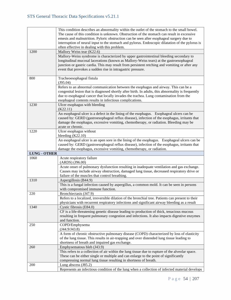
STS General Thoracic Data Specifications v5.21.1
P a g e 54 | 207
This condition describes an abnormality within the outlet of the stomach to the small bowel. The cause of this condition is unknown. Obstruction of the stomach can result in excessive emesis and malnutrition. Pyloric obstruction can be seen after esophageal surgery due to interruption of neural input to the stomach and pylorus. Endoscopic dilatation of the pylorus is often effective in dealing with this problem.
1200 Mallory Weiss tear (K22.6) Mallory-Weiss syndrome is characterized by upper gastrointestinal bleeding secondary to longitudinal mucosal lacerations (known as Mallory-Weiss tears) at the gastroesophageal junction or gastric cardia. This may result from persistent retching and vomiting or after any event that provokes a sudden rise in intragastric pressure.
800 Tracheoesophageal fistula (J95.04) Refers to an abnormal communication between the esophagus and airway. This can be a congenital lesion that is diagnosed shortly after birth. In adults, this abnormality is frequently due to esophageal cancer that locally invades the trachea. Lung contamination from the esophageal contents results in infectious complications.
1230 Ulcer esophagus with bleeding (K22.11) An esophageal ulcer is a defect in the lining of the esophagus. Esophageal ulcers can be caused by: GERD (gastroesophageal reflux disease), infection of the esophagus, irritants that damage the esophagus, excessive vomiting, chemotherapy, or radiation. Bleeding may be acute or chronic.
1220 Ulcer esophagus without bleeding (K22.10) An esophageal ulcer is an open sore in the lining of the esophagus. Esophageal ulcers can be caused by: GERD (gastroesophageal reflux disease), infection of the esophagus, irritants that damage the esophagus, excessive vomiting, chemotherapy, or radiation.
LUNG - OTHER 1060 Acute respiratory failure
(ARDS) (J96.00) Acute onset of pulmonary dysfunction resulting in inadequate ventilation and gas exchange. Causes may include airway obstruction, damaged lung tissue, decreased respiratory drive or failure of the muscles that control breathing.
1310 Aspergillosis (B44.9) This is a fungal infection caused by aspergillus, a common mold. It can be seen in persons with compromised immune function.
220 Bronchiectasis (J47.9) Refers to a localized, irreversible dilation of the bronchial tree. Patients can present to their physicians with recurrent respiratory infections and significant airway bleeding as a result
1340 Cystic fibrosis (E84.0) CF is a life-threatening genetic disease leading to production of thick, tenacious mucous resulting in frequent pulmonary congestion and infections. It also impacts digestive enzymes and function.
250 COPD/Emphysema (J44.9/J43.8) A form of chronic obstructive pulmonary disease (COPD) characterized by loss of elasticity of the lung tissue. This results in air-trapping and over distended lung tissue leading to shortness of breath and impaired gas exchange.
260 Emphysematous bleb (J43.9) This refers to a collection of air within the lung tissue due to rupture of the alveolar space. These can be either single or multiple and can enlarge to the point of significantly compressing normal lung tissue resulting in shortness of breath.
200 Lung abscess (J85.2) Represents an infectious condition of the lung when a collection of infected material develops

STS General Thoracic Data Specifications v5.21.1
P a g e 55 | 207
within the substance of the lung. 270 Interstitial lung disease/fibrosis
(J84.1) Refers to a number of conditions that lead to the progressive scarring of lung tissue. This scarring results in significant respiratory dysfunction and in its most severe form can lead to respiratory failure. In general, the scarring is irreversible
210 Pneumothorax (J93.1) This is a process that occurs when the lining of the lung parenchyma is disrupted and air leaks into the pleural space (the space between the lung and rib cage). This leads to varying degrees of lung collapse and subsequent symptomatology.
310 Solitary pulmonary nodule (not a tumor, e.g., granuloma, subpleural lymph node, pulmonary infarct) (R91.1) A solitary pulmonary nodule is defined as a discrete, well-marginated, rounded opacity less than or equal to 3 cm in diameter that is completely surrounded by lung parenchyma, does not touch the hilum or mediastinum, and is not associated with adenopathy, atelectasis, or pleural effusion. Lesions larger than 3 cm are considered masses and have a higher risk of malignancy.
1850 Atelectasis (J98.11) Atelectasis is the collapse or closure of a lung resulting in reduced or absent gas exchange. It may affect part or all of a lung. It is usually not bilateral. It is a condition where the alveoli are deflated down to little or no volume, as distinct from pulmonary consolidation, in which they are filled with liquid.
1860 Bronchopleural fistula (J98.09) Bronchopleural fistula (BPF) is a communication in the form of a sinus tract between the pleural space and the bronchial tree. BPF carries a high morbidity and mortality and is associated with prolonged hospital stay and thus high resource consumption. Surgical closure may be attempted, although cavernostomy/Eloesser flap may be required.
1880 Chronic respiratory failure (J96.1) Hypoxia (also known as hypoxiation) is a condition in which the body or a region of the body is deprived of adequate oxygen supply. Hypoxia may be classified as either generalized, affecting the whole body, or local, affecting a region of the body. Although hypoxia is often a pathological condition, variations in arterial oxygen concentrations can be part of the normal physiology, for example, during hypoventilation training or strenuous physical exercise.
140 Lung tumor, benign (e.g., hamartoma) (D14.30) These are masses within lung tissue that are not malignant. They can grow, but rarely cause symptoms. Benign lung tumors include hamartomas, chondromas, and fibromas.
280 Pneumonia (J18.9) A condition in which a portion of the lung is involved with an active infection. These can be due to bacterial, viral, or fungal organisms. Treatment is aimed at identifying the causative etiology and initiating appropriate antimicrobial therapy.
1380 Post inflammatory pulmonary fibrosis (J84.89) Post-inflammatory pulmonary fibrosis is a condition in which the tissues in the lungs thicken or become scarred. The lung tissues also become rigid, which makes breathing difficult. As post-inflammatory pulmonary fibrosis advances, lung tissue becomes more damaged, and shortness of breath worsens. Post-inflammatory pulmonary fibrosis typically occurs after an infection that causes serious damage to the lung tissues. There is no cure for post-inflammatory pulmonary fibrosis, but medications like corticosteroid drugs may be helpful in managing inflammation and swelling. Damage to the lungs caused by post-inflammatory pulmonary fibrosis is permanent, and those with significant damage may need a lung transplant.
1390 Primary pulmonary

STS General Thoracic Data Specifications v5.21.1
P a g e 56 | 207
hypertension (I27.0) Primary pulmonary hypertension (PPH) is a rare disease characterized by elevated pulmonary artery pressure with no apparent cause. PPH is also termed pre-capillary pulmonary hypertension or, as is currently preferred, idiopathic pulmonary arterial hypertension (IPAH). Untreated IPAH leads to right-sided heart failure and death.
290 Postprocedural Respiratory Failure (J95.82) This refers to a diffuse inflammatory process that typically involves all lung tissue. This condition can lead to severe impairment of gas exchange within the lung.
1070 Pulmonary sequestration (Q33.2) Pulmonary sequestration (also called accessory lung) refers to aberrant formation of segmental lung tissue that has no connection with the bronchial tree or pulmonary arteries. It is a bronchopulmonary foregut malformation (BPFM).
1400 Transplanted lung complication(s) (T86.8XX) Some complications are related to the operation itself, others are a result of immunosuppressive medication, which is needed to prevent rejection. Complications may include bleeding, rejection, bronchiolitis obliterans syndrome, post-transplantation lymphoproliferative disorder, infection, or side effects of long-term use of immunosuppressants.
1080 Gangrene and necrosis of lung (J85.0) Death of lung tissue due to loss of blood supply. Primary causes include pneumonia, pulmonary embolism, neoplasm (tumor). Secondary causes include trauma, surgery disrupting blood supply, lobar torsion, septic emboli, systemic infection, and lung toxicity of chemotherapeutic agents, radiation effect, and foreign body aspiration. Treatment and prognosis depend on the etiology and extent of lung damage.
300 Hemothorax (J94.2) The presence of blood within the pleural space. This may be due to a traumatic event with damage to the chest wall or lung. Treatment may require drainage with a chest tube or surgical intervention to address the bleeding source.
130 Lung tumor, metastatic (C78.00) This condition includes all cancers of the body that spread to the lungs, including other primary lung cancer. Aug 2021: Metastatic lung cancer from a lung primary should be captured here, however new primary lung cancer or synchronous primary lung cancers should be captured with the appropriate lung cancer category of disease and not with C78.00.
1720 Lung nodule/Mass/Other disorders of lung (J98.4) In radiology, a solitary pulmonary nodule (SPN) or coin lesion is a mass in the lung smaller than 3 centimeters in diameter. It can be an incidental finding found in up to 0.2% of chest v-rays and around 1% of CT Scans.
PLEURA 230 Empyema with fistula (J86.0)
This describes an infectious process within the pleural space with evidence of a communication between the bronchial tree within the lung and the pleural space. Treatment involves appropriate antibiotics with drainage of the pleural infection and correction of the bronchopleural fistula.
240 Empyema without fistula (J86.9) This describes an infectious process within the pleural space without evidence of a communication between the bronchial tree within the lung and the pleural space. Pleural infection is usually due to pneumonia within the lung tissue. Treatment involves appropriate antibiotics with drainage of the pleural infection.
500 Pleural effusion, malignant
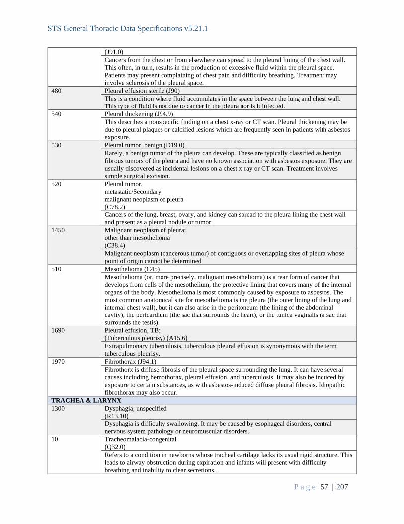
STS General Thoracic Data Specifications v5.21.1
P a g e 57 | 207
(J91.0) Cancers from the chest or from elsewhere can spread to the pleural lining of the chest wall. This often, in turn, results in the production of excessive fluid within the pleural space. Patients may present complaining of chest pain and difficulty breathing. Treatment may involve sclerosis of the pleural space.
480 Pleural effusion sterile (J90) This is a condition where fluid accumulates in the space between the lung and chest wall. This type of fluid is not due to cancer in the pleura nor is it infected.
540 Pleural thickening (J94.9) This describes a nonspecific finding on a chest x-ray or CT scan. Pleural thickening may be due to pleural plaques or calcified lesions which are frequently seen in patients with asbestos exposure.
530 Pleural tumor, benign (D19.0) Rarely, a benign tumor of the pleura can develop. These are typically classified as benign fibrous tumors of the pleura and have no known association with asbestos exposure. They are usually discovered as incidental lesions on a chest x-ray or CT scan. Treatment involves simple surgical excision.
520 Pleural tumor, metastatic/Secondary malignant neoplasm of pleura (C78.2) Cancers of the lung, breast, ovary, and kidney can spread to the pleura lining the chest wall and present as a pleural nodule or tumor.
1450 Malignant neoplasm of pleura; other than mesothelioma (C38.4) Malignant neoplasm (cancerous tumor) of contiguous or overlapping sites of pleura whose point of origin cannot be determined
510 Mesothelioma (C45) Mesothelioma (or, more precisely, malignant mesothelioma) is a rear form of cancer that develops from cells of the mesothelium, the protective lining that covers many of the internal organs of the body. Mesothelioma is most commonly caused by exposure to asbestos. The most common anatomical site for mesothelioma is the pleura (the outer lining of the lung and internal chest wall), but it can also arise in the peritoneum (the lining of the abdominal cavity), the pericardium (the sac that surrounds the heart), or the tunica vaginalis (a sac that surrounds the testis).
1690 Pleural effusion, TB; (Tuberculous pleurisy) (A15.6) Extrapulmonary tuberculosis, tuberculous pleural effusion is synonymous with the term tuberculous pleurisy.
1970 Fibrothorax (J94.1) Fibrothorx is diffuse fibrosis of the pleural space surrounding the lung. It can have several causes including hemothorax, pleural effusion, and tuberculosis. It may also be induced by exposure to certain substances, as with asbestos-induced diffuse pleural fibrosis. Idiopathic fibrothorax may also occur.
TRACHEA & LARYNX 1300 Dysphagia, unspecified
(R13.10) Dysphagia is difficulty swallowing. It may be caused by esophageal disorders, central nervous system pathology or neuromuscular disorders.
10 Tracheomalacia-congenital (Q32.0) Refers to a condition in newborns whose tracheal cartilage lacks its usual rigid structure. This leads to airway obstruction during expiration and infants will present with difficulty breathing and inability to clear secretions.

STS General Thoracic Data Specifications v5.21.1
P a g e 58 | 207
50 Tracheostomy-hemorrhage (J95.01) Describes excessive bleeding as a result of a tracheostomy tube. This may be due to granulation tissue within the airway or may represent the presence of a communication between the trachea and innominate artery or tracheoinnominate fistula.
120 Vocal cord paralysis unspecified (J38.00) Vocal cord paresis (or paralysis) is weakness of one or both of the vocal folds. Symptoms of paresis include hoarseness; vocal fatigue’ mild to severe reduction in vocal volume; pain in the throat when speaking; shortness of breath’ aspiration (foods of liquids going down the trachea) with frequent resultant coughing, and in extreme cases may cause death.
1040 Vocal cord paralysis, unilateral (J38.01) One of the two vocal cords is immobile or has extremely limited movement. This often impacts speech and swallowing.
1050 Vocal cord paralysis, bilateral (J38.02) Both vocal cords are immobile, often stuck partially open. This impacts speech and can lead to difficulty swallowing and aspiration.
TRAUMA 880 Flail chest (S22.5)
Describes a condition when a segment of ribs becomes separated from the rest of the chest wall as a result of multiple rib fractures. Patients often experience respiratory compromise as a result of impaired breathing mechanics.
860 Rib fracture (S22.39xa) Injury to the chest wall may result in rib fractures. Alone, these injuries are usually self-limited. However, rib fractures can cause a pneumothorax or hemothorax.
1240 Rib fractures, multiple (S22.49) Fractures involving more than one rib, typically caused by trauma
870 Sternal fracture (S22.20) These can be caused by blunt trauma to the chest and may herald more serious injuries. If significantly displaced, surgical fixation may be necessary.
890 Tracheal injury (S12.8) This life-threatening injury may be due to blunt or penetrating trauma to the neck or chest. Airway obstruction can result as a consequence. Surgical intervention is often required to address the airway injury.
900 Traumatic pneumothorax (S27.0) Collapse of a lung may occur as a result of either blunt or penetrating trauma to the chest. Chest tube placement is frequently needed to drain the pleural space.
MISCELLANEOUS 1030 Abnormal radiologic finding
(R91) This is a generalized explanation to describe atypical imaging results reported by a radiologist. Abnormal radiologic findings may initiate diagnostic procedures to determine the exact nature of the lesion identified.
1290 Chylothorax (I89.8) Chylothorax refers to the presence of lymphatic fluid in the pleural space secondary to leakage from the thoracic duct or one of its main tributaries.
1540 Disruption of internal operation, surgical wound (T81.32) Disruption or dehiscence of closure of: fascia, superficial or muscular, muscle or muscle flap, ribs or rib cage, or sternum or sternotomy. Do not assign this code when the surgeon purposely leaves the wound open.

STS General Thoracic Data Specifications v5.21.1
P a g e 59 | 207
1550 Hemorrhage complicating a procedure (multiple codes) Bleeding related to the surgical procedure. Do not assign hemorrhage as a complication of a procedure when the blood loss is from the disease itself, such as bleeding esophageal varices or angiodysplasia.
1560 Hematoma complicating a procedure (multiple codes) A hematoma is a localized collection of blood outside the blood vessels, usually in liquid form within the tissue in this case resulting from a surgical procedure.
1570 Hemoptysis unspecified (R04.2) Hemoptysis is the coughing up of blood or bloody sputum from the lungs or airway. It may be either self-limiting or recurrent. Hemoptysis can be caused by a range of disorders: infections (pneumonia; tuberculosis; aspergillosis; and parasitic diseases), tumors that erode blood vessel walls, cocaine abuse, trauma, vascular disorders, bronchitis, foreign bodies in airway, coagulopathies, or as a result of invasive procedures.
1250 Hyperhidrosis, focal axilla (L74.510) Hyperhidrosis is a condition characterized by excessive sweat production. It may involve the hands, axillae, or feet. Disruption of the sympathetic chain via thoracoscopic techniques is a treatment option.
1260 Hyperhidrosis, focal, face (L74.511) Hyperhidrosis is a condition characterized by excessive sweat production. It may involve the hands, axillae, or feet. Disruption of the sympathetic chain via thoracoscopic techniques is a treatment option.
1270 Hyperhidrosis, focal, palms (L74.512) Hyperhidrosis is a condition characterized by excessive sweat production. It may involve the hands, axillae, or feet. Disruption of the sympathetic chain via thoracoscopic techniques is a treatment option.
1020 Lymphadenopathy (R59.9) This refers to enlargement of a lymph node or group of lymph nodes and may be due to benign processes or metastatic cancer.
1600 Malignant poorly differentiated neuroendocrine carcinoma, any site (C74.1) Neuroendocrine tumors are a heterogeneous group of solid tumors that originate from neuroendocrine cells found throughout the body.
1610 Other complication of procedure, not elsewhere specified (i.e., Non-healing surgical wound) (T81.89) A non-healing or chronic wound is defined as a wound that does not improve after four weeks or does not heal in eight weeks.
1620 Other post- op infection (T81.4XXA) Infection acquired following surgery not otherwise listed
1630 Persistent post-op fistula not otherwise classified (T81.83) A fistula is an abnormal connection between two epithelialized surfaces. Fistulas are usually caused by injury or surgery, but they can also result from an infection or inflammation.
1640 Post-operative air leak (J95.812) A post-operative air leak may follow lung surgery and involves air escaping into the pleural

STS General Thoracic Data Specifications v5.21.1
P a g e 60 | 207
space. This usually resolves with chest tube therapy. A prolonged air leak is an air leak that lasts beyond postoperative day 5.
1650 Secondary malignant neoplasm of other specified sites (C79.89) A cancerous tumor in a site or organ separate from the primary tumor, does not include lymph node metastasis. Include diaphragm tumor, metastatic.
1660 Shortness of breath (R06.02) Shortness of breath (dyspnea) is a subjective experience of breathing discomfort that consists of qualitatively distinct sensations that vary in intensity. Distinct sensations include effort/work, chest tightness, and air hunger (the feeling of not enough oxygen). Dyspnea is a normal symptom of heavy exertion but becomes pathological if it occurs in unexpected situations. It may result from asthma, pneumonia, cardiac ischemia, interstitial lung disease, congestive heart failure, chronic obstructive pulmonary disease, diaphragm dysfunction, deconditioning or psychogenic causes such as panic disorder and anxiety.
1280 Other unlisted category of disease Diagnosis not in any of the listed categories.
SeqNo: 1260 Long Name: Category Of Disease - Primary - Other Specify Short Name: CategoryPrimOth Format: Text Definition: Indicate the PRIMARY diagnosis (category of disease) not listed for which the procedure was performed. Choose from the list, when possible if the category of disease is not listed, enter free text. ParentLongName: Category Of Disease - Primary ParentShortName: CategoryPrim ParentValue: 1280 ParentHarvestCodes: = "Other unlisted category of disease" Intent/Clarification: Capture unlisted primary diagnosis here after carefully reviewing choices above. SeqNo: 1270 Long Name: Category Of Disease - Primary - Other ICD Short Name: CategoryPrimOthICD Format: Text Definition: Enter ICD-9(April 9, 2021) or ICD-10 code, if known, of other primary diagnosis (category of disease) not listed. ParentLongName: Category Of Disease - Primary ParentShortName: CategoryPrim ParentValue: 1280 ParentHarvestCodes: = "Other unlisted category of disease" Intent/Clarification: Indicate the ICD-10 code of the other primary diagnosis not listed.

STS General Thoracic Data Specifications v5.21.1
P a g e 61 | 207
----------------------------------------------------------------------------------------------------------------------------------------------------------------- Operative -------------------------------------------------------------------------------------------------------------------------------------- SeqNo: 1310 Long Name: Date Of Surgery Short Name: SurgDt Format: Date in mm/dd/yyyy format Definition: Indicate the date of surgery, which equals the date the patient enters the operating room. Intent/Clarification: This field is required for record inclusion. If missing data, the entire record will be excluded from the analysis. SeqNo: 1320 Long Name: OR Entry Time Short Name: OREntryT Format: Time in 24-hour hh:mm format Definition: Indicate to the nearest minute (using 24 hour clock) the time the patient enters the operating room.
Intent/Clarification: This should be collected from the same place every time (i.e., always from anesthesia report). Even if the thoracic surgeon was present only part of the case, code the entire OR time.
This field is required for record inclusion. If missing data, the entire record will be excluded from the analysis. SeqNo: 1330 Long Name: OR Exit Time Short Name: ORExitT Format: Time in 24-hour hh:mm format Definition: Indicate to the nearest minute (using 24 hour clock) the time the patient exits the operating room.
Intent/Clarification: This should be collected from the same place every time (i.e., always from anesthesia report). Even if the thoracic surgeon was present only part of the case, code the entire OR time.
This field is required for record inclusion. If missing data, the entire record will be excluded from the analysis. SeqNo: 1340 Long Name: Anesthesia Start Time Short Name: AnesthStartT Format: Time in 24-hour hh:mm format

STS General Thoracic Data Specifications v5.21.1
P a g e 62 | 207
Definition: Indicate the time of anesthesia induction. Intent/Clarification: This should be collected from the same place every time (i.e., always from anesthesia report). This is the start of anesthetic management, placing lines, induction of anesthesia. This time should be recorded on the anesthesia record. SeqNo: 1350 Long Name: Anesthesia End Time Short Name: AnesthEndT Format: Time in 24-hour hh:mm format Definition: Indicate the anesthesia end time documented in the medical record. The definition of
anesthesia end time is when the anesthesiologist is no longer in personal attendance, that is, when the patient is safely placed under post-anesthesia supervision.
Intent/Clarification: This should be collected from the same place every time (i.e., always from anesthesia report). The time may be in the Recovery Room or ICU; when it is documented that anesthesia care has ended. SeqNo: 1360 Long Name: Procedure Start Time Short Name: ProcStartT Format: Time in 24-hour hh:mm format Definition: Indicate the time the procedure started. Intent/Clarification: This should be collected from the same place every time. The Anesthesia record is the preferred source document for this field.
This field is required for record inclusion. If missing data, the entire record will be excluded from the analysis. SeqNo: 1370 Long Name: Procedure End Time Short Name: ProcEndT Format: Time in 24-hour hh:mm format Definition: Indicate the time the procedure ended. Intent/Clarification: This should be collected from the same place every time. The Anesthesia record is the preferred source document for this field. This field is required for record inclusion. If missing data, the entire record will be excluded from the analysis. SeqNo: 1380 Long Name: Multi-Day Operation Short Name: MultiDay Format: Text (categorical values specified by STS) Definition: Indicate whether the operation continued through midnight from one day to the next.

STS General Thoracic Data Specifications v5.21.1
P a g e 63 | 207
Harvest Codes: Code: Value: 1 Yes 2 No Intent/Clarification: Indicate if the procedure start time (ProcStartT – seq 1360) and procedure end time (ProcEndT – seq 1370) continue through midnight. SeqNo: 1390 Long Name: Status Of Operation Short Name: Status Format: Text (categorical values specified by STS) Definition: Indicate the status that best describes the clinical status of the patient at the time of the primary surgical procedure. 1. Emergent: The surgical procedure must be performed within 24 hours of presentation. 2. Urgent: All of the following conditions are met: a. Not elective status b. Not emergent status. c. Procedure required during same hospitalization in order to minimize chance of further clinical deterioration. 3. Elective: The patient has been stable in the days or weeks prior to the operation. 4. Palliative: procedure intended to provide comfort or relief. Harvest Codes: Code: Value: 1 Emergent 2 Urgent 3 Elective 4 Palliative Intent/Clarification:
- Emergent status is coded for cases that require immediate intervention to prevent life threatening deterioration or death such as (but not limited to) esophageal perforation, severe hemorrhage, or massive hemoptysis.
- Urgent status is coded for cases in which the operation must be performed before the patient can be discharged. Examples of urgent cases would include bronchopleural fistula, pneumothorax, or decortication for empyema.
- Elective status is coded for cases that are performed during the same hospitalization for convenience would not be considered urgent. A medical patient with an incidental CXR finding who undergoes a diagnostic bronchoscopy or mediastinoscopy prior to discharge would have the procedure status coded as elective.
- Palliative status is coded for treatment intended to provide comfort or relief during end-of-life care. Treatment of malignant pleural effusions is often palliative and may include pleurodesis or placement of a chronic indwelling pleural drain (e.g., Pleurx catheter).
SeqNo: 1400 Long Name: Robotic Technology Assisted Short Name: Robotic

STS General Thoracic Data Specifications v5.21.1
P a g e 64 | 207
Format: Text (categorical values specified by STS) Definition: Indicate whether the thoracic surgery was assisted by robotic technology. Harvest Codes: Code: Value: 1 Yes 2 No Intent/Clarification: This field is to capture the use of robotics at any time during the procedure, including cases where the approach was converted (i.e., converted to open (UnanticConv – seq 1430)). In addition to coding this field (Robotic – seq 1400), please also code the use of robotics within the procedures performed section (Proc – seq 1470) by selecting Robotic-Assisted Surgery (capture as an additional code) (S2900). SeqNo: 1410 Long Name: Unanticipated Surgical Approach Conversion Short Name: UnanticConv Format: Text (categorical values specified by STS) Definition: Indicate whether or not there was an unanticipated conversion of the surgical approach. Some surgeons put a scope in / VATS to have a look and make sure there isn't wide spread disease. If disease is not widespread, their plan is to operate via thoracotomy. These are not conversions and should be listed as thoracotomy. If the plan was to try the resection via VATS , and they convert to thoracotomy for any reason, it should be listed as a conversion. Discuss with the surgeon to determine if the intent was to complete by VATs. If the answer is "yes" but could not, then it is a conversion. Of note, all surgeons counsel patients there is a chance of conversion for every case. Because they counsel the patient and "planned for it" by putting it on a consent does not preclude this from counting the case as a conversion. Harvest Codes: Code: Value: 1 Yes, VATS to open 2 Yes, Robotic to VATS 3 Yes, Robotic to open 4 No
Intent/Clarification: Conversion in a procedure does not mean something always went wrong or was not anticipated preoperatively; many times it is done for better visibility, need to palpate structures, or inability to reach a vital area, etc. This should not be viewed as a punitive data element.
Example: Patient to operating room for VATS Lobectomy. Unable to complete lobectomy as VATS. Converted to Thoracotomy. Code as ‘Yes, VATS to open’ and capture this as a thoracotomy in the procedure section (Proc – seq 1470). Example: For cases where the initial approach is a VATS and then converted to mini-thoracotomy, only consider this a conversion if retractors are used. Example: Lobe is removed via VATS then conversion must occur. Converted to Thoracotomy. Code as ‘Yes, VATS to open’ and capture the lobectomy as a thoracotomy in the procedure section (Proc – seq 1470).

STS General Thoracic Data Specifications v5.21.1
P a g e 65 | 207
In instances where the specimen/utility bag is too large to pass through the incision, only code as a conversion if retractors are used to facilitate the removal of the specimen/utility bag. For cases where the initial procedure is a RATS (Robotic Assisted Thoracoscopic Surgery) converted to open; code this as “Yes, Robotic to Open.” SeqNo: 1420 Long Name: Unanticipated Surgical Approach Conversion Type Short Name: UnanticConvTy Format: Text (categorical values specified by STS) Definition: Indicate the type of surgical approach conversion. ParentLongName: Unanticipated Surgical Approach Conversion ParentShortName: UnanticConv ParentValue: 1|2|3 ParentHarvestCodes: = "Yes, VATS to open", "Yes, Robotic to VATS" or "Yes, Robotic to open" Harvest Codes: Code: Value: 1 Elective 2 Emergent Intent/Clarification: Bleeding or injury to an adjacent structure which effects patient stability are a couple of examples of when conversion would be emergent. Please check with your surgeon when coding this field if there is a question as to whether a conversion was performed electively or emergently. SeqNo: 1430 Long Name: Unanticipated Surgical Approach Conversion Reason Short Name: UnanticConvRsn Format: Text (categorical values specified by STS) Definition: Indicate the reason for the surgical approach conversion. ParentLongName: Unanticipated Surgical Approach Conversion ParentShortName: UnanticConv ParentValue: 1|2|3 ParentHarvestCodes: = "Yes, VATS to open", "Yes, Robotic to VATS" or "Yes, Robotic to open" Harvest Codes: Code: Value: 1 Vascular 2 Anatomy 3 Lymph nodes 5 Other Intent/Clarification: - Vascular: pulmonary artery or vein injury, intercostal or other vascular injury - Anatomy: adhesions, visualization issues, tumor size or location, inability to tolerate single lung ventilation - Lymph nodes: bulky, sticky, or calcified lymph nodes - Other: staple misfire, equipment malfunction

STS General Thoracic Data Specifications v5.21.1
P a g e 66 | 207
If the conversion is performed due to ‘desaturation’ and no other reason is coded (i.e., vascular injury) then code this as ‘other.’ SeqNo: 1440 Long Name: Intraoperative Packed Red Blood Cells Short Name: IntraopPRBC Format: Text (categorical values specified by STS) Definition: Indicate whether the patient received packed Red Blood Cells intraoperatively. Harvest Codes: Code: Value: 1 Yes 2 No Intent/Clarification: Intraoperatively is defined as any blood started inside of the OR (between Procedure Start and Procedure Stop times). For these Intraop Blood Product data fields the intent is to ONLY collect blood products that were transfused any time Intra-operatively during THIS SURGERY. Whole blood is also captured here. SeqNo: 1450 Long Name: Intraoperative Packed Red Blood Cells - Number Short Name: IntraopPRBCNum Format: Integer Definition: Indicate the number of units of packed Red Blood Cells the patient received intraoperatively. Low Value: 1 High Value: 300 ParentLongName: Intraoperative Packed Red Blood Cells ParentShortName: IntraopPRBC ParentValue: 1 ParentHarvestCodes: = "Yes" Intent/Clarification: This can be found in the EMR, anesthesia or operative record or blood transfusion records. SeqNo: 1460 Long Name: ASA Classification Short Name: ASA Format: Text (categorical values specified by STS) Definition: Indicate the patient’s American Society of Anesthesiologists Risk Scale for this surgical procedure. Harvest Codes: Code: Value: Definition: 1 I. Normal, healthy patient 2 II Mild systemic disease 3 III Severe systemic disease 4 IV Life threatening severe systemic disease 5 V Moribund, not expected to survive without operation

STS General Thoracic Data Specifications v5.21.1
P a g e 67 | 207
6 VI Declared brain dead, organ donor
Intent/Clarification: ASA Classification is determined by the anesthesiologist of the procedure based on the patient’s condition. This is a standard risk scale for patients undergoing anesthesia.
- I = A normal healthy patient - II = A patient with mild systemic disease - III = A patient with severe systemic disease - IV = A patient with severe systemic disease that is a constant threat to life - V = A moribund patient who is not expected to survive without the operation - VI = A declared brain-dead patient whose organs are being removed for donor purposes This field is required for record inclusion. If missing data, the entire record will be excluded from the analysis. SeqNo: 1470 Long Name: Procedure Short Name: Proc Format: Text (categorical values specified by STS) Definition: Indicate the general thoracic procedures being performed during this operating room visit. Intent/Clarification: Check ALL the procedures that were performed. Complete Primary (Primary – seq 1480) to indicate Primary procedure. Each procedural section (i.e., Lung Cancer Resection, Concomitant Lung, Esophagectomy, etc.) has been assigned a weight. If procedures are performed from multiple procedural sections during the same OR encounter, code the highest weighted procedure as primary (Primary 1480).
- 70 – Esophagectomy - 60 – Lung Cancer Resection - 50 – Thymus/Mediastinal - 40 – Tracheal Resection - 30 – Hiatal Hernia/GERD - 20 – Concomitant Lung - 10 – Minor Procedures - Example: In the rare instance, that a patient has a primary lung and a primary esophageal cancer resected
concurrently: A thoracotomy wedge resection (32505) and a partial esophagectomy with thoracotomy (43112) are performed within the same visit to the OR. The esophagectomy has a higher weight (70) versus the thoracotomy/wedge resection (60); Code the esophagectomy as the primary procedure at Primary – seq 1480.
- Example: A thymectomy is performed with en bloc wedge resection of the lung to excise the thymic mass
completely. Thymus/Mediastinal (50) would be selected as the primary procedure versus the concomitant lung procedure (20).
The General Thoracic Surgery Database requires a separate data collection form for every OR/procedural area visit for major general thoracic procedure(s). Note: Not all procedures will have an assigned procedure code, if not listed please use ‘other’ or ‘other minor procedure.’
• Remember that billing codes do not always accurately capture the clinical procedure. Check with the surgeon if clarification is necessary.

STS General Thoracic Data Specifications v5.21.1
P a g e 68 | 207
• When trying to determine thoracotomy vs thoracoscopy, remember that if a rib spreader is used, the case is considered an open case (thoracotomy) regardless of the incision size.
Non-analyzed procedures are located in the Minor/Non-Analyzed Procedures (Concomitant Procedures) on the DCF. This field is required for Record Inclusion. If missing data, the entire record will be excluded from the analysis. Harvest Codes:
Code: Value: Lung Cancer Resection (Required)
Weight = 60
2800
Thoracoscopy, surgical; with lobectomy (32663) This is therapeutic procedure to remove an anatomic lobe of the lung requiring vascular and bronchial dissection done via small incisions (approximately 1 to 3 cm.) with a scope and other instruments. A rib spreader is not used.
4070
Thoracoscopy with therapeutic wedge resection (e.g., mass or nodule) initial, unilateral (32666) Minimally invasive removal of a section of diseased (typically cancerous) lung tissue. Thoracoscopy, sometimes abbreviated as ‘VATS’ (video assisted thoracoscopy) is performed through several small openings rather than a large chest wall incision.
4100
Thoracoscopy with removal of lung segment(s) (segmentectomy) (32669) Minimally invasive removal of a segment of lung tissue, larger than a wedge but smaller than a lobe, with segmental bronchus and pulmonary artery division.
4110
Thoracoscopy with removal of two lobes (bilobectomy) (32670) Minimally invasive excision of two lobes of the right lung, either right upper and middle or right lower and middle lobes.
4120 Thoracoscopy with removal of lung, pneumonectomy (32671) Minimally invasive excision of one lung.
4140
Thoracotomy with therapeutic wedge resection (e.g., mass nodule) initial (32505) Removal of a wedge of lung tissue with pathology (typically cancer) using an open surgical approach (thoracotomy).
2470
Removal of lung, total pneumonectomy; (32440) Resection of the entire lung most commonly for primary lung cancer, although there are other indications such as metastatic or inflammatory disease. Intrapericardial pneumonectomy describes when the major blood vessels are isolated and divided within the pericardial sac. The procedures may be performed by VATS, thoracotomy, or sternotomy.
2480
Removal of lung, sleeve (carinal) pneumonectomy (32442) Pneumonectomy with removal of both main stem bronchi with reconstruction of the remaining bronchus to the trachea by sutured anastomosis. This is usually done for primary airway tumors, such as adenoid cystic or mucoepidermoid carcinomas. Right-sided resection is performed through a right thoracotomy and left-sided resection requires bilateral thoracotomies. Less commonly, a sternotomy may give access for either side.
2500 Removal of lung, single lobe (lobectomy) (32480) Resection of a lobe of the lung most commonly for primary lung cancer. It can be performed by thoracotomy or sternotomy.
2510 Removal of lung, two lobes (bilobectomy) (32482) Removal of either the right upper and middle or the middle and lower lobes of the lung typically for lung cancer involving both adjacent lobes. It may be performed by VATS, thoracotomy, or

STS General Thoracic Data Specifications v5.21.1
P a g e 69 | 207
sternotomy.
2520
Removal of lung, single segment (segmentectomy) (32484) Describes resection of an anatomic segment within a lobe. It is performed for lesions occupying a segment as defined by a separate pulmonary artery, bronchus and segmental venous drainage that follows the fissures between segments. The indications also include benign tumors, metastatic and primary lung cancers. It can be performed by VATS, thoracotomy, or sternotomy.
2530
Removal of lung, sleeve lobectomy (32486) Defined as a lobectomy with removal of additional airway supplying a neighboring segment or lobe of the lung or the entire lung and reconstruction of the airway by direct suturing. It is usually performed when a tumor or disease process is involving only a portion of the adjacent airway while sparing the lung parenchyma, as in squamous cell lung cancer and primary airway tumors such as carcinoids or mucoepidermoid carcinoma. This is typically performed via thoracotomy but can also be performed via VATS/RATS.
2540
Removal of lung, completion pneumonectomy (32488) Resection of the entire lung in a re-operative setting following a previous lung resection, usually a lobectomy. It is performed most commonly for primary lung cancer, although there are other indications such as metastatic or inflammatory disease.
2570
Resection and repair of portion of bronchus (bronchoplasty) when performed at time of lobectomy or segmentectomy (32501) This refers to removal of a portion of the airway beyond the anatomic confines of either a lobe or segment during anatomic resection followed by primary repair of the airway in order to preserve lung tissue unaffected by the disease process. Bronchoplasty is typically performed through a thoracotomy.
2580
Resection of apical lung tumor (e.g., Pancoast tumor), including chest wall resection, without chest wall reconstruction(s) (32503) Describes resection of a primary lung tumor, usually NSCLC, located in the superior sulcus (anterior or posterior) with simultaneous removal of the involved ribs without prosthetic reconstruction. The lung resection is usually a lobectomy but may also be a segmentectomy or wedge resection depending on the size of the lesion and respiratory capacity of the patient.
2590
Resection of apical lung tumor(e.g., Pancoast tumor), including chest wall resection, with chest wall reconstruction (32504) Describes resection of a primary lung tumor, usually NSCLC, located in the superior sulcus (anterior or posterior) with simultaneous removal of the involved ribs with prosthetic reconstruction. The lung resection is usually a lobectomy but may also be a segmentectomy or wedge resection depending on the size of the lesion and respiratory capacity of the patient.
4800 Resection of lung with resection of chest wall Use this code for major lung resections requiring chest wall resection but does not meet criteria of apical lung tumor.
Concomitant Lung Procedures (Weight = 20)
4080
Thoracoscopy with therapeutic wedge resection (e.g., mass or nodule) each additional resection, ipsilateral (32667) List separately in addition to primary procedure code Minimally invasive removal of additional lung tissue wedges on the same side as the initial wedge resection. This is coded in addition to primary procedure.
4170
Thoracoscopy with mediastinal and regional lymphadenectomy (+32674) List separately in addition to primary procedure code Removal of lymph nodes using a minimally invasive approach from the mediastinum. Lymphadenectomy or lymph node dissection is the surgical removal of one or more groups of lymph

STS General Thoracic Data Specifications v5.21.1
P a g e 70 | 207
nodes. Do not code for removal of one lymph node. It is almost always performed as part of the surgical management of cancer. Do not code as primary procedure.
Please note that lymphadenectomy is already captured in the nodal section. This value will be removed in version 5.24 (2024). Only code this for thoracoscopic lymphadenectomy, do not use for open lymphadenectomy – the STS is not capturing open lymphadenectomy (38746).
4150
Thoracotomy with therapeutic wedge resection (e.g., mass nodule) each additional resection, ipsilateral (+32506) List separately in addition to primary procedure code Removal of multiple wedges of lung tissue with pathology (typically cancer) using an open surgical approach Ipsilateral = same side as primary resection. Do not code this as a primary procedure.
4160
Thoracotomy with diagnostic wedge resection followed by anatomic lung resection (+32507), List separately in addition to primary proc code Open surgical removal of a lung tissue sample for biopsy/diagnosis prior to therapeutic resection. Do not code this as a primary procedure.
4090
Thoracoscopy with diagnostic wedge resection followed by anatomic lung resection (32668), List separately in addition to primary procedure code Minimally invasive removal of a lung tissue sample for biopsy/diagnosis prior to therapeutic resection. Do not code this as a primary procedure.
Esophageal Cancer Procedures (Required) (Weight = 70)
3320
Transhiatal-Total esophagectomy, without thoracotomy, with cervical esophagogastrostomy (43107) Removal of the esophagus through an upper midline laparotomy and a neck incision. Intestinal continuity is restored by the formation of a gastric tube with an anastomosis between the gastric tube and remaining cervical esophagus.
3380
Total esophagectomy without thoracotomy; with colon interposition or small intestine reconstruction (43108) Removal of the esophagus through an upper midline laparotomy and a neck incision. Intestinal continuity is restored by the formation of a colonic or small bowel conduit with an anastomosis between the conduit and the remaining cervical esophagus.
3330
Three Incision -Total esophagectomy with thoracotomy; with cervical esophagogastrostomy (43112) Removal of the esophagus through an upper midline laparotomy, a right thoracotomy, and a neck incision. Intestinal continuity is restored by the formation of a gastric tube with an anastomosis between the gastric tube and remaining cervical esophagus.
3390
Total esophagectomy with thoracotomy; with colon interposition or small intestine reconstruction (43113) Removal of the esophagus through an upper midline laparotomy, a right thoracotomy, and a neck incision. Intestinal continuity is restored by the formation of a colonic or small intestine tube with an anastomosis between the gastric tube and remaining cervical esophagus.
3400 Partial esophagectomy,

STS General Thoracic Data Specifications v5.21.1
P a g e 71 | 207
cervical, with free intestinal graft, including microvascular anastomosis (43116) Removal of a short segment of cervical esophagus through a neck incision with or without sternal extension. Intestinal continuity is restored by the free transfer of small bowel requiring anastomosis between the conduit and the remaining proximal and distal esophagus. Blood flow must also be established to the small bowel segment by arterial and venous micro-anastomoses.
3340
Ivor Lewis-Partial esophagectomy, distal two- thirds, with thoracotomy and separate abdominal incision (43117) Removal of the distal two thirds of the esophagus through an upper midline laparotomy and a right thoracotomy. Intestinal continuity is restored by the formation of a gastric tube with an anastomosis between the gastric tube and remaining esophagus within the right chest.
3410
Partial esophagectomy, with thoracotomy and separate abdominal incision with colon interposition or small intestine (43118) Removal of the distal two thirds of the esophagus through an upper midline laparotomy and a thoracotomy. Intestinal continuity is restored by the formation of a colon or small intestine conduit with anastomosis between the conduit and remaining esophagus within the chest.
3420
Partial esophagectomy, distal two-thirds, with thoracotomy only (43121) Removal of the distal esophagus through a left thoracotomy approach with anastomosis of the stomach to the distal esophagus in the left chest.
3350
Thoracoabdominal-Partial esophagectomy, thoracoabdominal approach (43122) Removal of the distal esophagus through a left thoracoabdominal approach with anastomosis of the stomach to the distal esophagus in the left chest.
3430
Partial esophagectomy, thoracoabdominal with colon interposition or small intestine (43123) Removal of the distal esophagus through a left thoracoabdominal approach. Intestinal continuity is restored by the formation of a colonic or small intestine tube with an anastomosis between the conduit and remaining esophagus within the left chest.
3440
Total or partial esophagectomy, without reconstruction with cervical esophagostomy (43124) Removal of the esophagus without re-establishment of intestinal continuity. An end cervical esophagostomy or “spit fistulae” is created.
4190
Minimally invasive three incision esophagectomy (McKeown) (43288) The three-hole technique consists of thoracic mobilization of the esophagus, laparoscopic construction of a gastric conduit and a cervical esophagogastrostomy via minimally invasive approach.
3360 Minimally invasive esophagectomy, Ivor Lewis approach (43287)
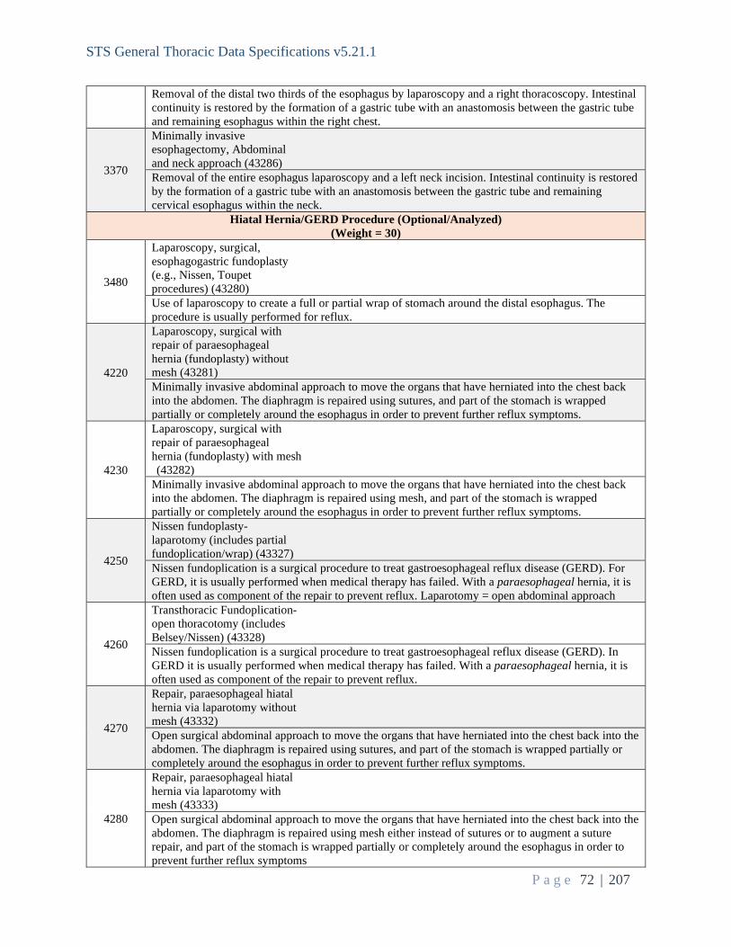
STS General Thoracic Data Specifications v5.21.1
P a g e 72 | 207
Removal of the distal two thirds of the esophagus by laparoscopy and a right thoracoscopy. Intestinal continuity is restored by the formation of a gastric tube with an anastomosis between the gastric tube and remaining esophagus within the right chest.
3370
Minimally invasive esophagectomy, Abdominal and neck approach (43286) Removal of the entire esophagus laparoscopy and a left neck incision. Intestinal continuity is restored by the formation of a gastric tube with an anastomosis between the gastric tube and remaining cervical esophagus within the neck.
Hiatal Hernia/GERD Procedure (Optional/Analyzed) (Weight = 30)
3480
Laparoscopy, surgical, esophagogastric fundoplasty (e.g., Nissen, Toupet procedures) (43280) Use of laparoscopy to create a full or partial wrap of stomach around the distal esophagus. The procedure is usually performed for reflux.
4220
Laparoscopy, surgical with repair of paraesophageal hernia (fundoplasty) without mesh (43281) Minimally invasive abdominal approach to move the organs that have herniated into the chest back into the abdomen. The diaphragm is repaired using sutures, and part of the stomach is wrapped partially or completely around the esophagus in order to prevent further reflux symptoms.
4230
Laparoscopy, surgical with repair of paraesophageal hernia (fundoplasty) with mesh (43282) Minimally invasive abdominal approach to move the organs that have herniated into the chest back into the abdomen. The diaphragm is repaired using mesh, and part of the stomach is wrapped partially or completely around the esophagus in order to prevent further reflux symptoms.
4250
Nissen fundoplasty- laparotomy (includes partial fundoplication/wrap) (43327) Nissen fundoplication is a surgical procedure to treat gastroesophageal reflux disease (GERD). For GERD, it is usually performed when medical therapy has failed. With a paraesophageal hernia, it is often used as component of the repair to prevent reflux. Laparotomy = open abdominal approach
4260
Transthoracic Fundoplication- open thoracotomy (includes Belsey/Nissen) (43328) Nissen fundoplication is a surgical procedure to treat gastroesophageal reflux disease (GERD). In GERD it is usually performed when medical therapy has failed. With a paraesophageal hernia, it is often used as component of the repair to prevent reflux.
4270
Repair, paraesophageal hiatal hernia via laparotomy without mesh (43332) Open surgical abdominal approach to move the organs that have herniated into the chest back into the abdomen. The diaphragm is repaired using sutures, and part of the stomach is wrapped partially or completely around the esophagus in order to prevent further reflux symptoms.
4280
Repair, paraesophageal hiatal hernia via laparotomy with mesh (43333) Open surgical abdominal approach to move the organs that have herniated into the chest back into the abdomen. The diaphragm is repaired using mesh either instead of sutures or to augment a suture repair, and part of the stomach is wrapped partially or completely around the esophagus in order to prevent further reflux symptoms

STS General Thoracic Data Specifications v5.21.1
P a g e 73 | 207
4290
Repair, paraesophageal hiatal hernia via thoracotomy without mesh (43334) Open surgical thoracic approach to move the organs that have herniated into the chest back into the abdomen. The diaphragm is repaired using sutures, and part of the stomach is wrapped partially or completely around the esophagus in order to prevent further reflux symptoms.
4300
Repair, paraesophageal hiatal hernia via thoracotomy with mesh (43335) Open surgical thoracic approach to move the organs that have herniated into the chest back into the abdomen. The diaphragm is repaired using mesh, and part of the stomach is wrapped partially or completely around the esophagus in order to prevent further reflux symptoms.
4310
Repair, paraesophageal hiatal hernia via thoracoabdominal approach without mesh (43336) Open surgical thoracoabdominal approach to move the organs that have herniated into the chest back into the abdomen. The diaphragm is repaired using sutures, and part of the stomach is wrapped partially or completely around the esophagus in order to prevent further reflux symptoms.
4320
Repair, paraesophageal hiatal hernia via thoracoabdominal approach with mesh (43337) Open surgical abdominal approach to move the organs that have herniated into the chest back into the abdomen. The diaphragm is repaired using mesh, and part of the stomach is wrapped partially or completely around the esophagus in order to prevent further reflux symptoms.
4580 LINX Procedure (43284 ) Implantation of the LINXTM device for management of GERD
Tracheal Resection (Optional/Analyzed) (Weight = 40)
2220
Carinal reconstruction (31766) A complex airway reconstruction for a disease process that involves the carina (the bifurcation of the trachea into the two main bronchi). Usually done for tracheal tumors but (rarely) can be done for benign diagnoses as well. The carina is resected and then the three airway ends (the trachea and the two main bronchi) are reconstructed. This operation can be performed via a right thoracotomy, a sternotomy, or a clamshell incision. Institution of cardiopulmonary bypass may be necessary during this operation.
2240
Excision tracheal stenosis, cervical (31780) The operation performed for both benign obstructive lesions of the cervical tracheal. The involved trachea is resected, and the two normal ends of the trachea are anastomosed together. This code would be used for those procedures conducted via a neck incision.
2250
Excision tracheal stenosis, thoracic (31781) Another approach to address benign tracheal pathology where, due to disease location, a partial or complete sternotomy is performed in addition to the neck incision.
2260
Tracheal tumor or carcinoma excision; cervical (31785) Resection of a tracheal tumor via a cervical approach. Involves resecting the section of trachea with the tumor and anastomosing the two divided ends of the trachea together.
2270
Tracheal tumor or carcinoma excision; thoracic (31786) Resection of an intrathoracic tracheal tumor. Usually done via a complete sternotomy or a right thoracotomy. May include a limited cervical incision as well. Involves resecting the section of trachea with the tumor and anastomosing the two divided ends of the trachea together.
Thymus/Mediastinal Mass Resection (Optional/Analyzed) (Weight = 50)

STS General Thoracic Data Specifications v5.21.1
P a g e 74 | 207
2790
Thoracoscopy, surgical; with excision of mediastinal cyst, tumor, or mass (32662) This is a procedure to remove a cyst, tumor or mass from the mediastinum done via small incisions (approximately 1 to 3 cm.) with a scope and other instruments.
4180
Thymus, resection via Thoracoscopy unilateral or bilateral (32673) Minimally invasive approach to resection of the thymus gland (one or both sides).
3210
Mediastinal tumor, excision, open, Transthoracic approach (39220) Most commonly these refer to Schwannomas, teratomas, or other types of malignancies (thymectomy for Thymoma or thymic carcinoma has separate codes). These are almost always solid in nature and may require VATS or open technique for complete resection.
3840
Thymectomy, transcervical approach (60520) This approach uses a collar incision and a retracting arm to gain access to the anterior mediastinum dissecting the thymus up and removing through this neck incision. It is more frequently used for “normal” thymus glands and not for thymomas or tumors.
3850
Thymectomy, transthoracic approach (60521) Almost always refers to a sternotomy and approach similar to a heart surgery with removal of the thymus via this wide exposure. Most frequent approach for larger tumors.
3860
Thymectomy, transthoracic approach, with radical mediastinal dissection (60522) Same as 60521 but with additional resection of pericardium, innominate vein, phrenic nerve, and lymph nodes. Do not code both 60521 and 60522.
MINOR/NON-Analyzed Procedures Trachea, Bronchi, Larynx (Optional/Non-analyzed)
(Weight = 10)
3880 Laryngectomy, partial (31370) Removal of part of the larynx, usually done in conjunction with a tracheal resection and reconstruction
2280
Tracheal wound or injury suture repair; cervical (31800) Partial disruption of the tracheal wall often requires direct surgical repair. When this injury is corrected in the neck, this code should be used.
2290
Tracheal wound or injury suture repair; intrathoracic (31805) Describes direct surgical repair of the intrathoracic trachea, usually performed via a right thoracotomy.
2300 Unlisted procedure, trachea, bronchi (31899)
2880
Bronchopleural fistula closure (32906) Bronchopleural fistula (BPF) is a communication in the form of a sinus tract between the pleural space and the bronchial tree. BPF carries a high morbidity and mortality and is associated with prolonged hospital stay and thus high resource consumption. Surgical closure may be attempted, although cavernostomy/Eloesser flap may be required.
4440 Bronchogenic cyst removal

STS General Thoracic Data Specifications v5.21.1
P a g e 75 | 207
Bronchogenic cysts are abnormal growths of tissue that are congenital (present from birth). They typically have thin walls and are filled with fluid or mucous. Most bronchogenic cysts are found in the mediastinum. Thoracotomy, VATs or robotic approaches may be used for removal.
4450 Bronchial laceration suture Surgical repair of laceration of the bronchus using suture
4470 Bronchoplasty, graft repair (31770) Surgical repair of a defect in the bronchus using tissue or synthetic graft material
2230
Bronchoplasty; excision stenosis and anastomosis (31775) An operation for a localized stenosis (stricture) of one of the major bronchi. Usually done for a benign process such as histoplasmosis or as a result of a stricture after a sleeve lobectomy. Usually done via a thoracotomy. The stenotic bronchus is resected, and the two bronchial ends are then anastomosed together.
4410
Tracheostomy replacement (tube change) prior to est. of fistula tract (31502) Trach placement involves a fistula tract from the skin of the anterior neck to the trachea. If the trach tube must be changed before the tract is fully established (usually after about seven days), report 31502.
3980
Tracheostomy, planned (31600) A planned surgical procedure to create a tracheostomy, an opening through the neck into the trachea (windpipe), a tube is usually placed through this opening to provide an airway and to remove secretions from the lungs.
4420 Tracheostomy revision simple, without flap (31613) Surgical procedure to revise an existing tracheostoma, often enlargement
4510 Tracheostomy revision complex, with flap (31614) Revision of the tracheostoma using a tissue flap or pedicle.
2200 Tracheoplasty; cervical (31750) A rarely performed operation for a deformed trachea to restore its normal shape. Tracheoplasty is usually done for tracheomalacia limited to the cervical region.
2210
Tracheoplasty; intrathoracic (31760) An operation performed for a deformed and softened trachea via a right thoracotomy. The posterior membranous wall of the trachea is plicated and fixed to a piece of mesh to restore the normal “C” shaped trachea.
4460 Bronchial sleeve resection A lung resection in which a section of the proximal bronchus is removed along with diseased lung tissue after which the proximal and distal ends of the bronchus are anastomosed
4520
Tracheostomy mediastinal An anterior mediastinal tracheostomy involves the construction of a tracheostomy stoma on the anterior chest wall using the intrathoracic trachea when there is insufficient length to reanastomose the remaining trachea or to bring the trachea out of the superior mediastinum for a standard suprasternal stoma. The procedure involves laryngectomy (if not done previously) and resection of the upper sternum, the medial third of the clavicles, and the first and usually second ribs. The primary indications for this operation are mostly limited to advanced cervicothoracic neoplasms in the superior mediastinum, although it is done occasionally for benign disease.
4430
Rigid stent removal Stents in the trachea or bronchus are often considered permanent but can be removed surgically or via bronchoscopy.
Bronchoscopy (Optional/Non-Analyzed)

STS General Thoracic Data Specifications v5.21.1
P a g e 76 | 207
(Weight = 10)
2960
Tracheobronchoscopy through established tracheostomy incision (31615) Airway evaluation with a bronchoscope that is performed through a previously placed tracheostomy tube.
2970
Endobronchial ultrasound (EBUS) during bronchoscopy diagnostic or therapeutic intervention(s) (31620) Describes usage of an endoscopic ultrasound probe to evaluate structures outside of the tracheobronchial tree.
2980
Bronchoscopy, diagnostic, with or without cell washing (31622) Describes endoscopic evaluation of the tracheobronchial tree with or without washing the airway for cytological or microbiologic evaluation. Performed as a matter of routine during a majority of thoracic surgery.
2990
Bronchoscopy, with brushing or protected brushings (31623) Describes endoscopic evaluation of the tracheobronchial tree with the use of a cytological brush to determine the etiology of an endobronchial abnormality.
3000
Bronchoscopy, with bronchial alveolar lavage (BAL) (31624) Describes endoscopic evaluation of the tracheobronchial tree with a thorough lavage of a bronchial tree.
3010
Bronchoscopy, with bronchial or endobronchial biopsy(s), single or multiple sites (31625) Describes endoscopic evaluation of the tracheobronchial tree with forceps biopsy of a directly visualized abnormality. This is done through the working channel of the bronchoscope.
3990
Bronchoscopy, with placement of Fiducial markers (31626) Fiducial markers are metallic markers that are implanted in and/or around a soft tissue tumor, or within the bony spine, to act as a radiologic landmark, to define the target lesion's position with millimeter precision. These are placed during bronchoscopy in preparation for radiation therapy.
4000
Bronchoscopy, navigational (31627) Navigational bronchoscopy is used to reach tumors located in the periphery of the lungs, where smaller bronchi are not wide enough to allow passage of a traditional bronchoscope. Navigational bronchoscopy can be used to find lung tumors, take biopsies, and administer treatment.
3020
Bronchoscopy, with transbronchial lung biopsy(s), single lobe (31628) Describes endoscopic evaluation of the tracheobronchial tree with forceps biopsy of a lesion outside of the bronchial tree. Often performed with x-ray guidance during the procedure.
3030
Bronchoscopy, with transbronchial needle aspiration biopsy(s) (31629) Describes endoscopic evaluation of the tracheobronchial tree with a needle biopsy of a lesion outside of the bronchial tree. Often performed with x-ray guidance during the procedure.
3040
Bronchoscopy, with tracheal/bronchial dilation or closed reduction of fracture (31630) Describes endoscopic evaluation of the tracheobronchial tree with a needle biopsy of a lesion outside

STS General Thoracic Data Specifications v5.21.1
P a g e 77 | 207
of the bronchial tree. Often performed with x-ray guidance during the procedure.
3050
Bronchoscopy, with placement of tracheal stent(s) (includes tracheal/bronchial dilation as required) (31631) Describes endoscopic evaluation of the tracheobronchial tree with dilatation of a stenotic tracheal lesion with placement of a tracheal stent.
3060
Bronchoscopy, with transbronchial lung biopsy(s), each additional lobe (31632) Code use for each additional lobe in which a transbronchial biopsy is performed.
3070
Bronchoscopy, with transbronchial needle aspiration biopsy(s), each additional lobe (31633) Code use for each additional lobe in which a transbronchial needle aspiration biopsy is performed.
3080
Bronchoscopy, with removal of foreign body (31635) Describes endoscopic evaluation of the tracheobronchial tree with removal of a foreign body within the airway.
3090
Bronchoscopy, with placement of bronchial stent(s) (includes tracheal/bronchial dilation as required), initial bronchus (31636) Describes endoscopic evaluation of the tracheobronchial tree with dilatation of a stenotic bronchial lesion with placement of a bronchial stent.
3100
Bronchoscopy, each additional major bronchus stented (31637) Code use for each additional major bronchus in which a stent is placed.
3110
Bronchoscopy, with revision of tracheal or bronchial stent inserted at previous session (31638) Describes endoscopic evaluation of the tracheobronchial tree with revision of a previously placed airway stent.
3120
Bronchoscopy, with excision of tumor (31640) Describes endoscopic evaluation of the tracheobronchial tree with destruction of an airway tumor by direct excision either by forceps or with rigid bronchoscopic techniques.
3130
Bronchoscopy, with destruction of tumor or relief of stenosis by any method other than excision (e.g., laser therapy) (31641) Describes endoscopic evaluation of the tracheobronchial tree with laser or photodynamic therapy treatment of an airway obstruction.
3140
Bronchoscopy, with placement of catheter(s) for intracavitary radioelement application (31643)

STS General Thoracic Data Specifications v5.21.1
P a g e 78 | 207
Describes endoscopic evaluation of the tracheobronchial tree with placement of a catheter to deliver endobronchial radiation therapy (brachytherapy).
3150
Bronchoscopy, with therapeutic aspiration of tracheobronchial tree, initial (drainage of lung abscess) (31645) Describes endoscopic evaluation of the tracheobronchial tree with the establishment of drainage of a lung abscess within the bronchial tree.
3160
Bronchoscopy, with therapeutic aspiration of tracheobronchial tree, subsequent (31646) Describes endoscopic evaluation of the tracheobronchial tree for any other repeat lung abscess drainage procedures on the same patient.
Pleural Space and Lung (Optional/Non-analyzed) Weight = 10
2310
Thoracostomy; with rib resection for empyema (32035) This refers to opening the chest and removal of one or more ribs to drain an infected, intrapleural infection. It may be performed either when the lung is fixed to the chest wall or over a chest tube that is left in until pleural space stabilization has occurred. The goal is progressive obliteration of the space over time with granulation tissue formation.
2320
Thoracostomy; with open flap drainage for empyema (32036) This describes the classic Eloesser flap, an open drainage of intrapleural infection with removal of several ribs and sewing of the skin and subcutaneous tissue to the endothoracic fascia in order to maintain long-term patency of the defect. This is typically performed in the setting of any large infected space, particularly following pneumonectomy.
4040
Thoracotomy with biopsy(s) lung infiltrate(s) (e.g., wedge), unilateral (32096) Retrieval of lung tissue for diagnostic assessment of a lung infiltrate via surgical incision, unilateral= one side
4050
Thoracotomy with biopsy(s) lung nodule(s) or masses (e.g. incisional), unilateral (32097) Retrieval of lung mass or nodule for diagnostic purposes via surgical incision, unilateral= one side
4060
Thoracotomy with biopsy(s) of pleura (32098) Synonymous with open lung biopsy, this is usually performed via a small anterior incision with the patient in the prone position. A small representative portion of lung is removed by wedge resection.
2340
Thoracotomy, with exploration (32100) Opening of the chest with rib spreading for the purposes of performing biopsies of either the lung or pleura. This is usually performed in anticipation of more extensive resection.
2350
Thoracotomy, major; with control of traumatic hemorrhage and/or repair of lung tear (32110) Refers to opening the chest with rib spreading following traumatic injury in order to ascertain any sites of vascular injury for repair either by primary repair or resection. Concomitant parenchymal

STS General Thoracic Data Specifications v5.21.1
P a g e 79 | 207
lung injury may also be sutured or resected either by wedge or larger anatomic resection.
2360
Thoracotomy, major; for postoperative complications (32120) Describes opening the chest in order to address complications from a previous surgical procedure. It can be performed any time after the initial procedure depending on the nature of the complication (hemorrhage, infection, fistula, chyle leak, etc.)
4530
Pneumolysis, any approach (32124) Open surgical lysis of adhesions in the pleural space. Surgical separation of the lung and costal pleura from the endothoracic fascia; formerly used in collapse therapy for tuberculosis.
2370
Thoracotomy, major; with cyst(s) removal, with or without a pleural procedure (32140) Open removal of a congenital cyst, either bronchogenic, esophageal, or pericardial with or without pleural flap reinforcement.
2380
Thoracotomy, major; with excision-plication of bullae, with or without any pleural procedure (32141) Open removal of bullae, air spaces whose walls are made up of destroyed lung, in order to re-establish ventilation and perfusion of the adjacent, normal, compressed lung. The bulla is opened, and the fibrous area resected using the walls to reinforce the staple line.
2390
Thoracotomy, major; with removal of intrapleural foreign body or hematoma (32150) Refers to opening the chest for evacuation of a large hematoma or removal of a retained foreign body, either traumatic or iatrogenic.
2400
Thoracotomy with cardiac massage (32160) This is a left-sided, anterolateral, rib-spreading incision usually performed in the setting of a traumatic arrest. The pericardial is opened for manual cardiac massage and placement of a large-bore right atrial catheter for rapid infusion. The descending aorta may also be clamped from the left chest incision.
2420
Decortication, pulmonary, total (32220) Refers to removal of fibrous scar tissue from the entire surface of the lung, typically in the setting of a chronic empyema and trapped lung. The goal is to expand the entire lung. This is typically performed through a thoracotomy.
2410
Pleural scarification for repeat pneumothorax (32215) This describes mechanical abrasion of the parietal pleura in order to induce pleurodesis (adhesion formation and obliteration of the pleural space). It is most commonly performed for recurrent, spontaneous pneumothorax, but may be done for other indications, such as recurrent pleural effusion or for treatment of chylothorax. It may be done via video-assisted thoracic surgery (VATS) or thoracotomy.
2430
Decortication, pulmonary, partial (32225) Removal of fibrous scar tissue from a localized portion of the lung. This is usually done in the setting of less extensive empyema, chronic pleural effusion or organized hemothorax. This may be done via VATS or thoracotomy.
2440
Pleurectomy, parietal (32310) Describes removal of the parietal pleura, usually through a thoracotomy. It is most commonly performed for malignant pleural mesothelioma, although it is still occasionally performed as prophylaxis for malignant pleural effusion in the setting of incidental metastatic pleural disease.

STS General Thoracic Data Specifications v5.21.1
P a g e 80 | 207
2450
Decortication and parietal pleurectomy (32320) This refers to removal of the entire parietal and visceral pleural surfaces most commonly for malignant pleural mesothelioma. It is performed via thoracotomy.
2550
Removal of lung, excision- plication of emphysematous lung(s) for lung volume reduction (LVRS) (32491) Resection of the most severely emphysematous lung in patients with heterogenous disease distribution and evidence of severe airflow obstruction and hyperinflation of the lungs despite optimal medical management. This is usually performed bilaterally by VATS or sternotomy for upper lobe predominant disease.
2830
Insertion indwelling tunneled pleural catheter (32550) Usually done for malignant pleural effusions under local anesthesia. Using a seldinger technique (a needle and a guide wire placed thru the needle) a small plastic tube is inserted into the pleural space and is anchored with a cuff in the subcutaneous tissue. It is then connected to a vacuum drainage bottle to collect the pleural fluid. Often left in for weeks to months. The most common trade name of the catheter used is the Pleurx catheter.
2610
Thoracoscopy, diagnostic lungs and pleural space, without biopsy (32601) Examination of pleural space and/or lungs with a thoracoscope through a small incision between the ribs. No biopsy specimens are obtained.
4010
Thoracoscopy, diagnostic; with biopsy(s) of lung infiltrate(s) (e.g., wedge), unilateral (32607) Minimally invasive retrieval of lung tissue sample from one side for diagnostic evaluation of a lung infiltrate. Thoracoscopy, sometimes abbreviated as ‘VATS’ (video assisted thoracoscopy) is performed through several small openings rather than a large chest wall incision.
4020
Thoracoscopy, diagnostic; with biopsy(s) of lung nodule(s) or mass(es) (e.g., incisional), unilateral (32608) Minimally invasive retrieval of lung mass or nodule tissue sample from one side for diagnostic purposes. Thoracoscopy, sometimes abbreviated as ‘VATS’ (video assisted thoracoscopy) is performed through several small openings rather than a large chest wall incision.
4030
Thoracoscopy, diagnostic; with biopsy(s) of pleura (32609) Minimally invasive retrieval of a pleural tissue sample from one side for diagnostic purposes. Thoracoscopy, sometimes abbreviated as ‘VATS’ (video assisted thoracoscopy) is performed through several small openings rather than a large chest wall incision
2670
Thoracoscopy, surgical; with pleurodesis (e.g., mechanical or chemical) (32650) This is a therapeutic procedure to promote the sealing (desis) of the lungs and chest wall (pleurodesis). It is performed through small incisions using a thoracoscope and an abrasive or irritating agent. Common abrasives are Bovie scratch pads or gauze pads. Common irritants are sterile talc or doxycycline. Bleomycin could be used but would be rare for a surgical procedure. A chest tube is left to evacuate any residual air or fluid. This is usually done under a general anesthetic. It is done for either air or fluid problems within the pleural space.
2680
Thoracoscopy, surgical; with partial pulmonary decortication (32651) This is therapeutic procedure to re-expand a part of one lung done via small incisions (approximately 1 to 3 cm.) with a scope and other instruments to remove a fibrous peel from the surface of the lung.

STS General Thoracic Data Specifications v5.21.1
P a g e 81 | 207
This peel initially restricts the expansion of lung. Its removal allows the lung to re-expand and fill the pleural space. One or more chest tubes are placed at the end of the procedure to drain fluid and air. Common indications for this procedure are chronic pleural effusions, parapneumonic effusions and malignant effusions.
2690
Thoracoscopy, surgical; with total pulmonary decortication (32652) This is therapeutic procedure to re-expand a complete lung on one side done via small incisions (approximately 1 to 3 cm.) with a scope and other instruments to remove a fibrous peel from the surface of the lung. This peel initially restricts the expansion of lung. Its removal allows the lung to re-expand and fill the pleural space. One or more chest tubes are placed at the end of the procedure to drain fluid and air. Common reasons to do this procedure are chronic pleural effusions, parapneumonic effusions and malignant effusions. The complete lung needs to be freed.
2700
Thoracoscopy, surgical; with removal of intrapleural foreign body or fibrin deposit (32653) This is therapeutic procedure to re-expand the lung done via small incisions (approximately 1 to 3 cm.) with a scope and other instruments to remove a gelatinous or fibrinous deposit from within the pleural space. The surface of the lung is not or only slightly involved and can spontaneously expand once the deposit is removed from the pleural space. This deposit initially restricts the expansion of lung. Its removal allows the lung to re-expand and fill the pleural space. One or more chest tubes are placed at the end of the procedure to drain fluid and air. Common reasons to do this procedure are chronic pleural effusions, parapneumonic effusions and malignant effusions.
2710
Thoracoscopy, surgical; with control of traumatic hemorrhage (32654) This is therapeutic procedure done via small incisions (approximately 1 to 3 cm.) with a scope and other instruments to control bleeding from within the thoracic cavity. This typically involves clipping, suturing, ligating, or cauterizing the lung or chest wall.
2720
Thoracoscopy, surgical; with excision-plication of bullae, including any pleural procedure (32655) This is therapeutic procedure done via small incisions (approximately 1 to 3 cm.) with a scope and other instruments to control bleeding from within the thoracic cavity. This typically involves clipping, suturing, ligating, or cauterizing the lung or chest wall.
2730
Thoracoscopy, surgical; with parietal pleurectomy (32656) This is therapeutic procedure to remove the pleural lining from the surface of the chest wall done via small incisions (approximately 1 to 3 cm.) with a scope and other instruments. The goal of this technique is to have the lung form adhesions to the chest wall to prevent further collapse of the lung, pneumothorax, or pleural effusion.
4130
Thoracoscopy with resection- plication for emphysematous lung (bullous or non-bullous) for lung volume reduction- LVRS, unilateral including any pleural procedure (32672) In lung volume reduction surgery (LVRS), a large area of damaged lung is removed to allow the remaining lung tissue to expand. This surgery is done only for people with severe chronic obstructive pulmonary disease (COPD) or with certain types of emphysema. Unilateral = one side
2840
Repair lung hernia through chest wall (32800) An uncommon operation usually done after trauma and more rarely after a previous thoracotomy. The procedure addresses lung tissue which protrudes between missing or separated ribs. An incision is made over the defect which is then repaired. It usually involves reconstructing the missing ribs

STS General Thoracic Data Specifications v5.21.1
P a g e 82 | 207
with mesh material.
2850
Closure of chest wall following open flap drainage for empyema (Clagett type procedure) (32810) An uncommon operation usually done after trauma and more rarely after a previous thoracotomy. The procedure addresses lung tissue which protrudes between missing or separated ribs. An incision is made over the defect which is then repaired. It usually involves reconstructing the missing ribs with mesh material.
2890
Total lung lavage (for alveolar proteinosis) (32997) An uncommon procedure for a rare medical condition (alveolar proteinosis) in which a large amount of abnormal protein is deposited in the alveoli of the lung impairing lung function. Using general anesthesia and a double lumen endotracheal tube, the lungs are washed until no more protein comes out of the lungs. Usually, 2-5 liters of saline are used for each lung. Can be performed on one or both lungs.
2900
Radio-frequency ablation (RFA) lung tumor (32998) This procedure can be done by either radiologists or thoracic surgeons. Usually done under local anesthesia using CT scan guidance. Using image guidance, a long needle is placed in a lung tumor (either lung cancer or a lung metastasis) and then energy is transmitted to the tip of the needle which makes the tip hot. The transmitted heat kills the tumor. Can also be done via VATS or open thoracotomy.
2490
Removal of lung, total pneumonectomy; extrapleural (32445) This describes pneumonectomy coupled with resection of the visceral and parietal pleura. It is typically done for malignant pleural mesothelioma and occasionally for other cancers with isolated pleural metastases (lung, thymoma). If performed for neoplastic disease, it may involve diaphragm and/or pericardial resection and reconstruction using prosthetic material. The procedure is usually performed via thoracotomy or sternotomy.
Lung – Other Procedure (Optional/Non-analyzed) Weight = 10
2860
Open closure of major bronchial fistula (32815) Usually performed for a postoperative bronchopleural fistula (BPF) after a pulmonary resection but it can also be done for rare cases of cancer or infections causing a BPF. The BPF must involve a major bronchus (i.e.; the main bronchus after pneumonectomy or the right lower lobe bronchus after lower lobectomy). This code should not be used to close a lung parenchymal air leak after a previous pulmonary resection (not a major bronchus). The bronchus can be sutured or stapled. A muscle or omental flap may be used to buttress the repair (code that as a secondary procedure).
2910 Single lung transplant (32851) Involves excision of poorly functioning lung and implantation of a new donor lung (do not code for the pneumonectomy). Usually done for emphysema or interstitial lung disease.
2920
Single lung transplant with CPB (32852) A single lung transplant done with the aid of cardiopulmonary bypass (do not code for the pneumonectomy).
4600 Cryoablation (32994)
4620 Pulmonary artery arterioplasty (33926)
2930 Double lung transplant (32853)

STS General Thoracic Data Specifications v5.21.1
P a g e 83 | 207
Excision of both lungs and replacement with two new donor lungs (do not code for the bilateral pneumonectomies). Usually done for cystic fibrosis, emphysema, bronchiectasis, interstitial lung disease.
2940
Double lung transplant with CPB (32854) Excision of both lungs and replacement with two new donor lungs (do not code for the bilateral pneumonectomies) with the aid of cardiopulmonary bypass. Usually done for cystic fibrosis, emphysema, bronchiectasis, interstitial lung disease.
2950 Unlisted procedure, lung (32999) Use for novel operations that do not fit in other lung codes.
Mediastinum and Diaphragm (Optional/Non-Analyzed) Weight = 10
2660
Thoracoscopy, diagnostic; mediastinal space, with biopsy (32606) Examination of the mediastinum, the space between the lungs/pleural space containing lymph nodes, adipose tissue, thymus, great vessels, heart from the pleural space. Access is via small incisions between the ribs. Specifically, this is not a midline or subxiphoid approach. Specimens of lymph nodes, adipose tissue and/or thymus are obtained.
3180
Mediastinotomy with exploration or biopsy; cervical approach (39000) A rarely used procedure to approach the superior mediastinum either for lymph nodes or anterior mass that was not diagnosed. If a resection such as thymectomy or substernal thyroid goiter is performed than this code should not be used.
3190
Mediastinotomy with exploration or biopsy; transthoracic approach (39010) Often this is referred to as a Chamberlain Procedure or anterior mediastinotomy. It is usually performed through the 2
nd or 3
rd interspace just lateral to the sternum. It is used to approach anterior
mediastinal masses or aortopulmonary window adenopathy on the left side. It typically involved use of a mediastinoscope to biopsy through the lighted channel. Many surgeons perform VATS or thoracoscopy for this type of biopsy because of the superior visualization offered with thoracoscopy.
3200
Mediastinal cyst, excision, open, Transthoracic approach (39200) These cysts can originate from the thymus, pericardium, bronchogenic or esophageal duplication cysts. All of these are mediastinal, and the common element of a cyst is it is fluid filled and lined with an epithelial wall (almost always benign). These also are frequently removed using VATS.
3220
Mediastinoscopy, with or without biopsy (39400) This refers to a commonly performed cervical mediastinoscopy (video-assisted also being performed). This procedure is used to sample/biopsy mediastinal lymph nodes most frequently to stage lung cancer but also to diagnose conditions with enlarged mediastinal lymph nodes both benign (histoplasmosis/sarcoidosis) and malignant (Lymphoma/Metastatic cancer from other sites than lung). Applies to any kind of cervical mediastinoscopy.
3230 Unlisted procedure, mediastinum (39499) Any mediastinal procedure not fitting into a described category).
3240 Diaphragm, laceration repair, any approach (39501) A procedure usually performed in the setting of trauma, can be performed through the chest

STS General Thoracic Data Specifications v5.21.1
P a g e 84 | 207
(thoracotomy/thoracoscopic) or the abdomen (laparotomy/laparoscopy. This refers to an acute injury that is amenable to primary suture repair. If a prosthetic patch is necessary, refer to 39540 (repair of diaphragmatic hernia – traumatic.
3260
Diaphragmatic hernia repair (other than neonatal), traumatic; acute (39540) Almost always associated with blunt trauma and may be approached through the abdomen or chest. Can be a simple repair with sutures or with a patch as needed.
3270
Diaphragmatic hernia repair (other than neonatal), traumatic; chronic (39541) Same as above except that the traumatic incident occurred in the past. A patch is more frequently required.
3280
Diaphragm imbrication (i.e., plication) of (39545) This is a procedure that is performed for diaphragmatic paralysis that can result in an elevated diaphragm that may impair lung function. The procedure can be performed via Thoracotomy or VATS or laparoscopy. The principle is to reef or plicate the flaccid diaphragmatic muscle stretching it flat to lower it and allow the lung to expand and ventilate better.
3290
Diaphragm; resection with simple repair (e.g., primary suture) (39560) Usually performed for cancer or malignant involvement. Primary tumors of the diaphragm are very rare. More frequently lung cancer surgery is being performed and the diaphragm must be removed for a complete enbloc resection. As a side note – removal of the diaphragm and reconstruction during an extrapleural pneumonectomy (as for mesothelioma) is not considered a separate procedure but part of the extrapleural pneumonectomy.
3300
Diaphragm; resection with complex repair (e.g., prosthetic material, local muscle flap) (39561) Same as 39560 but requiring a reconstruction with a patch instead of just primary repair with sutures.
3310
Unlisted procedure, diaphragm (39599) Diaphragmatic procedures in and of themselves are rare. This should be used for any surgeries involving the diaphragm not covered above.
Esophagoscopy (Optional/Non-Analyzed) Weight = 10
3640
Esophagoscopy (43200) Use of a flexible or rigid esophagoscope to examine the internal lumen of the esophagus. This differs from an upper GI endoscopy (43235), which examines the esophagus, stomach, pylorus, and proximal duodenum. Do not code both 43200 and 43245.
3650
Esophagoscopy with biopsy (43202) Use of a flexible or rigid esophagoscope to obtain a biopsy of the esophageal mucosa or of an esophageal lesion. Do not code both 43202 and 43239.
3660
Esophagoscopy with removal of foreign body (43215) Use of a flexible or rigid esophagoscope to remove a foreign body from the internal lumen of the esophagus. Do not code both 43215 and 43247.
3670
Esophagoscopy with insertion of stent (43219) Use of a flexible or rigid esophagoscope to place a stent to allow the passage of oral intake through a benign or malignant esophageal stenosis or obstruction. Do not code both 43219 and 43256.
3680 Esophagoscopy with balloon dilation (43220)

STS General Thoracic Data Specifications v5.21.1
P a g e 85 | 207
Use of a flexible or rigid esophagoscope with a balloon dilator to address a benign or malignant stenosis or obstruction. Do not code both 43220 and 43249.
3690
Esophagoscopy with insertion of guide wire followed by dilation over guide wire (43226) Use of a flexible or rigid esophagoscope with guide wire placement which enables progressive esophageal dilatation with the use of enlarging rubber dilating instruments. Do not code both 43226 and 43248.
3700
Esophagoscopy with ablation of tumor (43228) Use of a flexible or rigid esophagoscope and a device to locally destroy an esophageal malignancy. Types include photodynamic therapy (PDT), Nd-Yag laser, and radiofrequency ablation. Do not code both 43228 and 43258.
3710
Esophagoscopy with endoscopic ultrasound examination (EUS) (43231) Use of a flexible or rigid esophagoscope with an endoscopic ultrasound probe. This is used to determine the depth of tumor invasion and to assess the presence of paraesophageal lymph nodes with both enable the proper staging of esophageal cancer. Do not code both 43231 and 43237.
3720
Esophagoscopy with transendoscopic ultrasound- guided fine needle aspiration (43232) Real-time fine-needle aspiration (FNA) may be performed with ultrasound guidance to prove the presence or absence of cancer within paraesophageal lymph nodes. Do not code both 43232 and 43238.
3730
Upper gastrointestinal endoscopy, diagnostic (43235) Use of a flexible endoscope to examine the esophagus, stomach, pylorus, and proximal duodenum. This differs from Esophagoscopy (43200) which involves examination of the esophagus alone. Do not code both 43200 and 43235.
4670 Endoflip endoluminal balloon (91040)
3740
Upper gastrointestinal endoscopy with endoscopic ultrasound examination limited to the esophagus (43237) Same as esophagoscopy with EUS (43231), except entire upper GI tract is evaluated with endoscope.
3750
Upper gastrointestinal endoscopy with transendoscopic ultrasound- guided FNA (43238) Same as Esophagoscopy with transendoscopic ultrasound-guided fine needle aspiration (43232), except entire upper GI tract is evaluated with endoscope. Do not code both 43232 and 43238.
3760
Upper gastrointestinal endoscopy with biopsy (43239) Same as Esophagoscopy with biopsy (43202), except entire upper GI tract is evaluated with endoscope. Do not code both 43202 and 43239.
3770 Upper gastrointestinal endoscopy with dilation of
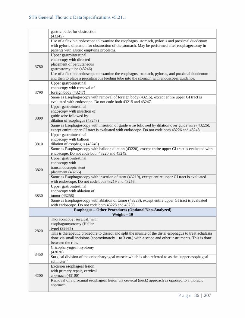
STS General Thoracic Data Specifications v5.21.1
P a g e 86 | 207
gastric outlet for obstruction (43245) Use of a flexible endoscope to examine the esophagus, stomach, pylorus and proximal duodenum with pyloric dilatation for obstruction of the stomach. May be performed after esophagectomy in patients with gastric emptying problems.
3780
Upper gastrointestinal endoscopy with directed placement of percutaneous gastrostomy tube (43246) Use of a flexible endoscope to examine the esophagus, stomach, pylorus, and proximal duodenum and then to place a percutaneous feeding tube into the stomach with endoscopic guidance.
3790
Upper gastrointestinal endoscopy with removal of foreign body (43247) Same as Esophagoscopy with removal of foreign body (43215), except entire upper GI tract is evaluated with endoscope. Do not code both 43215 and 43247.
3800
Upper gastrointestinal endoscopy with insertion of guide wire followed by dilation of esophagus (43248) Same as Esophagoscopy with insertion of guide wire followed by dilation over guide wire (43226), except entire upper GI tract is evaluated with endoscope. Do not code both 43226 and 43248.
3810
Upper gastrointestinal endoscopy with balloon dilation of esophagus (43249) Same as Esophagoscopy with balloon dilation (43220), except entire upper GI tract is evaluated with endoscope. Do not code both 43220 and 43249.
3820
Upper gastrointestinal endoscopy with transendoscopic stent placement (43256) Same as Esophagoscopy with insertion of stent (43219), except entire upper GI tract is evaluated with endoscope. Do not code both 43219 and 43256.
3830
Upper gastrointestinal endoscopy with ablation of tumor (43258) Same as Esophagoscopy with ablation of tumor (43228), except entire upper GI tract is evaluated with endoscope. Do not code both 43228 and 43258.
Esophagus – Other Procedures (Optional/Non-Analyzed) Weight = 10
2820
Thoracoscopy, surgical; with esophagomyotomy (Heller type) (32665) This is therapeutic procedure to dissect and split the muscle of the distal esophagus to treat achalasia done via small incisions (approximately 1 to 3 cm.) with a scope and other instruments. This is done between the ribs.
3450
Cricopharyngeal myotomy (43030) Surgical division of the cricopharyngeal muscle which is also referred to as the “upper esophageal sphincter.”
4200
Excision esophageal lesion with primary repair, cervical approach (43100) Removal of a proximal esophageal lesion via cervical (neck) approach as opposed to a thoracic approach

STS General Thoracic Data Specifications v5.21.1
P a g e 87 | 207
4340
Excision Esophageal lesion with primary repair, thoracic approach (e.g.: leiomyoma) (43101) Removal of an esophageal lesion and repair of the esophagus using a thoracic (chest) approach
3460
Diverticulectomy of hypopharynx or esophagus, with or without myotomy; cervical approach (43130) Removal of a diverticulum through a neck incision. The procedure most commonly includes a cricopharyngeal myotomy and is usually performed for a Zenker’s diverticulum of the esophagus.
3470
Diverticulectomy of esophagus, with or without myotomy; thoracic approach (43135) Removal of a diverticulum through a neck incision. The procedure most commonly includes a cricopharyngeal myotomy and is usually performed for a Zenker’s diverticulum of the esophagus.
3490
Laparoscopic esophageal myotomy (Heller Myotomy, with or without fundoplication) (43279) Use of laparoscopy to perform an esophageal myotomy (longitudinal division of the esophageal wall muscle while leaving the underlying esophageal mucosa intact). The procedure is done for esophageal motility disorders including achalasia.
4240
Laparoscopy, surgical, esophageal lengthening procedure (Collis) (43283) Secondary Procedure code Secondary Procedure code: Collis gastroplasty is a technique for lengthening a "shortened" esophagus, a condition that often results from gastroesophageal reflux disease (GERD). The stomach acid that flows back into the esophagus in GERD causes tissue changes, inflammation and scarring that can sometimes shorten the esophageal size. It is typically done in conjunction with a fundoplication procedure to prevent reflux. Laparoscopy is a minimally invasive abdominal approach.
4210 Unlisted laparoscopy, esophagus (43289) Minimally invasive abdominal procedure of the esophagus, not covered above
4350
Esophagoplasty with repair of TEF, cervical approach (43305) Esophageal reconstruction/repair as part of repair of a tracheoesophageal fistula via cervical (neck) approach
4360
Esophagoplasty with repair TEF, thoracic approach (43312) Esophageal reconstruction/repair as part of a repair of a tracheoesophageal fistula via thoracic (chest) approach
4370
Esophagomyotomy (Heller type); thoracic approach (43331) Longitudinal division of the esophageal wall muscle while preserving the underlying esophageal mucosa performed thru a thoracotomy.
3600
Free jejunum transfer with microvascular anastomosis (43496) This refers to utilizing a piece of small bowel as a “free flap” to restore gastrointestinal continuity after esophagectomy. This code should be used when the vascular supply of the small bowel conduit

STS General Thoracic Data Specifications v5.21.1
P a g e 88 | 207
is divided in the abdomen and then recreated utilizing blood vessels within the neck or chest.
3630 Unlisted procedure, esophagus (43499) Any surgery involving the esophagus not covered above.
3530
Esophagostomy, fistulization of esophagus, external; cervical approach (43352) This refers to the creation of a “spit fistula”, where either the end or side of the esophagus is brought out to exit on the skin of the neck. A drainage bag is often placed to drain saliva that is swallowed and exits onto the skin.
3540
Gastrointestinal reconstruction for previous esophagectomy with stomach (43360) In patients who undergo esophagectomy, delayed restoration of gastrointestinal continuity may be performed. Reasons for not undergoing immediate reconstruction include mediastinal contamination from a perforation and hemodynamic instability. This code should be used when the stomach is utilized as the conduit for reconstruction.
3550
Gastrointestinal reconstruction for previous esophagectomy with colon interposition or small intestine (43361) In patients who undergo esophagectomy, delayed restoration of gastrointestinal continuity may be performed. Reasons for not undergoing immediate reconstruction include mediastinal contamination from a perforation and hemodynamic instability. This code should be used when either the colon or small intestine is utilized as the conduit for reconstruction. Here, the blood vessels supplying either the colon or small bowel are left attached in their normal location within the abdomen.
3570
Suture of esophageal wound or injury; cervical approach (43410) Traumatic injuries to the esophagus may be addressed through direct suture repair. This code should be used when the esophageal injury is located within the neck.
3580
Suture of esophageal wound or injury; transthoracic or transabdominal approach (43415) Traumatic injuries to the esophagus may be addressed through direct suture repair. This code should be used when the esophageal injury is located within the chest or abdomen.
3590
Closure of esophagostomy or fistula; cervical approach (43420) This describes a local closure of a previously placed loop cervical esophagostomy which was created to divert oral secretions onto the neck and away from the distal esophagus.
3610
Total gastrectomy with esophagoenterostomy (43620) Refers to total resection of the stomach with gastrointestinal continuity restored with the remaining small bowel in an end-to-end fashion.
3620
Total gastrectomy with Roux- en-Y reconstruction (43621) Refers to total resection of the stomach with gastrointestinal continuity restored with the remaining small bowel in an end-to-end fashion.
4540
Conduit revision s/p esophagectomy Reoperation on a patient with a previous esophagectomy to revise the conduit
4560 Per oral endoscopic myotomy (POEM)

STS General Thoracic Data Specifications v5.21.1
P a g e 89 | 207
Reoperation on a patient with a previous esophagectomy to revise the conduit
4550
Trans oral fundoplication Transoral incisionless fundoplication (TIF) is an endoscopic approach to reflux performed through the esophagus. TIF creates a wrap of stomach around the end of the esophagus creating a 240-degree partial wrap from the inside of the stomach.
4330
Esophageal lengthening procedure - open (Collis) Secondary Procedure code (43338) Collis gastroplasty is a technique for lengthening a "shortened" esophagus, a condition that often results from gastroesophageal reflux disease (GERD). The stomach acid that flows back into the esophagus in GERD causes tissue changes, inflammation and scarring that can sometimes shorten the esophageal size. It is typically done in conjunction with a fundoplication procedure to prevent reflux. Code the fundoplasty/fundoplication as primary. “Open” refers to a traditional surgical incision on the abdomen rather than a minimally invasive approach.
3560
Ligation or stapling at gastroesophageal junction for esophageal perforation (43405) This procedure describes the division of the esophagus at the gastroesophageal junction to address an esophageal perforation. The esophagus is typically resected, and a cervical esophagostomy is created. Often, tubes are placed within the stomach and small bowel to drain and enable enteral nutrition, respectively.
Chest Wall and Neck (Optional/Non-Analyzed) Weight = 10
2000
Muscle flap, neck (15732) Surgeon rotates a neck muscle flap as an adjunct to surgery, typically used to buttress or augment a suture line, anastomosis or fill a space. Commonly used neck muscles are strap muscles, sternocleidomastoid muscle, levator scapulae.
2010
Muscle flap; trunk (i.e., intercostal, pectoralis or serratus muscle) (15734) Used where a surgeon rotates a neck muscle flap as an adjunct to surgery, typically used to buttress or augment a suture line, anastomosis or fill the pleural space. Commonly used trunk muscles are the intercostal, serratus, pectoralis, or latissimus dorsi.
2020
Excision of chest wall tumor including ribs (19260) Excision of ribs and attached muscles for a benign or malignant tumor of the chest wall. When three or less ribs are taken or if the defect is covered by the scapula, reconstruction may not be necessary.
2030
Excision of chest wall tumor involving ribs, with reconstruction (19271) Resection of the chest wall tumor with reconstruction of the defect, usually with plastic mesh (marlex, prolene), methylmethracralate/mesh sandwich or a muscle flap. Usually used for larger resections.
2040
Excision tumor, soft tissue of neck or thorax; subcutaneous (21555) Excision of a tumor in the skin/fat of the chest wall; typically, a lipoma.
2050
Excision tumor, soft tissue of neck or thorax; deep, subfascial, intramuscular (21556) Excision of a deep chest wall tumor that involves the muscles but not the ribs. These would usually be benign tumors such as a fibroma or a deep lipoma.
2060 Radical resection of tumor

STS General Thoracic Data Specifications v5.21.1
P a g e 90 | 207
(e.g., malignant neoplasm), soft tissue of neck or thorax (21557) En-bloc, radical excision of a cancer of the chest wall muscles, involving the skin, fat, and muscles. Typically, it would be a desmoid tumor or a sarcoma (MFH-malignant fibrous histiocytoma, rhabdomyosarcoma).
2070 Excision of rib, partial (21600) Removal of a part of a rib (but not the first for thoracic outlet syndrome), usually for a small tumor.
2080 Excision first and/or cervical rib (21615) Removal of the first rib or a cervical rib for TOS (Thoracic Outlet Syndrome).
2090
Excision first and/or cervical rib; with sympathectomy (21616) Rarely done now. Usually for Thoracic Outlet Syndrome with chronic arm pain from RSD (Reflex Sympathetic Dystrophy).
2870
Major reconstruction, chest wall (posttraumatic) (32820) An operation conducted for the reconstruction of a large (greater than two ribs) posttraumatic defect in the chest wall. The ribs are usually replaced with mesh or PTFE, although metallic rib struts or fasteners can be used as well.
2190 Unlisted procedure, neck or thorax (21899) Unlisted procedure not described above.
2100 Radical resection of sternum (21630) Involves radical removal of the sternum for either a tumor or severe sternal infection.
2110
Radical resection of sternum; with mediastinal lymphadenectomy (21632) Involves resection of the sternum and mediastinal lymph node dissection.
2120
Hyoid myotomy and suspension (21685) secondary procedure code Typically done as a suprahyoid laryngeal release to reduce tension on a cervical tracheal resection anastomosis. The hyoid bone is cut laterally on both sides to allow it to drop down and thus lower the larynx and trachea.
2130
Division of scalenus anticus; without resection of cervical rib (21700) Usually done for a Thoracic Outlet Syndrome (TOS) variant where the muscle or a band from it impinges on the brachial plexus.
2140
Division of scalenus anticus; with resection of cervical rib (21705) Usually done for a Thoracic Outlet Syndrome (TOS) variant where the muscle or a band from it impinges on the brachial plexus along with resection of the abnormal cervical rib.
2150
Reconstructive repair of pectus excavatum or carinatum; open (21740) Repair of either of these two congenital chest wall deformities. Usually involves resecting several costal cartilages, a partial osteotomy of the sternum, and often placement of a temporary bar for stabilization (also known as a Ravitch repair.)
2160 Reconstructive repair of pectus, minimally invasive

STS General Thoracic Data Specifications v5.21.1
P a g e 91 | 207
approach (Nuss procedure), without thoracoscopy (21742) Placement of a Nuss transverse chest wall bar to push the sternum forward to repair pectus excavatum.
2180
Open treatment of sternum fracture with or without skeletal fixation (21825) Repair of a sternal fracture with sutures, wires, plates, or bars.
4570 Removal of sternal wire(s) Sternotomy incisions are typically closed with a series of wires to support the bone during healing. These are left in place unless the patient experiences irritation or infection.
2170
Reconstructive repair of pectus, minimally invasive approach (Nuss procedure), with thoracoscopy (21743) Placement of a Nuss transverse chest wall bar to push the sternum forward to repair pectus excavatum with the visual aid of thoracoscopy.
4680 Intercostal nerve block (64220-1)
Miscellaneous (Optional/Non-Analyzed) Weight = 10
2640
Thoracoscopy, diagnostic pericardial sac, with biopsy (32604) Minimally invasive approach to remove a sample of pericardial tissue for diagnostic purposes.
2750
Thoracoscopy, surgical; with removal of clot or foreign body from pericardial sac (32658) This is a therapeutic procedure to remove clot or a foreign object (such as a bullet) from the pericardium done via small incisions (approximately 1 to 3 cm.) with a scope and other instruments. A drain is commonly left.
2760
Thoracoscopy, surgical; with creation of pericardial window or partial resection of pericardial sac for drainage (32659) This is therapeutic procedure to drain fluid from the pericardium and remove a segment of the pericardium done via small incisions (approximately 1 to 3 cm.) with a scope and other instruments. A drain is commonly left.
2770
Thoracoscopy, surgical; with total pericardiectomy (32660) This is an uncommon therapeutic procedure to remove the entire pericardium done via small incisions (approximately 1 to 3 cm.) with a scope and other instruments.
2780
Thoracoscopy, surgical; with excision of pericardial cyst, tumor, or mass (32661) This is a procedure to remove a cyst, tumor or mass from the pericardium done via small incisions (approximately 1 to 3 cm.) with a scope and other instruments. The important distinction is the complete removal of abnormal tissue.
2810
Thoracoscopy, surgical; with thoracic sympathectomy (32664) This is therapeutic procedure to divide or interrupt the sympathetic chain in the chest. It is commonly done to treat hyperhidrosis. The technique involves using small incisions (approximately 1 to 3 cm.)
STS General Thoracic Data Specifications v5.21.1
P a g e 92 | 207
with a scope and other instruments.
4390
Stereotactic radiosurgery (SRS) and stereotactic body radiotherapy (SBRT),surgeon participation (32701) Stereotactic radiosurgery (SRS) is a highly precise form of radiation therapy initially developed to treat small brain tumors and functional abnormalities of the brain. The principles of cranial SRS, namely high precision radiation where delivery is accurate to within one to two millimeters, are now being applied to the treatment of body tumors with a procedure known as stereotactic body radiotherapy (SBRT). Despite its name, SRS is a non-surgical procedure that delivers precisely-targeted radiation at much higher doses, in only a single or few treatments, as compared to traditional radiation therapy. This treatment is only possible due to the development of highly advanced radiation technologies that permit maximum dose delivery within the target while minimizing dose to the surrounding healthy tissue. The goal is to deliver doses that will destroy the tumor and achieve permanent local control.
3940
Tube pericardiostomy (33015) This involves opening the pericardium and placing a tube into the pericardial space for drainage - may be placed percutaneously via needle and guide wire, via thoracoscopy or thoracotomy or subxiphoid. If no tube placed in the pericardial space, see: Thoracoscopy (VATS), surgical; with creation of pericardial window or partial resection of pericardial sac for drainage.
4790 Insertion of Tunneled CV Catheter (36561)
3970 Other Any procedure not covered by any of the above descriptions.
3960 SVC resection and reconstruction (34502) Removal of part or all of the superior vena cava with or without reconstruction.
3890
Ligation thoracic duct (38381) Tying off or clipping the main lymph channel in the chest. Usually performed at a level just above the diaphragm on the right side and is commonly done for a chyle leak (chylothorax); can be approached by VATS or open methods. Also includes obliterating or ligating the cisterna chyli.
3910
Omental flap (49904) Omentum (usually the greater omentum) is brought through a subcutaneous tunnel or the diaphragm to a cover soft tissue defect, bronchial stump, or other structure to stimulate granulation and promote healing.
3920
Transthoracic thyroidectomy (60270) Removing part or all of the thyroid gland via a thoracic incision. Adding an upper sternal split to facilitate resection of a substernal goiter would not be in this definition (see below). Removing part or all of the thyroid gland by VATS would also be a transthoracic thyroidectomy.
3930
Removal substernal thyroid, cervical approach (60271) Removal of part or all of the thyroid gland via a cervical incision. The use of an upper sternal split to facilitate a thyroidectomy which is partially substernal would still be considered a cervical approach, since this is the dominant incision.
4380
Application of wound vac (97605, 97606) Negative pressure wound therapy (NPWT) is a therapeutic technique using a vacuum dressing to promote healing in acute or chronic wounds. The therapy involves the controlled application of sub-atmospheric pressure to the local wound environment, using a sealed wound dressing connected to a vacuum pump. The continued vacuum draws out fluid from the wound and increases blood flow to the area. The vacuum may be applied continuously or intermittently, depending on the type of wound being treated and the clinical objectives.
3950 Pericardial window (33025) Opening a draining the pericardial space by making a small (usually 1 to 4 cm in diameter) hole in

STS General Thoracic Data Specifications v5.21.1
P a g e 93 | 207
the pericardium. Done via thoracotomy or subxiphoid approach; if VATS used see: Thoracoscopy (VATS), surgical; with creation of pericardial window or partial resection of pericardial sac for drainage. If a tube is placed see Tube pericardiostomy above.
4400 Other Minor Procedure Unlisted minor/Non-Analyzed procedure.
4810
Robotic-Assisted Surgery (capture as an additional code) (S2900) In This field is to capture the use of robotics at any time during the procedure, including cases where the approach was converted (i.e., converted to open (UnanticConv – seq 1430)). In addition to coding Robotic-Assisted Surgery (capture as an additional code) (S2900), please also code Assisted by Robotic Technology (Robotic -seq 1400). Code this field in addition to the primary procedure.
SeqNo: 1480 Long Name: Primary Procedure Short Name: Primary Format: Text (categorical values specified by STS) Definition: Indicate whether this is the primary surgical procedure. Harvest Codes: Code: Value: 1 Yes 2 No Intent/Clarification: Each procedural section (i.e., Lung Cancer Resection, Concomitant Lung, Esophagectomy, etc.) has been assigned a weight. If procedures are performed from multiple procedural sections during the same OR encounter, code the highest weighted procedure as primary (Primary 1480).
- 70 – Esophageal Cancer Resection - 60 – Lung Cancer Resection - 50 – Thymus/Mediastinal - 40 – Tracheal Resection - 30 – Hiatal Hernia/GERD - 20 – Concomitant Lung - 10 – Minor Procedures - Example: A thoracotomy wedge resection (32505) and a partial esophagectomy with thoracotomy (43112)
are performed within the same visit to the OR. The esophagectomy has a higher weight (70) versus the thoracotomy/wedge resection (60); Code the esophagectomy as the primary procedure at Primary – seq 1480.
- Example: A thymectomy for thymoma and en bloc wedge resection are performed within the same visit to
the OR. The thymectomy has a higher weight than the concomitant lung procedure; code the thymectomy as the primary procedure at Primary – seq 1480.
This field is required for record inclusion. If missing data, the entire record will be excluded from the analysis. SeqNo: 1490

STS General Thoracic Data Specifications v5.21.1
P a g e 94 | 207
Long Name: Procedure Unlisted - Specify Short Name: ProcOth Format: Text Definition: Indicate the general thoracic procedure(s) not listed being performed during this operating room visit, free text up to 150 characters. ParentLongName: Procedure ParentShortName: Proc ParentValue: 2190|2300|2950|3230|3310|3630|3970|4210|4400 ParentHarvestCodes: = "Unlisted procedure, neck or thorax (21899)", "Unlisted procedure, trachea, bronchi (31899)", "Unlisted procedure, lung (32999)", "Unlisted procedure, mediastinum (39499)", "Unlisted procedure, diaphragm (39599)", "Unlisted procedure, esophagus (43499)", "Other", "Unlisted laparoscopy, esophagus (43289)" or "Other Minor Procedure" Intent/Clarification: SeqNo: 1500 Long Name: Procedure Unlisted - CPT Short Name: ProcOthCPT Format: Text Definition: Indicate 5 digit CPT code(s) of unlisted procedure(s). ParentLongName: Procedure ParentShortName: Proc ParentValue: 2190|2300|2950|3230|3310|3630|3970|4210|4400 ParentHarvestCodes: = "Unlisted procedure, neck or thorax (21899)", "Unlisted procedure, trachea, bronchi (31899)", "Unlisted procedure, lung (32999)", "Unlisted procedure, mediastinum (39499)", "Unlisted procedure, diaphragm (39599)", "Unlisted procedure, esophagus (43499)", "Other", "Unlisted laparoscopy, esophagus (43289)" or "Other Minor Procedure" Intent/Clarification: SeqNo: 1505 Long Name: Procedure Laterality Short Name: Laterality Format: Text (categorical values specified by STS) Definition: Indicate the laterality of the procedure performed or N/A if not applicable. Harvest Codes: Code: Value: 1 Right 2 Left 3 Bilateral 4 Not applicable Intent/Clarification: This field is intended to capture the side surgery was performed on. If there is no laterality, then code N/A.

STS General Thoracic Data Specifications v5.21.1
P a g e 95 | 207
Aug 2021: Lung resections have laterality, most hernia repairs and esophagectomies do not and will be coded as N/A. SeqNo: 1510 Long Name: Primary Lung Cancer Resection Performed Short Name: LungCancer Format: Text (categorical values specified by STS) Definition: Indicate whether a major lung resection was performed for a primary lung cancer (e.g. wedge, segment, lobe, pneumonectomy), open or VATS. If yes complete clinical and pathological staging. Harvest Codes: Code: Value: 1 Yes 2 No Intent/Clarification: Only primary lung cancer resections are to be entered in this field. A primary lung cancer is a tumor that originated/started in the anatomical location of the lung where surgery is being performed. If ‘yes,’ please complete section F. There are two reasons for performing procedures: diagnostic and therapeutic. Diagnostic resections are those procedures intended to confirm a diagnosis or to better understand the disease process and are not captured here. Therapeutic procedures are performed to treat the disease and are captured here. Differentiating between diagnostic and therapeutic intent can be difficult. If you are uncertain, please submit a clinical question for guidance. This field is required for record inclusion. If missing data, the entire record will be excluded from the analysis. SeqNo: 1530 Long Name: Esophageal Cancer Resection Performed Short Name: EsophCancer Format: Text (categorical values specified by STS) Definition: Indicate whether an esophagectomy was performed for esophageal cancer. If yes complete clinical and pathological staging. Harvest Codes: Code: Value: 1 Yes 2 No Intent/Clarification: Only primary esophageal cancer resections are to be entered in this field. A primary esophageal cancer is a tumor that originated/started in the anatomical location of the esophagus where surgery is being performed. If ‘Yes,’ please complete section G.

STS General Thoracic Data Specifications v5.21.1
P a g e 96 | 207
Esophagectomies are sometimes performed for benign disease. These procedures are not captured here. If you are uncertain whether an esophagectomy is required for entry in the database, please submit a clinical question for guidance. This field is required for record inclusion. If missing data, the entire record will be excluded from the analysis. SeqNo: 1540 Long Name: Thymus/Mediastinal Mass Resection/Myasthenia Gravis Short Name: ThymusMediastinalData Format: Text (categorical values specified by STS) Definition: Indicate whether a surgical procedure was performed for the thymus, a mediastinal mass, or Myasthenia Gravis. Harvest Codes: Code: Value: 1 Yes 2 No Intent/Clarification: Procedures on the thymus, for a mediastinal mass, or for Myasthenia Gravis are captured here. (Sept 2021) Intent/Clarification - For Thymus/Mediastinal Mass cases • Collect detailed info on thymectomies for myasthenia including open, cervical or VATS route • Collect all thymectomies for myasthenia regardless of whether they have thymoma • Collect detailed info on thymectomies for thymoma including open or VATS Robotics should be coded with thoracoscopic procedures, also code S2900 under miscellaneous procedures. If ‘Yes,’ please complete section H. This field is required for record inclusion. If missing data, the entire record will be excluded from the analysis. SeqNo: 1550 Long Name: Tracheal Resection Short Name: TrachealData Format: Text (categorical values specified by STS) Definition: Indicate if the surgical procedure was a tracheal resection. Harvest Codes: Code: Value: 1 Yes 2 No Intent/Clarification: Tracheal resections performed for any reason are captured here. If ‘Yes,’ please complete section I.

STS General Thoracic Data Specifications v5.21.1
P a g e 97 | 207
This field is required for record inclusion. If missing data, the entire record will be excluded from the analysis. SeqNo: 1560 Long Name: Hiatal Hernia / Diaphragmatic Hernia or GERD Short Name: HiatalHerniaData Format: Text (categorical values specified by STS) Definition: Indicate if the surgical procedure was performed for a hiatal hernia/diaphragmatic hernia/GERD. Harvest Codes: Code: Value: 1 Yes 2 No Intent/Clarification: There are various types of hernias. This field is intended to capture procedures performed for hiatal hernias, diaphragmatic hernias, and procedures for gastroesophageal reflux disease (GERD). Hiatal hernias occur when the upper part of the stomach bulges through the large muscle separating the abdomen and chest (diaphragm). The diaphragm has a small opening (hiatus) through which the esophagus passes connecting to the stomach. In a hiatal hernia, the stomach pushed up through that opening into the chest. https://www.mayoclinic.org/diseases-conditions/hiatal-hernia/symptoms-causes/syc-20373379 Diaphragmatic hernias can be either congenital or acquired. The purpose of this field is to capture acquired diaphragmatic hernias (ADH). Acquired diaphragmatic hernias occurs when one or more abdominal organs move upward into the chest cavity through a defect in the diaphragm. ADH usually occurs as a result of blunt force trauma (i.e., traffic accident, falls), surgical procedures of the chest or abdomen, stab, or gunshot wounds. These types of hernias are usually acute in nature and require near-immediate surgical intervention.

STS General Thoracic Data Specifications v5.21.1
P a g e 98 | 207
------------------------------------------------------------------------------------------------------------------------------------------------------------------------------------ Lung Cancer ------------------------------------------------------------------------------------------------------------------------------------------------------------------------------------ SeqNo: 1600 Long Name: Clinical Staging Done For Lung Cancer Short Name: ClinStagDoneLung Format: Text (categorical values specified by STS) Definition: Indicate whether clinical staging was performed on this patient related to this lung procedure. ParentLongName: Primary Lung Cancer Resection Performed ParentShortName: LungCancer ParentValue: 1 ParentHarvestCodes: = "Yes" Harvest Codes: Code: Value: 1 Yes 2 No Intent/Clarification: Clinical staging is based on evidence/testing gathered prior to therapeutic surgery for primary treatment. This may occur in the form of preoperative biopsies or bronchial washings, among other diagnostic or radiological testing. Diagnostic and/or radiologic tests are performed to determine the type and extent of the cancer and used to guide treatment decisions. Diagnostic resections are those procedures intended to confirm a diagnosis or to better understand the disease process. Diagnostic procedures are not captured here. Therapeutic procedures are performed to treat the disease. If a procedure is scheduled as a diagnostic procedure (which is not captured here) and then, based on specimens removed during the procedure, turns into a therapeutic procedure, code no to ClinStagDoneLung (seq 1600), unless other preoperative clinical staging was performed. This field is required for record inclusion. If missing data, the entire record will be excluded from the analysis.

STS General Thoracic Data Specifications v5.21.1
P a g e 99 | 207
SeqNo: 1620 Long Name: Clinical Staging Methods Short Name: ClinStagMeth Format: Multi-Select Definition: Identify the clinical staging methods utilized to confirm primary lung cancer. ParentLongName: Clinical Staging Done For Lung Cancer ParentShortName: ClinStagDoneLung ParentValue: 1 ParentHarvestCodes: = "Yes" Harvest Codes: Code: Value: 1 PET/CT 2 CT 3 Brain CT 4 Brain MRI 5 Invasive Mediastinal Staging Performed Intent/Clarification: Select the type(s) of clinical staging methods performed. Timeframe: Please collect all clinical staging procedures used during the work-up of the current disease process.
1. PET/CT a. Positron emission tomography, also called PET imaging or a PET scan, is a type of nuclear
medicine imaging. Nuclear medicine or radionuclide imaging procedures are noninvasive and, with the exception of intravenous injections, are usually painless medical tests that help diagnose medical conditions. These imaging scans use radioactive materials called radiopharmaceuticals or radiotracers.
b. PET or PET/CT Skull scans are not the same as CT of the Brain. PET or PET/CT skull scans are capture here. CT of the brain is captured by selecting Brain CT.
c. PET or PET/CT scans are captured here
2. CT a. Computed tomography (CT) scan, also called computerized axial tomography (CAT) scan, is used
to create cross-sectional images of structures in the body. In this procedure, x-rays are taken from many different angles and processed through a computer to produce a three-dimensional (3-D) image called a tomogram.
b. CT or CT Angiograms are captured here
3. Brain (Head) CT with contrast a. CT scan of the head with contrast is an acceptable means of staging the brain for cancer. Only
capture CT of the head with contrast. b. A CT scan of the head without contrast is not useful for staging the brain. Do not include CT of
the head without contrast. c. CT of the head is not the same as PET or PET/CT skull scans. CT of the head is captured here,
PET or PET/CT skull scans are captured by selecting PET/CT.
4. Brain MRI a. Magnetic resonance imagining (MRI) is a medical imagining technique that uses a magnetic field
and computer-generated radio waves to create detailed images of the organs and tissues in the body.
b. An MRI of the brain is an acceptable means of staging the brain.
5. Invasive Mediastinal Staging Performed

STS General Thoracic Data Specifications v5.21.1
P a g e 100 | 207
a. Indicate if the patient underwent biopsies of mediastinal lymph nodes. b. If a mediastinoscopy is performed during the same OR trip as a therapeutic lung procedure, it can
be captured here. c. VATS/thoracotomy mediastinal lymph node dissection performed with lung resection is not
captured here. It is captured under seq 1880 LungNodesAsses. d. A biopsy of lymph node tissue is required to code this option.
Octreotide scans, which are used to locate primary neuroendocrine tumors, are not captured here. Aug 2021: Question - How do I capture a Core Needle Biopsy of the lung mass itself preop? It is not a mediastinal lymph node biopsy? Answer – core needle biopsies of the lung mass are not captured in V5.21. SeqNo: 1630 Long Name: Mediastinal Lymph Node Sampling Staging Method Short Name: MedLymNodSam Format: Multi-Select Definition: Identify the method(s) of the mediastinal lymph node sampling staging that was performed. Select all that apply. ParentLongName: Clinical Staging Methods ParentShortName: ClinStagMeth ParentValue: contains(5) ParentHarvestCodes: Contains ("Invasive Mediastinal Staging Performed") Harvest Codes: Code: Value: 1 EBUS 2 IR Needle Biopsy 3 EUS 4 Chamberlain 5 Mediastinoscopy 6 VATS/Lymph Node Biopsy 7 Other Intent/Clarification: Select the method(s) used to obtain mediastinal lymph node sampling for staging purposes.
1. EBUS a. EBUS is an invasive procedure in which physicians use ultrasound devices on the end of a special
bronchoscope or placed through a bronchoscope to examine the airways and the lung for exploration of the structures of airway walls, the surrounding mediastinum, and the lungs. It is commonly used to biopsy lymph nodes outside the airway wall.
b. This does not include super dimensional bronchoscopy. c. EBUS done in the OR prior to surgery can be included here. d. EBUS without lymph node biopsy is not sufficient to code this field.
2. IR Needle Biopsy
a. Interventional radiology (IR) procedures include imaging-guided biopsies to obtain samples for cytologic or pathologic testing without affecting adjacent structures. Only code IR Needle Biopsies of lymph nodes here.
3. EUS a. EUS is a procedure that combines endoscopy and ultrasound to obtain images and information

STS General Thoracic Data Specifications v5.21.1
P a g e 101 | 207
about the digestive tract and the surrounding tissue and organs. In EUS a small ultrasound transducer is installed on the tip of the endoscope placed into the esophagus (not the airway) allowing the transducer to get closer to internal organs. This generally permits more accurate and detailed images of those organs than ones obtained by traditional ultrasound done from the surface of the body.
4. Chamberlain a. The Chamberlain procedure is used to biopsy lymph nodes in the center of the chest, or to biopsy a
mass in the center of the chest. The Chamberlain procedure differs from a cervical mediastinoscopy by the location of the incision, and the location of the lymph nodes or mass to be biopsied. The Chamberlain procedure is used to biopsy lymph nodes or masses in the aorto-pulmonary window on the left side of the chest, or nodes in the hilar areas of the lung. (In contrast, the cervical mediastinoscopy procedure is used to biopsy nodes or masses to the front or side of the trachea, or windpipe.) The aorto-pulmonary window is the area in the center of the chest bound by the aorta superiorly, and the pulmonary artery inferiorly. This area contains lymph nodes that filter lymph coming from the left lung, especially the left upper lobe. If a lung cancer is present in the left lung, the Chamberlain procedure is useful for staging the cancer (determining the extent of spread.) The hilar areas of the lung (the hilum) are the areas of the lung where the pulmonary artery and vein (the blood supply) join the lung.
5. Mediastinoscopy a. Mediastinoscopy is a procedure that enables visualization of the contents of the mediastinum,
usually for the purpose of obtaining a biopsy. Mediastinoscopy is often used for staging of lymph nodes of lung cancer or for diagnosing other conditions affecting structures in the mediastinum such as sarcoidosis or lymphoma. Mediastinoscopy involves making an incision approximately 1 cm above the suprasternal notch of the sternum, or breastbone. Dissection is carried out down to the pretracheal space and down to the carina. A scope (mediastinoscope) is then advanced into the created tunnel which provides a view of the mediastinum. The scope may provide direct visualization or may be attached to a video monitor.
b. Mediastinoscopy done in the OR just prior to resection can be included as clinical staging. All nodes from the path report count for the path staging.
6. VATS/Lymph Node Biopsy
a. Video-assisted thoracoscopic surgery (VATS) is a minimally invasive surgical technique used to diagnose and treat problems in the chest. During this surgery, a tiny camera (thoracoscope) and surgical instruments are inserted in the chest through small incisions. The thoracoscope transmits images of the inside of the chest onto a video monitor, guiding the surgeon performing the procedure. Video-assisted thoracoscopic surgery (VATS) can be used for many purposes, ranging from a biopsy to removal of tumors or entire lobes from the lung.
b. VATS without lymph node biopsy is not sufficient to code this field. c. VATS with a wedge resection of a primary lesion followed by a lobectomy as a result of a positive
wedge is NOT captured here. d. Lymph node resection as part of a planned procedure is NOT captured here. e. A biopsy must be performed in order to capture this field.
7. Other
a. Indicate if any other method/technology was used for clinical staging.
SeqNo: 1800 Long Name: Clinical Staging Lung Cancer Tumor Size In cm Short Name: LungCaTumSz Format: Real Definition: Indicate the tumor size of the dominant/most concerning lesion in centimeters.

STS General Thoracic Data Specifications v5.21.1
P a g e 102 | 207
Low Value: 0.00 High Value: 50.00 ParentLongName: Clinical Staging Done For Lung Cancer ParentShortName: ClinStagDoneLung ParentValue: 1 ParentHarvestCodes: = "Yes" Intent/Clarification: Size of tumor should be taken from CT scan or PET scan. If neo-adjuvant treatment was completed, always use tumor size prior to treatment. Approximately 40% of people over the age of 50 will have small lung nodules which are not malignant. If there is no biopsy, the PET or PET/CT is negative, or the nodules are less than 5mm and the surgeon/oncologist chooses not to address these, do not consider them when staging. For tumors that are part solid, report on the surgeon’s documentation of the clinical staging. If the surgeon does not address this, then you may refer to the radiology report. For all other tumors, use the size provided on the preoperative imaging. If the size is not available on the preoperative imaging, then leave this field as blank. Multiple imagining may be available to code this field. If a PET scan and a CT are available, then use the CT if it is less than 3 months old. If the CT is greater than 3 months and the PET is less than 3 months, use the PET. If both exams are over 3 months old, use the CT. SeqNo: 1810 Long Name: Lung Cancer T Stage Short Name: ClinStageLungTumor Format: Text (categorical values specified by STS) Definition: Indicate the appropriate descriptor for lung cancer tumor staging. Clinical staging is based on the PRE-TREATMENT ESTIMATED staging workup which may include CT scan, PET scan, endoscopic ultrasound, etc. (Tis - T4). ParentLongName: Clinical Staging Done For Lung Cancer ParentShortName: ClinStagDoneLung ParentValue: 1 ParentHarvestCodes: = "Yes" Harvest Codes: Code: Value: 1 Tis 2 T1 3 T2 4 T3 5 T4 Intent/Clarification: The stage of a cancer tells you how big the tumor is and whether it has spread. Knowing the stage helps your doctor decide which treatment you need. The TNM (Tumor, Node, Metastasis) staging system is the most common way for doctors to stage lung cancer. Each staging has four categories. Use the most recent scan for documenting T-stage.

STS General Thoracic Data Specifications v5.21.1
P a g e 103 | 207
The T-stage of the TNM staging system is captured in this field and related to the size of the tumor (area of cancer).
There are 5 categories – Tis to T4. Since TX and T0 are not indicative of cancer, they are not captured as lung cancer. TX means the main cancer (primary) can’t be assessed. It doesn't show on scans but there might be cancer cells present in spit or in fluid taken from the lung. T0 means there is no sign of cancer.
1. Tis a. Carcinoma In Situ b. Tis means that the cancer cells are only growing in the layer of cells where they started, without
spreading or growing into deeper layers. This may also be called in situ cancer or pre-cancer
2. T1 Tumor 3 cm or less in greatest dimension, surrounded by lung or visceral pleura, without bronchoscopic evidence of invasion more proximal than the lobar bronchus (i.e., not in the main bronchus)
3. T2 a. The tumor is between 3cm and 5cm across. b. Or the cancer has one or more of the following features:
i. Involves main bronchus regardless of distance to the carina, but without involving the carina.
ii. Invades visceral pleura iii. Associated with atelectasis or obstructive pneumonitis that extends to the hilar region,
either involving part of the lung or the entire lung.
4. T3 a. The tumor is between 5cm to 7cm. b. Or there is more than one tumor in the same lobe of the lung. c. Or the cancer has grown into one or more of these structures:
i. Chest Wall / Parietal Pleura (the protective structure around the lungs and other organs in the chest). Includes superior sulcus tumors.
ii. Phrenic Nerve (the nerve closest to the lung iii. Parietal Pericardium (the outer covering of the heart)
5. T4
a. The tumor is more than 7cm. b. Separate tumor nodule(s) in a different ipsilateral lobe to that of the primary tumor. c. Invades one or more of the following structures:
i. Diaphragm (the muscle under the lungs) ii. Mediastinum (the area between the lungs in the middle of the chest)
iii. Heart iv. Great Vessels v. Trachea
vi. Recurrent Laryngeal Nerve (the nerve that controls the voice box) vii. Esophagus
viii. Vertebral Body ix. Carina (the area where the main airway divides to go to each lung)
https://radiologyassistant.nl/chest/lung-cancer/tnm-classification-8th-edition SeqNo: 1820 Long Name: Lung Cancer Nodes - N

STS General Thoracic Data Specifications v5.21.1
P a g e 104 | 207
Short Name: ClinStageLungN Format: Text (categorical values specified by STS) Definition: Indicate the appropriate descriptor for the lung cancer nodal metastases. All nodes > 1cm on CT or PET/CT are considered positive. All PET positive nodes are considered positive. Results of previous invasive staging (EBUS, Mediastinoscopy) should be included here. Clinical staging is based on the PRE-TREATMENT ESTIMATED staging workup which may include CT scan, PET scan, endoscopic ultrasound, etc. (8th Edition) ParentLongName: Clinical Staging Done For Lung Cancer ParentShortName: ClinStagDoneLung ParentValue: 1 ParentHarvestCodes: = "Yes" Harvest Codes and Value Definitions: Value: Definition: 1 N0 - No regional lymph node metastasis 2 N1 - Metastasis in ipsilateral peribronchial or hilar and intrapulmonary nodes. Includes direct extension. 3 N2 - Metastasis in ipsilateral mediastinal and/or subcarinal lymph nodes 4 N3 - Metastasis in contralateral mediastinal or contralateral hilar nodes, ipsilateral or contralateral scalene or supraclavicular nodes Intent/Clarification: Use the most recent scan for documenting N-stage. The N-stage of the TNM staging system is captured in this field and describes whether the cancer has spread to the lymph nodes. NX means that the lymph nodes can’t be assessed and is not capture here.
1. N0 2. N0 means that the lymph nodes don’t contain cancer cells. 2. N1 3. N1 means there are cancer cells in lymph nodes within the lung or in lymph nodes in the area where the
lungs join the airway (the hilum). 4. N2 5. N2 means there is cancer in lymph nodes: 6. in the center of the chest (mediastinum) on the same side as the affected lung (ipsilateral) or 7. just under where the windpipe branches off to each lung 8. N3 9. N3 means there is cancer in lymph nodes: 10. on the opposite side of the chest from the affected lung (contralateral) or above the collar bone or at the top
of the lung
Pathology results overrule radiological findings. For example, a PET/CT scan is positive for lymph nodal metastases, but biopsies obtained from a mediastinoscopy are negative. Code the results from the mediastinoscopy. If the lymph node is larger than 1 cm on the CT and PET negative and a biopsy is not performed, the lymph nodes

STS General Thoracic Data Specifications v5.21.1
P a g e 105 | 207
are considered positive. If this generates discrepancy with the surgeon’s documentation, please clarify with your surgeon. https://www.cancerresearchuk.org/about-cancer/lung-cancer/stages-types-grades/tnm-staging SeqNo: 1830 Long Name: Lung Cancer Metastasis - M Short Name: ClinStageLungM Format: Text (categorical values specified by STS) Definition: Indicate the appropriate descriptor for the lung cancer distant metastases. Clinical staging is based on the PRE- TREATMENT ESTIMATED staging workup which may include CT scan, PET scan, endoscopic ultrasound, etc. ParentLongName: Clinical Staging Done For Lung Cancer ParentShortName: ClinStagDoneLung ParentValue: 1 ParentHarvestCodes: = "Yes" Harvest Codes and Value Definitions: Code: Definition: 1 M0 - No distant metastasis 4 M1 - Distant Metastasis
Intent/Clarification: Clinical staging is based on the PRE- TREATMENT ESTIMATED staging workup which may include CT scan, PET scan, endoscopic ultrasound, etc.
The stage of a cancer tells you how big the tumor is and whether it has spread. Knowing the stage helps your doctor decide which treatment you need. The TNM (Tumor, Node, Metastasis) staging system is the most common way for doctors to stage lung cancer. Each staging has four categories. Use the most recent scan for documenting M-stage. The M-stage of the TNM staging system is captured in this field and describes whether the cancer has spread to a different part of the body. There are 2 stages of metastasis – M0 and M1.
1. M0 a. means the cancer hasn’t spread to another lobe of the lung or any other part of the body.
2. M1 means the cancer has spread to other areas of the body. Includes metastasis to contralateral lobe. It is split into M1a, M1b and M1c. a. M1a means one or more of the following:
i. there is cancer in both lungs (exception would be synchronous primary lung cancers) ii. there are areas of cancer in the lining around the lung or the lining around the heart iii. there is fluid around the lung or heart that contains cancer cells – this is called a malignant pleural effusion or a malignant pericardial effusion
b. M1b means that there is a single area of cancer outside the chest in an organ (such as the liver or brain) or a lymph node.
c. M1c means that there is more than one area of cancer in one or several organs.
https://www.cancerresearchuk.org/about-cancer/lung-cancer/stages-types-grades/tnm-staging

STS General Thoracic Data Specifications v5.21.1
P a g e 106 | 207
SeqNo: 1841 Long Name: Clinical Staging - Lung Cancer Tumor Present Short Name: ClinStageTumorPres Format: Text (categorical values specified by STS) Definition: Indicate whether the diagnosis for lung cancer as reported in the final pathology report indicated the presence of a tumor. ParentLongName: Primary Lung Cancer Resection Performed ParentShortName: LungCancer ParentValue: 1 ParentHarvestCodes: = "Yes" Harvest Codes: Code: Value: 1 Yes 2 No
Intent/Clarification: The intent is to capture only primary lung cancer cases. This section (section F) is not to be captured for metastatic or benign processes.
Sept 2021: If the patient had neoadjuvant therapy and had a complete response with the final surgical pathology report indicating T0 or no T stage is provided, then code yes to seq 1841 “ClinStageTumorPres ’ and code T0 for seq 1850 ‘PathStageLungT’. SeqNo: 1850 Long Name: Pathologic Staging - Lung Cancer - T Short Name: PathStageLungT Format: Text (categorical values specified by STS) Definition: Indicate the appropriate descriptor for the lung cancer primary tumor based on final pathology report. ParentLongName: Clinical Staging - Lung Cancer Tumor Present ParentShortName: ClinStageTumorPres ParentValue: 1 ParentHarvestCodes: = "Yes" Harvest Codes: Code: Value: Definition: 7 TX Primary Tumor cannot be assessed, or tumor proven by the presence of malignant cells in sputum or bronchial washings but not visualized by imaging or bronchoscopy 8 T0 No evidence of primary tumor 9 Tis Carcinoma in situ; squamous cell carcinoma in situ (SCIS); Adenocarcinoma in situ (AIS): adenocarcinoma with pure lepidic pattern, <3 cm in greatest dimension 10 T1mi Minimally invasive adenocarcinoma: adenocarcinoma (<3 cm in greatest dimension) with a predominantly

STS General Thoracic Data Specifications v5.21.1
P a g e 107 | 207
lepidic pattern and <5 mm invasion in greatest dimension. 11 T1a Tumor <1 cm in greatest dimension. A superficial, spreading tumor of any size whose invasive component is limited to the bronchial wall and may extend proximal to the main bronchus also is classified as T1a, but these tumors are uncommon. 12 T1b Tumor > 1 cm but < 2 cm in greatest dimension 13 T1c Tumor > 2 cm but < 3 cm in greatest dimension 14 T2a Tumor > 3 cm but < 4 cm at greatest dimension, or having any of the following features: 1. involves the main bronchus regardless of distance to the carina, 2. but without involvement of the carina; invades visceral pleura (PL1 or PL2); 3. associated with atelectasis or obstructive pneumonitis that extends to the hilar region, involving part or all of the lung. Includes tumors with visceral pleural involvement that are less than 4cm. 15 T2b Tumor > 4 cm but < 5 cm at greatest dimension 16 T3 Tumor > 5 cm but < 7 cm in greatest dimension or directly invading any of the following: parietal pleura (PL3), chest wall (including superior sulcus tumors), phrenic nerve, parietal pericardium; or separate tumor nodule(s) in the same lobe as the primary 17. T4 Tumor > 7 cm or tumor of any size invading one or more of the following: diaphragm, mediastinum, heart, great vessels, trachea, recurrent laryngeal nerve, esophagus, vertebral body, or carina; separate tumor nodule(s) in an ipsilateral lobe different from that of the primary Intent/Clarification: Indicate the appropriate descriptor for the lung cancer primary tumor based on final pathology report. If two or more tumors are dissected out during the same procedure, code the most aggressive disease noted on the pathology report. Consultation with pathology may be necessary to determine. Use the pathology report associated with the procedure from which it originated. If two separate procedures are performed and two separate pathology reports are obtained, use the pathology report with the corresponding procedure. Sept 2021: If the patient had neoadjuvant therapy and had a complete response with the final surgical pathology report indicating T0 or no T stage is provided, then code yes to seq 1841 “ClinStageTumorPres ’ and code T0 for seq 1850 ‘PathStageLungT’. SeqNo: 1860 Long Name: Lung - Final pathological staging - Visceral Pleura Invasion Short Name: VisPleuraInv Format: Text (categorical values specified by STS)

STS General Thoracic Data Specifications v5.21.1
P a g e 108 | 207
Definition: Indicate if the final pathology report specifies visceral pleura invasion present. ParentLongName: Pathologic Staging - Lung Cancer - T ParentShortName: PathStageLungT ParentValue: 14|15 ParentHarvestCodes: = "T2a" or "T2b" Harvest Codes: Code: Value: 1 Yes 2 No Intent/Clarification: Visceral pleural invasion is indicated by PL1 or PL2 on the final path report. There must be clear evidence that there is visceral pleural invasion. If two or more tumors are dissected out during the same procedure, code the most aggressive disease noted on the pathology report. Consultation with pathology may be necessary to determine. SeqNo: 1875 Long Name: Lung Cancer- Invasion of Adjacent Structures - Multi-Select Short Name: LCInvAdjStrMulti Format: Multi-Select Definition: Indicate the adjacent structure(s) the tumor invades. Select all that apply or 'none'. ParentLongName: Clinical Staging - Lung Cancer Tumor Present ParentShortName: ClinStageTumorPres ParentValue: 1 ParentHarvestCodes: = "Yes" Harvest Codes: Code: Value: 1 None 2 Phrenic Nerve 3 Chest Wall 4 Esophagus 5 Heart 6 Pericardium 7 Diaphragm 8 Recurrent Laryngeal Nerve 9 Great Vessels 10 Vetebral Body Intent/Clarification: Select the adjacent structure(s) that the tumor invades. If more than one tumor is invading adjacent structures, capture all. SeqNo: 1880 Long Name: Lung Cancer Nodes Assessed Short Name: LungNodeAsses Format: Text (categorical values specified by STS) Definition: Indicate whether the nodes were assessed. ParentLongName: Clinical Staging - Lung Cancer Tumor Present

STS General Thoracic Data Specifications v5.21.1
P a g e 109 | 207
ParentShortName: ClinStageTumorPres ParentValue: 1 ParentHarvestCodes: = "Yes" Harvest Codes: Code: Value: 1 Yes 2 No Intent/Clarification: SeqNo: 1890 Long Name: Nodal Station 1
STS General Thoracic Data Specifications v5.21.1
P a g e 110 | 207
Short Name: NS1 Format: Text (categorical values specified by STS) Definition: Indicate the assessment of nodal station 1. ParentLongName: Lung Cancer Nodes Assessed ParentShortName: LungNodeAsses ParentValue: 1 ParentHarvestCodes: = "Yes" Harvest Codes: Code: Value: 1 Not Sampled 2 Malignant 3 Benign Intent/Clarification: Indicate if any portion of nodal station 1 was sampled and if it was either malignant (positive for cancer) or benign. If any nodes from this station are positive, code malignant regardless if additional nodes from the same station are benign. SeqNo: 1900 Long Name: Nodal Station 2 Short Name: NS2 Format: Text (categorical values specified by STS) Definition: Indicate the assessment of nodal station 2. ParentLongName: Lung Cancer Nodes Assessed ParentShortName: LungNodeAsses ParentValue: 1 ParentHarvestCodes: = "Yes" Harvest Codes: Code: Value: 1 Not Sampled 2 Malignant 3 Benign Intent/Clarification: Indicate if any portion of nodal station 2 was sampled and if it was either malignant (positive for cancer) or benign. If any nodes from this station are positive, code malignant regardless if additional nodes from the same station are benign. SeqNo: 1910 Long Name: Nodal Station 3 Short Name: NS3 Format: Text (categorical values specified by STS) Definition: Indicate the assessment of nodal station 3. ParentLongName: Lung Cancer Nodes Assessed ParentShortName: LungNodeAsses ParentValue: 1 ParentHarvestCodes: = "Yes"

STS General Thoracic Data Specifications v5.21.1
P a g e 111 | 207
Harvest Codes: Code: Value: 1 Not Sampled 2 Malignant 3 Benign Intent/Clarification: Indicate if any portion of nodal station 3 was sampled and if it was either malignant (positive for cancer) or benign. If any nodes from this station are positive, code malignant regardless if additional nodes from the same station are benign. SeqNo: 1920 Long Name: Nodal Station 4 Short Name: NS4 Format: Text (categorical values specified by STS) Definition: Indicate the assessment of nodal station 4. ParentLongName: Lung Cancer Nodes Assessed ParentShortName: LungNodeAsses ParentValue: 1 ParentHarvestCodes: = "Yes" Harvest Codes: Code: Value: 1 Not Sampled 2 Malignant 3 Benign Intent/Clarification: Indicate if any portion of nodal station 4 was sampled and if it was either malignant (positive for cancer) or benign. If any nodes from this station are positive, code malignant regardless if additional nodes from the same station are benign. SeqNo: 1930 Long Name: Nodal Station 5 Short Name: NS5 Format: Text (categorical values specified by STS) Definition: Indicate the assessment of nodal station 5. ParentLongName: Lung Cancer Nodes Assessed ParentShortName: LungNodeAsses ParentValue: 1 ParentHarvestCodes: = "Yes" Harvest Codes: Code: Value: 1 Not sampled 2 Malignant 3 Benign Intent/Clarification: Indicate if any portion of nodal station 5 was sampled and if it was either malignant (positive for cancer) or benign. If any nodes from this station are positive, code malignant regardless if additional nodes from the same station are benign.

STS General Thoracic Data Specifications v5.21.1
P a g e 112 | 207
SeqNo: 1940 Long Name: Nodal Station 6 Short Name: NS6 Format: Text (categorical values specified by STS) Definition: Indicate the assessment of nodal station 6. ParentLongName: Lung Cancer Nodes Assessed ParentShortName: LungNodeAsses ParentValue: 1 ParentHarvestCodes: = "Yes" Harvest Codes: Code: Value: 1 Not Sampled 2 Malignant 3 Benign Intent/Clarification: Indicate if any portion of nodal station 6 was sampled and if it was either malignant (positive for cancer) or benign. If any nodes from this station are positive, code malignant regardless if additional nodes from the same station are benign. SeqNo: 1950 Long Name: Nodal Station 7 Short Name: NS7 Format: Text (categorical values specified by STS) Definition: Indicate the assessment nodal station 7. ParentLongName: Lung Cancer Nodes Assessed ParentShortName: LungNodeAsses ParentValue: 1 ParentHarvestCodes: = "Yes" Harvest Codes: Code: Value: 1 Not Sampled 2 Malignant 3 Benign Intent/Clarification: Indicate if any portion of nodal station 7 was sampled and if it was either malignant (positive for cancer) or benign. If any nodes from this station are positive, code malignant regardless if additional nodes from the same station are benign. SeqNo: 1960 Long Name: Nodal Station 8 Short Name: NS8 Format: Text (categorical values specified by STS) Definition: Indication the assessment of nodal station 8. ParentLongName: Lung Cancer Nodes Assessed ParentShortName: LungNodeAsses

STS General Thoracic Data Specifications v5.21.1
P a g e 113 | 207
ParentValue: 1 ParentHarvestCodes: = "Yes" Harvest Codes: Code: Value: 1 Not Sampled 2 Malignant 3 Benign Intent/Clarification: Indicate if any portion of nodal station 8 was sampled and if it was either malignant (positive for cancer) or benign. If any nodes from this station are positive, code malignant regardless if additional nodes from the same station are benign. SeqNo: 1970 Long Name: Nodal Station 9 Short Name: NS9 Format: Text (categorical values specified by STS) Definition: Indicate the assessment of nodal station 9. ParentLongName: Lung Cancer Nodes Assessed ParentShortName: LungNodeAsses ParentValue: 1 ParentHarvestCodes: = "Yes" Harvest Codes: Code: Value: 1 Not Sampled 2 Malignant 3 Benign Intent/Clarification: Indicate if any portion of nodal station 9 was sampled and if it was either malignant (positive for cancer) or benign. If any nodes from this station are positive, code malignant regardless if additional nodes from the same station are benign. SeqNo: 1980 Long Name: Nodal Station 10 Short Name: NS10 Format: Text (categorical values specified by STS) Definition: Indicate the assessment of nodal station 10. ParentLongName: Lung Cancer Nodes Assessed ParentShortName: LungNodeAsses ParentValue: 1 ParentHarvestCodes: = "Yes" Harvest Codes: Code: Value: 1 Not Sampled 2 Malignant 3 Benign Intent/Clarification: Indicate if any portion of nodal station 10 was sampled and if it was either malignant (positive for cancer) or benign. If any nodes from this station are positive, code malignant regardless if additional nodes from

STS General Thoracic Data Specifications v5.21.1
P a g e 114 | 207
the same station are benign. SeqNo: 1990 Long Name: Nodal Station 11 Short Name: NS11 Format: Text (categorical values specified by STS) Definition: Indicate the assessment of nodal station 11. ParentLongName: Lung Cancer Nodes Assessed ParentShortName: LungNodeAsses ParentValue: 1 ParentHarvestCodes: = "Yes" Harvest Codes: Code: Value: 1 Not Sampled 2 Malignant 3 Benign Intent/Clarification: Indicate if any portion of nodal station 11 was sampled and if it was either malignant (positive for cancer) or benign. If any nodes from this station are positive, code malignant regardless if additional nodes from the same station are benign. SeqNo: 2000 Long Name: Nodal Stations 12-14 Short Name: NS12_14 Format: Text (categorical values specified by STS) Definition: Indicate the assessment of nodal stations 12-14. ParentLongName: Lung Cancer Nodes Assessed ParentShortName: LungNodeAsses ParentValue: 1 ParentHarvestCodes: = "Yes" Harvest Codes: Code: Value: 1 Not Sampled 2 Malignant 3 Benign Intent/Clarification: Indicate if any portion of nodal stations 12-14 were sampled and if they were either malignant (positive for cancer) or benign. If any nodes from these stations are positive, code malignant regardless if additional nodes from these same stations are benign. SeqNo: 2010 Long Name: Nodal Stations Contralateral Short Name: NSContraLat Format: Text (categorical values specified by STS) Definition: Indicate the assessment of the contralateral nodal stations.

STS General Thoracic Data Specifications v5.21.1
P a g e 115 | 207
ParentLongName: Lung Cancer Nodes Assessed ParentShortName: LungNodeAsses ParentValue: 1 ParentHarvestCodes: = "Yes" Harvest Codes: Code: Value: 1 Not Sampled 2 Malignant 3 Benign Intent/Clarification: Indicate if any contralateral (opposite side of operative area) stations were sampled and if they were either malignant (positive for cancer) or benign. If any nodes from a contralateral station are positive, code malignant regardless if additional contralateral nodes are benign. SeqNo: 2020 Long Name: Number of Malignant Nodes Short Name: NumMaligNodes Format: Integer Definition: Indicate the number of malignant nodes. Low Value: 0 High Value: 60 ParentLongName: Lung Cancer Nodes Assessed ParentShortName: LungNodeAsses ParentValue: 1 ParentHarvestCodes: = "Yes" Intent/Clarification: Indicate the total number of malignant nodes. This is not the same as the number of malignant nodal stations. Total number of malignant nodes will be listed on your final pathology report. Use the final pathology report from the day of surgery (resection) for the number of malignant nodes. Node fragments – Differentiating node fragments from separate lymph nodes is a very difficult problem. Ideally the surgeon will count lymph nodes during the case and create a system to label specimens with the count as they leave the OR. This will permit the pathologist to report the actual lymph node count during the case in the path report. If the pathologist cannot, they will often report “lymph node fragments” which implies they cannot provide a lymph node count. In this circumstance, we have to conservatively assume that all those fragments come from a single node. If they report 10 fragments from station 7 as being malignant or a mixture of benign and malignant, then count this as one node positive for malignancy since they all are being reported as coming from the same station. Code the highest severity in these situations where segments coming from the same station are reported as both malignant and benign. Please encourage your surgeons to develop a way of counting nodes in the case that can be conveyed in the pathology report. SeqNo: 2030 Long Name: Lung Cancer - Number of Nodes Short Name: LungCANodes Format: Integer Definition: Indicate the total number of nodes sampled/harvested.

STS General Thoracic Data Specifications v5.21.1
P a g e 116 | 207
Low Value: 1 High Value: 60 ParentLongName: Lung Cancer Nodes Assessed ParentShortName: LungNodeAsses ParentValue: 1 ParentHarvestCodes: = "Yes" Intent/Clarification: Indicate the total number of nodes sampled. This is not the same as number of nodal stations. Total number of nodes will be listed on your final pathology report. Use the final pathology report from the day of surgery (resection) for the number of nodes. Capture the total number of nodes harvested during surgery. Only count the number of nodes that were actually harvested. If nodes examined but not harvested or not found, do not count. Node fragments – Differentiating node fragments from separate lymph nodes is a very difficult problem. Ideally the surgeon will count lymph nodes during the case and create a system to label specimens with the count as they leave the OR. This will permit the pathologist to report the actual lymph node count during the case in the path report. If the pathologist cannot, they will often report “lymph node fragments” which implies they cannot provide a lymph node count. In this circumstance, we have to conservatively assume that all those fragments come from a single node. If they report 10 fragments from station 7, then count this as one node since they all are being reported as coming from the same station. Please encourage your surgeons to develop a way of counting nodes in the case that can be conveyed in the pathology report. Sept 2021: If there is discrepancy in the final pathology report between gross description and summary, use the summary to determine the total number of nodes sampled. SeqNo: 2040 Long Name: Pathologic Staging - Lung Cancer - N Short Name: PathStageLungN Format: Text (categorical values specified by STS) Definition: Indicate the appropriate descriptor for the lung cancer regional nodes based on final pathology report. ParentLongName: Lung Cancer Nodes Assessed ParentShortName: LungNodeAsses ParentValue: 1 ParentHarvestCodes: = "Yes" Harvest Codes: Code: Value: Definition: 1 N0 No regional lymph node metastasis/All Benign 2 N1 Metastasis in ipsilateral peribronchial and/or ipsilateral hilar lymph nodes and intrapulmonary nodes, includes involvement by direct extension/No N2 or N3 marked malignant 3 N2 Metastasis in ipsilateral mediastinal and/or subcarinal lymph node(s)/No N3 marked malignant 4 N3 Metastasis in contralateral mediastinal, contralateral hilar, ipsilateral or contralateral scalene or supraclavicular lymph node(s)/Any N3 marked malignant 5 NX Regional lymph nodes cannot be assessed/Not Sampled Intent/Clarification: Identify the N classification.

STS General Thoracic Data Specifications v5.21.1
P a g e 117 | 207
If no lymph nodes or lymph node fragments are sampled, then code NX. If lymph node(s)/fragments are sampled and all specimens come back benign (negative for cancer or malignancy) then code N0. If lymph nodes(s)/fragments are sampled and any node or fragment comes back positive for malignancy then use the below classification system. Code the highest severity, with N1 being the lowest and N3 being the highest.
Nodal Station: ‘N’ Classification: NS1 N3 NS2 N2 NS3 N2 NS4 N2 NS5 N2 NS6 N2 NS7 N2 NS8 N2 NS9 N2 NS10 (Hilar) N1 NS11 (Interlobar) N1 NS12-14 (Lobar) N1 Contralateral N3
Use the final pathology report from the specimens collected during the current surgery. SeqNo: 2060 Long Name: Lung CA Metastases Short Name: PathStageLungM Format: Text (categorical values specified by STS) Definition: Indicate the appropriate descriptor for the lung cancer metastases based on final pathology report. ParentLongName: Clinical Staging - Lung Cancer Tumor Present ParentShortName: ClinStageTumorPres ParentValue: 1 ParentHarvestCodes: = "Yes" Harvest Codes: Code: Value: Definition: 1 M0 No distant metastasis 2 M1a Malignant pleural or pericardial effusion; pleural or pericardial nodules or separate tumor nodule(s) in contralateral lobe 3 M1b Single extrathoracic metastasis 5 M1c Multiple extrathoracic metastases (1 or >1organ)
Intent/Clarification: The intent of the M-stage (metastasis stage) is to determine if the cancer has spread from the primary tumor. See the Harvest Definitions above for a description of the choices within this field. Indicate the appropriate M-stage (metastasis stage) based on the final pathology report of the current surgery. If a patient has no known metastasis found prior to or during current procedure but is found with metastasis following the procedure, then code this as M0.

STS General Thoracic Data Specifications v5.21.1
P a g e 118 | 207
Example: No known metastasis prior to procedure, undergoes procedure and final pathology report does not report any known metastasis, then CT of brain is performed following the procedure and is positive for brain metastasis. This is to be coded as M0 since the metastasis was found after current procedure.
MX is sometimes reported for cancers that could not be evaluated for distant metastasis. If MX is reported, this is to be coded as M0.
SeqNo: 2070 Long Name: Lung Cancer Histology Short Name: LungCAHist Format: Text (categorical values specified by STS) Definition: Indicate the appropriate descriptor for the lung cancer histology based on final pathology report. ParentLongName: Clinical Staging - Lung Cancer Tumor Present ParentShortName: ClinStageTumorPres ParentValue: 1 ParentHarvestCodes: = "Yes" Harvest Codes: Code: Value: 2 Adenocarcinoma 3 Squamous cell 4 Large cell 5 Small cell 7 Mixed 8 Low grade Neuroendocrine (typical carcinoid) 9 Intermediate grade neuroendocrine, atypical carcinoid 1 Carcinoma in situ 10 Other Intent/Clarification: Provide the lung cancer histology based upon the final pathology report. For large cell high grade neuroendocrine tumors, code ‘Large Cell’ for LungCAHist (seq 2070) and ‘High Grade’ for LungCAHistGrade (seq 2080). When multiple tumors are present, code the histology of the most advanced in staging.
Example: Pathology is given for two lung nodules. One nodule is Large cell neuroendocrine carcinoma pT1cN1MX and the second is invasive adenocarcinoma, acinar predominant pT2N0MX. Large cell is more aggressive, therefore code ‘Large Cell’ for LungCAHist (seq 2070).
When a tumor has a mixed histology, code ‘Mixed’ for LungCAHist (seq 2070).
Example: Final path report showed histologic type to be atypical carcinoid tumor and typical carcinoid tumor pT1b pN1. This is one tumor with two types of histology. Code this as ‘Mixed’ for LungCAHist (seq 2070).
When a tumor has two histologies reported but both are of the same type, code the type.

STS General Thoracic Data Specifications v5.21.1
P a g e 119 | 207
Example: Final path report showed mixed invasive mucinous and non-mucinous adenocarcinoma. These are both adenocarcinomas and should be captured as such. Code this ‘Adenocarcinoma’ for LungCAHist (seq 2070).
Use the final pathology report from the specimens collected during the current surgery. SeqNo: 2080 Long Name: Lung Cancer Histology Grade Short Name: LungCAHistGrade Format: Text (categorical values specified by STS) Definition: Indicate the appropriate descriptor for lung cancer grade based on the final pathology report. ParentLongName: Clinical Staging - Lung Cancer Tumor Present ParentShortName: ClinStageTumorPres ParentValue: 1 ParentHarvestCodes: = "Yes" Harvest Codes: Code: Value: 1 Low grade (well differentiated) 2 Intermediate grade (moderately differentiated) 3 High grade (poorly differentiated) 4 Unknown / Not reported Intent/Clarification: Provide the lung cancer grading based upon the final pathology report. For large cell high grade neuroendocrine tumors, code ‘Large Cell’ for LungCAHist (seq 2070) and ‘High Grade’ for LungCAHistGrade (seq 2080). For pathology reports that report High grade/undifferentiated, code as ‘High grade (poorly differentiated)’ for LungCAHistGrade (seq 2080). Use the final pathology report from the specimens collected during the current surgery. SeqNo: 2100 Long Name: Lung Cancer - Pathology Margins Short Name: LungCAPathMarg Format: Text (categorical values specified by STS) Definition: Indicate whether pathology report indicated positive surgical margins. ParentLongName: Clinical Staging - Lung Cancer Tumor Present ParentShortName: ClinStageTumorPres ParentValue: 1 ParentHarvestCodes: = "Yes" Harvest Codes: Code: Value: 1 Yes 2 No

STS General Thoracic Data Specifications v5.21.1
P a g e 120 | 207
Intent/Clarification: Provide if margins were positive on the final pathology report. Use the final pathology report from the specimens collected during the current surgery. SeqNo: 2110 Long Name: Lung Cancer - Pathology Margins - Residual Tumor Short Name: LungCAPathMargPosR Format: Text (categorical values specified by STS) Definition: Indicate whether the positive surgical margins indicated in the final pathology report are R1 or R2. ParentLongName: Lung Cancer - Pathology Margins ParentShortName: LungCAPathMarg ParentValue: 1 ParentHarvestCodes: = "Yes" Harvest Codes: Code: Value: Definition: 1 R1 Microscopic residual tumor present 2 R2 Macroscopic (gross) residual tumor present Intent/Clarification: If margin(s) were positive and LungCAPathMarg (seq 2100) is coded as ‘Yes,’ provide if the residual tumor was present microscopically or macroscopically (gross/visible by the naked eye). If visibility is not obvious on the pathology report, then verify with the surgeon or pathologist. Only code ‘R2 – Macroscopic (gross) residual tumor present,’ if it is present in the surgical report. This must be documented on the operative note, please work with the surgeon to include this in the operative note if it is provided on the pathology report and not on the operative note. ------------------------------------------------------------------------------------------------------------------------------------------------------------------------------------ Esophageal Cancer ------------------------------------------------------------------------------------------------------------------------------------------------------------------------------------ SeqNo: 2120 Long Name: Weight Loss of 10% or more Short Name: WtLos10Pct Format: Text (categorical values specified by STS) Definition: Indicate whether the patient experienced unanticipated weight loss of 10% or greater within the last three months prior to surgery. ParentLongName: Esophageal Cancer Resection Performed ParentShortName: EsophCancer ParentValue: 1 ParentHarvestCodes: = "Yes" Harvest Codes: Code: Value: 1 Yes 2 No 3 Not Documented

STS General Thoracic Data Specifications v5.21.1
P a g e 121 | 207
Intent/Clarification: Capturing must be based on Provider documentation of sustained weight loss and not calculated. If the patient loses weight, but then regains the weight it is not captured. SeqNo: 2135 Long Name: Clinical Staging Performed For Esophageal Cancer - Multi-Select Short Name: ClinStagDoneEsophMulti Format: Multi-Select Definition: Indicate the type of clinical staging done. Select all that apply or 'none'. ParentLongName: Esophageal Cancer Resection Performed ParentShortName: EsophCancer ParentValue: 1 ParentHarvestCodes: = "Yes" Harvest Codes: Code: Value: 1 None 2 PET/CT 3 CT 4 Bronchoscopy 5 EUS 6 VATS (for staging) 7 Laparoscopy (for staging) 8 Endoscopic 9 Other Intent/Clarification: This field is required for record inclusion. If missing data, the entire record will be excluded from the analysis.
1. None
a. No clinical staging was performed.
2. PET/CT a. Positron emission tomography, also called PET imaging or a PET scan, is a type of nuclear
medicine imaging. Nuclear medicine or radionuclide imaging procedures are noninvasive and, with the exception of intravenous injections, are usually painless medical tests that help diagnose medical conditions. These imaging scans use radioactive materials called radiopharmaceuticals or radiotracers.
i. PET or PET/CT Skull scans are not the same as CT of the Brain. PET or PET/CT skull scans are capture here. CT of the brain (i.e. head CT with contrast) is captured by selecting CT.
ii. PET or PET/CT scans are captured here
3. CT a. Computed tomography (CT) scan, also called computerized axial tomography (CAT) scan, is used
to create cross-sectional images of structures in the body. In this procedure, x-rays are taken from many different angles and processed through a computer to produce a three-dimensional (3-D) image called a tomogram.
i. CT or CT Angiograms are captured here ii. Brain (Head) CT with contrast is captured here. Head CT without contrast is not useful
for staging and is not captured here.
STS General Thoracic Data Specifications v5.21.1
P a g e 122 | 207
4. Bronchoscopy
a. Bronchoscopy is a procedure in which a cylindrical fiberoptic scope is inserted into the airways. This scope allows the visual examination of the trachea, main bronchi and central airways. During a bronchoscopy, a physician can visually examine the airways, including the larynx, trachea and 2 to 3 generations of bronchi.
b. For staging of esophageal cancer, the procedure is used to examine the mucosal surface of the central airways for abnormalities that might be associated with the cancer invading these airways which would render them nonresectable.
c. This staging test is most critical for esophageal tumors of the upper and middle third of the thoracic esophagus which is typically from 15 to 27 cm from the incisors.
5. EUS
a. EUS is a procedure that combines endoscopy and ultrasound to obtain images and information about the digestive tract and the surrounding tissue and organs. In EUS a small ultrasound transducer is installed on the tip of the endoscope placed into the esophagus (not the airway) allowing the transducer to get closer to internal organs. This generally permits more accurate and detailed images of those organs than ones obtained by traditional ultrasound done from the surface of the body.
6. VATS (for staging)
a. Video-assisted thoracoscopic surgery (VATS) is a minimally invasive surgical technique used to diagnose, stage and treat problems in the chest. During this surgery, a tiny camera (thoracoscope) and surgical instruments are inserted in the chest through small incisions. The thoracoscope transmits images of the inside of the chest onto a video monitor, guiding the surgeon performing the procedure. Video-assisted thoracoscopic surgery (VATS) can be used for many purposes, ranging from a biopsy to removal of tumors.
7. Laparoscopy (for staging)
a. Laparoscopy is a minimally invasive procedure used as a diagnostic tool and surgical procedure that is performed to examine the abdominal and pelvic organs. Tissue samples and peritoneal washings can be collected using laparoscopy and malignancies treated when it is combined with other therapies.
8. Endoscopic Mucosal/Submucosal Resection • An Endoscopic Mucosal Resection is a diagnostic procedure during which fluid is injected into
the esophageal wall to raise the mucosa up and away from the esophageal muscle. This “island” of raised mucosa can then be removed much like a polyp providing a larger and thicker sample to judge the depth of penetration of cancer into the esophageal wall. This is a potentially therapeutic procedure. An EMR is commonly done for very small esophageal cancers located on/in the mucosa (inner lining of the esophagus). If the cancer is completely removed and other criteria on the pathology report are met, the procedure is therapeutic.
• An endoscopic mucosal/submucosal resection is not the same as an endoscopy with biopsy.
9. Other a. Indicate if any other clinical staging method was performed that is not listed above.
SeqNo: 2220 Long Name: Esophageal Cancer Tumor - T Short Name: ClinStageEsophT Format: Text (categorical values specified by STS) Definition: Record T status based on EUS report. If EUS not done, estimate T based on CT or PET/CT.
• No esophageal thickening = T1.

STS General Thoracic Data Specifications v5.21.1
P a g e 123 | 207
• If esophageal thickening is present, use T2. • If stricture is noted on endoscopy or barium swallow or the patient
is experiencing dysphagia, code as T3 • If CT or PET/CT indicated invasion of adjacent structures, use T4.
ParentLongName: Clinical Staging Performed For Esophageal Cancer - Multi-Select ParentShortName: ClinStagDoneEsophMulti ParentValue: contains (2|3|4|5|6|7|8|9) ParentHarvestCodes: Contains ("PET/CT", "CT", "Bronchoscopy", "EUS", "VATS (for staging)", "Laparoscopy (for staging)", "Endoscopic Mucosal/Submucosal Resection" or "Other") Harvest Codes: Code: Value: Definition: 1 T0 No evidence of primary tumor 2 Tis High grade dysplasia 9 T1 Tumor invades lamina propria, mucosa or submucosa 3 T1a Tumor invades the lamina propria or muscularis mucosae 4 T1b Tumor invades the submucosa 13 T1 unspecified 5 T2 Tumor invades muscularis propria 6 T3 Tumor invades adventitia 10 T4 Tumor invades adjacent structures Intent/Clarification: Consult with the surgeon if T-stage is not clear. If the patient had preoperative chemo or radiation therapy, then use the T-stage prior to chemo/radiation therapy. SeqNo: 2230 Long Name: Clinical Diagnosis of Nodal Involvement Short Name: ClinStageEsophNode Format: Text (categorical values specified by STS) Definition: Indicate whether there was a clinical diagnosis of N1, N2 or N3 nodal involvement. ParentLongName: Clinical Staging Performed For Esophageal Cancer - Multi-Select ParentShortName: ClinStagDoneEsophMulti ParentValue: contains(2|3|4|5|6|7|8|9) ParentHarvestCodes: Contains ("PET/CT", "CT", "Bronchoscopy", "EUS", "VATS (for staging)", "Laparoscopy (for staging)", "Endoscopic Mucosal/Submucosal Resection" or "Other") Harvest Codes: Code: Value: 1 Yes (N1, N2, or N3) 2 No Intent/Clarification: Indicate nodal status. Nodes > 1cm on CT or PET/CT or EUS are considered positive. All positive PET nodes are considered positive. Count biopsy positive nodes. Include any comments about involved or suspicious nodes. If the lymph node is larger than 1 cm on the CT and PET negative and a biopsy is not performed, the lymph nodes are considered positive. If this generates discrepancy with the surgeons documentation, please clarify with your surgeon. SeqNo: 2240 Long Name: Esophageal Cancer Metastasis - M

STS General Thoracic Data Specifications v5.21.1
P a g e 124 | 207
Short Name: ClinStageEsophM Format: Text (categorical values specified by STS) Definition: Indicate the appropriate descriptor for the esophageal cancer distant metastasis. Clinical staging is based on the PRE-TREATMENT ESTIMATED staging workup which may include CT scan, PET scan, endoscopic ultrasound, etc. ParentLongName: Clinical Staging Performed For Esophageal Cancer - Multi-Select ParentShortName: ClinStagDoneEsophMulti ParentValue: contains(2|3|4|5|6|7|8|9) ParentHarvestCodes: Contains ("PET/CT", "CT", "Bronchoscopy", "EUS", "VATS (for staging)", "Laparoscopy (for staging)", "Endoscopic Mucosal/Submucosal Resection" or "Other") Harvest Codes: Code: Value: Definition: 1 M0 No Distant Metastasis 2 M1 Distant Metastasis Intent/Clarification: Metastasis or metastatic disease (sometimes abbreviated mets), is the spread of cancer from one organ to another non-adjacent organ or tissue. A positive (+) cervical node is M1 disease if the primary tumor is in the lower thoracic esophagus or at GE junction. Similarly, a positive (+) left gastric node would be M1 disease if the primary cancer involved only the cervical or upper thoracic esophagus. SeqNo: 2250 Long Name: Esophageal Tumor Location Short Name: EsoTumLoc Format: Multi-Select Definition: Indicate the location of the esophageal tumor(s). Select all that apply. ParentLongName: Clinical Staging Performed For Esophageal Cancer - Multi-Select ParentShortName: ClinStagDoneEsophMulti ParentValue: contains(2|3|4|5|6|7|8|9) ParentHarvestCodes: Contains ("PET/CT", "CT", "Bronchoscopy", "EUS", "VATS (for staging)", "Laparoscopy (for staging)", "Endoscopic Mucosal/Submucosal Resection" or "Other") Harvest Codes: Code: Value: 1 Cervical Esophagus (15- <20cm) 2 Upper Thoracic (20-<25cm) 3 Middle Thoracic (25-<30cm) 4 Lower Thoracic, including EG Junction (30-42cm) Intent/Clarification:
1. Cervical Esophagus
Indicate whether tumor existed in the cervical esophagus (from 15cm up to, but not including 20cm) per the diagnostic reports.
2. Upper Thoracic

STS General Thoracic Data Specifications v5.21.1
P a g e 125 | 207
Indicate whether tumor existed in the upper thoracic (from and including 20cm up to, but not including 30cm) per the diagnostic reports.
3. Middle Thoracic
Indicate whether tumor existed in the middle thoracic (from and including 25cm up to, but not including 30cm) per the diagnostic reports.
4. Lower Thoracic, including esophagogastric (EG) junction
Indicate whether tumor existed in the lower thoracic, including esophagogastric (EG) junction, (from and including 30cm up to 42cm) per the diagnostic reports.
If tumor is more than one location, then select all that apply. SeqNo: 2300 Long Name: Planned, staged procedure Short Name: PlanStageProc Format: Text (categorical values specified by STS)
Definition: Indicate if the patient's surgery is a planned, staged procedure. A procedure that is planned to occur in two stages which require the patient leave the operating room and return at a preplanned time on a subsequent day in order to complete the case. ParentLongName: Esophageal Cancer Resection Performed ParentShortName: EsophCancer ParentValue: 1 ParentHarvestCodes: = "Yes" Harvest Codes: Code: Value: 1 Yes 2 No
Intent/Clarification: The intent is to capture when a single major esophageal surgical procedure needs to be completed in two OR trips. Diagnostic procedures prior to a major procedure are not considered planned, staged procedures. Staged procedures are rare and must be documented prior to first procedure as the intended plan of care.
SeqNo: 2310 Long Name: Esophageal Neck Approach Short Name: EsoNeckAppr Format: Text (categorical values specified by STS) Definition: Indicate whether a neck approach was used for the esophageal resection procedure. ParentLongName: Esophageal Cancer Resection Performed ParentShortName: EsophCancer ParentValue: 1 ParentHarvestCodes: = "Yes" Harvest Codes: Code: Value: 1 None 2 Open

STS General Thoracic Data Specifications v5.21.1
P a g e 126 | 207
3 Cervicoscopic 4 Robotic Intent/Clarification: Indicate neck approach. If no neck approach was used, select none. If the procedure was started with one approach and then converted to a different approach, capture the final approach. SeqNo: 2320 Long Name: Neck Lymphadenectomy Performed Short Name: NeckLymphAden Format: Text (categorical values specified by STS) Definition: Indicate whether a neck lymphadenectomy was performed. ParentLongName: Esophageal Neck Approach ParentShortName: EsoNeckAppr ParentValue: 2|3|4 ParentHarvestCodes: = "Open", "Cervicoscopic" or "Robotic" Harvest Codes: Code: Value: 1 Yes - With neck dissection 2 Yes - Without neck dissection 3 No Intent/Clarification: If a neck approach was used, indicate if lymphadenectomy was performed with or without neck dissection. Neck dissection is a major surgical procedure performed to remove cancer that has spread to the lymph nodes in the neck. Neck dissection refers to the removal of lymph nodes and the surrounding tissue from the neck. SeqNo: 2330 Long Name: Esophageal Thorax Approach Short Name: EsoThorAppr Format: Text (categorical values specified by STS) Definition: Indicate whether a thorax approach was used for this esophageal procedure. ParentLongName: Esophageal Cancer Resection Performed ParentShortName: EsophCancer ParentValue: 1 ParentHarvestCodes: = "Yes" Harvest Codes: Code: Value: 1 None 2 Thoracotomy 3 Thoracoscopic 4 Robotic 5 Thoracoabdominal Intent/Clarification: Indicate thorax approach. If no thorax approach was used, select none. If the procedure was started with one approach and then converted to a different approach, capture the final approach. For example, the procedure began as a thoracoscopy but was converted to a thoracotomy, code thoracotomy. Also, if the conversion was unanticipated, capture the conversion at UnanticConv (seq 1410,) UnanticConvTy (seq 1420), and UnanticConvRsn (seq 1430).

STS General Thoracic Data Specifications v5.21.1
P a g e 127 | 207
SeqNo: 2340 Long Name: Esophageal Abdominal Approach Short Name: EsoAbdAppr Format: Text (categorical values specified by STS) Definition: Indicate whether an abdominal approach was used for this esophageal procedure. ParentLongName: Esophageal Cancer Resection Performed ParentShortName: EsophCancer ParentValue: 1 ParentHarvestCodes: = "Yes" Harvest Codes: Code: Value: 1 None 2 Laparotomy 3 Laparoscopic 4 Robotic Intent/Clarification: Indicate abdominal approach. If no abdominal approach was used, select none. If the procedure was started with one approach and then converted to a different approach, capture the final approach.
- Laparotomy: A laparotomy is a surgical procedure involving small incisions through the abdominal wall to gain access into the abdominal cavity. This is considered an ‘open’ procedure.
- Laparoscopic (laparoscopy): A laparoscopy is done with a laparoscope, a thin, flexible tube with a light and a small video camera on the end. The tube is put in a small cut made through the abdominal wall near the navel. Additional incision(s) may also be made in other parts of the abdominal cavity or thoracic cavity to put in other instruments. Laparoscopy is also known as minimally invasive surgery.
SeqNo: 2350 Long Name: Esophageal Abdominal Approach Hand Assist Short Name: EsoAbdApprHandAss Format: Text (categorical values specified by STS) Definition: Indicate whether hand assist was used for abdominal approach laparoscopic or robotic methods. ParentLongName: Esophageal Abdominal Approach ParentShortName: EsoAbdAppr ParentValue: 3|4 ParentHarvestCodes: = "Laparoscopic" or "Robotic" Harvest Codes: Code: Value: 1 Yes 2 No Intent/Clarification: If the abdominal approach was laparoscopic or robotic, indicate if hand-assist was used. This involves the surgeon inserting their hand into the body cavity to assist during either type of minimally invasive procedure. This is not considered a conversion to an open procedure.
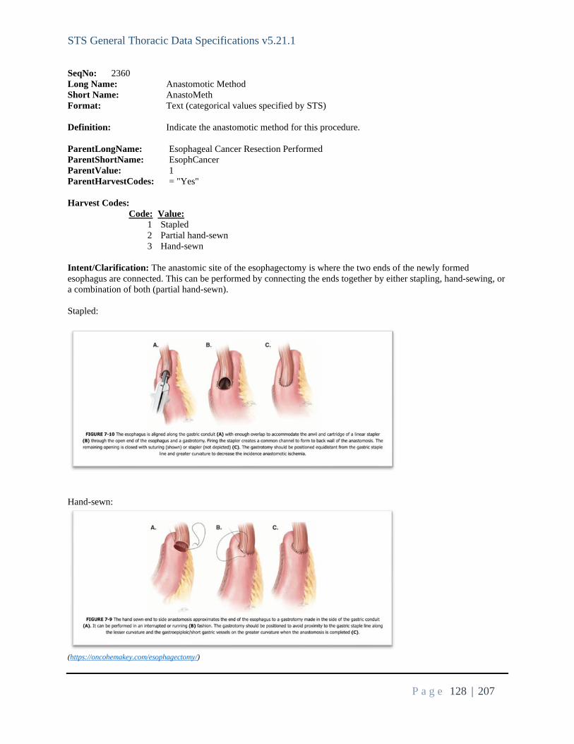
STS General Thoracic Data Specifications v5.21.1
P a g e 128 | 207
SeqNo: 2360 Long Name: Anastomotic Method Short Name: AnastoMeth Format: Text (categorical values specified by STS) Definition: Indicate the anastomotic method for this procedure. ParentLongName: Esophageal Cancer Resection Performed ParentShortName: EsophCancer ParentValue: 1 ParentHarvestCodes: = "Yes" Harvest Codes: Code: Value: 1 Stapled 2 Partial hand-sewn 3 Hand-sewn Intent/Clarification: The anastomic site of the esophagectomy is where the two ends of the newly formed esophagus are connected. This can be performed by connecting the ends together by either stapling, hand-sewing, or a combination of both (partial hand-sewn). Stapled:
Hand-sewn:
(https://oncohemakey.com/esophagectomy/)

STS General Thoracic Data Specifications v5.21.1
P a g e 129 | 207
SeqNo: 2370 Long Name: Esophageal Conduit Short Name: EsopConduit Format: Text (categorical values specified by STS) Definition: Indicate the type of esophageal conduit. ParentLongName: Esophageal Cancer Resection Performed ParentShortName: EsophCancer ParentValue: 1 ParentHarvestCodes: = "Yes" Harvest Codes: Code: Value: 1 Stomach 2 Small bowel 3 Colon 4 Supercharged small bowel 5 Supercharged colon Intent/Clarification: Indicate the type of gastrointestinal tissue used to reconstruct the esophagus. A “supercharged” small or large bowel interposition conduit is defined as one in which the pedicle blood supply has been augmented with additional arterial and venous connections, thus restoring more of the native blood flow. (https://www.annalsthoracicsurgery.org/article/S0003-4975(13)00105-7/pdf) SeqNo: 2380 Long Name: Pylorus Management Short Name: PylorusManage Format: Multi-Select Definition: Indicate the type of pylorus management provided. ParentLongName: Esophageal Cancer Resection Performed ParentShortName: EsophCancer ParentValue: 1 ParentHarvestCodes: = "Yes" Harvest Codes: Code: Value: 1 None 2 Botox Injection 3 Balloon Dilation 4 Pyloroplasty 5 Pyloromyotomy Intent/Clarification: The pylorus is the opening from the stomach to the duodenum (small intestine).

STS General Thoracic Data Specifications v5.21.1
P a g e 130 | 207
Indicate if Botox, balloon dilatation, pyloroplasty, pyloromyotomy.
- Balloon Dilation: Insertion and expansion of a balloon to widen the pyloric opening.
(https://link.springer.com/chapter/10.1007/978-981-13-1184-0_16)
- Pyloroplasty: A surgical procedure to widen the opening between the stomach and duodenum by bypassing the pyloric sphincter and allowing direct emptying of the stomach into the small intestine. This oftentimes results in gastric dumping.

STS General Thoracic Data Specifications v5.21.1
P a g e 131 | 207
(https://link.springer.com/chapter/10.1007/978-3-319-96122-4_67)
- Pyloromyotomy: A surgical procedure where an incision is made in the wall of the pylorus and the lining of the pylorus bulges through the incision, thereby opening up the channel.
(https://www.mayoclinic.org/diseases-conditions/pyloric-stenosis/multimedia/pyloromyotomy/img-20006399)
SeqNo: 2390 Long Name: J-Tube Placement Short Name: JTubePlac
STS General Thoracic Data Specifications v5.21.1
P a g e 132 | 207
Format: Text (categorical values specified by STS) Definition: Indicate the occurrence of a jejunostomy tube (J-tube) placement ParentLongName: Esophageal Cancer Resection Performed ParentShortName: EsophCancer ParentValue: 1 ParentHarvestCodes: = "Yes" Harvest Codes: Code: Value: 1 None 2 Pre-existing 3 During surgery 4 Post surgery Intent/Clarification: A jejunostomy tube (J-tube) is a soft flexible tube placed through the skin of the abdomen directly into the small intestine. It is used to provide nutrients and medication to the patient, bypassing the esophagus and stomach. Indicated if a J-tube placed either pre-existing, during or post-surgery, within 30 days of procedure. If a J-tube was pre-existing, removed during this operation, and then another J-tube was placed, code this as pre-existing. SeqNo: 2401 Long Name: Esophageal Cancer Present Short Name: EsophCancerPres Format: Text (categorical values specified by STS) Definition: Indicate whether esophageal cancer is present as indicated by the results of the final pathologic diagnosis. ParentLongName: Esophageal Cancer Resection Performed ParentShortName: EsophCancer ParentValue: 1 ParentHarvestCodes: = "Yes" Harvest Codes: Code: Value: 1 Yes 2 No Intent/Clarification: Although redundant to EsophCancer (seq 1530), this field is the parent to the below fields and required on all esophageal cancer cases. This field is required for record inclusion. If missing data, the entire record will be excluded from the analysis. SeqNo: 2410 Long Name: Pathologic Staging - Esophageal Cancer - T Short Name: PathStageEsophT Format: Text (categorical values specified by STS) Definition: Indicate the appropriate descriptor for the esophageal cancer primary tumor based on

STS General Thoracic Data Specifications v5.21.1
P a g e 133 | 207
final pathology report. ParentLongName: Esophageal Cancer Present ParentShortName: EsophCancerPres ParentValue: 1 ParentHarvestCodes: = "Yes" Harvest Codes: Code: Value: Definition: 1 T0 No evidence of primary tumor 2 Tis High Grade dysplasia, defined as malignant cells confined to the epithelium by the basement membrane 3 T1a Tumor invades lamina propria or muscularis mucosa 4 T1b Tumor invades submucosa 5 T2 Tumor invades muscularis propria 6 T3 Tumor invades adventitia 7 T4a Tumor invades pleura, pericardium, azygos vein, diaphragm or peritoneum 8 T4b Tumor invades other adjacent structures such as aorta, vertebral body, or airway. Intent/Clarification: The TNM (Tumor, Node, Metastasis) staging system is the most common way for doctors to stage esophageal cancer. Each staging has four categories. Use the most recent scan for documenting T-stage. The T-stage of the TNM staging system is captured in this field and related to the size of the tumor (area of cancer).
There are 4 categories – T1 to T4. Since TX is not not indicative of cancer, it is not captured as esophageal cancer. TX means the main cancer (primary) can’t be assessed. It doesn't show on scans but there might be cancer cells present in spit or in fluid taken from the lung.
1. T0 : a. No evidence of primary tumor b. Indicates no pathologic response
2. Tis a. High Grade dysplasia, defined as malignant cells confined to the epithelium by the basement
membrane b. High-grade dysplasia includes all noninvasive neoplastic epithelial lesions formerly called
carcinoma in situ; that term is no longer used for columnar mucosae anywhere in the gastrointestinal tract.
c. 3. T1a
a. Tumor invades lamina propria or muscularis mucosa 4. T1b
a. Tumor invades submucosa 5. T2
a. Tumor invades muscularis propria 6. T3
a. Tumor invades adventitia 7. T4a
1) Tumor invades pleura, pericardium, azygos vein, diaphragm or peritoneum 8. T4b
a. Tumor invades other adjacent structures such as aorta, vertebral body, or airway.

STS General Thoracic Data Specifications v5.21.1
P a g e 134 | 207
If the patient had induction therapy and had a complete response with path report indicating T0 or no T stage is provided, check ‘tumor present’ and code T0. If the tumor was resected at a separate endoscopic mucosal resection (EMR) procedure, use the t-stage from the EMR procedure. SeqNo: 2420 Long Name: Pathologic Staging - Esophageal Cancer - N Short Name: PathStageEsophN Format: Text (categorical values specified by STS) Definition: Indicate the appropriate descriptor for the esophageal cancer regional lymph nodes based on final pathology report. ParentLongName: Esophageal Cancer Present ParentShortName: EsophCancerPres ParentValue: 1 ParentHarvestCodes: = "Yes" Harvest Codes: Code: Value: Definition: 7 NX Regional lymph nodes cannot be assessed 1 N0 No regional lymph node metastasis 8 N1 Metastasis in 1-2 regional nodes 9 N2 Metastasis in 3-6 regional lymph nodes 10 N3 Metastasis in 7 or more regional lymph nodes Intent/Clarification: Indentify the N classification. If no lymph nodes or lymph node fragments are sampled, then code NX. If lymph node(s)/fragments are sampled and all specimens come back benign (negative for cancer or malignancy) then code N0. Use the final pathology report from the specimens collected during the current surgery. SeqNo: 2430 Long Name: Pathologic Staging - Esophageal Cancer - M Short Name: PathStageEsophM Format: Text (categorical values specified by STS) Definition: Indicate the appropriate descriptor for the esophageal cancer distant metastases based on final pathology report. ParentLongName: Esophageal Cancer Present ParentShortName: EsophCancerPres ParentValue: 1 ParentHarvestCodes: = "Yes" Harvest Codes: Code: Value: Definition: 1 M0 No distant metastasis

STS General Thoracic Data Specifications v5.21.1
P a g e 135 | 207
2 M1 Distant metastasis Intent/Clarification: The intent of the M-stage (metastasis stage) is to determine if the cancer has spread from the primary tumor. See the Harvest Definitions above for a description of the choices within this field. Indicate the appropriate M-stage (metastasis stage) based on the final pathology report of the current surgery. If a patient has no known metastasis found prior to or during current procedure but is found with metastasis following the procedure, then code this as M0.
Example: No known metastasis prior to procedure, undergoes procedure and final pathology report does not report any known metastasis, then CT of brain if performed following the procedure and is positive for brain metastasis. This is to be coded as M0 since the metastasis was found after current procedure.
MX is sometimes reported for cancers that could not be evaluated for distant metastasis. If MX is reported, this is to be coded as M0.
Use the final pathology report from the specimens collected during the current surgery. SeqNo: 2440 Long Name: Pathologic Staging - Esophageal Cancer - H Short Name: PathStageEsophH Format: Text (categorical values specified by STS) Definition: Indicate the appropriate descriptor for the esophageal cancer histopathologic type based on final pathology report. ParentLongName: Esophageal Cancer Present ParentShortName: EsophCancerPres ParentValue: 1 ParentHarvestCodes: = "Yes" Harvest Codes: Code: Value: 1 H1 Squamous Carcinoma 2 H2 Adenocarcinoma 3 Other Intent/Clarification: Tumor histology is determined by pathologic evaluation of the specimen. Use the final pathology report from the specimens collected during the current surgery. However, if final pathology report lists T0 and no histologic grade, then select histologic grade from the pre-surgical biopsy. SeqNo: 2450 Long Name: Pathologic Staging - Esophageal Cancer - G Short Name: PathStageEsophG Format: Text (categorical values specified by STS) Definition: Indicate the appropriate descriptor for the esophageal cancer histologic grade based on final pathology report. If a range of differentiation is reported, choose the worst differentiation. ParentLongName: Esophageal Cancer Present ParentShortName: EsophCancerPres ParentValue: 1 ParentHarvestCodes: = "Yes"

STS General Thoracic Data Specifications v5.21.1
P a g e 136 | 207
Harvest Codes: Code: Value: Definition: 1 GX Grade cannot be assessed 2 G1 Well differentiated 3 G2 Moderately differentiated 4 G3 Poorly differentiated, undifferentiated Intent/Clarification: Use the grading from the final pathology report from the current surgery unless the patient had induction therapy. If the patient had induction therapy, then use the pre-induction therapy biopsy report. SeqNo: 2460 Long Name: Esophageal Cancer - Number of Nodes Short Name: EsophCANodes Format: Integer Definition: Indicate the total number of nodes sampled/harvested. Low Value: 0 High Value: 60 ParentLongName: Esophageal Cancer Present ParentShortName: EsophCancerPres ParentValue: 1 ParentHarvestCodes: = "Yes" Intent/Clarification: Total number of nodes will be listed on your final pathology report. Use the final pathology report from the day of surgery (resection) for the number of nodes. Capture the total number of nodes harvested during surgery. Only count the number of nodes that were actually harvested. If nodes examined but not harvested or not found, do not count. Node fragments – Differentiating node fragments from separate lymph nodes is a very difficult problem. Ideally the surgeon will count lymph nodes during the case and create a system to label specimens with the count as they leave the OR. This will permit the pathologist to report the actual lymph node count during the case in the path report. If the pathologist cannot, they will often report “lymph node fragments” which implies they cannot provide a lymph node count. In this circumstance, we have to conservatively assume that all those fragments come from a single node. If they report multiple fragments from a single station, then count this as one node since they all are being reported as coming from the same station. Please encourage your surgeons to develop a way of counting nodes in the case that can be conveyed in the pathology report. SeqNo: 2470 Long Name: Esophageal Cancer - Pathology Margins Short Name: EsophCAPathMarg Format: Text (categorical values specified by STS) Definition: Indicate whether pathology report indicated positive surgical margins. ParentLongName: Esophageal Cancer Present ParentShortName: EsophCancerPres ParentValue: 1 ParentHarvestCodes: = "Yes"

STS General Thoracic Data Specifications v5.21.1
P a g e 137 | 207
Harvest Codes: Code: Value: 1 Yes 2 No Intent/Clarification: Margins, also known as ‘margins of resection,’ refer to the distance between a tumor and the edge of the surrounding tissue that's removed along with it. ‘Positive margins’ indicate cancer cells extend to the edge of resected tissue. SeqNo: 2480 Long Name: Location of Positive Margins Short Name: PosMargLocate Format: Multi-Select Definition: Indicate the location(s) of the positive margin(s). ParentLongName: Esophageal Cancer - Pathology Margins ParentShortName: EsophCAPathMarg ParentValue: 1 ParentHarvestCodes: = "Yes" Harvest Codes: Code: Value: 1 Proximal (Esophageal) 2 Distal (Gastric) 3 Radial Intent/Clarification: If there were positive margins, indicate if the location of the positive margin was proximal (toward the head), distal (toward the stomach), and/or radial (circumferential). Select al that apply. Proximal Distal ------------------------------------------------------------------------------------------------------------------------------------------------------------------------------------ Thymoma/Thymectomy/Mediastinal Mass/Myasthenia Gravis ------------------------------------------------------------------------------------------------------------------------------------------------------------------------------------

STS General Thoracic Data Specifications v5.21.1
P a g e 138 | 207
Instructions - For Thymus/Mediastinal Mass cases
• Collect detailed info on thymectomies for myasthenia including open, cervical or VATS route • Collect all thymectomies for myasthenia regardless of whether they have thymoma • Collect detailed info on thymectomies for thymoma including open or VATS
Robotics should be coded with thoracoscopic procedures, also code S2900 under miscellaneous procedures. SeqNo: 2490 Long Name: Symptomatic myasthenia Short Name: MyastheniaSympt Format: Text (categorical values specified by STS) Definition: Indicate whether the patient has symptomatic Myasthenia Gravis ParentLongName: Thymus/Mediastinal Mass Resection/Myasthenia Gravis ParentShortName: ThymusMediastinalData ParentValue: 1 ParentHarvestCodes: = "Yes" Harvest Codes: Code: Value: 1 Yes 2 No Intent/Clarification: SeqNo: 2500 Long Name: Chronical Medical Treatement Short Name: ChronMedTreat Format: Multi-Select Definition: Indicate the chronic medical treatment the patient received. Select all that apply. ParentLongName: Symptomatic myasthenia ParentShortName: MyastheniaSympt ParentValue: 1 ParentHarvestCodes: = "Yes" Harvest Codes: Code: Value: 1 Mestinon 2 Steroids 3 Other Immunosuppressive Therapy Intent/Clarification:
- Mestinon o Indicate whether patient uses Mestinon (pyridostigmine) for the treatment of myasthenia gravis.
- Steroids o Indicate whether patient uses steroids for the treatment of myasthenia gravis.
Only include steroid use for treatment of myasthenia gravis. Do not capture steroids used for other reasons or one-time doses for procedural/imagining

STS General Thoracic Data Specifications v5.21.1
P a g e 139 | 207
support here (i.e., prednisone prior to CT scan is not captured here). - Other Immunosuppressive Therapy
o Indicate whether patient uses another immunosuppressive therapy for the treatment of myasthenia gravis.
Only include immunosuppressive therapy for the treatment of myasthenia gravis. Examples are azathioprine (Imuran), mycophenolate mofetil (CellCept), cyclosporine
(Sandimmune, Neoral), methotrexate (Trexall) or tacrolimus (Prograf) and Rituxan. Please note that neoadjuvant therapies with chemotherapy (i.e., CAP, ADOC, PE, VIP) are not to be captured here. They are to be captured under History of Cancer (HistCancer – seq 650); Preoperative Chemotherapy/Immunotherapy. SeqNo: 2540 Long Name: Pre-operative management - IVIG Short Name: IVIG Format: Text (categorical values specified by STS) Definition: Indicate whether the patient has had IVIG pre-operatively. ParentLongName: Thymus/Mediastinal Mass Resection/Myasthenia Gravis ParentShortName: ThymusMediastinalData ParentValue: 1 ParentHarvestCodes: = "Yes" Harvest Codes: Code: Value: 1 Yes 2 No Intent/Clarification: Indicate if the patient’s peroperative management included intravenous immunoglobulin (IVIG). SeqNo: 2550 Long Name: Pre-operative management - Plasmaphereis Short Name: Plasmaphereis Format: Text (categorical values specified by STS) Definition: Indicate whether the patient has had plasmaphereis pre-operatively. ParentLongName: Thymus/Mediastinal Mass Resection/Myasthenia Gravis ParentShortName: ThymusMediastinalData ParentValue: 1 ParentHarvestCodes: = "Yes" Harvest Codes: Code: Value: 1 Yes 2 No Intent/Clarification: Indicate if the patient’s preoperative management included plasmapheresis. SeqNo: 2560 Long Name: Thymus / Mediastinal Mass Size Known

STS General Thoracic Data Specifications v5.21.1
P a g e 140 | 207
Short Name: MassSizeKnown Format: Text (categorical values specified by STS) Definition: Indicate whether the size of the thymus / mediastianl mass is known. ParentLongName: Thymus/Mediastinal Mass Resection/Myasthenia Gravis ParentShortName: ThymusMediastinalData ParentValue: 1 ParentHarvestCodes: = "Yes" Harvest Codes: Code: Value: 1 Yes 2 No Intent/Clarification: SeqNo: 2571 Long Name: Size of Mass In cm Short Name: MassSizeCm Format: Real Definition: Indicate the largest diameter in cm derived from preop axial, coronal or sagittal imagining. Low Value: 0.00 High Value: 50.00 ParentLongName: Thymus / Mediastinal Mass Size Known ParentShortName: MassSizeKnown ParentValue: 1 ParentHarvestCodes: = "Yes"
Intent/Clarification: Tumor size can be found on contrast-enhanced chest CT or MRI. If the tumor size is greater than 50cm, then enter 50cm.
SeqNo: 2580 Long Name: Thymus / Mediastinal Mass - Initial Surgical Approach Short Name: ThyInitSurgAp Format: Text (categorical values specified by STS) Definition: Indicate the initial surgical approach used by the surgeon. ParentLongName: Thymus/Mediastinal Mass Resection/Myasthenia Gravis ParentShortName: ThymusMediastinalData ParentValue: 1 ParentHarvestCodes: = "Yes" Harvest Codes: Code: Value: 1 Full Sternotomy 2 Clamshell or Hemiclamshell 3 Transcervical 4 Partial Sternotomy 5 Robotic

STS General Thoracic Data Specifications v5.21.1
P a g e 141 | 207
6 VATS 7 Thoracotomy Intent/Clarification: SeqNo: 2590 Long Name: Thymus / Mediastinal Mass - Robotic / VATS Location Short Name: ThyRobVATSLoc Format: Text (categorical values specified by STS) Definition: Indicate the location of the robotic or VATS procedure. ParentLongName: Thymus / Mediastinal Mass - Initial Surgical Approach ParentShortName: ThyInitSurgAp ParentValue: 5|6|7 ParentHarvestCodes: = "Robotic", "VATS" or "Thoracotomy" Harvest Codes: Code: Value: 1 Right 2 Left 3 Bilateral Intent/Clarification: SeqNo: 2600 Long Name: Thymus / Mediastinal Mass - Conversion To Open Approach Short Name: ThyConvToOpen Format: Text (categorical values specified by STS) Definition: Indicate if the approach was converted to an open approach during the procedure. ParentLongName: Thymus / Mediastinal Mass - Initial Surgical Approach ParentShortName: ThyInitSurgAp ParentValue: 3|5|6 ParentHarvestCodes: = "Transcervical", "Robotic" or "VATS" Harvest Codes: Code: Value: 1 Yes, planned 2 Yes, unplanned 3 No Intent/Clarification: The intent is to capture if the approach was converted to open and if it was planned (indicated prior to the start of the operation) or unplanned. To code as planned, it must be documented in the plan of care prior to OR Entry. SeqNo: 2610 Long Name: Thymus / Mediastinal Mass - Conversion Approach Short Name: ThyConvAp Format: Text (categorical values specified by STS) Definition: Indicate the final surgical approach for the thymus / mediastinal mass resection.

STS General Thoracic Data Specifications v5.21.1
P a g e 142 | 207
ParentLongName: Thymus / Mediastinal Mass - Conversion To Open Approach ParentShortName: ThyConvToOpen ParentValue: 1|2 ParentHarvestCodes: = "Yes, planned" or "Yes, unplanned" Harvest Codes: Code: Value: 1 Sternotomy 2 Clamshell 3 Thoracotomy Intent/Clarification: If a conversion was performed, indicate the conversion approach. SeqNo: 2620 Long Name: Intentional resection of functioning phrenic nerve Short Name: PhrenicNerveResect Format: Text (categorical values specified by STS) Definition: Indicate if functioning phrenic nerve was resected intentionally by the surgeon during the procedure. ParentLongName: Thymus/Mediastinal Mass Resection/Myasthenia Gravis ParentShortName: ThymusMediastinalData ParentValue: 1 ParentHarvestCodes: = "Yes" Harvest Codes: Code: Value: 1 Yes 2 No Intent/Clarification: SeqNo: 2630 Long Name: Thymoma Procedure Short Name: ThymomaProc Format: Text (categorical values specified by STS) Definition: Indicate whether a thymectomy was performed. ParentLongName: Thymus/Mediastinal Mass Resection/Myasthenia Gravis ParentShortName: ThymusMediastinalData ParentValue: 1 ParentHarvestCodes: = "Yes" Harvest Codes: Code: Value: 1 Yes 2 No Intent/Clarification: This field is required for record inclusion. If missing data, the entire record will be excluded from the analysis.

STS General Thoracic Data Specifications v5.21.1
P a g e 143 | 207
SeqNo: 2640 Long Name: Pathologic Staging (from pathology report) Short Name: PathRptStage Format: Text (categorical values specified by STS) Definition: Indicate the pathological stage as reported on the final Pathology report. ParentLongName: Thymoma Procedure ParentShortName: ThymomaProc ParentValue: 1 ParentHarvestCodes: = "Yes" Harvest Codes: Code: Value: Definition: 8 No Cancer Found 1 Stage I Grossly and microscopically encapsulated. Also called a noninvasive thymoma. That is, it has not spread beyond the thymus. 2 Stage II The thymoma invades beyond the capsule (outer boundary of the thymus) and into the nearby fatty tissue or to the pleura (outer covering of the lung). Sometimes divided into: Stage IIa or Stage IIb. 3 Stage IIa Microscopic transcapsular invasion 4 Stage IIb Macroscopic capsular invasion 5 Stage III Macroscopic invasion of neighboring organs. The 6 Stage IVa Pleural or pericardial dissemination. The thymoma has spread widely throughout the pleura and/or pericardium. 7 Stage IVb Hematogenous or lymphatic dissemination. The thymoma has spread to distant organs. Intent/Clarification:
- If no cancer is found/no thymoma, please code ‘No Cancer Found.’ - For purposes of consistency, STS uses the Masaoka/Modified Masaoka staging system.
o Example: Pathology report provides Modified Masaoka Stage: IIa; Moran Stage I. Please code Stage IIa.
- If no staging is provided on the pathology report, then leave blank.
SeqNo: 2650 Long Name: WHO classification Short Name: ThymomaType Format: Text (categorical values specified by STS) Definition: Indicate the WHO classification as reported on the final Pathology Report. ParentLongName: Pathologic Staging (from pathology report) ParentShortName: PathRptStage ParentValue: <>8 And Is Not Missing ParentHarvestCodes: Is Not "No Cancer Found" And Is Not Missing

STS General Thoracic Data Specifications v5.21.1
P a g e 144 | 207
Harvest Codes: Code: Value: 1 Type A 2 Type AB 3 Type B1 4 Type B2 5 Type B3 6 Thymic Carcinoma or Type C Intent/Clarification: Use the final pathology report for coding of this field.
- If a two-part tumor is described on the pathology report as Type A and Type AB, code Type AB. - If a tumor is described with multiple morphologies, such as Type A, Type B3, Type B2, then code Type
AB. SeqNo: 2660 Long Name: Completeness of resection (from operative note or pathology report) Short Name: ResectCompleteness Format: Text (categorical values specified by STS) Definition: Indicate the completeness of the resection as reported on the Operative Note or final Pathology Report. ParentLongName: Thymus/Mediastinal Mass Resection/Myasthenia Gravis ParentShortName: ThymusMediastinalData ParentValue: 1 ParentHarvestCodes: = "Yes" Harvest Codes: Code: Value: 1 R0 2 R1 3 R2 Intent/Clarification: Indicate if the margins of the resected tumor were positive or negative for malignant cells.
- R0 – Complete resection with negative margins - R1 – Microscopically positive margins - R2 – Grossly positive margins. Evident to the naked-eye.
If visibility is not obvious on the pathology report, then verify with the surgeon or pathologist. Only code ‘R2 – Macroscopic (gross) residual tumor present,’ if it is present in the surgical report. This must be documented on the operative note, please work with the surgeon to include this in the operative note if it is provided on the pathology report and not on the operative note. SeqNo: 2670 Long Name: Patient Alive 30 Days Post Procedure Short Name: PtAlive30Day Format: Text (categorical values specified by STS) Definition: Indicate if the patient is alive at 30 days post-operative. ParentLongName: Thymus/Mediastinal Mass Resection/Myasthenia Gravis

STS General Thoracic Data Specifications v5.21.1
P a g e 145 | 207
ParentShortName: ThymusMediastinalData ParentValue: 1 ParentHarvestCodes: = "Yes" Harvest Codes: Code: Value: 1 Yes 2 No Intent/Clarification: Indicate if the patient is alive at 30-days post-op. SeqNo: 2680 Long Name: Myasthenic crisis requiring return to ICU or intervention (intubation, plasmapheresis) - Post-Operative event (30 day) Short Name: MYAL Format: Text (categorical values specified by STS) Definition: Indicate whether the patient experience myasthenic crisis after surgery. ParentLongName: Patient Alive 30 Days Post Procedure ParentShortName: PtAlive30Day ParentValue: 1 ParentHarvestCodes: = "Yes" Harvest Codes: Code: Value: 1 Yes 2 No Intent/Clarification: Myasthenic crisis is a complication of myasthenia gravis characterized by worsening of muscle weakness, resulting in respiratory failure that requires intubation and mechanical ventilation. This field is intended to capture those patients who experience myasthenia crisis requiring return to ICU or intervention (intubation, plasmapheresis) within 30 days of surgery. (https://www.ncbi.nlm.nih.gov/pmc/articles/PMC3726100/#:~:text=Myasthenic%20crisis%20is%20a%20complication,rate%20associated%20with%20myasthenic%20crisis.) SeqNo: 2690 Long Name: Unitenional phrenic nerve palsy - Post-Operative event (30 day) Short Name: PhrenicNervePalsy Format: Text (categorical values specified by STS) Definition: Indicate if the patient experienced unintentional phrenic nerve palsy in the post operative period. Unintentional means phrenic nerve palsy without having undergone intentional resection of the phrenic nerve. ParentLongName: Patient Alive 30 Days Post Procedure ParentShortName: PtAlive30Day ParentValue: 1 ParentHarvestCodes: = "Yes" Harvest Codes: Code: Value: 1 Yes

STS General Thoracic Data Specifications v5.21.1
P a g e 146 | 207
2 No Intent/Clarification: Time frame is from OR Exit to post-op day 30. SeqNo: 2700 Long Name: Patient Alive 90 Days Post Procedure Short Name: PtAlive90Day Format: Text (categorical values specified by STS) Definition: Indicate if the patient is alive at 90 days post-operative. ParentLongName: Patient Alive 30 Days Post Procedure ParentShortName: PtAlive30Day ParentValue: 1 ParentHarvestCodes: = "Yes" Harvest Codes: Code: Value: 1 Yes 2 No Intent/Clarification: Indicate if the patient is alive at 90-days post-op. SeqNo: 2710 Long Name: Adjuvant thoracic radiation - Post-Operative event (90 day) Short Name: ThoracicRadiation Format: Text (categorical values specified by STS) Definition: Indicate whether the patient had adjuvant thoracic radiation within 90 days post operatively. ParentLongName: Patient Alive 90 Days Post Procedure ParentShortName: PtAlive90Day ParentValue: 1 ParentHarvestCodes: = "Yes" Harvest Codes: Code: Value: 1 Yes 2 No Intent/Clarification: The adjuvant thoracic radiation therapy must occur within the 90-day postoperative window to code this field as ‘Yes.’ SeqNo: 2720 Long Name: Persistent unitenional phrenic nerve palsy - Post-Operative event (90 day) Short Name: PhrenNrvPalsyPersis Format: Text (categorical values specified by STS) Definition: Indicate if the patient has persistent diaphragm dysfunction due to phrenic nerve palsy 90 days following surgery. ParentLongName: Patient Alive 90 Days Post Procedure

STS General Thoracic Data Specifications v5.21.1
P a g e 147 | 207
ParentShortName: PtAlive90Day ParentValue: 1 ParentHarvestCodes: = "Yes" Harvest Codes: Code: Value: 1 Yes 2 No
Intent/Clarification: Unintentional means phrenic nerve palsy without having undergone intentional resection of the phrenic nerve.
------------------------------------------------------------------------------------------------------------------------------------------------------------------------------------ Tracheal Resection ------------------------------------------------------------------------------------------------------------------------------------------------------------------------------------ SeqNo: 2730 Long Name: Current Airway - Pre-Operative Short Name: AirwayCurr Format: Text (categorical values specified by STS) Definition: Indicate the patient's airway status prior to surgery. ParentLongName: Tracheal Resection ParentShortName: TrachealData ParentValue: 1 ParentHarvestCodes: = "Yes" Harvest Codes: Code: Value: 1 Native 2 Oral ETT 3 Trach 4 T-Tube Intent/Clarification: SeqNo: 2740 Long Name: Prior tracheostomy - Pre-Operative Short Name: TracheostomyPrior Format: Text (categorical values specified by STS) Definition: Indicate if the patient has had a prior tracheostomy. ParentLongName: Tracheal Resection ParentShortName: TrachealData ParentValue: 1 ParentHarvestCodes: = "Yes"

STS General Thoracic Data Specifications v5.21.1
P a g e 148 | 207
Harvest Codes: Code: Value: 1 Yes 2 No Intent/Clarification: At any time in the past including open or percutaneous tracheostomy or cricothyroidostomy. SeqNo: 2750 Long Name: Prior intubation - Pre-Operative Short Name: IntubatePrior Format: Text (categorical values specified by STS) Definition: Indicate if the patient has had a prior intubation at any point in their life. ParentLongName: Tracheal Resection ParentShortName: TrachealData ParentValue: 1 ParentHarvestCodes: = "Yes" Harvest Codes: Code: Value: 1 Yes 2 No Intent/Clarification: SeqNo: 2760 Long Name: Prior Tracheal Resection - Pre-Operative Short Name: TrachealResectPrior Format: Text (categorical values specified by STS) Definition: Indicate if the patient has ever had a prior tracheal resection. ParentLongName: Tracheal Resection ParentShortName: TrachealData ParentValue: 1 ParentHarvestCodes: = "Yes" Harvest Codes: Code: Value: 1 Yes 2 No Intent/Clarification: SeqNo: 2770 Long Name: Recent Bronchoscopic Intervention (within 6 weeks) Short Name: BronchInt6Wks Format: Text (categorical values specified by STS) Definition: Indicate if the patient has had any bronchoscopic interventions within the last 6 weeks. This includes, for example, core out, dilation, ablation, stent.

STS General Thoracic Data Specifications v5.21.1
P a g e 149 | 207
ParentLongName: Tracheal Resection ParentShortName: TrachealData ParentValue: 1 ParentHarvestCodes: = "Yes" Harvest Codes: Code: Value: 1 Yes 2 No Intent/Clarification: SeqNo: 2780 Long Name: Recurrent Nerves Intact Preoperatively Short Name: RecurrNervesIntact Format: Text (categorical values specified by STS) Definition: Indicate whether the recurrent nerves were intact preoperatively. ParentLongName: Tracheal Resection ParentShortName: TrachealData ParentValue: 1 ParentHarvestCodes: = "Yes" Harvest Codes: Code: Value: 1 Yes 2 No 3 Unknown Intent/Clarification: SeqNo: 2790 Long Name: Recurrent Nerves Not Intact Short Name: RecurrNervNotIntact Format: Text (categorical values specified by STS) Definition: Indicate which recurrent nerve is not intact. ParentLongName: Recurrent Nerves Intact Preoperatively ParentShortName: RecurrNervesIntact ParentValue: 2 ParentHarvestCodes: = "No" Harvest Codes: Code: Value: 1 Right 2 Left 3 Both Intent/Clarification: SeqNo: 2800

STS General Thoracic Data Specifications v5.21.1
P a g e 150 | 207
Long Name: Air Way Management During Tracheal Resection Short Name: ArWyMgtDurngTrachResc Format: Multi-Select Definition: Indicate the patients airway management during tracheal resection. Select all that apply or 'none'. ParentLongName: Tracheal Resection ParentShortName: TrachealData ParentValue: 1 ParentHarvestCodes: = "Yes" Harvest Codes: Code: Value: 1 None 2 Cross-table ventilation 3 VA ECMO 4 Jet ventilation 5 VV ECMO 6 Cardiopulmonary bypass Intent/Clarification: Indicate the patient’s airway management during tracheal resection. Select all that apply or 'none'.
- VA ECMO stands for Veno-arterial Extracorporeal Membrane Oxygenation. This process takes deoxygenated blood from a central vein or the right atrium, pumps it past the oxygenator, and then returns the oxygenated blood, under pressure, to the arterial side of the circulation (typically to the aorta).
- Jet ventilation refers to delivery of oxygen via high pressure jet ventilator
- VV ECMO stands for Veno-venus Extracorporeal Membrane Oxygenation. This process takes blood from a large vein, pumps it past the oxygenator, and returns oxygenated blood back to a large vein.
- Cardiopulmonary bypass is a technique that temporarily takes over the function of the heart and lungs
during surgery, maintaining the circulation of blood and the oxygen content of the patient's body SeqNo: 2860 Long Name: Tracheal Resection Incision Short Name: TrachIncis Format: Multi-Select Definition: Indicate the type of incision(s) made for the tracheal resection procedure. ParentLongName: Tracheal Resection ParentShortName: TrachealData ParentValue: 1 ParentHarvestCodes: = "Yes" Harvest Codes: Code: Value: 1 Cervical 2 Partial sternotomy 3 Full sternotomy 4 Right thoracotomy 5 Clamshell

STS General Thoracic Data Specifications v5.21.1
P a g e 151 | 207
Intent/Clarification: Indicate the type of incision(s) made for the tracheal resection procedure. Select all that apply. SeqNo: 2920 Long Name: Length of tracheal resection in cm (Surgical or pathological measurement acceptable) Short Name: TrachealResectLen Format: Real Definition: Indicate the length of the tracheal resection in cm as reported on the pathology or surgical report. Low Value: 0.00 High Value: 10.00 ParentLongName: Tracheal Resection ParentShortName: TrachealData ParentValue: 1 ParentHarvestCodes: = "Yes" Intent/Clarification: SeqNo: 2930 Long Name: Cricoid resection required Short Name: CricoidResect Format: Text (categorical values specified by STS) Definition: Indicate whether a cricoid resection was performed. ParentLongName: Tracheal Resection ParentShortName: TrachealData ParentValue: 1 ParentHarvestCodes: = "Yes" Harvest Codes: Code: Value: 1 Yes 2 No Intent/Clarification: SeqNo: 2940 Long Name: Carinal resection required Short Name: CarinalResect Format: Text (categorical values specified by STS) Definition: Indicate whether a carinal resection was performed. ParentLongName: Tracheal Resection ParentShortName: TrachealData ParentValue: 1 ParentHarvestCodes: = "Yes" Harvest Codes: Code: Value:

STS General Thoracic Data Specifications v5.21.1
P a g e 152 | 207
1 Yes 2 No Intent/Clarification: SeqNo: 2960 Long Name: Release Maneuver - Type Short Name: ReleaseManeuverType Format: Text (categorical values specified by STS) Definition: Indicate what type of release maneuver was performed. ParentLongName: Tracheal Resection ParentShortName: TrachealData ParentValue: 1 ParentHarvestCodes: = "Yes" Harvest Codes: Code: Value: 6 None 1 Suprahyoid 2 Suprathyroid 3 Hilar 4 Suprahyoid - Hilar 5 Suprathyroid - Hilar Intent/Clarification: SeqNo: 2970 Long Name: Tracheal Procedures Additional Post-Op Events Short Name: TrachAddlPOEve Format: Multi-Select Definition: Indicate additional post-operative events following the tracheal procedure. Select all that apply or 'none'. ParentLongName: Tracheal Resection ParentShortName: TrachealData ParentValue: 1 ParentHarvestCodes: = "Yes" Harvest Codes: Code: Value: 1 None 2 Anastomotic dehiscence requiring drainage, revision, stent, tracheostomy, T-tube 3 Anastomotic stricture requiring intervention 4 Airway obstruction requiring intervention 5 Recurrent nerve palsy

STS General Thoracic Data Specifications v5.21.1
P a g e 153 | 207
Intent/Clarification: Indicate if the patient experienced any of the above within 30 days of surgery or during same admission if not discharged within 30 days. Select all that apply or ‘none.’ If the patient had pre-operative recurrent laryngeal nerve palsy do not capture here.
SeqNo: 2980 Long Name: Recurrent nerve palsy Short Name: NervePalsyRecurr Format: Text (categorical values specified by STS) Definition: Indicate the location of the recurrent nerve palsy that occurred in the post-operative period. ParentLongName: Tracheal Procedures Additional Post-Op Events ParentShortName: TrachAddlPOEve ParentValue: contains(5) ParentHarvestCodes: Contains ("Recurrent nerve palsy") Harvest Codes: Code: Value: 1 Right 2 Left 3 Bilateral Intent/Clarification: SeqNo: 3020 Long Name: Patient left hospital with tracheal appliance Short Name: TrachealAppliance Format: Text (categorical values specified by STS) Definition: Indicate if the patient was discharged from the acute care hospital with a tracheal
STS General Thoracic Data Specifications v5.21.1
P a g e 154 | 207
appliance in place; such as a tracheostomy or T-tube. ParentLongName: Tracheal Resection ParentShortName: TrachealData ParentValue: 1 ParentHarvestCodes: = "Yes" Harvest Codes: Code: Value: 1 Yes 2 No 3 Patient died in hospital Intent/Clarification: Use the final disposition from the acute care setting. If the patient was transferred to another acute care hospital, use the status at discharge from the final acute care hospital. SeqNo: 3040 Long Name: Patient Is Stent/Tube Free At 30 Days Postoperative Short Name: StentTubeFree30days Format: Text (categorical values specified by STS) Definition: Indicate if the patient is free of a stent or tracheal tube at 30 days post operatively. ParentLongName: Tracheal Resection ParentShortName: TrachealData ParentValue: 1 ParentHarvestCodes: = "Yes" Harvest Codes: Code: Value: 1 Yes 2 No 3 Patient died within 30 days of procedure Intent/Clarification: Time frame is within 30 days following surgery. Day of surgery = day 0, 1st day post-op = day 1, 2nd day post-op = day 3, etc. SeqNo: 3060 Long Name: Patient Is Stent/Tube Free At 90 Days Postoperative Short Name: StentTubeFree90days Format: Text (categorical values specified by STS) Definition: Indicate if the patient is free of a stent or tracheal tube at 90 days post operatively. ParentLongName: Patient Is Stent/Tube Free At 30 Days Postoperative ParentShortName: StentTubeFree30days ParentValue: 2 ParentHarvestCodes: = "No" Harvest Codes: Code: Value: 1 Yes 2 No

STS General Thoracic Data Specifications v5.21.1
P a g e 155 | 207
3 Patient died within 90 days of procedure Intent/Clarification: ------------------------------------------------------------------------------------------------------------------------------------------------------------------------------------ Hiatal Hernia/GERD ------------------------------------------------------------------------------------------------------------------------------------------------------------------------------------ SeqNo: 3070 Long Name: Hiatal Hernia/GERD Symptoms Short Name: HiatHernSymp Format: Multi-Select Definition: Indicate the patient's hiatal hernia/GERD symptoms. Select all that apply or 'none'. ParentLongName: Hiatal Hernia / Diaphragmatic Hernia or GERD ParentShortName: HiatalHerniaData ParentValue: 1 ParentHarvestCodes: = "Yes" Harvest Codes: Code: Value: 1 None 2 Heartburn 3 Cough 4 Regurgitation 5 Hoarseness 6 Dysphagia 7 Sore throat 8 Epigastric/chest pain 9 Asthma 10 Early satiety 11 Reflux laryngitis 12 Anemia Intent/Clarification: Include chronic symptoms, even if present prior to the diagnosis of Hiatal Hernia/ Diaphragmatic Hernia or GERD. SeqNo: 3190 Long Name: Plan-position-indication - PPI Use Short Name: PPIUse Format: Text (categorical values specified by STS) Definition: Indicate if the patient used PPIs pre-operatively - at the time of the evaluation. ParentLongName: Hiatal Hernia / Diaphragmatic Hernia or GERD ParentShortName: HiatalHerniaData ParentValue: 1 ParentHarvestCodes: = "Yes" Harvest Codes: Code: Value:
STS General Thoracic Data Specifications v5.21.1
P a g e 156 | 207
1 Yes 2 No Intent/Clarification: Proton-pump inhibitors (PPIs) are a class of medications that cause a profound and prolonged reduction of stomach acid production. They do so by irreversibly inhibiting the stomach's H+/K+ ATPase proton pump. Capture only PPIs in this field. Do not capture H2-receptor antagonists. Common PPI’s include: omeprazole (Prilosec), Esomeprazole (Nexium), Lansoprazole (Prevacid), Pantoprazole (Protonix) and Zegerid, many do not require a prescription. SeqNo: 3200 Long Name: Plan-position-indication - PPI Relief Short Name: PPIRelief Format: Text (categorical values specified by STS) Definition: Indicate if the patient obtained relief of symptoms. Indicate 'partial' if the patient had relief in the past but does not receive relief currently. ParentLongName: Plan-position-indication - PPI Use ParentShortName: PPIUse ParentValue: 1 ParentHarvestCodes: = "Yes" Harvest Codes: Code: Value: 1 Complete 2 Partial 3 No Intent/Clarification: Indicate ‘no’ if the patient had no relief, 'partial' if the patient had a decrease in symptoms (some relief), or ‘complete’ if the patient no longer had symptoms while taking PPIs (Proton Pump Inhibitors). SeqNo: 3210 Long Name: EDG Done Short Name: EGDDone Format: Text (categorical values specified by STS) Definition: Indicate whether an EDG was performed. ParentLongName: Hiatal Hernia / Diaphragmatic Hernia or GERD ParentShortName: HiatalHerniaData ParentValue: 1 ParentHarvestCodes: = "Yes" Harvest Codes: Code: Value: 1 Yes 2 No Intent/Clarification: Time Frame: Within 1 year. Code the value closest to procedure date. SeqNo: 3220

STS General Thoracic Data Specifications v5.21.1
P a g e 157 | 207
Long Name: EGD - Esophagitis Short Name: Esophagitis Format: Text (categorical values specified by STS) Definition: Indicate if the patient has esophagitis. ParentLongName: EDG Done ParentShortName: EGDDone ParentValue: 1 ParentHarvestCodes: = "Yes" Harvest Codes: Code: Value: 1 Yes 2 No Intent/Clarification: SeqNo: 3230 Long Name: Esophagitis - LA Grade Short Name: LAGrade Format: Text (categorical values specified by STS) Definition: Indicate the LA Grade. ParentLongName: EGD - Esophagitis ParentShortName: Esophagitis ParentValue: 1 ParentHarvestCodes: = "Yes" Harvest Codes: Code: Value: 1 A 2 B 3 C 4 D Intent/Clarification: The Los Angeles (LA) Classification System is used to grade esophagitis using a scale from A-D, with A being the least severe and D being the most severe form. Code the highest severity if more than one is provided. Example: If esophagitis is graded as A-B, code B because this is the highest severity
- Grade A: One or more mucosal breaks no longer than 5mm, not bridging the tops of the mucosal folds. - Grade B: One or more mucosal breaks longer than 5mm, not bridging the tops of the mucosal folds. - Grade C: One or more mucosal breaks bridging the tops of mucosal folds involving <75% of the
circumference. - Grade D: One or more mucosal breaks bridging the tops of the mucosal folds involving ≥75% of the
circumference.

STS General Thoracic Data Specifications v5.21.1
P a g e 158 | 207
__________________________________________________________________________ SeqNo: 3240 Long Name: Barrett's metaplasia Short Name: MetaplasiaBarrett Format: Text (categorical values specified by STS) Definition: Indicate if the patient has Barrett's metaplasia, and if it is low or high grade dysplasia. ParentLongName: EDG Done ParentShortName: EGDDone ParentValue: 1 ParentHarvestCodes: = "Yes" Harvest Codes: Code: Value: 4 Yes 3 No Intent/Clarification: Indicate if the patient has Barrett’s metaplasia. Barrett’s esophagus or Barrett’s metaplasia occurs when reflux of stomach acid into the lower esophagus goes on for a long time. It leads to damage to the inner lining of the esophagus. This causes the squamous cells that normally line the esophagus to be replaced with gland cells. These gland cells usually look like the cells that line the stomach and the small intestine and are more resistant to stomach acid. The gland cells in Barrett’s esophagus can become more abnormal over time. This can result in dysplasia, a pre-cancerous condition. Dysplasia is graded by how abnormal the cells look under the microscope. Low-grade dysplasia looks more like normal cells, while high-grade dysplasia is more abnormal. High-grade dysplasia is linked to the highest risk of cancer.

STS General Thoracic Data Specifications v5.21.1
P a g e 159 | 207
SeqNo: 3250 Long Name: Barretts Metaplasia Grade Short Name: BarMetGrade Format: Text (categorical values specified by STS) Definition: Indicate the grade of Barrett's Metaplasia. ParentLongName: Barrett's metaplasia ParentShortName: MetaplasiaBarrett ParentValue: 4 ParentHarvestCodes: = "Yes" Harvest Codes: Code: Value: 1 Low grade dysplasia 2 High grade dysplasia 3 Indeterminate for dysplasia 4 Without dysplasia Intent/Clarification: For patients with a diagnosis of Barrett’s metaplasia, code the grade of dysplasia. If no dysplasia is present, code ‘Without dysplasia.’ SeqNo: 3260 Long Name: pH Testing Short Name: pHTest Format: Text (categorical values specified by STS) Definition: Indicate if the patient had pH Testing done. ParentLongName: Hiatal Hernia / Diaphragmatic Hernia or GERD ParentShortName: HiatalHerniaData ParentValue: 1 ParentHarvestCodes: = "Yes" Harvest Codes: Code: Value: 1 Yes 2 No Intent/Clarification: An esophageal pH test measures how often stomach contents reflux into the lower esophagus and how much acid the reflux contains. Time Frame: Within 1 year. Code the value closest to procedure date. SeqNo: 3270 Long Name: DeMeester score Short Name: DeMeesterScore Format: Real Definition: Indicate the patient's DeMeester score. Low Value: 0.00 High Value: 200.00 ParentLongName: pH Testing

STS General Thoracic Data Specifications v5.21.1
P a g e 160 | 207
ParentShortName: pHTest ParentValue: 1 ParentHarvestCodes: = "Yes" Intent/Clarification: Capture the final DeMeester score. SeqNo: 3280 Long Name: Manometry performed Short Name: Manometry Format: Text (categorical values specified by STS) Definition: Indicate if Manometry was performed. ParentLongName: Hiatal Hernia / Diaphragmatic Hernia or GERD ParentShortName: HiatalHerniaData ParentValue: 1 ParentHarvestCodes: = "Yes" Harvest Codes: Code: Value: 1 Yes 2 No Intent/Clarification: Esophageal Manometry measures the function of the lower esophageal sphincter and the muscles of the esophagus indicating if food is able to move to the stomach normally. Time Frame: Within 1 year. Code the value closest to procedure date. SeqNo: 3290 Long Name: Manometry motility Short Name: Motility Format: Text (categorical values specified by STS) Definition: Indicate the patient's motility. ParentLongName: Manometry performed ParentShortName: Manometry ParentValue: 1 ParentHarvestCodes: = "Yes" Harvest Codes: Code: Value: 1 Normal 2 Decreased 3 Aperistalsis Intent/Clarification: For patients with increased motility, answer ‘Yes’ to Manometry (seq 3280) and leave Motility (seq 3290) blank. SeqNo: 3300 Long Name: Lower esophageal segment (LES) resting pressure in mmHg Short Name: RestPressure Format: Real

STS General Thoracic Data Specifications v5.21.1
P a g e 161 | 207
Definition: Indicate the patient's LES resting pressure. Low Value: 0.00 High Value: 200.00 ParentLongName: Manometry performed ParentShortName: Manometry ParentValue: 1 ParentHarvestCodes: = "Yes" Intent/Clarification: Lower esophageal segment (LES) resting pressure will be found in the manometry report. The LES resting pressure may also be reported as Basal pressure, OES, respiratory mean. Enter ‘0’ if a negative number is reported. SeqNo: 3310 Long Name: Percent of failed swallows Short Name: SwallowFail Format: Integer Definition: Indicate the patient's percentage of failed swallows. Low Value: 0 High Value: 100 ParentLongName: Manometry performed ParentShortName: Manometry ParentValue: 1 ParentHarvestCodes: = "Yes" Intent/Clarification: Percentage of failed swallows will be found on manometry report. SeqNo: 3320 Long Name: Imaging performed Short Name: ImagePerform Format: Text (categorical values specified by STS) Definition: Indicate if any imaging was performed. ParentLongName: Hiatal Hernia / Diaphragmatic Hernia or GERD ParentShortName: HiatalHerniaData ParentValue: 1 ParentHarvestCodes: = "Yes" Harvest Codes: Code: Value: 1 Yes 2 No Intent/Clarification: Indicate if the patient had any of the following imagining performed during the preoperative work-up. Barium swallow/Upper GI, CT Scan, CXR. Time Frame: Within 1 year. Code the value closest to procedure date. SeqNo: 3331

STS General Thoracic Data Specifications v5.21.1
P a g e 162 | 207
Long Name: Type of Imaging performed - Multi-Select Short Name: ImageTypeMulti Format: Multi-Select Definition: Indicate all of the types of imaging that were performed. ParentLongName: Imaging performed ParentShortName: ImagePerform ParentValue: 1 ParentHarvestCodes: = "Yes" Harvest Codes: Code: Value: 1 Barium swallow / Upper GI 2 CT Scan 3 CXR Intent/Clarification: If more than one imaging test was performed in the preoperative work-up, choose all that apply. Barium swallow/Upper GI does not include EGDs. EGDs are captured at EGDDone (seq 3210). SeqNo: 3350 Long Name: Hiatal hernia type Short Name: HerniaType Format: Text (categorical values specified by STS) Definition: Indicate the type of hiatal hernia. ParentLongName: Hiatal Hernia / Diaphragmatic Hernia or GERD ParentShortName: HiatalHerniaData ParentValue: 1 ParentHarvestCodes: = "Yes" Harvest Codes: Code: Value: 1 I 2 II 3 III 4 IV Intent/Clarification: Hiatal hernias are divided into 4 types. See picture below. Use the intraoperative diagnosis, if available. Otherwise, use the preoperative diagnosis.
• Type I - sliding hiatal hernia; GE junction is above the diaphragmatic hiatus • Type II - paraesophageal hernia; GE junction is in normal position, but a portion of the gastric fundus is
above the diaphragmatic hiatus • Type III - mixed - both the GE junction and gastric fundus are above the diaphragmatic hiatus • Type IV - presence of other abdominal viscera in the hernia sac in addition to the stomach

STS General Thoracic Data Specifications v5.21.1
P a g e 163 | 207
https://www.researchgate.net/figure/Normal-anatomy-of-the-esophageal-hiatus-shown-with-examples-of-different-types-of-hiatal_fig2_277018613 SeqNo: 3360 Long Name: Hernia Repair Status Short Name: HerniaRepStat Format: Text (categorical values specified by STS) Definition: Indicate the status of the hernia repair procedure. ParentLongName: Hiatal Hernia / Diaphragmatic Hernia or GERD ParentShortName: HiatalHerniaData ParentValue: 1 ParentHarvestCodes: = "Yes" Harvest Codes: Code: Value: 1 Primary repair 2 Re-operation Intent/Clarification: Current hernia repairs with a previous laparoscopic fundoplications are coded as a reoperation. SeqNo: 3370 Long Name: Initial Hernia Procedure Surgical Approach Short Name: HerniaReopApp Format: Text (categorical values specified by STS)

STS General Thoracic Data Specifications v5.21.1
P a g e 164 | 207
Definition: Indicate the approach used in the initial procedure. ParentLongName: Hernia Repair Status ParentShortName: HerniaRepStat ParentValue: 2 ParentHarvestCodes: = "Re-operation" Harvest Codes: Code: Value: 1 Laparoscopic 2 Laparotomy 3 Thoracotomy 5 LINX 4 Not documented Intent/Clarification: For cases coded as REOPERATION at HerniaRepStat (seq 3360), indicate the surgical approach used for the previous procedure. Field HHProcAppro (seq 3380) is used to capture the procedural approach for the current procedure.
- Laparoscopic is a procedure where multiple small incisions are made into the abdomen, a camera is inserted, and the procedure is performed through these small incisions. This may be referred to as a minimally invasive technique. Do not capture LINX procedures here.
- Laparotomy is considered an open procedure of the abdomen where larger incision(s) are made. The surgeon has ‘naked-eye’ visualization of the surgical field.
- Thoracotomy is considered an open procedure of the thoracic cavity where larger incision(s) are made. The surgeon has ‘naked-eye’ visualization of the surgical field.
- LINX procedure is a minimally invasive procedure where laparoscopic incisions are made into the abdomen followed by the placement of a LINX device around the esophagus.
SeqNo: 3380 Long Name: Hiatal Hernia Procedural Approach Short Name: HHProcAppro Format: Multi-Select Definition: Indicate the procedural approach used for this procedure. Select all that apply. ParentLongName: Hiatal Hernia / Diaphragmatic Hernia or GERD ParentShortName: HiatalHerniaData ParentValue: 1 ParentHarvestCodes: = "Yes" Harvest Codes: Code: Value: 1 Laparoscopic 2 Robotic Chest 3 Robotic Abdomen 4 Laparotomy 5 Thoracotomy 6 Thoracoscopic Intent/Clarification: Indicate the surgical approach for the CURRENT procedure. Select all that apply.
- Laparoscopic is a procedure where multiple small incisions are made into the abdomen, a camera is inserted, and the procedure is performed through these small incisions. This may be referred to as a minimally invasive technique.

STS General Thoracic Data Specifications v5.21.1
P a g e 165 | 207
- Robotic Chest is the use of a robotic approach into the thoracic cavity. - Robotic Abdomen is the use of a robotic approach into the abdominal cavity. - Laparotomy is considered an open procedure of the abdomen where larger incision(s) are made. The
surgeon has ‘naked-eye’ visualization of the surgical field. - Thoracotomy is considered an open procedure of the thoracic cavity where larger incision(s) are made.
The surgeon has ‘naked-eye’ visualization of the surgical field. - Thoracoscopic is a procedure where multiple small incisions are made into the thoracic cavity, a camera is
inserted, and the procedure is performed through these small incisions. This may be referred to as a minimally invasive technique.
SeqNo: 3430 Long Name: Hiatal Hernia / GERD Fundoplication Short Name: ProcFundoplicate Format: Text (categorical values specified by STS) Definition: Indicate if a fundoplication was performed. ParentLongName: Hiatal Hernia / Diaphragmatic Hernia or GERD ParentShortName: HiatalHerniaData ParentValue: 1 ParentHarvestCodes: = "Yes" Harvest Codes: Code: Value: 1 Yes 2 No Intent/Clarification: A fundoplication is a surgical procedure used to treat gastroesophageal reflux where a portion of the stomach is wrapped around the esophagus to increase pressure. This extra pressure at the base of the esophagus decrease reflux of stomach acid into the esophagus. SeqNo: 3440 Long Name: Type of Fundoplication Short Name: FundoplicateType Format: Text (categorical values specified by STS) Definition: Indicate the type of fundoplication that was performed. ParentLongName: Hiatal Hernia / GERD Fundoplication ParentShortName: ProcFundoplicate ParentValue: 1 ParentHarvestCodes: = "Yes" Harvest Codes: Code: Value: 1 Partial 2 Complete Intent/Clarification: A fundoplication can be either partial or complete. Partial fundoplications include Dor and Touet. Complete fundoplications include the Nissen fundoplication.

STS General Thoracic Data Specifications v5.21.1
P a g e 166 | 207
SeqNo: 3450 Long Name: Hiatal Hernia / GERD Gastroplasty Short Name: ProcGastroplasty Format: Text (categorical values specified by STS) Definition: Indicate if a gastroplasty was performed. ParentLongName: Hiatal Hernia / Diaphragmatic Hernia or GERD ParentShortName: HiatalHerniaData ParentValue: 1 ParentHarvestCodes: = "Yes" Harvest Codes: Code: Value: 1 Yes 2 No Intent/Clarification: SeqNo: 3460 Long Name: Hiatal Hernia / GERD Mesh Short Name: ProcMesh Format: Text (categorical values specified by STS) Definition: Indicate if mesh was utilized. ParentLongName: Hiatal Hernia / Diaphragmatic Hernia or GERD ParentShortName: HiatalHerniaData ParentValue: 1 ParentHarvestCodes: = "Yes" Harvest Codes: Code: Value: 1 Yes 2 No
STS General Thoracic Data Specifications v5.21.1
P a g e 167 | 207
Intent/Clarification: SeqNo: 3470 Long Name: Hiatal Hernia / GERD Relaxing incision Short Name: ProcRelaxIncision Format: Text (categorical values specified by STS) Definition: Indicate if a relaxing incision was used. ParentLongName: Hiatal Hernia / Diaphragmatic Hernia or GERD ParentShortName: HiatalHerniaData ParentValue: 1 ParentHarvestCodes: = "Yes" Harvest Codes: Code: Value: 1 Yes 2 No Intent/Clarification: Diaphragmatic relaxing incisions adjacent to the crura allow the primary hiatal defect the ability to come together without tension. They can be done on either the right or the left side of the diaphragm, depending on the patient's anatomy. SeqNo: 3480 Long Name: Magenetic Sphincter Augmentation (LINX) Short Name: MagSphAugmen Format: Text (categorical values specified by STS) Definition: Indicate if a magnetic sphincter augmentation (LINX) was performed. ParentLongName: Hiatal Hernia / Diaphragmatic Hernia or GERD ParentShortName: HiatalHerniaData ParentValue: 1 ParentHarvestCodes: = "Yes" Harvest Codes: Code: Value: 1 Yes 2 No Intent/Clarification:

STS General Thoracic Data Specifications v5.21.1
P a g e 168 | 207
SeqNo: 3490 Long Name: Hiatal Hernia / GERD - Patient Alive 30 Days After Procedure Short Name: GERDPtAliveMth Format: Text (categorical values specified by STS) Definition: Indicate whether the patient was alive at 30 days post operatively. ParentLongName: Hiatal Hernia / Diaphragmatic Hernia or GERD ParentShortName: HiatalHerniaData ParentValue: 1 ParentHarvestCodes: = "Yes" Harvest Codes: Code: Value: 1 Yes 2 No Intent/Clarification: SeqNo: 3500 Long Name: Hiatal Hernia 30 day Follow-up Short Name: HH30dFU Format: Multi-Select Definition: Indicate the patient's 30 day post-operative procedural status. Select all that apply or 'none'. ParentLongName: Hiatal Hernia / GERD - Patient Alive 30 Days After Procedure ParentShortName: GERDPtAliveMth ParentValue: 1 ParentHarvestCodes: = "Yes" Harvest Codes: Code: Value: 1 None 2 Radiographic reocurrence 3 Symptomatic reocurrence 4 Endoscopic intervention 5 Redo operation Intent/Clarification: For patients that are alive at post-op day 30, indicate if any of the following occurred within the 30 days postoperatively, including post-procedure acute care stay prior to discharge for the index operation. It is
STS General Thoracic Data Specifications v5.21.1
P a g e 169 | 207
important for sites to follow-up with patients after discharge to investigate if any of the below occurred within 30 days of the index operation. If no documentation of assessment, leave blank – do not select ‘none’ unless assessment occurred.
- Radiographic recurrent: Indicate if patient has radiographic recurrence as defined by the presence of >10% or 2 cm of the stomach located above the level of the diaphragm on barium esophagram or CT scan within one month of surgery.
- Symptomatic reocurrence: Indicate if the patient has recurrent symptoms similar to his/her preoperative symptoms within 30 days of index operation.
- Endoscopic intervention: Indicate if the patient required endoscopic intervention for surgery related problems within 30 days of index operation.
- Redo operation: Indicate whether the patient required a redo hiatal hernia repair within 30 days of index procedure.
For procedures that occur within 30 days of operations that are captured at HH30dFU (seq 3500) and also in the Post-Operative Events section, capture in both sections. SeqNo: 3550 Long Name: Hiatal Hernia / GERD - Patient Alive 1 Year After Procedure Short Name: GERDPtAliveYr Format: Text (categorical values specified by STS) Definition: Indicate if the patient is alive at one year post procedure. ParentLongName: Hiatal Hernia / GERD - Patient Alive 30 Days After Procedure ParentShortName: GERDPtAliveMth ParentValue: 1 ParentHarvestCodes: = "Yes" Harvest Codes: Code: Value: 1 Yes 2 No Intent/Clarification: Follow-up must be at least 1 year from date of index operation. SeqNo: 3560 Long Name: Hiatal Hernia One Year Follow-up Short Name: HH1yFU Format: Multi-Select Definition: Indicate the patient's post procedural status at one year. Select all that apply or 'none'. ParentLongName: Hiatal Hernia / GERD - Patient Alive 1 Year After Procedure ParentShortName: GERDPtAliveYr ParentValue: 1 ParentHarvestCodes: = "Yes" Harvest Codes: Code: Value: 1 None 2 Radiographic reocurrence 3 Symptomatic reocurrence 4 Endoscopic intervention

STS General Thoracic Data Specifications v5.21.1
P a g e 170 | 207
5 Redo operation Intent/Clarification: For patients that are alive one year post-op, indicate if any of the following occurred within the one-year postoperatively. It is important for sites to follow-up with patients after discharge to investigate if any of the below occurred within one year of the index operation. If no documentation of assessment, leave blank – do not code ‘none’ unless assessment has occurred.
- Radiographic recurrent: Indicate if patient has radiographic recurrence as defined by the presence of >10% or 2 cm of the stomach located above the level of the diaphragm on barium esophagram or CT scan within one month of surgery.
- Symptomatic reocurrence: Indicate if the patient has recurrent symptoms similar to his/her preoperative symptoms within one year of index operation.
- Endoscopic intervention: Indicate if the patient required endoscopic intervention for surgery related problems within one year of index operation.
- Redo operation: Indicate whether the patient required a redo hiatal hernia repair within one year of index procedure.
Follow-up must be one year or more following the index operation. Less than one year is not acceptable to capture these fields. ----------------------------------------------------------------------------------------------------------------------------------------------------------------- Post-Operative Events -------------------------------------------------------------------------------------------------------------------------------------- SeqNo: 3660 Long Name: Postoperative Events Occurred Short Name: POEvents Format: Text (categorical values specified by STS) Definition: Indicate whether the patient experienced a postoperative event at any time during this hospital visit regardless of length of stay, and/or events that occur within 30 days of surgery if discharged from the hospital. Harvest Codes: Code: Value: 1 Yes 2 No post operative events 3 No, Patient died in OR Intent/Clarification: This field is meant to capture any instance of postoperative events listed below that the patient developed. These need to have occurred anytime during the patient’s entire hospital stay or until 30 days post-op if they were discharged. This does not include events that occurred during the index operation or that were present preoperatively. All post-operative events can be captured on the index case, or they can be collected on each following case. Either way is acceptable, just be consistent in how this is done at your facility. For procedures performed that are NOT related to a complication of the index procedure, please capture on a separate DCF.

STS General Thoracic Data Specifications v5.21.1
P a g e 171 | 207
ONLY COMPLICATIONS ARE CAPTURED HERE. This field is required for record inclusion. If missing data, the entire record will be excluded from the analysis. SeqNo: 3670 Long Name: Post Op Procedure Through New or Existing Incision Short Name: PostOpProc Format: Text (categorical values specified by STS) Definition: Indicate if the patients required another operation through a new or existing incision related to the index procedure. ParentLongName: Postoperative Events Occurred ParentShortName: POEvents ParentValue: 1 ParentHarvestCodes: = "Yes" Harvest Codes: Code: Value: 1 Yes 2 No Intent/Clarification: Capture any other operation through a new or existing incision that is related to the index operation. SeqNo: 3680 Long Name: Bleeding Requiring Reoperation Short Name: BleedOperate Format: Text (categorical values specified by STS) Definition: Indicate if the patient's reoperation was related to bleeding. ParentLongName: Post Op Procedure Through New or Existing Incision ParentShortName: PostOpProc ParentValue: 1 ParentHarvestCodes: = "Yes" Harvest Codes: Code: Value: 1 Yes 2 No Intent/Clarification: Code ‘yes’ for any procedure through a new or existing incision related to bleeding, including anticoagulant related events. SeqNo: 3690 Long Name: Air Leak Greater Than Five Days Short Name: AirLeak5 Format: Text (categorical values specified by STS) Definition: Indicate whether the patient experienced a postoperative air leak for more than five

STS General Thoracic Data Specifications v5.21.1
P a g e 172 | 207
days. ParentLongName: Postoperative Events Occurred ParentShortName: POEvents ParentValue: 1 ParentHarvestCodes: = "Yes" Harvest Codes: Code: Value: 1 Yes 2 No Intent/Clarification: Air leaks pre-op do not count toward the 5-day limit. Days must be consecutive. Count from the day the air leak was documented to when the chest tube comes out, even if the patient went home. For patients who develop an air leak post-discharge that last more than 5 days, code ‘yes’. This includes reoccurrence of air leaks. For example, a patient develops a small air leak last 3 days which resolves. Two days later develops another air leak requiring pig-tail catheter placement and lasting longer than 5 days. Code this as ‘yes’ to air leak >5 days. If any air leak lasting greater than 5 days occurs within the post-operative time period, then code 'yes'. Time frame: From OR Exit of index operation to discharge or 30-days post op, whichever is longer. SeqNo: 3700 Long Name: Post Operative Therapeutic Bronchoscopy Short Name: POTherBronc Format: Text (categorical values specified by STS) Definition: Indicate if the patient had a post operative therapeutic bronchoscopy. ParentLongName: Postoperative Events Occurred ParentShortName: Poevents ParentValue: 1 ParentHarvestCodes: = "Yes" Harvest Codes: Code: Value: 1 Yes 2 No Intent/Clarification: A therapeutic bronchoscopy is performed for a variety of reasons and is intended to treat the patient. This is different than a diagnostic bronchoscopy, which is intended to provide a diagnosis. Only therapeutic bronchoscopies, including bronchoscopies for pulmonary toilet, are captured here. SeqNo: 3710 Long Name: Post-op-Pleural Effusion Requiring Drainage Short Name: CPlEff Format: Text (categorical values specified by STS) Definition: Indicate whether a postoperative pleural effusion required drainage via thoracentesis or chest tube insertion occurred ParentLongName: Postoperative Events Occurred

STS General Thoracic Data Specifications v5.21.1
P a g e 173 | 207
ParentShortName: POEvents ParentValue: 1 ParentHarvestCodes: = "Yes" Harvest Codes: Code: Value: 1 Yes 2 No Intent/Clarification: Include only effusions requiring drainage with thoracentesis or chest tube, even if unsuccessful in draining the effusion. Do not code medically managed effusions. SeqNo: 3720 Long Name: Pneumonia Short Name: Pneumonia Format: Text (categorical values specified by STS) Definition: Indicate if the patient experienced pneumonia in the postoperative period. ParentLongName: Postoperative Events Occurred ParentShortName: POEvents ParentValue: 1 ParentHarvestCodes: = "Yes" Harvest Codes: Code: Value: 1 Yes 2 No Intent/Clarification: Pneumonia is defined as meeting three of five characteristics and physician documentation:
- Fever (> 100.4 F or 38 C) - Leukocytosis - CXR or other lung imaging with infiltrate/opacity/consolidation - Positive culture from sputum - Treatment with antibiotics Do not capture pneumonia present preoperatively, unless resolved prior to index operation and reoccurred in the post-operative period.
SeqNo: 3730 Long Name: Post Operative Grade for Pneumonia Short Name: POGrdPnu Format: Text (categorical values specified by STS) Definition: Indicate the severity of post operative pneumonia. ParentLongName: Pneumonia ParentShortName: Pneumonia ParentValue: 1 ParentHarvestCodes: = "Yes" Harvest Codes and Value Definitions: Code: Value: Definition:

STS General Thoracic Data Specifications v5.21.1
P a g e 174 | 207
1 Grade 2 Moderate symptoms; oral intervention indicated (oral antibiotics, antifungal, or antiviral) 2 Grade 3 Invasive intervention indicated; IV antibiotic, antifungal, or antiviral intervention indicated 3 Grade 4 Urgent Intervention indicated; Life threatening consequences (escalation of care/intubation/hemodynamic support) 4 Grade 5 Death Intent/Clarification: Code the most severe grade of pneumonia the patient developed post-operatively. Minimal grade is a grade 2 for patients diagnosed with pneumonia. General Note for coding the grade of complications: Grade refers to the severity of the adverse event (AE). The Common Terminology Criteria for Adverse Events (CTCAE v5.0) displays Grades 1 through 5 with unique clinical descriptions of severity for each AE based on this general guideline:
- Grade 1* Mild; asymptomatic or mild symptoms; clinical or diagnostic observations only; intervention not indicated.
- Grade 2 Moderate; minimal, local, or noninvasive intervention indicated; limiting age appropriate instrumental ADL.
- Grade 3 Severe or medically significant but not immediately life-threatening; hospitalization or prolongation of hospitalization indicated; disabling; limiting self-care ADL.
- Grade 4 Life-threatening consequences; urgent intervention indicated. - Grade 5*,** Death related to AE.
A Semi-colon indicates ‘or’ within the description of the grade. A single dash (-) indicates a Grade is not available. Not all Grades are appropriate for all AEs. Therefore, some AEs are listed with fewer than five options for Grade selection. Please note:
- * Not all grades are not captured for every adverse event where grading is required. - ** If the patient expires, only code Grade 5 (Death) if there is clear indication as to the cause of death,
including death certificate. For patients with multiple post-operative complications who expire, only code one Grade 5. If you are unsure as to the cause of death, check with the surgeon. If there is still doubt, do not code any complications as grade 5.
https://ctep.cancer.gov/protocoldevelopment/electronic_applications/docs/CTCAE_v5_Quick_Reference_5x7.pdf SeqNo: 3740 Long Name: Acute Respiratory Distress Syndrome Short Name: ARDS Format: Text (categorical values specified by STS) Definition: Indicate whether the patient has evidence of ARDS (Acute respiratory distress syndrome). ParentLongName: Postoperative Events Occurred ParentShortName: POEvents ParentValue: 1 ParentHarvestCodes: = "Yes" Harvest Codes: Code: Value: 1 Yes 2 No

STS General Thoracic Data Specifications v5.21.1
P a g e 175 | 207
Intent/Clarification: Acute respiratory distress syndrome (ARDS) is a life-threatening lung injury that allows fluid to enter the lungs. The onset is acute and is often life-threatening. Code ‘yes’ to ARDS if documented in patient record, code ‘no’ to ARDS for documentation of possible or rule-out ARDS. SeqNo: 3750 Long Name: Post-operative grade for ARDS Short Name: POGrdARDS Format: Text (categorical values specified by STS) Definition: Indicate the severity of post-operative ARDS. ParentLongName: Acute Respiratory Distress Syndrome ParentShortName: ARDS ParentValue: 1 ParentHarvestCodes: = "Yes" Harvest Codes and Value Definitions: Code: Value: Definition: 1 Grade 3 Present with radiologic findings; intubation not indicated 2 Grade 4 Life-threatening respiratory or hemodynamic compromise; intubation or urgent intervention indicated 3 Grade 5 Death Intent/Clarification: Code the most severe grade of acute respiratory distress syndrome (ARDS) the patient developed post-operatively. Minimal grade is a grade 3 for patients diagnosed with ARDS. General Note for coding the grade of complications: Grade refers to the severity of the adverse event (AE). The Common Terminology Criteria for Adverse Events (CTCAE v5.0) displays Grades 1 through 5 with unique clinical descriptions of severity for each AE based on this general guideline:
- Grade 1* Mild; asymptomatic or mild symptoms; clinical or diagnostic observations only; intervention not indicated.
- Grade 2 Moderate; minimal, local, or noninvasive intervention indicated; limiting age appropriate instrumental ADL.
- Grade 3 Severe or medically significant but not immediately life-threatening; hospitalization or prolongation of hospitalization indicated; disabling; limiting self-care ADL.
- Grade 4 Life-threatening consequences; urgent intervention indicated. - Grade 5*,** Death related to AE.
A Semi-colon indicates ‘or’ within the description of the grade. A single dash (-) indicates a Grade is not available. Not all Grades are appropriate for all AEs. Therefore, some AEs are listed with fewer than five options for Grade selection. Please note:
- * Not all grades are not captured for every adverse event where grading is required. - ** If the patient expires, only code Grade 5 (Death) if there is clear indication as to the cause of death,
including death certificate. For patients with multiple post-operative complications who expire, only code one Grade 5. If you are unsure as to the cause of death, check with the surgeon. If there is still doubt, do not code any complications as grade 5.
https://ctep.cancer.gov/protocoldevelopment/electronic_applications/docs/CTCAE_v5_Quick_Reference_5x7.pdf SeqNo: 3760
STS General Thoracic Data Specifications v5.21.1
P a g e 176 | 207
Long Name: Respiratory Failure Short Name: RespFail Format: Text (categorical values specified by STS) Definition: Indicate whether the patient experienced respiratory failure in the postoperative period requiring reintubation. ParentLongName: Postoperative Events Occurred ParentShortName: POEvents ParentValue: 1 ParentHarvestCodes: = "Yes" Harvest Codes: Code: Value: 1 Yes 2 No Intent/Clarification: Respiratory failure occurs when there is inadequate gas exchange resulting in hypoxia and or hypercarbia.
- Collect reintubation here, including immediate reintubations following a procedure - Do not count BiPAP as reintubation - Ventilator support ends with the removal of the endotracheal tube or if the patient has a tracheostomy tube,
until no longer ventilator dependent. - Do not count intubations that occur in the OR following the procedure prior to OR Exit. - Only count those intubations or reintubations that occur after OR Exit. - Do not include intubations required for subsequent surgical procedures unless the patient remains intubated
after OR Exit of the subsequent procedure. SeqNo: 3770 Long Name: Bronchopleural Fistula Short Name: Bronchopleural Format: Text (categorical values specified by STS) Definition: Indicate if the patient experienced a documented bronchopleural fistula in the postoperative period. Bronchopleural fistula is defined as a major bronchial air leak requiring intervention such as a chest tube, operation, or other procedure. ParentLongName: Postoperative Events Occurred ParentShortName: POEvents ParentValue: 1 ParentHarvestCodes: = "Yes" Harvest Codes: Code: Value: 1 Yes 2 No Intent/Clarification: There may be a complete or partial dehiscence of the bronchial stump in the postoperative period. SeqNo: 3780 Long Name: Pulmonary Embolus Short Name: PE Format: Text (categorical values specified by STS)

STS General Thoracic Data Specifications v5.21.1
P a g e 177 | 207
Definition: Indicate whether the patient experienced a Pulmonary Embolus in the postoperative period as experienced by a V/Q scan, angiogram, or spiral CT. ParentLongName: Postoperative Events Occurred ParentShortName: POEvents ParentValue: 1 ParentHarvestCodes: = "Yes" Harvest Codes: Code: Value: 1 Yes 2 No Intent/Clarification: If no testing is available with a confirmed diagnosis, code ‘no.’ SeqNo: 3790 Long Name: Post Operative Grade Pulmonary Embolus Short Name: POGrdPE Format: Text (categorical values specified by STS) Definition: Indicate the severity of the pulmonary embolus. ParentLongName: Pulmonary Embolus ParentShortName: PE ParentValue: 1 ParentHarvestCodes: = "Yes" Harvest Codes and Value Definitions: Code: Value: Definition: 1 Grade 1 Medical intervention not indicated 2 Grade 2 Medical intervention indicated 3 Grade 3 Urgent medical intervention indicated 4 Grade 4 Life-threatening consequences with hemodynamic or neurologic instability 5 Grade 5 Death Intent/Clarification: Code the most severe grade of pulmonary embolus (PE) the patient developed post-operatively. Minimal grade is a grade 1 for patients diagnosed with a PE. General Note for coding the grade of complications: Grade refers to the severity of the adverse event (AE). The Common Terminology Criteria for Adverse Events (CTCAE v5.0) displays Grades 1 through 5 with unique clinical descriptions of severity for each AE based on this general guideline:
- Grade 1* Mild; asymptomatic or mild symptoms; clinical or diagnostic observations only; intervention not indicated.
- Grade 2 Moderate; minimal, local, or noninvasive intervention indicated; limiting age appropriate instrumental ADL.
- Grade 3 Severe or medically significant but not immediately life-threatening; hospitalization or prolongation of hospitalization indicated; disabling; limiting self-care ADL.
- Grade 4 Life-threatening consequences; urgent intervention indicated. - Grade 5*,** Death related to AE.

STS General Thoracic Data Specifications v5.21.1
P a g e 178 | 207
A Semi-colon indicates ‘or’ within the description of the grade. A single dash (-) indicates a Grade is not available. Not all Grades are appropriate for all AEs. Therefore, some AEs are listed with fewer than five options for Grade selection. Please note:
- * Not all grades are not captured for every adverse event where grading is required. - ** If the patient expires, only code Grade 5 (Death) if there is clear indication as to the cause of death,
including death certificate. For patients with multiple post-operative complications who expire, only code one Grade 5. If you are unsure as to the cause of death, check with the surgeon. If there is still doubt, do not code any complications as grade 5.
https://ctep.cancer.gov/protocoldevelopment/electronic_applications/docs/CTCAE_v5_Quick_Reference_5x7.pdf SeqNo: 3800 Long Name: Pneumothorax req. CT Short Name: Pneumo Format: Text (categorical values specified by STS) Definition: Indicate whether the patient experienced a postoperative pneumothorax requiring chest tube reinsertion. ParentLongName: Postoperative Events Occurred ParentShortName: POEvents ParentValue: 1 ParentHarvestCodes: = "Yes" Harvest Codes: Code: Value: 1 Yes 2 No Intent/Clarification:
- Only code a pneumothorax that required reinsertion or new insertion of a chest tube (CT) - Do not code pneumothorax mentioned on x-ray or CT but not treated. - Imaging does not need to be performed to identify a pneumothorax prior to CT placement
o Example: Patient has CTs removed POD 2, subsequently develops severe subcutaneous emphysema requiring a pig-tail catheter. Code ‘yes’ to Pneumothorax requiring CT placement (Pneumo – seq 3800).
SeqNo: 3810 Long Name: Initial Vent Support >48 Hours Short Name: Vent Format: Text (categorical values specified by STS) Definition: Indicate if the patient initially was ventilated greater than 48 hours in the postoperative period. If the patient is reintubated, select the postoperative event "Reintubation". Do not select this element even if ventilator support post-reintubation is > 48 hours. Ventilator support ends with the removal of the endotracheal tube or if the patient has
a tracheostomy tube, until no longer ventilator dependent. ParentLongName: Postoperative Events Occurred ParentShortName: POEvents

STS General Thoracic Data Specifications v5.21.1
P a g e 179 | 207
ParentValue: 1 ParentHarvestCodes: = "Yes" Harvest Codes: Code: Value: 1 Yes 2 No
Intent/Clarification: This field is specific to the initial intubation period, from OR Exit to initial extubation. Exclude breathing trials, the patient must be fully extubated or with a tracheostomy tube without mechanical ventilatory support. If a patient was ventilated prior to the index case and remains intubated for more than 48 hours post procedure, code no to seq 3810 vent.
Bi-Pap is not included as ventilatory support. SeqNo: 3820 Long Name: Tracheostomy Short Name: Trach Format: Text (categorical values specified by STS) Definition: Indicate whether the patient required a tracheostomy in the postoperative period whether performed in the ICU or the OR. ParentLongName: Postoperative Events Occurred ParentShortName: POEvents ParentValue: 1 ParentHarvestCodes: = "Yes" Harvest Codes: Code: Value: 1 Yes 2 No Intent/Clarification:
- Do not include changing out a tracheostomy tube that was present preoperatively or tracheostomy done intraoperatively, during the initial operation.
- Prophylactic mini tracheostomy performed during surgery should not be considered a complication. SeqNo: 3830 Long Name: Atrial Arrhythmia Requiring Treatment Short Name: AtrialArryth Format: Text (categorical values specified by STS) Definition: Indicate whether the patient had a new onset of atrial fibrillation, atrial flutter, or atrial fibrillation/flutter requiring treatment. Does not include recurrence of atrial arrhythmias which had been present preoperatively. ParentLongName: Postoperative Events Occurred ParentShortName: POEvents ParentValue: 1 ParentHarvestCodes: = "Yes" Harvest Codes:

STS General Thoracic Data Specifications v5.21.1
P a g e 180 | 207
Code: Value: 1 Yes 2 No Intent/Clarification: This field is intended to capture new onset of atrial arrhythmias (atrial fibrillation/flutter, supraventricular tachycardia (SVT), or other atrial dysrhythmia) following surgery and requiring treatment.
- Treatment may include medications to slow the heart rate, cardioversions, or any anticoagulant administered for embolic prophylaxis related to the atrial arrhythmia.
- This does not include those patients with a preoperative history of atrial arrhythmias. - Capture A-Fib lasting longer than one hour and/or requiring treatment. - Do not include A-Fib not lasting longer than one hour without treatment. - Do not include events that occur in the OR prior to OR Exit. If the event persists past OR Exit, then code
‘yes.’ SeqNo: 3840 Long Name: Discharged on Anticoagulation Short Name: DCAntiCoag Format: Text (categorical values specified by STS) Definition: Indicate whether the patients was discharged on anticoagulation. ParentLongName: Atrial Arrhythmia Requiring Treatment ParentShortName: AtrialArryth ParentValue: 1 ParentHarvestCodes: = "Yes" Harvest Codes: Code: Value: 1 Yes 2 No Intent/Clarification: This field is to capture if the patient was discharged on an anticoagulant for treatment of a new atrial arrhythmia. SeqNo: 3850 Long Name: Ventricular Arrhythmia Requiring Treatment Short Name: VentArryth Format: Text (categorical values specified by STS) Definition: Indicate whether the patient, in the postoperative period, experienced sustained ventricular tachycardia and/or ventricular fibrillation that has been clinically documented and treated with any of the following treatment modalities: 1. ablation therapy 2. AICD 3. permanent pacemaker 4. pharmacologic treatment 5. cardioversion ParentLongName: Postoperative Events Occurred ParentShortName: POEvents ParentValue: 1 ParentHarvestCodes: = "Yes"

STS General Thoracic Data Specifications v5.21.1
P a g e 181 | 207
Harvest Codes: Code: Value: 1 Yes 2 No Intent/Clarification: Atrial fibrillation with rapid ventricular response (RVR) is not a ventricular arrhythmia. Treated SVT should be collected as an atrial arrhythmia post-operative event along with treated, new onset a fib/flutter. SeqNo: 3870 Long Name: Myocardial Infarct Short Name: MI Format: Text (categorical values specified by STS) Definition: Indicate if the patient experienced a MI postoperatively as evidenced by: 1. Transmural infarction: Defined by the appearance of a new Q wave in two or more contiguous leads on ECG, or 2. Subendocardial infarction: (non-Q wave) Infarction, which is considered present in a patient having clinical, angiographic, electrocardiographic, and/or 3. Laboratory biomarker (CPK, Troponin) evidence of myocardial necrosis with an ECG showing no new Q waves ParentLongName: Postoperative Events Occurred ParentShortName: POEvents ParentValue: 1 ParentHarvestCodes: = "Yes" Harvest Codes: Code: Value: 1 Yes 2 No Intent/Clarification: Physician documentation of post-operative myocardial infarction is required to capture this field. If there is inconsistency between documentation, verify with the surgeon. SeqNo: 3880 Long Name: Post Operative Grade MI Short Name: POGrdMI Format: Text (categorical values specified by STS) Definition: Indicate the severity of the post operative myocardial infarction. ParentLongName: Myocardial Infarct ParentShortName: MI ParentValue: 1 ParentHarvestCodes: = "Yes" Harvest Codes and Value Definitions: Code: Value: Definition: 1 Grade 2 Asymptomatic and cardiac enzymes minimally abnormal and no evidence of ischemic ECG changes 2 Grade 3 Severe symptoms; cardiac enzymes abnormal; hemodynamically stable; ECG changes consistent with infarction

STS General Thoracic Data Specifications v5.21.1
P a g e 182 | 207
3 Grade 4 Life-threatening consequences; hemodynamically unstable 4 Grade 5 Death Intent/Clarification: Code the most severe grade of a myocardial infarction (MI) the patient developed post-operatively. Minimal grade is a grade 2 for patients diagnosed with an MI. General Note for coding the grade of complications: Grade refers to the severity of the adverse event (AE). The Common Terminology Criteria for Adverse Events (CTCAE v5.0) displays Grades 1 through 5 with unique clinical descriptions of severity for each AE based on this general guideline:
- Grade 1* Mild; asymptomatic or mild symptoms; clinical or diagnostic observations only; intervention not indicated.
- Grade 2 Moderate; minimal, local, or noninvasive intervention indicated; limiting age appropriate instrumental ADL.
- Grade 3 Severe or medically significant but not immediately life-threatening; hospitalization or prolongation of hospitalization indicated; disabling; limiting self-care ADL.
- Grade 4 Life-threatening consequences; urgent intervention indicated. - Grade 5*,** Death related to AE.
A Semi-colon indicates ‘or’ within the description of the grade. A single dash (-) indicates a Grade is not available. Not all Grades are appropriate for all AEs. Therefore, some AEs are listed with fewer than five options for Grade selection. Please note:
- * Not all grades are not captured for every adverse event where grading is required. - ** If the patient expires, only code Grade 5 (Death) if there is clear indication as to the cause of death,
including death certificate. For patients with multiple post-operative complications who expire, only code one Grade 5. If you are unsure as to the cause of death, check with the surgeon. If there is still doubt, do not code any complications as grade 5.
https://ctep.cancer.gov/protocoldevelopment/electronic_applications/docs/CTCAE_v5_Quick_Reference_5x7.pdf SeqNo: 3890 Long Name: DVT Requiring Treatment Short Name: DVT Format: Text (categorical values specified by STS) Definition: Indicate whether the patient has experienced a deep venous thrombosis (DVT) confirmed by doppler study, contrast study, or other study that required treatment. ParentLongName: Postoperative Events Occurred ParentShortName: POEvents ParentValue: 1 ParentHarvestCodes: = "Yes" Harvest Codes: Code: Value: 1 Yes 2 No Intent/Clarification: The patient must receive DVT treatment to capture this field. Patients who have a “follow up” for a DVT, confirmed in the postoperative phase as “chronic” or dictation states “no significant interval change” should not be counted, even if the patient requires anticoagulation.

STS General Thoracic Data Specifications v5.21.1
P a g e 183 | 207
SeqNo: 3900 Long Name: Ileus Short Name: Ileus Format: Text (categorical values specified by STS) Definition: Indicate whether the patient experienced an ileus lasting > 3 days as defined by limited GI motility requiring treatment (e.g., nasogastric tube insertion for decompression, etc.) in the postoperative period. ParentLongName: Postoperative Events Occurred ParentShortName: POEvents ParentValue: 1 ParentHarvestCodes: = "Yes" Harvest Codes: Code: Value: 1 Yes 2 No Intent/Clarification: SeqNo: 3910 Long Name: Delayed conduit emptying requiring intervention Short Name: DelayCondEmp Format: Text (categorical values specified by STS) Definition: Indicate whether delayed conduit emptying required intervention such as pyloric dilation, Botox injection, and/or maintenance of NG drainage for more than seven days. ParentLongName: Postoperative Events Occurred ParentShortName: POEvents ParentValue: 1 ParentHarvestCodes: = "Yes" Harvest Codes: Code: Value: 1 Yes 2 No Intent/Clarification: Post-operative days are counted with the surgical day = day 0, the day following surgery starting at 12:01am is post-operative day 1. SeqNo: 3920 Long Name: Esophagogastric leak from anastomosis following esophageal surgery Short Name: PosOpProcAL Format: Text (categorical values specified by STS) Definition: Indicate if the patient had an anastomotic leak following esophageal surgery. ParentLongName: Postoperative Events Occurred ParentShortName: POEvents

STS General Thoracic Data Specifications v5.21.1
P a g e 184 | 207
ParentValue: 1 ParentHarvestCodes: = "Yes" Harvest Codes: Code: Value: 1 Yes 2 No Intent/Clarification: Esophagogastric leak from anastomosis, staple line or localized conduit necrosis resulting in a leak. For patients with a conduit necrosis resulting in an anastomotic leak, code both PosOpProcAL – seq 3920 AND POConNec – seq 3940. SeqNo: 3930 Long Name: Post Operative Anastomotic Leak Type Short Name: POTypeAnasLeak Format: Text (categorical values specified by STS) Definition: Indicate the type of post operative anastomotic leak. ParentLongName: Esophagogastric leak from anastomosis following esophageal surgery ParentShortName: PosOpProcAL ParentValue: 1 ParentHarvestCodes: = "Yes" Harvest Codes and Value Definitions: Code: Value: Definition: 1 Type 1 Local defect requiring no change in therapy or treated medically or with dietary modification 2 Type 2 Localized defect requiring interventional but not surgical therapy 3 Type 3 Localized defect requiring surgical therapy Intent/Clarification: Code the most severe grade of the anastomotic leak the patient developed post-operatively. Minimal grade is a grade 1 for patients diagnosed with an anastomotic leak. General Note for coding the grade of complications: Grade refers to the severity of the adverse event (AE). The Common Terminology Criteria for Adverse Events (CTCAE v5.0) displays Grades 1 through 5 with unique clinical descriptions of severity for each AE based on this general guideline:
- Grade 1* Mild; asymptomatic or mild symptoms; clinical or diagnostic observations only; intervention not indicated.
- Grade 2 Moderate; minimal, local, or noninvasive intervention indicated; limiting age appropriate instrumental ADL.
- Grade 3 Severe or medically significant but not immediately life-threatening; hospitalization or prolongation of hospitalization indicated; disabling; limiting self-care ADL.
- Grade 4 Life-threatening consequences; urgent intervention indicated. - Grade 5*,** Death related to AE.
A Semi-colon indicates ‘or’ within the description of the grade. A single dash (-) indicates a Grade is not available. Not all Grades are appropriate for all AEs. Therefore, some AEs are listed with fewer than five options for Grade selection. Please note:

STS General Thoracic Data Specifications v5.21.1
P a g e 185 | 207
- * Not all grades are not captured for every adverse event where grading is required. - ** If the patient expires, only code Grade 5 (Death) if there is clear indication as to the cause of death,
including death certificate. For patients with multiple post-operative complications who expire, only code one Grade 5. If you are unsure as to the cause of death, check with the surgeon. If there is still doubt, do not code any complications as grade 5.
https://ctep.cancer.gov/protocoldevelopment/electronic_applications/docs/CTCAE_v5_Quick_Reference_5x7.pdf SeqNo: 3940 Long Name: Post Operative Conduit Necrosis Short Name: POConNec Format: Text (categorical values specified by STS) Definition: Indicate whether conduit necrosis or failure occurred in the post operative period. ParentLongName: Postoperative Events Occurred ParentShortName: POEvents ParentValue: 1 ParentHarvestCodes: = "Yes" Harvest Codes: Code: Value: 1 Yes 2 No Intent/Clarification: SeqNo: 3950 Long Name: Post Operative Conduit Necrosis Type Short Name: POCondNecType Format: Text (categorical values specified by STS) Definition: Indicate the type of post operative conduit necrosis. ParentLongName: Post Operative Conduit Necrosis ParentShortName: POConNec ParentValue: 1 ParentHarvestCodes: = "Yes" Harvest Codes and Value Definitions: Code: Value: Definition: 1 Type 1 Identified endoscopically; additional monitoring or non- surgical therapy 2 Type 2 Identified endoscopically; not associated w/ free anastomotic or conduit leak; surgical therapy w/out esophageal diversion 3 Type 3 Extensive necrosis; conduit resection/diversion Intent/Clarification: Code the most severe grade of conduit necrosis the patient developed post-operatively. Minimal grade is a grade 1 for patients diagnosed with a conduit necrosis. For patients with a conduit necrosis resulting in an anastomotic leak, code both PosOpProcAL – seq 3920 AND POConNec – seq 3940. General Note for coding the grade of complications: Grade refers to the severity of the adverse event (AE). The Common Terminology Criteria for Adverse Events (CTCAE v5.0) displays Grades 1 through 5 with unique clinical descriptions of severity for each AE based on this general guideline:
STS General Thoracic Data Specifications v5.21.1
P a g e 186 | 207
- Grade 1* Mild; asymptomatic or mild symptoms; clinical or diagnostic observations only; intervention not
indicated. - Grade 2 Moderate; minimal, local, or noninvasive intervention indicated; limiting age appropriate
instrumental ADL. - Grade 3 Severe or medically significant but not immediately life-threatening; hospitalization or
prolongation of hospitalization indicated; disabling; limiting self-care ADL. - Grade 4 Life-threatening consequences; urgent intervention indicated. - Grade 5*,** Death related to AE.
A Semi-colon indicates ‘or’ within the description of the grade. A single dash (-) indicates a Grade is not available. Not all Grades are appropriate for all AEs. Therefore, some AEs are listed with fewer than five options for Grade selection. Please note:
- * Not all grades are not captured for every adverse event where grading is required. - ** If the patient expires, only code Grade 5 (Death) if there is clear indication as to the cause of death,
including death certificate. For patients with multiple post-operative complications who expire, only code one Grade 5. If you are unsure as to the cause of death, check with the surgeon. If there is still doubt, do not code any complications as grade 5.
https://ctep.cancer.gov/protocoldevelopment/electronic_applications/docs/CTCAE_v5_Quick_Reference_5x7.pdf SeqNo: 3970 Long Name: Postoperative Packed Red Blood Cells Short Name: PostopPRBC Format: Text (categorical values specified by STS) Definition: Indicate whether the patient received packed Red Blood Cells (RBC) postoperatively. ParentLongName: Postoperative Events Occurred ParentShortName: POEvents ParentValue: 1 ParentHarvestCodes: = "Yes" Harvest Codes: Code: Value: 1 Yes 2 No Intent/Clarification: Do not count packed red blood cells given or started in the OR during the initial operation, blood given in the OR during subsequent procedures would be counted here. SeqNo: 3980 Long Name: Postoperative Packed Red Blood Cells - Units Short Name: PostopPRBCUnits Format: Integer Definition: Indicate the number of packed RBC units the patient received postoperatviely prior to discharge. Low Value: 1 High Value: 300 ParentLongName: Postoperative Packed Red Blood Cells

STS General Thoracic Data Specifications v5.21.1
P a g e 187 | 207
ParentShortName: PostopPRBC ParentValue: 1 ParentHarvestCodes: = "Yes" Intent/Clarification: Do not count packed red blood cells given or started in the OR during the initial operation, blood given in the OR during subsequent procedures would be counted here. SeqNo: 3990 Long Name: Urinary Tract Infection Short Name: UTI Format: Text (categorical values specified by STS) Definition: Indicate if the patient experienced a urinary tract infection (with positive urine cultures postoperatively) requiring treatment. ParentLongName: Postoperative Events Occurred ParentShortName: POEvents ParentValue: 1 ParentHarvestCodes: = "Yes" Harvest Codes: Code: Value: 1 Yes 2 No Intent/Clarification: Positive urine culture and treatment required. Do not code based on urinalysis results only. SeqNo: 4000 Long Name: Urinary retention req. Catheterization Short Name: UrinRetent Format: Text (categorical values specified by STS) Definition: Indicate whether the patient experienced urinary retention requiring catheterization. ParentLongName: Postoperative Events Occurred ParentShortName: POEvents ParentValue: 1 ParentHarvestCodes: = "Yes" Harvest Codes: Code: Value: 1 Yes 2 No Intent/Clarification: Include straight catheterization unless the patient performed self-catheterizations at home prior to index procedure. SeqNo: 4010 Long Name: Discharged With Foley Catheter Short Name: DischFoley Format: Text (categorical values specified by STS) Definition: Indicate whether the patient was discharged with a Foley Catheter in place.

STS General Thoracic Data Specifications v5.21.1
P a g e 188 | 207
ParentLongName: Postoperative Events Occurred ParentShortName: POEvents ParentValue: 1 ParentHarvestCodes: = "Yes" Harvest Codes: Code: Value: 1 Yes 2 No Intent/Clarification: Code ‘no’ if patient was admitted to the hospital with an indwelling foley catheter. SeqNo: 4020 Long Name: Empyema Requiring Treatment Short Name: Empyema Format: Text (categorical values specified by STS) Definition: Indicate whether the patient experienced an empyema requiring treatment in the postoperative period (i.e., chest tube drainage by interventional radiology, etc.). ParentLongName: Postoperative Events Occurred ParentShortName: POEvents ParentValue: 1 ParentHarvestCodes: = "Yes" Harvest Codes: Code: Value: 1 Yes 2 No Intent/Clarification: Empyema refers to an infected pleural space requiring additional antibiotic coverage or placement of additional chest tubes/drains. Diagnosis of empyema should be confirmed by thoracentesis or drain placement: frank pus or cloudy fluid may be aspirated from the pleural space. The fluid typically has leukocytosis, low pH (<7.2), low glucose (<60mg/dL), high LDH, elevat4ed protein, and may contain infectious organisms. Every empyema is an organ space infection. It is not necessary to capture both empyema and SSI. Capture empyema as it is more specific than SSI SeqNo: 4030 Long Name: Surgical Site Infection Short Name: SurgSiteInfect Format: Text (categorical values specified by STS) Definition: Indicate the extent of surgical site infection if one was present within 30 days of surgery. ParentLongName: Postoperative Events Occurred ParentShortName: POEvents ParentValue: 1 ParentHarvestCodes: = "Yes" Harvest Codes: Code: Value:

STS General Thoracic Data Specifications v5.21.1
P a g e 189 | 207
1 None 2 Superficial 3 Deep 4 Organ space Intent/Clarification: Surgical Site Infection (SSI): Superficial incisional SSI Must meet the following criteria: Date of event for infection occurs within 30 days after any NHSN operative procedure (where day 0 = the procedure date) AND involves only skin and subcutaneous tissue of the incision AND patient has at least one of the following:
a. purulent drainage from the superficial incision. b. organisms identified from an aseptically obtained specimen from the superficial incision or subcutaneous tissue by a culture or non-culture based microbiologic testing method which is performed for purposes of clinical diagnosis or treatment (for example, not Active Surveillance Culture/Testing (ASC/AST). c. superficial incision that is deliberately opened by a surgeon, attending physician** or other designee and culture or non-culture based testing is not performed. AND patient has at least one of the following signs or symptoms: pain or tenderness; localized swelling; erythema; or heat. d. diagnosis of a superficial incisional SSI by the surgeon or attending physician** or other designee.
There are two specific types of superficial incisional SSIs: 1. Superficial Incisional Primary (SIP) – a superficial incisional SSI that is identified in the primary incision in a patient that has had an operation with one or more incisions (for example, C-section incision or chest incision for CBGB) 2. Superficial Incisional Secondary (SIS) – a superficial incisional SSI that is identified in the secondary incision in a patient that has had an operation with more than one incision (for example, donor site incision for CBGB)
An infected burn wound is classified as BURN and is not an SSI. Deep incisional SSI: Must meet the following criteria: The date of event for infection occurs within 30 days after the NHSN operative procedure (where day 0 = the procedure date) AND involves deep soft tissues of the incision (for example, fascial and muscle layers) AND patient has at least one of the following:
a. purulent drainage from the deep incision. b. a deep incision that spontaneously dehisces, or is deliberately opened or aspirated by a surgeon, attending physician** or other designee AND organism is identified by a culture or non-culture based microbiologic testing method which is performed for purposes of clinical diagnosis or treatment (for example, not Active Surveillance Culture/Testing (ASC/AST) or culture or non-culture based microbiologic testing method is not performed AND patient has at least one of the following signs or symptoms: fever (>38°C); localized pain or tenderness. A culture or non-culture based test that has a negative finding does not meet this criterion. c. an abscess or other evidence of infection involving the deep incision that is detected on gross anatomical or histopathologic exam, or imaging test.
There are two specific types of deep incisional SSIs: 1. Deep Incisional Primary (DIP) – a deep incisional SSI that is identified in a primary incision in a patient that has had an operation with one or more incisions (for example, C-section incision or chest incision for CBGB) 2. Deep Incisional Secondary (DIS) – a deep incisional SSI that is identified in the secondary incision in a patient that has had an operation with more than one incision (for example, donor site incision for CBGB)
Organ/Space SSI: Must meet the following criteria: Date of event for infection occurs within 30 days after operative procedure (where day 0 = the procedure date) AND infection involves any part of the body deeper than the fascial/muscle layers, that is opened or manipulated during the operative procedure AND patient has at least one of the following:
1. purulent drainage from a drain that is placed into the organ/space(for example, closed suction drainage system, open drain, T-tube drain, CT guided drainage) 2. organisms are identified from fluid or tissue in the organ/space by a culture or non-culture based microbiologic testing method which is performed for purposes of clinical diagnosis or treatment (for example, not Active Surveillance Culture/Testing (ASC/AST).

STS General Thoracic Data Specifications v5.21.1
P a g e 190 | 207
3. an abscess or other evidence of infection involving the organ/space that is detected on gross anatomical or histopathologic exam, or imaging test evidence suggestive of infection. AND meets at least one criterion for a specific organ/space infection of Mediastinitis (see below).
MED-Mediastinitis: Mediastinitis must meet at least one of the following criteria:
1. Patient has organism(s) identified from mediastinal tissue or fluid by a culture or non-culture based microbiologic testing method which is performed for purposes of clinical diagnosis or treatment, for example, not Active Surveillance Culture/Testing (ASC/AST) 2. Patient has evidence of mediastinitis on gross anatomic or histopathologic exam. 3. Patient has at least one of the following signs or symptoms: fever (>38.0°C), chest pain*, or sternal instability* And at least one of the following: 1. purulent drainage from mediastinal area 2. mediastinal widening on imaging test
Patient ≤1 year of age has at least one of the following signs or symptoms: fever (>38.0°C), hypothermia (<36.0°C), apnea*, bradycardia*, or sternal instability* AND at least one of the following:
- purulent drainage from mediastinal area - mediastinal widening on imaging test
* With no other recognized cause The mediastinal space is the area under the sternum and in front of the vertebral column, containing the heart and its large vessels, trachea, esophagus, thymus, lymph nodes, and other structures and tissues. It is divided into anterior, middle, posterior, and superior regions. Report mediastinitis (MED) following cardiac surgery that is accompanied by osteomyelitis as SSI-MED. CDC, Surgical Site Infection (SSI) Event, January 2018, https://www.cdc.gov/nhsn/pdfs/pscmanual/9pscssicurrent.pdf SeqNo: 4040 Long Name: Sepsis Short Name: Sepsis Format: Text (categorical values specified by STS) Definition: Indicate whether the patient experienced sepsis (septicemia) requiring positive blood cultures in the postoperative period. ParentLongName: Postoperative Events Occurred ParentShortName: POEvents ParentValue: 1 ParentHarvestCodes: = "Yes" Harvest Codes: Code: Value: 1 Yes 2 No Intent/Clarification: Sepsis is defined as evidence of serious infection accompanied by a deleterious systemic response. In the time period of the first 48 postoperative or post procedural hours, the diagnosis of sepsis requires the presence of a Systemic Inflammatory Response Syndrome (SIRS) resulting from a proven infection (such as bacteremia, fungemia or urinary tract infection). In the time period after the first 48 postoperative or post procedural

STS General Thoracic Data Specifications v5.21.1
P a g e 191 | 207
hours, sepsis may be diagnosed by the presence of a SIRS resulting from suspected or proven infection. During the first 48 hours, a SIRS may result from the stress associated with surgery and/or cardiopulmonary bypass. Thus, the clinical criteria for sepsis during this time period should be more stringent. A systemic inflammatory response syndrome (SIRS) is present when at least two of the following criteria are present: hypo‐ or hyperthermia (>38.5°C or <36.0°C), tachycardia or bradycardia, tachypnea, leukocytosis or leukopenia, or thrombocytopenia. SeqNo: 4050 Long Name: Other Infection Requiring IV Antibiotics Short Name: OtherInfect Format: Text (categorical values specified by STS) Definition: Inidicate whether the patient experienced any other infection requiring IV antibiotics. ParentLongName: Postoperative Events Occurred ParentShortName: POEvents ParentValue: 1 ParentHarvestCodes: = "Yes" Harvest Codes: Code: Value: 1 Yes 2 No Intent/Clarification: Capture other infections not otherwhere captured in the post-operative complications section. The patient must receive IV antibiotics to code this field as ‘yes.’ Do not capture oral antibiotics here. If an infection is present pre-operatively and treated post-operatively, it is not a post op event. For all patients, if an infection develops post-operatively, then it is a post op event. SeqNo: 4060 Long Name: New Central Neurological Event Short Name: CentNeuroEvt Format: Text (categorical values specified by STS) Definition: Indicate whether the patient experienced any of the following neurological events in the postoperative period that was not present preoperatively: 1. A central neurologic deficit persisting postoperatively for > 72 hours. 2. A postoperatively transient neurologic deficit (TIA recovery within 24 hours; 3. RIND recovery within 72 hours). 4. New postoperative coma that persists for at least 24 hours secondary to anoxic/ischemic and/or metabolic encephalopathy, thromboembolic event or cerebral bleed. ParentLongName: Postoperative Events Occurred ParentShortName: POEvents ParentValue: 1 ParentHarvestCodes: = "Yes" Harvest Codes: Code: Value: 1 Yes 2 No

STS General Thoracic Data Specifications v5.21.1
P a g e 192 | 207
Intent/Clarification: Transient Ischemic Attack (TIA): A TIA is a transient neurologic event that lasts less than 24 hours, sometimes only for a few minutes. It occurs when the blood supply to part of the brain is briefly interrupted. TIA symptoms, which usually occur suddenly, are similar to those of stroke but do not last as long. Most symptoms of a TIA disappear within an hour, although they may persist for up to 24 hours. Symptoms can include numbness or weakness in the face, arm, or leg, especially on one side of the body; confusion or difficulty in talking or understanding speech; trouble seeing in one or both eyes; and difficulty with walking, dizziness, or loss of balance and coordination. Patients who have suffered a TIA have an increased risk of peripheral and coronary artery atherosclerosis, and an increased risk of subsequent heart attack and stroke. Reversible ischemic neurological deficit (RIND): A stroke whose clinical presentation lasts for a short time and then resolves. Despite the short duration of symptoms or signs, images of the brain taken after RIND often reveal infarction. Capture RIND if symptoms last less than 72 hours. Central neurologic deficit persisting postoperatively for > 72 hours (Stroke): Occurs when the blood supply to part of the brain is suddenly interrupted or when a blood vessel in the brain bursts, spilling blood into the spaces surrounding brain cells or blood flow is otherwise obstructed. Brain cells die when they no longer receive oxygen and nutrients from the blood or there is sudden bleeding into or around the brain. The symptoms of a stroke persist for 24 hours or more and may include sudden numbness or weakness, especially on one side of the body; sudden confusion or trouble speaking or understanding speech; sudden trouble seeing in one or both eyes; sudden trouble with walking, dizziness, or loss of balance or coordination; or sudden severe headache with no known cause. There are two forms of stroke: ischemic ‐ blockage of a blood vessel supplying the brain, and hemorrhagic ‐ bleeding into or around the brain. Central events are caused by embolic or hemorrhagic events. Neurological deficits such as confusion, delirium and/or encephalopathic (anoxic or metabolic) events are not to be coded in this field. Coma: Sometimes also called persistent vegetative state, is a profound or deep state of unconsciousness. Persistent vegetative state is not brain‐death. An individual in a state of coma is alive but unable to move or respond to his or her environment. Encephalopathy is a term for any diffuse disease of the brain that alters brain function or structure. Encephalopathy may be caused by infectious agent (bacteria, virus, or prion), metabolic or mitochondrial dysfunction, brain tumor or increased pressure in the skull, prolonged exposure to toxic elements (including solvents, drugs, radiation, paints, industrial chemicals, and certain metals), chronic progressive trauma, poor nutrition, or lack of oxygen or blood flow to the brain. The hallmark of encephalopathy is an altered mental state. Depending on the type and severity of encephalopathy, common neurological symptoms are progressive loss of memory and cognitive ability, subtle personality changes, inability to concentrate, lethargy, and progressive loss of consciousness. Other neurological symptoms may include myoclonus (involuntary twitching of a muscle or group of muscles), nystagmus (rapid, involuntary eye movement), tremor, muscle atrophy and weakness, dementia, seizures, and loss of ability to swallow or speak. Blood tests, spinal fluid examination, imaging studies, electroencephalograms, and similar diagnostic studies may be used to differentiate the various causes of encephalopathy. SeqNo: 4070 Long Name: Central Neurological Event Type Short Name: CentNeuroEvtTyp Format: Text (categorical values specified by STS) Definition: Indicate the type of new central neurological event which occurred post operatively. ParentLongName: New Central Neurological Event ParentShortName: CentNeuroEvt ParentValue: 1 ParentHarvestCodes: = "Yes" Harvest Codes: Code: Value: 1 Postoperative transient neurologic deficit (TIA recovery within 24 hours)

STS General Thoracic Data Specifications v5.21.1
P a g e 193 | 207
2 RIND recovery within 72 hours 3 Central neurologic deficit persisting postoperatively for > 72 hours 4 New postoperative coma that persists for at least 24 hours secondary to anoxic/ischemic and/or metabolic encephalopathy, thromboembolic event Intent/Clarification: Transient Ischemic Attack (TIA): A TIA is a transient neurologic event that lasts less than 24 hours, sometimes only for a few minutes. It occurs when the blood supply to part of the brain is briefly interrupted. TIA symptoms, which usually occur suddenly, are similar to those of stroke but do not last as long. Most symptoms of a TIA disappear within an hour, although they may persist for up to 24 hours. Symptoms can include numbness or weakness in the face, arm, or leg, especially on one side of the body; confusion or difficulty in talking or understanding speech; trouble seeing in one or both eyes; and difficulty with walking, dizziness, or loss of balance and coordination. Patients who have suffered a TIA have an increased risk of peripheral and coronary artery atherosclerosis, and an increased risk of subsequent heart attack and stroke. Reversible ischemic neurological deficit (RIND): A stroke whose clinical presentation lasts for a short time and then resolves. Despite the short duration of symptoms or signs, images of the brain taken after RIND often reveal infarction. Capture RIND if symptoms last less than 72 hours. Central neurologic deficit persisting postoperatively for > 72 hours (Stroke): Occurs when the blood supply to part of the brain is suddenly interrupted or when a blood vessel in the brain bursts, spilling blood into the spaces surrounding brain cells or blood flow is otherwise obstructed. Brain cells die when they no longer receive oxygen and nutrients from the blood or there is sudden bleeding into or around the brain. The symptoms of a stroke persist for 24 hours or more and may include sudden numbness or weakness, especially on one side of the body; sudden confusion or trouble speaking or understanding speech; sudden trouble seeing in one or both eyes; sudden trouble with walking, dizziness, or loss of balance or coordination; or sudden severe headache with no known cause. There are two forms of stroke: ischemic ‐ blockage of a blood vessel supplying the brain, and hemorrhagic ‐ bleeding into or around the brain. Central events are caused by embolic or hemorrhagic events. Neurological deficits such as confusion, delirium and/or encephalopathic (anoxic or metabolic) events are not to be coded in this field. Coma: Sometimes also called persistent vegetative state, is a profound or deep state of unconsciousness. Persistent vegetative state is not brain‐death. An individual in a state of coma is alive but unable to move or respond to his or her environment. Encephalopathy is a term for any diffuse disease of the brain that alters brain function or structure. Encephalopathy may be caused by infectious agent (bacteria, virus, or prion), metabolic or mitochondrial dysfunction, brain tumor or increased pressure in the skull, prolonged exposure to toxic elements (including solvents, drugs, radiation, paints, industrial chemicals, and certain metals), chronic progressive trauma, poor nutrition, or lack of oxygen or blood flow to the brain. The hallmark of encephalopathy is an altered mental state. Depending on the type and severity of encephalopathy, common neurological symptoms are progressive loss of memory and cognitive ability, subtle personality changes, inability to concentrate, lethargy, and progressive loss of consciousness. Other neurological symptoms may include myoclonus (involuntary twitching of a muscle or group of muscles), nystagmus (rapid, involuntary eye movement), tremor, muscle atrophy and weakness, dementia, seizures, and loss of ability to swallow or speak. Blood tests, spinal fluid examination, imaging studies, electroencephalograms, and similar diagnostic studies may be used to differentiate the various causes of encephalopathy. SeqNo: 4080 Long Name: Recurrent laryngeal nerve paresis - new onset Short Name: LaryngealNerve

STS General Thoracic Data Specifications v5.21.1
P a g e 194 | 207
Format: Text (categorical values specified by STS) Definition: Indicate whether the patient experienced in the postoperative period, paresis or paralysis of the recurrent laryngeal nerve that was not identified during the preoperative evaluation. ParentLongName: Postoperative Events Occurred ParentShortName: POEvents ParentValue: 1 ParentHarvestCodes: = "Yes" Harvest Codes: Code: Value: 1 Yes 2 No Intent/Clarification: The recurrent laryngeal nerve (RLN) is a branch of the vagus nerve (cranial nerve X) that supplies all the intrinsic muscles of the larynx, with the exception of the cricothyroid muscles. There are two recurrent laryngeal nerves, right and left, in the human body. The nerves emerge from the vagus nerve at the level of the arch of aorta, and then travel up the side of the trachea to the larynx. The recurrent laryngeal nerves may be injured as a result of trauma, during surgery, as a result of tumor spread, or due to other means. Injury to the recurrent laryngeal nerves can result in a weakened voice (hoarseness) or loss of voice (aphonia), aspiration or other problems in the respiratory tract. SeqNo: 4090 Long Name: Laryngeal Nerve Paresis Severity Short Name: LaryNerPareSev Format: Text (categorical values specified by STS) Definition: Indicate the severity of post operative new onset laryngeal nerve paresis. ParentLongName: Recurrent laryngeal nerve paresis - new onset ParentShortName: LaryngealNerve ParentValue: 1 ParentHarvestCodes: = "Yes" Harvest Codes: Code: Value: 1 Unilateral 2 Bilateral Intent/Clarification: Unilateral indicates paresis of one side. Bilateral indicate paresis of both side of the laryngeal nerve. SeqNo: 4100 Long Name: Laryngeal Nerve Paresis Grade Short Name: LaryNerParGrade Format: Text (categorical values specified by STS) Definition: Indicate the grade of the post operative new onset laryngeal nerve paresis. ParentLongName: Recurrent laryngeal nerve paresis - new onset ParentShortName: LaryngealNerve
STS General Thoracic Data Specifications v5.21.1
P a g e 195 | 207
ParentValue: 1 ParentHarvestCodes: = "Yes" Harvest Codes: Code: Value: 1 Medical Therapy/Dietary Modification Only 2 Elective Procedural Intervention 3 Emergent Procedural Intervention Intent/Clarification: Code the most severe grade of laryngeal nerve paresis the patient developed post-operatively. Minimal grade is a grade 1 for patients diagnosed with a laryngeal nerve paresis. General Note for coding the grade of complications: Grade refers to the severity of the adverse event (AE). The Common Terminology Criteria for Adverse Events (CTCAE v5.0) displays Grades 1 through 5 with unique clinical descriptions of severity for each AE based on this general guideline:
- Grade 1* Mild; asymptomatic or mild symptoms; clinical or diagnostic observations only; intervention not indicated.
- Grade 2 Moderate; minimal, local, or noninvasive intervention indicated; limiting age appropriate instrumental ADL.
- Grade 3 Severe or medically significant but not immediately life-threatening; hospitalization or prolongation of hospitalization indicated; disabling; limiting self-care ADL.
- Grade 4 Life-threatening consequences; urgent intervention indicated. - Grade 5*,** Death related to AE.
A Semi-colon indicates ‘or’ within the description of the grade. A single dash (-) indicates a Grade is not available. Not all Grades are appropriate for all AEs. Therefore, some AEs are listed with fewer than five options for Grade selection. Please note:
- * Not all grades are not captured for every adverse event where grading is required. - ** If the patient expires, only code Grade 5 (Death) if there is clear indication as to the cause of death,
including death certificate. For patients with multiple post-operative complications who expire, only code one Grade 5. If you are unsure as to the cause of death, check with the surgeon. If there is still doubt, do not code any complications as grade 5.
https://ctep.cancer.gov/protocoldevelopment/electronic_applications/docs/CTCAE_v5_Quick_Reference_5x7.pdf SeqNo: 4110 Long Name: Delirium Short Name: Delirium Format: Text (categorical values specified by STS) Definition: Indicate whether the patient experienced delirium in the postoperative period marked by illusions, confusion, cerebral excitement, and having a comparatively short course. ParentLongName: Postoperative Events Occurred ParentShortName: POEvents ParentValue: 1 ParentHarvestCodes: = "Yes" Harvest Codes: Code: Value:

STS General Thoracic Data Specifications v5.21.1
P a g e 196 | 207
1 Yes 2 No Intent/Clarification: Include delirium as a result of alcohol (ETOH) withdrawal; delirium trenems. Includes any documentation of confusion and/or disorientation post-operatively that was not documented as being present in the preoperative phase of case. SeqNo: 4120 Long Name: Renal Failure - RIFLE Criteria Short Name: RenFailRIFLE Format: Text (categorical values specified by STS) Definition: Indicate whether the patient had acute renal failure or worsening renal function resulting in ONE OR BOTH of the following: 1. Increase in serum creatinine level X 3.0, or serum creatinine > mg/dL 4.0 with at least a 0.5 mg/dL rise. 2. A new requirement for dialysis postoperatively. ParentLongName: Postoperative Events Occurred ParentShortName: POEvents ParentValue: 1 ParentHarvestCodes: = "Yes" Harvest Codes: Code: Value: 1 Yes 2 No Intent/Clarification: Indicate whether the patient had acute renal failure or worsening renal function resulting in ONE OR BOTH of the following: 1. Increase in serum creatinine level X 3.0, or serum creatinine > mg/dL 4.0 with at least a 0.5 mg/dL rise. 2. A new requirement for dialysis postoperatively. SeqNo: 4130 Long Name: Discharged on Dialysis Short Name: DCDialys Format: Text (categorical values specified by STS) Definition: Indicate whether the patient was discharged on dialysis. ParentLongName: Renal Failure - RIFLE Criteria ParentShortName: RenFailRIFLE ParentValue: 1 ParentHarvestCodes: = "Yes" Harvest Codes: Code: Value: 1 Yes 2 No 3 Patient died in hospital Intent/Clarification: For patients with new or worsening renal failure, indicate if they were discharged home on dialysis. Include patients who were ordered dialysis at discharge but refused.

STS General Thoracic Data Specifications v5.21.1
P a g e 197 | 207
SeqNo: 4140 Long Name: Unexpected Escalation of Care Short Name: POEscCare Format: Text (categorical values specified by STS) Definition: Indicate whether the patient's level of care was unexpectedly escalated in the post operative period. ParentLongName: Postoperative Events Occurred ParentShortName: POEvents ParentValue: 1 ParentHarvestCodes: = "Yes" Harvest Codes: Code: Value: 1 Yes 2 No Intent/Clarification: If a patient is removed from telemetry but must be placed back on telemetry code ‘yes’ to seq 4140 POEscCare. Escalation of care does not require the patient be moved to a new room, but rather is intended to capture a change in status of level of care required per the ADT. Sept 2021: Only code ‘yes’ to seq 4140 if there is unexpected escalation of care during the index admission. SeqNo: 4150 Long Name: Post Operative Chyle Leak Short Name: POChylLeak Format: Text (categorical values specified by STS) Definition: Indicate whether the patient had a post operative chyle leak. ParentLongName: Postoperative Events Occurred ParentShortName: POEvents ParentValue: 1 ParentHarvestCodes: = "Yes" Harvest Codes: Code: Value: 1 Yes 2 No Intent/Clarification: SeqNo: 4170 Long Name: Post Operative Chyle Leak Severity Short Name: POChyLeakSev Format: Text (categorical values specified by STS) Definition: Indicate the severity of the post operative chyle leak. ParentLongName: Post Operative Chyle Leak ParentShortName: POChylLeak ParentValue: 1 ParentHarvestCodes: = "Yes"

STS General Thoracic Data Specifications v5.21.1
P a g e 198 | 207
Harvest Codes: Code: Value: 1 <1 liter per day maximum 2 >= 1 liter per day maximum Intent/Clarification: SeqNo: 4180 Long Name: Post Operative Chyle Leak Grade Short Name: POChyLeakGrade Format: Text (categorical values specified by STS) Definition: Indicate the grade of the post operative chyle leak. ParentLongName: Post Operative Chyle Leak ParentShortName: POChylLeak ParentValue: 1 ParentHarvestCodes: = "Yes" Harvest Codes and Value Definitions: Code: Value: Definition: 1 Type I Enteric dietary modifications 2 Type II TPN 3 Type III Treatment Intent/Clarification: Code the most severe grade of chyle leak the patient developed post-operatively. Minimal grade is a grade 1 for patients diagnosed with a chyle leak. Type is used to mean grade in this section. General Note for coding the grade of complications: Grade refers to the severity of the adverse event (AE). The Common Terminology Criteria for Adverse Events (CTCAE v5.0) displays Grades 1 through 5 with unique clinical descriptions of severity for each AE based on this general guideline:
- Grade 1* Mild; asymptomatic or mild symptoms; clinical or diagnostic observations only; intervention not indicated.
- Grade 2 Moderate; minimal, local, or noninvasive intervention indicated; limiting age appropriate instrumental ADL.
- Grade 3 Severe or medically significant but not immediately life-threatening; hospitalization or prolongation of hospitalization indicated; disabling; limiting self-care ADL.
- Grade 4 Life-threatening consequences; urgent intervention indicated. - Grade 5*,** Death related to AE.
A Semi-colon indicates ‘or’ within the description of the grade. A single dash (-) indicates a Grade is not available. Not all Grades are appropriate for all AEs. Therefore, some AEs are listed with fewer than five options for Grade selection. Please note:
- * Not all grades are not captured for every adverse event where grading is required. - ** If the patient expires, only code Grade 5 (Death) if there is clear indication as to the cause of death,
including death certificate. For patients with multiple post-operative complications who expire, only code one Grade 5. If you are unsure as to the cause of death, check with the surgeon. If there is still doubt, do not code any complications as grade 5.
https://ctep.cancer.gov/protocoldevelopment/electronic_applications/docs/CTCAE_v5_Quick_Reference_5x7.pdf
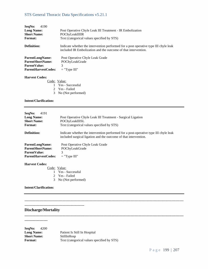
STS General Thoracic Data Specifications v5.21.1
P a g e 199 | 207
SeqNo: 4190 Long Name: Post Operative Chyle Leak III Treatment - IR Embolization Short Name: POChyLeakIIIIR Format: Text (categorical values specified by STS) Definition: Indicate whether the intervention performed for a post operative type III chyle leak included IR Embolization and the outcome of that intervention. ParentLongName: Post Operative Chyle Leak Grade ParentShortName: POChyLeakGrade ParentValue: 3 ParentHarvestCodes: = "Type III" Harvest Codes: Code: Value: 1 Yes - Successful 2 Yes - Failed 3 No (Not performed) Intent/Clarification: SeqNo: 4191 Long Name: Post Operative Chyle Leak III Treatment - Surgical Ligation Short Name: POChyLeakIIISL Format: Text (categorical values specified by STS) Definition: Indicate whether the intervention performed for a post-operative type III chyle leak included surgical ligation and the outcome of that intervention. ParentLongName: Post Operative Chyle Leak Grade ParentShortName: POChyLeakGrade ParentValue: 3 ParentHarvestCodes: = "Type III" Harvest Codes: Code: Value: 1 Yes - Successful 2 Yes - Failed 3 No (Not performed) Intent/Clarification: ----------------------------------------------------------------------------------------------------------------------------------------------------------------- Discharge/Mortality -------------------------------------------------------------------------------------------------------------------------------------- SeqNo: 4200 Long Name: Patient Is Still In Hospital Short Name: StillInHosp Format: Text (categorical values specified by STS)

STS General Thoracic Data Specifications v5.21.1
P a g e 200 | 207
Definition: Indicate if, at the time of data submission, the patient remains an inpatient in the hospital. Harvest Codes: Code: Value: 1 Yes 2 No Intent/Clarification: Indicate if the patient remains in the acute care setting following the index operation. This field is the parent to the discharge/mortality section. Answering ‘yes’ will not allow you to answer the discharge/mortality questions and will remove the case from analysis. Starting with version 5.21 - Patients discharge to hospice are considered an operative mortality, regardless of date of death. For patients discharged to hospice, code still in hospital (StillInHosp – seq 4200) = ‘yes’ until the patient expires. Then code MtDCStat (seq 4200) = Discharged to Hospice AND MortDate (seq 4300) with date of death. SeqNo: 4210 Long Name: Hospital Discharge Date Short Name: DischDt Format: Date in mm/dd/yyyy format Definition: Indicate the date the patient was discharged from the hospital (acute care). If the patient expired in the hospital; the discharge date is the date of death. ParentLongName: Patient Is Still In Hospital ParentShortName: StillInHosp ParentValue: 2 ParentHarvestCodes: = "No" Intent/Clarification: Indicate the patient was discharged from the acute care setting. If the patient was transferred to another acute care setting following the index operation, then this will be the date of discharge from the transfer hospital. It is expected and required that the patients are followed during their entire acute care stay. This field is required for record inclusion. If missing data, the entire record will be excluded from the analysis. SeqNo: 4220 Long Name: Hospital Discharge Status Short Name: MtDCStat Format: Text (categorical values specified by STS) Definition: Indicate whether the patient was alive or dead at discharge from the hospitalization in which the primary surgery procedure occurred. ParentLongName: Patient Is Still In Hospital ParentShortName: StillInHosp ParentValue: 2 ParentHarvestCodes: = "No" Harvest Codes: Code: Value: 3 Discharged Alive, last known status Alive (other than hospice)

STS General Thoracic Data Specifications v5.21.1
P a g e 201 | 207
4 Discharged Alive, died after discharge 5 Discharged to Hospice 6 Died in Hospital Intent/Clarification: Indicate the patient status at the time of discharge from the acute care setting. If the patient was transferred to another acute care setting following the index operation, then this will be the date of discharge from the transfer hospital. It is expected and required that the patients are followed during their entire acute care stay. Starting with version 5.21 - Patients discharge to hospice are considered an operative mortality, regardless of date of death. For patients discharged to hospice, code still in hospital (StillInHosp – seq 4200) = ‘yes’ until the patient expires. Then code MtDCStat (seq 4200) = Discharged to Hospice AND MortDate (seq 4300) with date of death. This field is required for record inclusion. If missing data, the entire record will be excluded from the analysis. SeqNo: 4230 Long Name: Discharge Location Short Name: DisLoctn Format: Text (categorical values specified by STS) Definition: Indicate the location to where the patient was discharged. ParentLongName: Hospital Discharge Status ParentShortName: MtDCStat ParentValue: 3|4 ParentHarvestCodes: = "Discharged Alive, last known status Alive (other than hospice)" or "Discharged Alive, died after discharge" Harvest Codes: Code: Value: 1 Home 2 Extended Care/Transitional Care Unit/Rehab 3 Other Hospital 4 Nursing Home 777 Other Intent/Clarification: Skilled Nursing Facilities (SNF) are nursing facilities with staff and equipment to give skilled nursing care and, in most cases, skilled rehabilitative care services. You will need to investigate the intent of discharge. If the intent of discharge is for a patient from the hospital to go to a SNF for rehabilitation and to eventually discharge home, then Code as Extended Care/Transitional Care/Rehab. If the intent of discharging to a SNF is for a higher level of care without the intent of discharging home, then chose nursing home. You will often be able to locate the intent within the discharge planning documents by Social Service, Case Management, and/or Physical Therapy. Home – Includes assisted living or temporarily, at the home of a relative. Includes return to jail for incarcerated individuals. Extended Care/Transitional Care Unit /Rehab – Includes short-term inpatient and outpatient rehab facilities and long-term acute care hospital and Acute Care Rehabilitation facilities (LTACH, LTAC, LTCH). Long-term acute care hospital is not part of the acute care stay. May include Skilled Nursing Facilities (SNF). Skilled Nursing Facilities (SNF) are nursing facilities with staff and equipment to give skilled nursing care and, in most cases, skilled rehabilitative care services. You will need to investigate the intent of discharge. If the intent of discharge is

STS General Thoracic Data Specifications v5.21.1
P a g e 202 | 207
for a patient from the hospital to go to a SNF for rehabilitation and to eventually discharge home, then Code as Extended Care/Transitional Care/Rehab. • Nursing Home - Nursing Home is a residence where skilled care or acute care is not required. May include SNF if the intent of discharging to a SNF is for a higher level of care without the intent of discharging home, then chose nursing home. • Left AMA – Left Against Medical Advice • Other – Includes prison, jail, homeless shelter, drug rehab facilities Other hospital was inadvertently left out as an option in v5.21.1, please disregard this as you should code ‘still in hospital’ for patients who are transferred to another acute care facility. This field is not available for patients discharged to hospice. SeqNo: 4235 Long Name: Discharged With Chest Tube Short Name: CTubeDis Format: Text (categorical values specified by STS) Definition: Indicate whether the patient was discharged with a chest tube for persistent air leak or to drain a postoperative effusion. ParentLongName: Hospital Discharge Status ParentShortName: MtDCStat ParentValue: 3|4 ParentHarvestCodes: = "Discharged Alive, last known status Alive (other than hospice)" or "Discharged Alive, died after discharge" Harvest Codes: Code: Value: 1 Yes 2 No Intent/Clarification: Capture this for all patients discharged after any procedure with any type of chest tube for a persistent air leak or postoperative effusion. This field is coded only based on the index admission/discharge. This field is not available for patients discharged to hospice. SeqNo: 4240 Long Name: Discharged with home O2 (new; not using O2 pre-op) Short Name: DischHomeO2 Format: Text (categorical values specified by STS) Definition: Indicate if the patient was discharged home with an order to use oxygen at home. If the Patient used oxygen at home prior to surgery check "no" to this field. ParentLongName: Hospital Discharge Status ParentShortName: MtDCStat ParentValue: 3|4 ParentHarvestCodes: = "Discharged Alive, last known status Alive (other than hospice)" or "Discharged Alive, died after discharge" Harvest Codes: Code: Value:

STS General Thoracic Data Specifications v5.21.1
P a g e 203 | 207
1 Yes 2 No Intent/Clarification: This field is coded only based on the index admission/discharge. This field is not available for patients discharged to hospice. SeqNo: 4260 Long Name: On Oxygen at 30 Days PostOp Short Name: OnOxygen30DayPOp Format: Text (categorical values specified by STS) Definition: Indicate if the patient is using home oxygen at 30 days post operatively. ParentLongName: Discharged with home O2 (new; not using O2 pre-op) ParentShortName: DischHomeO2 ParentValue: 1 ParentHarvestCodes: = "Yes" Harvest Codes: Code: Value: 1 Yes 2 No 4 Unknown 3 Patient died within 30 days postop Intent/Clarification: This field is not available for patients discharged to hospice. SeqNo: 4270 Long Name: Readmission within 30 days of Discharge Short Name: Readm30Dis Format: Text (categorical values specified by STS) Definition: Indicate whether patient was readmitted to any hospital within 30 days of discharge. ParentLongName: Hospital Discharge Status ParentShortName: MtDCStat ParentValue: 3|4 ParentHarvestCodes: = "Discharged Alive, last known status Alive (other than hospice)" or "Discharged Alive, died after discharge" Harvest Codes: Code: Value: 1 Yes 2 No 3 Unknown Intent/Clarification: This field is not available for patients discharged to hospice.

STS General Thoracic Data Specifications v5.21.1
P a g e 204 | 207
Aug 2021: Readmission applies to IP readmissions only. If a patient returns to the hospital and is in OP/OBS status for their entire stay, please code ‘no’ to 4270. SeqNo: 4290 Long Name: Substance Use Screening and Counseling Short Name: DCSubUseScre Format: Text (categorical values specified by STS) Definition: Indicate if substance use screening and appropriate counseling was performed. This is NQF measure 2597. Patients require screening on tobacco use, alcohol use, and illicit/non-prescription drug use. Patient must be screened for all three. If the patient screens positive for any listed substance use, appropriate counseling is required to choose yes for this field. ParentLongName: Hospital Discharge Status ParentShortName: MtDCStat ParentValue: 3|4 ParentHarvestCodes: = "Discharged Alive, last known status Alive (other than hospice)" or "Discharged Alive, died after discharge" Harvest Codes: Code: Value: 1 Yes 2 No 3 Not Applicable Intent/Clarification: This is NQF measure 2597. Indicate patients who were screened by a healthcare provider, nurse, or an allied healthcare provider at least once within the last 24 months for tobacco use, unhealthy alcohol use, non-medical prescription drug use, and illicit drug use AND who received an intervention for all positive screening results. Time frame 24 months prior to episode of care until discharge of episode of care. Patient must be screened for all three. If the patient screens positive for any listed substance use, appropriate counseling is required to code YES for this field. Tobacco use component: Patients who are screened for tobacco use and who received tobacco cessation intervention if identified as a tobacco user. Counseling should be provided to users of Cigarettes, Pipe, Cigars, Smokeless Cans, Other tobacco products (e‐cigs, orbs, strips, sticks, hookah, etc.) Unhealthy alcohol use component: Patients who were screened for unhealthy alcohol use (as determined by each facility) using a systematic screening method (as determined by each facility) and who received brief counseling if identified as an unhealthy alcohol user. Drug use component (nonmedical prescription drug use and illicit drug use): Patients who were screened for nonmedical prescription drug use and illicit drug use using a systematic screening method(as determined by each facility) and who received brief counseling if identified as a nonmedical prescription drug user or illicit drug user. If the patient is medically, mentally, or emotionally inappropriate for a referral, select “Not Applicable”. https://aspe.hhs.gov/report/review-medication-assisted-treatment-guidelines-andmeasures-opioid-and-alcohol-use/appendix-d-substance-use-medication-assistedtreatment-and-other-related-measures

STS General Thoracic Data Specifications v5.21.1
P a g e 205 | 207
Sept 2021: Per the CMS Measure - screening and counseling is specific to hospitals and the STS cannot determine your site process for method of screening or counseling. If you have documentation of screening and counseling that meets your hospital standard, then you have met the STS standard. SeqNo: 4300 Long Name: Mortality Date Short Name: MortDate Format: Date in mm/dd/yyyy format Definition: Indicate the patient's date of death (even if after discharge). ParentLongName: Hospital Discharge Status ParentShortName: MtDCStat ParentValue: 4|5 ParentHarvestCodes: = "Discharged Alive, died after discharge" or "Discharged to Hospice" Intent/Clarification: SeqNo: 4310 Long Name: Status 30 Days After Surgery Short Name: Mt30Stat Format: Text (categorical values specified by STS) Definition: Indicate whether the patient was alive or dead at 30 days post surgery (whether in the hospital or not). Harvest Codes: Code: Value: 1 Alive 2 Dead 3 Unknown Intent/Clarification: This field is required for record inclusion. If missing data, the entire record will be excluded from the analysis. SeqNo: 4580 Long Name: Temporary Yes/No Field #1 Short Name: TempYN1 Format: Text (categorical values specified by STS) Definition: This is a temporary field that should not be used for data collection until expressly instructed to by the STS. Harvest Codes: Code: Value: 1 Yes 2 No Intent/Clarification: Use only as directed by STS, do not add custom field here.

STS General Thoracic Data Specifications v5.21.1
P a g e 206 | 207
SeqNo: 4590 Long Name: Temporary Yes/No Field #2 Short Name: TempYN2 Format: Text (categorical values specified by STS) Definition: This is a temporary field that should not be used for data collection until expressly instructed to by the STS. Harvest Codes: Code: Value: 1 Yes 2 No Intent/Clarification: Use only as directed by STS, do not add custom field here. SeqNo: 4600 Long Name: Temporary Date Field Short Name: TempDt Format: Date in mm/dd/yyyy format Definition: This is a temporary field that should not be used for data collection until expressly instructed to by the STS. Intent/Clarification: To further understand the impact of Covid-19 on surgical patients, STS will begin collecting the date of positive PCR testing for Covid-19 patients with surgery dates starting May 1, 2020. If there is more than one positive test date, collect the date that is closest to the OR date. Positive antibody testing is not captured in this field. Sites have the option to retroactively collect this field back to January 1 if they choose to do so. To achieve this, the temporary field (TempDt) will be utilized for patients who have a confirmed Covid-19 diagnosis through PCR testing. Only code the date of a positive PCR test. Use only as directed by STS, do not add custom field here. SeqNo: 4610 Long Name: Temporary Coded Field Short Name: TempCode Format: Text (categorical values specified by STS) Definition: This is a temporary field that should not be used for data collection until expressly instructed to by the STS. Harvest Codes: Code: Value: 1 1 2 2 3 3 4 4 5 5 6 6 7 7 8 8 9 9 10 10 11 11

STS General Thoracic Data Specifications v5.21.1
P a g e 207 | 207
12 12 13 13 14 14 15 15 16 16 17 17 18 18 19 19 20 20 Intent/Clarification: This field will be used to collect data on Covid-19. Did the patient have a laboratory confirmed diagnosis of Covid-19? • No (Harvest code 10)
– the patient was not tested, or the patient was tested, and the test was negative • Yes, prior to hospitalization for this surgery (Harvest Code 11) • Yes, in hospital prior to surgery (Harvest Code 12) • Yes, in hospital after surgery (Harvest Code 13) • Yes, after discharge within 30 days of surgery (Harvest Code 14) There are many tests for different types of coronaviruses. The STS is only collecting data on the one that causes COVID 19 which is SARS-CoV-2. There is no preoperative timeframe in place for coding COVID-19 + patients. Any preoperative positive PCR patients is to be captured. Use only as directed by STS, do not add custom field here. Sept 2021: In the event a patient was COVID tested and was positive and later tested again and found to be COVID negative, please choose an appropriate ‘yes’ code for this sequence. Do not code ‘no’ even if the negative test was closer to the operative date. SeqNo: 4620 Long Name: Temporary Text Field Short Name: TempText Format: Text Definition: This is a temporary field that should not be used for data collection until expressly instructed to by the STS. Intent/Clarification: Use only as directed by STS, do not add custom field here.


![S4 - Measuring Variables & Data Collection [Dr a Bermudez]](https://img.pdfslide.net/doc/110x75/55cf8c575503462b138b9950/s4-measuring-variables-data-collection-dr-a-bermudez.jpg)