Embed Size (px)
Citation preview
Defensins and the dynamic genome: What we canlearn from structural variation at humanchromosome band 8p23.1Edward J. Hollox,1,4 John C.K. Barber,2 Anthony J. Brookes,1 and John A.L. Armour3
1Department of Genetics, University of Leicester, Leicester LE1 7RH, United Kingdom; 2Wessex Regional Genetics Laboratory,National Genetics Reference Laboratory (Wessex) and Human Genetics Division, School of Medicine, University of Southampton,Southampton SO17 1BJ, United Kingdom; 3Institute of Genetics, University of Nottingham, Nottingham NG7 2RD,United Kingdom
Over the past four years, genome-wide studies have uncovered numerous examples of structural variation in thehuman genome. This includes structural variation that changes copy number, such as deletion and duplication, andstructural variation that does not change copy number, such as orientation and positional polymorphism. Oneregion that contains all these types of variation spans the chromosome band 8p23.1. This region has been studied insome depth, and the focus of this review is to examine our current understanding of the variation of this region. Wealso consider whether this region is a good model for other structurally variable regions in the genome and what theimplications of this variation are for clinical studies. Finally, we discuss the bioinformatics challenges raised, discussthe evolution of the region, and suggest some future priorities for structural variation research.
Structural variation and its importance
Polymorphic variation in genome structure has been appreciatedfor many years. Structural variation was apparent in early studiesof human DNA polymorphisms, because of the ability to analyzeduplications and deletions using Southern blots, and the pheno-typic variation caused by copy number variation in genes of, forexample, the Rh locus, the opsin locus, and the alpha-globinlocus (Baine et al. 1976; Nathans et al. 1986; Colin et al. 1991;Wolf et al. 1999).
More recent studies have revealed the human genome to beconsiderably more variable than a single nucleotide polymor-phism (SNP)-focused eye would have us believe. Initial studiesfound parts of the genome over- or under-represented comparedto a reference genome and therefore variable in copy number,but the regions found in each study had minimal overlap witheach other, suggesting that each study had sampled only a smallproportion of the number of regions that are copy number vari-able (CNV) (Iafrate et al. 2004; Sebat et al. 2004; Sharp et al.2005). A comprehensive study on 270 individuals used for theHapMap project confirmed this, and ∼12% of the genomeshowed an increase or decrease in DNA dosage in at least one ofthese individuals (Redon et al. 2006).
While array-CGH (comparative genomic hybridization) hasdriven a boom in copy number variation discovery, the charac-terization of balanced structural variation, in which there is nonet gain or loss of sequence between individuals, has been lim-ited by current technology (Baptista et al. 2008). Inversion poly-morphisms have been discovered mostly from comparative se-quence analysis of humans and chimpanzees (Feuk et al. 2005)and from paired-end mapping, where orientations of sequencesfrom either end of a piece of DNA are inverted with respect to the
reference sequence (Tuzun et al. 2005; Korbel et al. 2007). Cur-rent inversion genotyping methods rely on identification of thebreakpoint and the assumption that all inverted alleles share thesame breakpoint. This will be true only when the inversion eventhas happened just once, and in these cases, the inversion can alsobe genotyped by typing surrogate SNPs within the inversion andin complete association with it (Stefansson et al. 2005). Inver-sions with breakpoints within large inverted repeats can be geno-typed using single-molecule haplotyping (Turner et al. 2006), butin complex repeat regions with many kilobases of inverted re-peats or with multiple inversion breakpoints, even this method isfallible. This is because the location of the inverted repeat medi-ating the inversion must be known. At present, molecular cyto-genetics and pulsed-field gel electrophoresis remain the onlymethods to genotype inversions with complex breakpoints ormultiple origins. Polymorphic translocations, in which a se-quence is polymorphically present at a different position, eitherwithin the chromosome or on a different chromosome, are com-mon in subtelomeric regions, but, again, cytogenetics remainsthe most reliable way to type them (Wong et al. 1990; Martin-Gallardo et al. 1995; Linardopoulou et al. 2005). In short, currenttechnologies are not sufficient for the analysis of medium- andlarge-scale structural variation in all its forms. Furthermore,small-scale structural variation is not yet fully characterized andrequires more method development.
Such extensive variation in genomic structure suggests thatat least some will have consequences on phenotype and diseasesusceptibility. These could be mediated in many ways, includingdirect DNA dosage or the unmasking of deleterious recessive mu-tations. Recent studies have implicated copy number variation asaffecting gene expression, protein expression, and phenotypicvariation in both genome-wide and locus-specific studies (Jo-hansson et al. 1993; Stranger et al. 2007). For example, increasedcopy number of the salivary amylase gene cluster increases ex-pression of the protein and ability to digest starch (Perry et al.2007). Similarly, increased copy number of the beta-defensin re-
4Corresponding author.E-mail [email protected]; fax 44-116-252-3378.Article is online at http://www.genome.org/cgi/doi/10.1101/gr.080945.108.
Review
1686 Genome Researchwww.genome.org
18:1686–1697 ©2008 by Cold Spring Harbor Laboratory Press; ISSN 1088-9051/08; www.genome.org
Cold Spring Harbor Laboratory Press on November 27, 2015 - Published by genome.cshlp.orgDownloaded from

gion increases risk of the inflammatory skin disease psoriasis(Hollox et al. 2008). In both cases, a direct gene dosage effect islikely to link diplotype to phenotype. Structural variation canalso act as a substrate for larger genomic rearrangements withprofound clinical consequences (Giglio et al. 2001, 2002; Linar-dopoulou et al. 2005).
The application of array CGH to clinical patients is alreadyrevealing a rising number of CNVs, many of which are initiallynovel (Menten et al. 2006). Detailed clinical analysis of the phe-notypes of such rare cases can give important insights into genefunction, and there are ongoing efforts to relate specific chromo-some gains and losses to dosage imbalance of individual genes.These are being facilitated by international databases such asDECIPHER (http://www.sanger.ac.uk/PostGenomics/decipher/)and ECARUCA (http://www.ecaruca.net).
There have been several excellent reviews recently that dis-cuss the current genome-wide CNV studies and make generalinferences from the pattern and nature of the CNVs found (Free-man et al. 2006; Beckmann et al. 2007; Cooper et al. 2007). Inthis review, we discuss the structural variation at the humanbeta-defensin region, what we know about it, and argue that thiscan inform studies on other structurally variable regions of hu-man and other mammalian genomes.
Defensins—Renaissance molecules
Defensins are short cationic peptides that are expressed in epi-thelia and leukocytes and have an important role in the innateimmune system. They are divided into three types: alpha, beta,and theta, depending on the pattern of disulfide-bridge forma-tion between the six conserved cysteine residues that define de-fensins (Ganz 2003). They were initially characterized as effectiveantimicrobial peptides, killing both Gram-positive and Gram-negative bacteria and fungi (Klotman and Chang 2006; Pazgier etal. 2006), and are thought to kill microbes by depolarizing andpermeabilizing the cell membrane (Lehrer et al. 1989; Kagan etal. 1990; Hill et al. 1991). Later studies showed that they haveextensive cell-signaling activity, recruiting immature dendriticcells to the site of infection, which have been mirrored by otherstudies suggesting that chemokines, such as CCL20, also havepotent antimicrobial activity (Yang et al. 1999, 2003; Biragyn etal. 2002). It has been suggested that these chemokines and de-fensins should be regarded as one functional group, termed“alarmins” (Oppenheim and Yang 2005). Alpha, beta, and thetadefensins have also been shown to have potent antiviral activity,in particular against HIV-1 (Cole et al. 2002; Mackewicz et al.2003; Quinones-Mateu et al. 2003; Chang and Klotman 2004).One beta-defensin signals through the melanocortin receptor indogs and humans and controls coat color in dogs (Candille et al.2007). Others have a function in reproduction; for example,DEFB126 coats the glycocalyx of sperm and is involved in attach-ing sperm to the oviduct epithelia (Yudin et al. 2005; Tollner etal. 2008). Beta-defensins have also evolved to become venom inthe platypus (Whittington et al. 2008). It is likely that differentdefensins are much more than antimicrobial, and further func-tional work will uncover different roles for what is increasinglyregarded as a multifunctional gene family.
Without analysis of synteny relationships, true orthologs ofdefensins can be difficult to identify because of the amino aciddifferences between defensins of different species. It is thereforedifficult to determine whether “defensins” in insects and plantsare true orthologs of vertebrate defensins. Nevertheless, analysis
of the defensin family within mammals reveals a considerableamount about the evolution of these genes. Rapid duplicationand divergence is a recurring theme, as would be expected in agene family central to host–pathogen interaction (Hughes 1999;Lynn et al. 2004). There is evidence of positive selection in mam-malian alpha-defensins and in rodent and primate beta-defensins(Morrison et al. 2003; Semple et al. 2005; Hollox and Armour2008). Moreover, in rodents, there has been duplication andpseudogenization of the beta-defensin family, generating a dis-tinct beta-defensin repertoire in the rat and mouse (Morrison etal. 2003).
The story is more complex than this may suggest. Despiteevidence for duplication and pseudogenization in the rodents,evidence from primates and other mammalian groups suggestsconservation of synteny (Patil et al. 2005). There has been posi-tive selection of the amino acid sequence of some, but not all,beta-defensins in primates: the genes involved in the CNV regionseem not to have changed much in the primate lineage (Holloxand Armour 2008). These genes are CNV in macaques as well ashumans, suggesting persistence of copy number variation formore than 35 million years (Myr) (Lee et al. 2008). It is possiblethat this variation has prevented gene sequence diversificationby facilitating gene conversion events between paralogs, al-though further investigation into the rates of gene conversionand selection pressure on these paralogs is required.
Defensin variation at 8p23.1
The alpha-defensin DEFA1A3 and the beta-defensins DEFB4,DEFB103, DEFB104, DEFB105, DEFB106, and DEFB107 map to8p23.1 and vary in copy number independently: the alpha-defensin as a 19-kb tandemly repeated unit and the beta-defensins on a copy number variable unit that is at least 250 kbin size, but the exact size and breakpoints of the copy numbervariable region are not known (Fig. 1). This latter repeat unit alsocontains SPAG11, an alternatively spliced gene formed by a head-to-head fusion of two beta-defensins, which codes for a proteinthat shows potent antimicrobial activity and is present on sper-matozoa, perhaps providing antimicrobial protection for its car-rier (Yenugu et al. 2003; Zanich et al. 2003). The other alpha-defensins—DEFA4, DEFA5, and DEFA6—and the beta-defensinDEFB1 do not show CNV (Hollox et al. 2003; Aldred et al. 2005;Linzmeier and Ganz 2005).
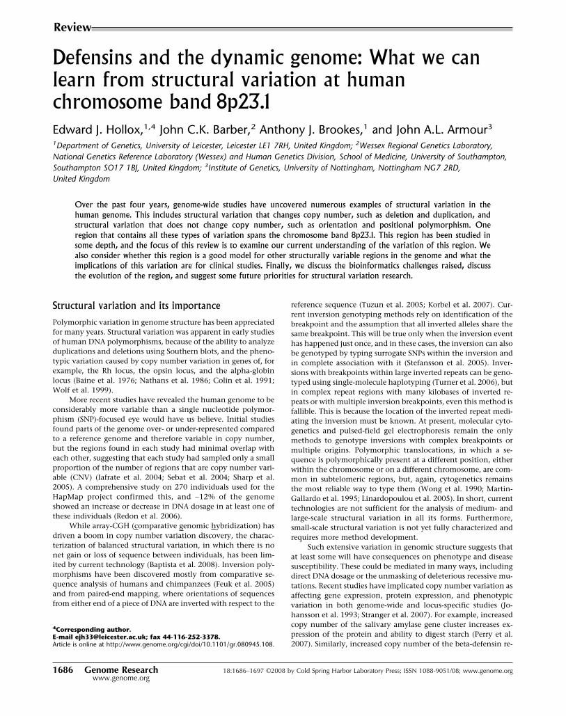
CNV of DEFA1 had been suspected from somatic cell hybridmapping of chromosome 8 (Mars et al. 1995). We, and others,showed that the DEFA1 and DEFA3 genes were variants of thesame gene that differed by one nucleotide (Aldred et al. 2005;Linzmeier and Ganz 2005). The renamed DEFA1A3 gene is en-coded on a tandem repeat that has a diploid copy number be-tween four and 11. The nucleotide change leading to DEFA3 ishuman-specific and is most frequently at the most proximal po-sition in the tandem array. This suggests a relatively recent originof this variant, although in 50% of arrays, the DEFA3 variant isnot at the proximal position but elsewhere in the array, suggest-ing a recent history of non-allelic recombination (NAHR) or geneconversion within this array (Fig. 2) (Aldred et al. 2005). In nativeUK individuals, DEFA3 variant is absent in 10% of the popula-tion, and in sub-Saharan Africans (Yoruba), it is absent in 37% ofthe population. DEFA3 absence is associated with one SNP hap-lotype in Europeans but shows no association with any SNP hap-lotype in other populations (Ballana et al. 2007). The theta-defensin DEFT1 is encoded on the same 19-kb repeat as
Defensins and the dynamic genome
Genome Research 1687www.genome.org
Cold Spring Harbor Laboratory Press on November 27, 2015 - Published by genome.cshlp.orgDownloaded from
DEFA1A3. In humans, gorillas, and chimpanzees, this is an inac-tive pseudogene, but is active in other primate lineages tested(Tang et al. 1999; Nguyen et al. 2003). DEFA1 is present in mul-tiple copies in other apes, making it likely that this has beenvariable in copy number since before the divergence of gibbonsand great apes around 25 million years ago (Mya) (Aldred et al.2005).
The beta-defensin genes involved in CNV are on a largegenomic repeat unit within a “sea” of more complex CNV in-volving retroviral elements and olfactory repeat (OR) regions,
collectively known as “REPD” (for repeat distal) (Giglio et al.2001). Another smaller OR region called “REPP” (repeat proxi-mal) is 5 Mb proximal on 8p23.1 and shares a high level ofidentity with REPD (Fig. 1; Sugawara et al. 2003). The total dip-loid copy number of the beta-defensin region ranges from one to12, and commonly between two and seven (Hollox et al. 2003).We have observed only one individual with a copy number ofone in more than 1500 individuals with DNA from blood, sug-gesting that the null allele exists but is very rare (allele fre-quency ∼ 0.2%). Given the functional importance of these genes,
Figure 1. Genome assembly of the 8p23.1 region Highlighted is the REPD region containing one of two beta-defensin repeat units assembled,separated by a gap. The repeat classes II–V are from Taudien et al. (2004). Sites for assays for beta-defensin copy number are also shown, including PRT,MAPH, MLPA, and REDVR (Hollox et al. 2008). EPEV-1–EPEV-3 refer to simple tandem repeats used for copy number and segregation analysis inpedigrees. The segmental duplication track is based on the data from Bailey et al. (2001); (light to dark gray) 90%–98% similarity with its duplicate;(yellow) 98%–99% similarity; (orange) >99% similarity. Based on UCSC Genome Browser (http://genome.ucsc.edu) build hg18.
Hollox et al.
1688 Genome Researchwww.genome.org
Cold Spring Harbor Laboratory Press on November 27, 2015 - Published by genome.cshlp.orgDownloaded from

the null homozygote may be strongly deleterious, and null allelesgenerated by recurrent mutation rapidly removed from the popu-lation by purifying selection. At the other end of the scale, exactdefinition of integer copy number becomes very difficult. Largediploid copy numbers are, at least in part, due to recent expan-sion of the beta-defensin copy number in independent lineages,and such expansions can be seen directly under G-band chromo-some staining as a euchromatic variant (Hollox et al. 2003; Bar-ber et al. 2005).
The repeat structure has been studied in detail, and fourclasses of repeat region have been identified (Classes II–V) (Fig. 1;Taudien et al. 2004). The main beta-defensin region appears tocorrespond to Class V, with more complex repeat-rich regionsflanking it (Fig. 1). At the ends of the beta-defensin genomicrepeat unit—perhaps at the shore of the sea of complex olfactoryreceptor repeats—are FAM90A gene clusters that are on Class IIIrepeats. FAM90A genes are on several other chromosomes andare expressed in several different tissues, although their functionis not known (Bosch et al. 2007).
Although the >250-kb repeat unit is too large to be capturedby current paired-end mapping approaches, such methods canreveal information on the flanking regions surrounding the re-peat unit (Korbel et al. 2007; Kidd et al. 2008). The fosmid paired-end mapping approach has the particular advantage that thestructural variant is cloned as a fosmid and can be sequenceddirectly, providing sequence-level resolution (Iafrate et al. 2004).The disadvantage is that it is a time-consuming and expensiveapproach, with only a few genomes analyzed so far. Data pre-sented so far in the Structural Variation Database (http://humanparalogy.gs.washington.edu/structuralvariation/) on four
genomes is consistent with the whole beta-defensin repeat unitvarying as a block with no internal small duplications or dele-tions. However, variation exists where the repeat unit meets theflanking sea of more complex repeats and sequencing of theseparticular fosmids may reveal, for the first time, some sequence-level information about CNV of the beta-defensin region. Hope-fully, paired-end approaches combined with novel sequencingtechnologies may shed light on the small-scale variation at thislocus, although novel approaches are needed to define the large-scale variation at a sequence-level resolution.
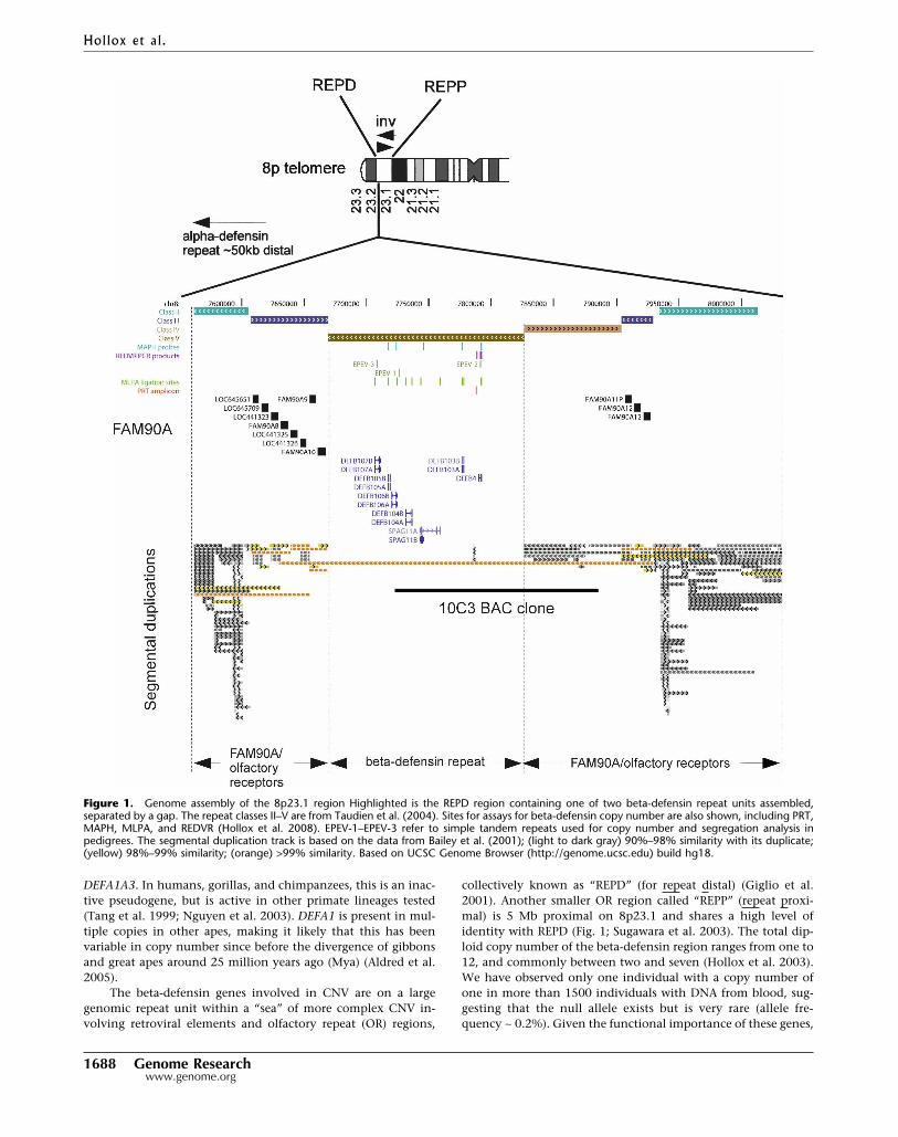
Sequence-based approaches and paired-end mapping can-not easily capture the large inversion between REPD and REPP.However, physical methods such as fluorescent in situ hybridiza-tion (FISH) and genetic analysis can type individuals for thisinversion polymorphism. The region between REPP and REPDhas been shown to be polymorphically inverted by FISH analysiswith a frequency of 25% in Europeans (Giglio et al. 2001). Thishas been confirmed genetically and has since been used to con-firm orientation of the region in certain CEPH individuals: re-combination between inversion homozygotes results in an ap-parent triple recombinant when the genetic markers are arrangedusing a map based on a standard (non-inverted) assembly (Fig. 3)(Broman et al. 2003). As expected, the inversion breakpoints mapto REPP and REPD, and analysis of informative markers withinthese repetitive regions may resolve the inversion breakpointswith greater accuracy. SNPs and other polymorphisms carried onan inverted region are expected to be isolated from recombina-tion with non-inverted regions and to form a non-recombining
Figure 2. Evolution of the DEFA1A3 CNV region. Following an initial denovo duplication at least 25 Mya, recurrent ectopic recombination gen-erates long arrays of DEFA1 repeats. A point mutation in humans gener-ates the DEFA3 gene, which is shuffled through the array by continuingectopic recombination.
Figure 3. Apparent triple recombinants reflect inverted marker orien-tation. (A,B) A single recombination within an inversion between twoinverted chromosomes is revealed as an apparent triple recombinant ifthe markers are assumed to be in the non-inverted orientation (Bromanet al. 2003). (C) Analysis of the apparent triple recombinants can localizethe site of recombination and the two breakpoints of the inversion.
Defensins and the dynamic genome
Genome Research 1689www.genome.org
Cold Spring Harbor Laboratory Press on November 27, 2015 - Published by genome.cshlp.orgDownloaded from
haplotype clade. However, we and others (Broman et al. 2003)have shown that multiple alleles at many loci are found on bothinverted and non-inverted haplotypes, suggesting a recurrent orfrequently reverting process of inversion.
Methods for typing defensin copy number
Accurately typing copy number is both more difficult than anddifferent from SNP genotyping; it should measure a quantitativedifference rather than a qualitative difference. It is clear thatprogress is limited by technology; just as discovery of CNVs waslimited until the development of array-CGH technology, accu-rate CNV typing is limited by the technologies available. Never-theless, progress has been made in both typing precision andaccuracy (Clayton et al. 2005; McCarroll and Altshuler 2007).The accuracy is primarily a consequence of correct normalizationand controls and is aided by the fact that we are confident thatthe underlying biological reality of germline copy number varia-tion at any one sequence position is gain and loss of integernumbers of copies (2, 3, 4, etc., no intermediates), and whereempirical data of sufficient accuracy have been examined in de-tail, this prediction has been borne out for somatic DNA (Armouret al. 2007). This has two important consequences: first, thereshould be adequate availability of well-characterized copy num-ber reference controls to allow for comparison of results acrosslaboratories, and, second, if any CNVs do show frequent somaticvariation, they will require even more effort to type accurately,since the assumption of integer copy number variation may notbe applicable.
Precision is a technical consequence of the method used fortyping and can be regarded as the reproducibility of the result onrepeated testing. Both precision and accuracy reflect on eachother: for example, a highly precise method will produce cluster-ing of results around integer copy number, and therefore cali-brating each copy number to the center of these clusters willovercome any accuracy differences between experiments. De-spite the importance of accuracy and precision in interpretingresults correctly, very few published studies so far provide anyanalysis on the accuracy and precision of their data sets (McCar-roll and Altshuler 2007; McCarroll 2008). Even with a precisemethod, difference in accuracy between cases and controls in anassociation study caused, for example, by inadequate normaliza-tion to known controls can result in a bias that is spuriouslyinterpreted as a significant association. Where data have beenshown, it is clear that, at present, no one method is quite goodenough for large case-control studies and that power comes fromcombining methods and repeat typing (Hollox et al. 2008). Wewill briefly discuss various copy number typing methods thathave been applied to the defensin loci.
Multiplex amplifiable probe hybridization (MAPH)
The first method used to measure beta-defensin copy numberwas multiplex amplifiable probe hybridization (Armour et al.2000; Hollox et al. 2003). We constructed several probes acrossthe beta-defensin region (Fig. 1) and showed that, with few ex-ceptions, copy number reported from each probe was equivalentand therefore that the repeat unit varied as a whole repeat unitwithout substantial heterogeneity in structure (Hollox et al.2005, 2008; Groth et al. 2008). The strength of the method is thatany sequence, except high copy number repeats such as Alus andextremely GC-rich or GC-poor regions, can be used as a probe,
and probes are straightforward to produce in a laboratory. Thedisadvantage is that each hybridization requires 1 µg of DNA,with duplicate hybridizations required for increased accuracy.This cannot be overcome by whole genome-amplification, whichis known to introduce bias in relative copy number (Hosonoet al. 2003). Without this, MAPH can seriously deplete DNAcollections. The technique also requires manipulation of verysmall dry filters, which is time-consuming and can lead to sampleloss.
Multiplex ligation-dependent probe amplification (MLPA)
This method, similar to MAPH, uses hybridization and ligation oftwo half-probes to specifically record the amount of sequence ina sample (Schouten et al. 2002). It is a single-tube assay andrequires less DNA (100–250 ng), although that amount is stillsignificant compared to PCR-based methods. A probe set for thebeta-defensin region is commercially available, with probes forseveral defensin genes across the variable region (Fig. 1). Whenthis method was used on 135 samples, it showed equivalent pre-cision to a single paralog ratio test (see below) and MAPH (Ar-mour et al. 2007).
Quantitative real-time PCR
This method, using fluorescent techniques such as TaqMan al-lowing the real-time measurement of PCR product accumulation,is increasingly popular in measuring copy number variation andhas been applied to the beta-defensin region (Linzmeier andGanz 2005; Chen et al. 2006; Fellermann et al. 2006). Its strengthis that, in theory, it can measure the copy number of any se-quence and requires DNA amounts sufficient for PCR, typically5–10 ng. However, typical studies using this method to type ge-nomic copy number typically do not present analysis of the errorrate or a thorough test of the method by comparison from cor-roborating data from other methods. Quantitative real-time PCRhas found its niche analyzing expression levels of genes, whichoften differs by greater than 10-fold (Bustin 2002). It is likely thatreliably distinguishing four and five copies, for example, basedon numbers inferred from a near-exponential curve, is beyondthe resolution capabilities of this approach.
Paralog ratio test (PRT)
The paralog ratio test (PRT) is essentially a development of com-parative PCR, but where test and reference loci are amplified bythe same primer pair (Deutsch et al. 2004; Armour et al. 2007).This improves reproducibility by making the amplification kinet-ics of test and reference loci very similar. This requires carefuldesign of primers, often using diverged dispersed repeat se-quences so that the primers amplify the sequence on the regionof interest, and one sequence elsewhere on the genome, prefer-ably on another chromosome to minimize potential gene con-version between the test and reference sequences. For example, aPRT assay for the beta-defensin region has been designed to spe-cifically amplify a heat-shock protein pseudogene on the copynumber variable region and on chromosome 5. Figure 1 showsthe location of the PRT assay relative to the genes on the repeatunit.
The sequence requirements for PRT assay developmentmean that it is limited to certain sequences, and an assay cannotnecessarily be designed for a small sequence region, for example,one specific exon. However, the precision and accuracy areequivalent to those of MLPA and MAPH, and it has the advantage
Hollox et al.
1690 Genome Researchwww.genome.org
Cold Spring Harbor Laboratory Press on November 27, 2015 - Published by genome.cshlp.orgDownloaded from

of potential high-throughput analysis and the small DNA re-quirement of PCR-based methods (Armour et al. 2007).
Simple tandem repeat (STR) analysis
Simple tandem repeats within CNV regions are likely to showmultiple alleles, up to a maximum of the number of copies ofCNV. These alleles, like single-copy STRs, can be amplified andresolved by length using electrophoresis. STR analysis is valuablefor tracking individual copies of a repeat through a pedigree be-cause of the high informativeness of these loci: each repeat has ahigh chance of carrying a different length allele (Hollox et al.2003). There is also the possibility of simply counting the allelesas a discrete measure of copy number, and three STRs have beenused to distinguish ambiguous copy number diplotypes (Fig. 1,shown as “EPEV”; Hollox et al. 2005).
Array-CGH
Although primarily intended for discovering CNV rather thantyping it, analysis of the precision of the array-CGH techniquesuggests that it is at least equivalent to other methods. The mainadvantage is its multiplicity, in that a whole-genome bacterialartificial chromosome (BAC) array, for example, analyzes thewhole genome for copy number variation and can, at least for thebeta-defensin locus, genotype it reasonably well (Redon et al.2006; Armour et al. 2007). It has several disadvantages, not leastbeing the cost per test and amount of DNA required. Anotherissue is that while other genotyping methods measure copy num-ber of a short segment, BAC array-CGH measures the copy num-ber of a large segment of DNA (100 kb), and hybridization inten-sities can be influenced by variation in other sequences. For ex-ample, the clone RPCI-11 10C3 overlaps regions that are thoughtto be more highly variable than the beta-defensin “island” (Fig.1), and this may account for differences in accuracy between, forexample, PRT and BAC array-CGH, especially at higher copynumbers (Armour et al. 2007).
Hybridization intensity from SNP genotyping chips
Hybridization chips used for genotyping single nucleotide poly-morphisms by comparative hybridization intensities of genomicDNA to the oligonucleotides representing each allele also giveinformation on copy number from the raw intensity signalstrength of the hybridization. This was the second platform usedby Redon et al. (2006) for their genome-wide analysis of CNV.The principal disadvantage for most chips is that they were de-signed to analyze SNPs not CNVs, and CNV-rich segmental du-plications, including the 8p23.1 region, have been deliberatelyunder-represented in the array. New chips have probes designedto map to known CNV regions, and it will be important to assesshow accurately and precisely they can genotype known multial-lelic CNVs such as the beta-defensin region.
The alpha-defensin copy number polymorphism distal tothe beta-defensin region has been the subject of less intensiveinvestigation, primarily because of the difficulty in accuratelydiplotyping a locus with a copy number between four and 11.The small 19-kb repeat element, and surrounding single copysequence, allows straightforward pulsed-field gel analysis of theregion and accurate genotyping of control samples, but this isnot a practical method for large sample numbers (Aldred et al.2005). Because of the small repeat size, loci suitable for PRT arelimited, and accurate diplotyping of this locus remains an area ofactive research.
Over the next few years, there is likely to be a plethora ofassociation studies using genotyping methods and study designsof varying quality. Statistical methods to deal with CNV in popu-lation- and family-based data sets, especially those that incorpo-rate a measure of the inaccuracy of the CNV genotyping method,will be an essential tool for such studies (Kosta et al. 2007; Ionita-Laza et al. 2008).
Disease studies and clinical relevance of structuralvariation at 8p23.1
Association with common multifactorial diseases
As discussed above, several copy number variable genes havebeen shown to affect susceptibility to disease. In addition, struc-tural variations predispose to large structural rearrangements.Both these mechanisms have been shown to cause disease in the8p23.1 region. The first positive study associated beta-defensincopy number with colonic Crohn’s disease, an inflammatory dis-ease of the bowel (Fellermann et al. 2006). While noting thecaveat that this was an unreplicated study of small sample size,the authors found an association between increased genomiccopy number with increased expression of DEFB4 in the gut andprotection against Crohn’s disease. They also provided a plau-sible mechanistic explanation for their results: low copy numberpredisposed to Crohn’s disease because of a lower anti-infectionbarrier. The next study genotyped beta-defensin copy number fortwo larger cohorts of psoriasis patients and controls from theNetherlands and Germany. Increasing beta-defensin copy num-ber was shown to increase susceptibility to psoriasis in both co-horts. The data showed a linear relationship between copy num-ber and disease relative risk, so that each additional copy in-creased the risk by about 34 percentage points (95% CI 25–43).This represents between a 2.2� and 3.1� difference in risk acrossthe common copy number variation of two to seven copies (Hol-lox et al. 2008).
Association of beta-defensin region copy number withdisease immediately suggests a mechanism for mediating itseffect: an increase in gene product reflected in gene dosage. How-ever, it does not suggest which gene on the repeat region isinvolved—there are at least seven, and a likely candidate mustbe inferred from other work. hBD2 protein, encoded by theDEFB4 gene, was initially discovered in extracts of psoriaticplaques (Harder et al. 1997) and is up-regulated in psoriatic skin,so this is the best candidate, but none of the other genes can beruled out. Functional studies are required to test each candidategene in turn. For DEFB4, there is a correlation with mRNA levelsin lymphoblastoid cell lines and in gut mucosa (Hollox et al.2003; Fellermann et al. 2006). The increase in risk in psoriasisincreases linearly with an increase in dosage, supporting the ideathat the risk factor is gene dosage itself and not some unknownsequence variant within the beta-defensin region (Hollox et al.2008).
The relationship between expression level and genomiccopy number may not always be so straightforward. ForDEFA1A3, expression level is not simply correlated with copynumber, yet the expression ratio of DEFA1:A3 is correlated withthe genomic ratio of DEFA1:A3 (at least in the small sample sizetested). We would predict that this ratio may be a risk factor fordisease rather than total copy number.
Crohn’s disease has not been associated with variation at8p23.1 in whole-genome association studies, and neither
Defensins and the dynamic genome
Genome Research 1691www.genome.org
Cold Spring Harbor Laboratory Press on November 27, 2015 - Published by genome.cshlp.orgDownloaded from
Crohn’s disease nor psoriasis has shown linkage to 8p23.1. Theabsence of an association can be explained by the low associationof multiallelic CNV diplotype with neighboring SNP genotypes(Redon et al. 2006), which means that CNV will not be effectivelyinterrogated by genotyping chips assaying tagSNPs flanking theCNV region. The absence of linkage can, at least in the psoriasisstudy, be attributed to the fact that the effect size is not strongenough to be detected. The common nature of the variation andthe difficulty of interpreting segregation patterns may also cloudany potential linkage to 8p23.1.
The crucial role that defensins play in the innate immuneresponse suggests that alpha- and beta-defensin genomic copynumber variations may be excellent candidate loci for other in-flammatory diseases. There have been several diseases linked to8p23.1, including type II diabetes (Kim et al. 2004; Pezzolesi et al.2004), asthma (Xu et al. 2001a; Dizier et al. 2003), and prostatecancer (Xu et al. 2001b; Wiklund et al. 2003). Given that linkagewill only detect strong effects, the difficulty in interpreting seg-regation patterns and the high frequency of variation, absence oflinkage signal does not preclude the CNV as a candidate locus forother diseases.
Predisposition to large-scale imbalances of 8p
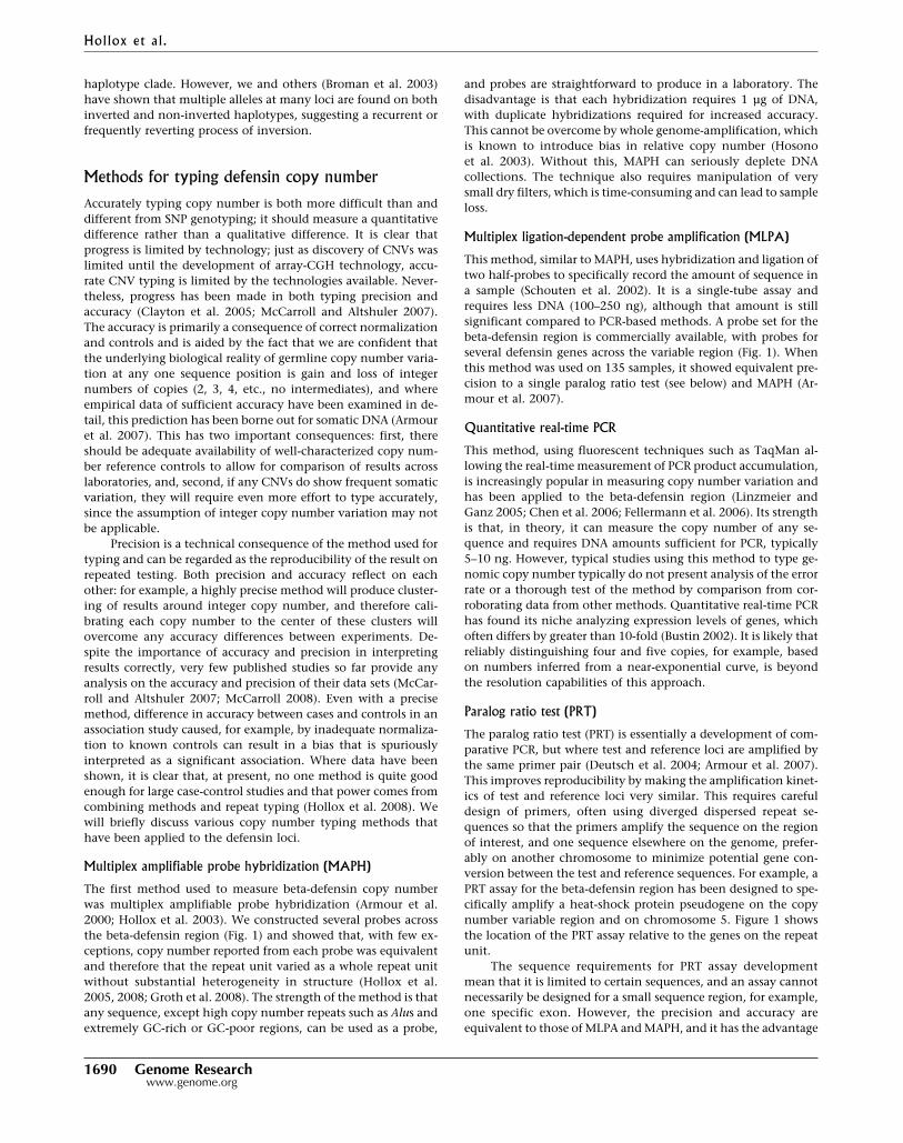
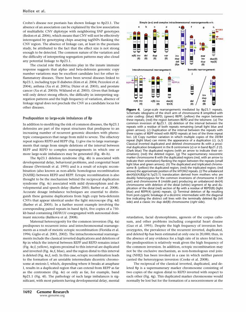
In addition to modifying the risk of common diseases, the 8p23.1defensins are part of the repeat structures that predispose to anincreasing number of recurrent genomic disorders with pheno-typic consequences that range from mild to severe. The olfactoryrepeat regions REPD and REPP (Fig. 1) give rise to large rearrange-ments that range from simple deletions of the interval betweenREPP and REPD to complex rearrangements in which one ormore large-scale imbalances may be generated (Fig. 4b–h).
The 8p23.1 deletion syndrome (Fig. 4b) is associated withdevelopmental delay, behavioral problems, and congenital heartdisease (Devriendt et al. 1995) and is a result of ectopic recom-bination (also known as non-allelic homologous recombination[NAHR]) between REPD and REPP. Ectopic recombination is alsothought to be the mechanism behind the reciprocal duplicationsyndrome (Fig. 4c) associated with mild dysmorphism and de-velopmental and speech delay (Barber 2005; Barber et al. 2008).Accurate dosage imbalance techniques are essential to distin-guish these genuine duplications from high copy beta-defensinCNVs that appear identical under the light microscope (Fig. 4d)(Barber et al. 2005). In a further recent example involving thecorresponding paired repeats in band 4p16, five copies of a 750-kb band containing DEFB131 cosegregated with autosomal dom-inant microtia (Balikova et al. 2008).
Maternal heterozygosity for the common inversion (Fig. 4a)predisposes to recurrent intra- and interchromosomal rearrange-ments as a result of meiotic ectopic recombination (Floridia et al.1996; Giglio et al. 2001, 2002). The intrachromosomal rearrange-ments include the classical inverted duplications and deletions of8p in which the interval between REPP and REPD remains intact(Fig. 4e,f, yellow), regions proximal to this interval are duplicatedand inverted (Fig. 4e,f, blue), and the region distal to this intervalis deleted (Fig. 4e,f, red). In this case, ectopic recombination leadsto the formation of an unstable intermediate dicentric chromo-some at meiosis I, which, depending where it breaks at anaphaseI, results in a duplicated region that can extend from REPP as faras the centromere (Fig. 4e) or only as far, for example, band8p21.1 (Fig. 4f). The pathology of such large imbalances is sig-nificant, with most patients having developmental delay, mental
retardation, facial dysmorphisms, agenesis of the corpus callo-sum, and other problems including congenital heart disease(Guo et al. 1995). Despite the high frequency of inversion het-erozygotes, the prevalence of the recurrent inverted, duplicated,and deleted 8p has been estimated at only one in 20,000; thus, inthe absence of any evidence for a high rate of in utero fetal loss,the predisposition is relatively weak given the high frequency ofthe common inversion. In addition, ectopic recombination maynot be the exclusive mechanism, as non-homologous end join-ing (NHEJ) has been invoked in a case in which neither parentcarried the heterozygous inversion (Cooke et al. 2008).
The reciprocal of the classical inverted, duplicated, and de-leted 8p is a supernumerary marker chromosome consisting oftwo copies of the region distal to REPD inverted with respect toeach other (Fig. 4g). This duplicated marker chromosome wouldnormally be lost but for the formation of a neocentromere at the
Figure 4. Large-scale rearrangements mediated by 8p23.1 repeats.Schematic idiograms of the short arm of chromosome 8 simplified withcolor coding: (blue) REPD; (green) REPP; (yellow) the region betweenthese repeats; (red) the region between REPD and the telomere. (a) Thecommon inversion of 8p23.1. (b) deletion of the interval between therepeats with a residue of both repeats remaining (small light blue andgreen arrows). (c) Duplication of the interval between the repeats withthree copies of REPP mixed with REPD repeats at two of the three repeatloci. (d) Copy number variation in which multiple copies of the DEFB4repeat (light blue) can mimic the appearance of a duplication (c). (e,f)Classical inverted duplicated and deleted chromosome 8s with a proxi-mal duplication breakpoint in the 8 centromere (e) or in band 8p21.2 (f).(Dark blue) The duplicated regions (with an arrow to indicate their ori-entation); (red) the deleted region. (g) The supernumerary neocentricmarker chromosome 8 with the duplicated regions (red, with an arrow toindicate their orientation) flanking the region between the repeats (smalllight blue and green arrows). (h) The duplicated and triplicated chromo-some 8: (yellow) the duplicated region; (red) the triplicated region; (redarrows) the approximate position of the MYOM2 repeats. (i) The unbalancedder(4)(t(4;8)(p16.1p23.1) translocation derived from mothers who aredoubly heterozygous for the common inversions of chromosome 4 (leftpair) and chromosome 8 (middle pair) producing an unbalanced der(4)chromosome with deletion of the distal (white) segment of 4p and du-plication of the distal (red) section of 8p with a residue of REPD(8) (lightblue) and REPP(4) (pink) repeats. (j) Terminal deletion of 8p resultingfrom post-zygotic breakage of a dicentric intermediate with the diagonalline indicating the distinct cell lines with the terminally deleted 8p (leftside) and a classic inv dup del(8) chromosome (right side).
Hollox et al.
1692 Genome Researchwww.genome.org
Cold Spring Harbor Laboratory Press on November 27, 2015 - Published by genome.cshlp.orgDownloaded from
site of the remaining REPP and REPD repeats at its center. Inaddition, REPP and REPD can interact with the more distalmyomesin 2 (MYOM2) repeats in 8p23.3 to produce a chromo-some that is duplicated for the interval between REPP and REPD(Fig. 4h, yellow) and triplicated for the interval between REPDand the MYOM2 repeat in 8p23.3 (Fig. 4h, red arrow) (Giorda etal. 2007).
REPD and REPP also mediate interchromosomal rearrange-ments such as the recurrent de novo unbalanced translocationbetween chromosomes 4 and 8 that is one of the causes of Wolf-Hirschhorn syndrome (Fig. 4i) (Giglio et al. 2002; Maas et al.2007). Remarkably, the parent of origin is consistently maternaland doubly heterozygous for the common 8p23.1 inversion aswell as another common inversion between olfactory repeat re-gions at ∼4 Mb and ∼9 Mb from the telomere of chromosome 4.The breakpoint in all the original cases investigated was at REPDon chromosome 8 and at either REPD or REPP on the short armof chromosome 4 (Giglio et al. 2002).
Somatic rearrangements involving 8p23.1 and cancer
Recently, it has become clearer that the dicentric intermediatethat leads to the classic inverted duplication and deletion of 8p[inv dup del(8)] may persist into the zygote, where subsequentearly post-zygotic mitotic events can lead to mosaicism for theinv dup del(8) and a second cell line with other breakage prod-ucts of the dicentric chromosome (Vermeesch et al. 2003; Pram-paro et al. 2004). These include terminal deletions with break-points from 8p21.1 (Fig. 4j) (Vermeesch et al. 2003) to 8p11.2(Pramparo et al. 2004). Interestingly, the cell lines with deletionsand duplications can complement for each other, resulting in amilder phenotype than might otherwise be expected. It is prob-able that this is a more common mechanism than previouslysuspected and that mosaicism may rescue conceptions thatwould otherwise be lost owing to in utero selection against largeimbalances.
Given the number of constitutional rearrangements medi-ated by REPP and REPD at meiosis, it might be expected that thesame repeats would predispose to somatic recombination events.Until, recently, there was little evidence for this. The REPP-to-REPD interval contains the malignant fibrous histiocytoma am-plified sequence 1 gene (MFHAS1, formerly MASL1 OMIM*605352), but the amplicon in this cancer has been only crudelymapped and may not be related to the 8p23.1 repeats (Sakabe etal. 1999). An 8p23.1-to-8p22 amplicon has also been reported inesophageal cancer, but the minimum size of this amplicon in twogastric tumors was 2.6 Mb between 10.1 and 12.7 Mb from the 8ptelomere (Vauhkonen et al. 2007), and is therefore unlikely tohave a breakpoint in either REPP or REPD. However, the break-points in 10 carcinoma cell lines have recently been mapped indetail, and “tumor break-prone segmental duplications” that cor-respond to REPP and REPD on 8p23.1 have been identified (Da-rai-Ramqvist et al. 2008). These, in turn, coincide with evolution-ary breakpoints and carcinoma-related chromosome rearrange-ment hotspots in the Mitelman Database of ChromosomeAberrations in Cancer (http://cgap.nci.nih.gov/Chromosomes/Mitelman).
Thus, the beta-defensin and olfactory receptor repeats in8p23.1 have established a paradigm in which paired repeats anda common polymorphism predispose to a collection of simpleand complex chromosomal imbalances with significant clinicalconsequences. These repeats also masquerade as cytogenetic du-
plications when amplified, may nucleate neocentromeres, andcould yet play a role in the formation of oncogenic rearrange-ments.
Bioinformatics and structural variation
Bioinformatics of structural variation will allow incorporation ofthat variation into the genomic context, and analysis of thatvariation will allow further hypotheses to be generated andtested. However, the main problem is that the data produced bycurrent laboratory methods are generally too ambiguous and in-complete for use in bioinformatics pipelines. For example, eventhe very latest genome build, which is based on sophisticatedcomputational assembly of quite deep sets of trace file sequenc-ing reads, still contains many gaps and regions of uncertainty,and these correlate significantly with the presence of complexand unstable segmental duplications and CNVs (Redon et al.2006). To improve on this situation and to locate shorter, rarer,and less similar CNVs, we and others are exploring more “tun-able” tools for alignment, assembly, and visualization of primarytrace file data. Including data from other laboratory methods,such as array-CGH and SNP genotyping, can also improve themap, but these methods are far from perfect as they only estimatethe nucleotide extent and copy number range for structurallyvariable regions. As mentioned previously in the context of thebeta-defensin region at 8p23.1 (Taudien et al. 2004), all of thischallenges the concept of a single reference genome assembly,and it almost certainly means that genome browsers will need topresent many alternative genomes as a reference set upon whichall possible structural variations can be displayed. As an example,there are alternative assemblies for the HLA region presented ingenome browsers, and this approach should be extended anddeepened to cover structurally variable regions, and possibly thewhole genome (Traherne et al. 2006).
We are therefore at a stage that is both exciting and frus-trating, in that we know that CNVs, inversions, and the like arevery common and important to genome function, but we areunable to draw a truly high-resolution map of these elements.This forces bioinformatics groups like the Database of GenomicVariants (DGV) (http://projects.tcag.ca/variation/) to make com-promises. For example, the DGV currently annotates each re-ported structural variant over its largest possible genome extent,and then considers all CNVs that seem to overlap as alternativediscoveries of the same structurally variable element (Zhang et al.2006). But, of course, in many cases, these structural variants willbe distinct and non-overlapping, or will overlap but have differ-ent genomic end-points and evolutionary origins.
The above uncertainties raise major problems for computa-tional and analytical handling of structural variation data in dis-ease studies, and this situation is made even worse by additionallimitations of method accuracy and precision (as discussedabove). The accuracy and precision of each method should beestimated, ideally empirically from real data, and such error ratesincorporated into bioinformatics pipelines. At present, the errorrates remain so high that the power of bioinformatics for com-bining and analyzing large amounts of data is diluted by theassociated error with each one of those pieces of data, and infer-ence of anything but large effects is difficult because of experi-mental noise. Given these complications, advanced bioinformat-ics could help solve the problem by enabling better assay design,thereby improving the ability of assays to count CNV copies andto distinguish between copies at distinct locations. But this solu-
Defensins and the dynamic genome
Genome Research 1693www.genome.org
Cold Spring Harbor Laboratory Press on November 27, 2015 - Published by genome.cshlp.orgDownloaded from
tion cannot be relied on until methods can first fully define allthe sequence versions and locations of CNVs, as input data forthe assay design software to operate on. The error rates of currentlaboratory methods are the major limiting factor in the develop-ment of bioinformatics for structural variation.
Evolution of the dynamic defensins: Genomedriven, biology driven, or both?
Highly duplicated regions, or regions with a high frequency ofretrotransposons that share high sequence similarity, provide anideal environment for high rates of ectopic recombination. In-deed, high amounts of structural variation are correlated withduplication-rich regions of the genome, and molecular studieson specific loci provide direct evidence of the importance of ec-topic recombination in generating structural variation (Lam andJeffreys 2006, 2007; Turner et al. 2008). But why does structuralvariation exist? Is it a consequence of genome architecture,which itself is a consequence of other evolutionary processes? Oris there a particular biological reason why certain genes in certainregions are structurally variable? A well-characterized 900-kb in-version on chromosome 17 has been shown to be under selec-tion, with higher fecundity in heterozygous females, which maysuggest that other inversions are selectively maintained as well(Stefansson et al. 2005). The distribution of CNVs in the genomecan argue both for their being mostly neutral or candidates forselection (Cooper et al. 2007). The association of CNVs with seg-mental duplications and other repeat-rich regions suggests thatCNV formation is a mechanistically driven consequence of a par-ticular genomic architecture, and therefore consistent with aneutralist perspective. The alternative view is that CNVs are bi-ased to certain functional classes of genes, often involved in en-vironment sensing and response, which suggests that CNV for-mation is not random and subject to selection (Redon et al.2006).
What role does selection have in maintaining or removingdiversity in CNVs? Studies on CNV in Drosophila suggest thatmost CNVs are in mutation–selection balance, with purifyingselection removing deleterious mutations generated by a highmutation rate as a consequence of the genomic architecture(Dopman and Hartl 2007; Emerson et al. 2008). In humans,population differences in CNV allele frequency such as thoseobserved for the CCL3L1 and amylase loci are indications thatpositive selection may be operating. In the amylase study, in-crease in copy number has been linked to a phenotypic effect(increase in production of salivary amylase) and a potential se-lective mechanism for increase in copy number (increase instarch in diets of certain populations) (Perry et al. 2007). Never-theless, it is extremely difficult to prove conclusively that selec-tion is responsible without an estimation of mutation rate andmutational model, and a justifiable model for the mechanism ofselection itself.
The beta-defensin locus provides some tentative clues as tothe processes involved. The very low frequency (∼0.2%) of nullalleles suggests that the homozygote nulls may be lethal, andtherefore purifying selection may be acting against this tail of thedistribution, which may be in mutation–selection balance. Aswith the amylase locus, a gene dosage effect provides phenotypicvariation for selection to act on. The correlation of copy numberwith disease suggests putative selective mechanisms actingagainst both sides of the allelic distribution: a double-edged
sword of susceptibility to infection of low copy number and aninappropriate inflammatory response of high copy number. Wesuggest that such a model may be applied to other immune CNVloci, such as the CCL3L1 locus. Further disease-association stud-ies will either reinforce this model or refute it.
In addition to structural variation, sequence variation be-tween paralogous repeats is an extra level of variation that mayshow signs of selection. Two paralogous genes may have twodifferent selective pressures acting on them—the pressure to di-verge in sequence and the pressure to maintain gene dosage byconcerted evolution driven by gene conversion between the twoparalogs. Most analyses have focused on duplicate genes assum-ing no CNV, analyzing distribution patterns of nucleotide varia-tion in the Rh locus (Innan 2003b) and the CMT1A repeats (Lind-say et al. 2006), for example.
All structural variation should be treated in a similar mannerto other variation: neutral unless proven otherwise. For sequencevariation within copy number variation, standard tests of neu-trality based on the neutral coalescent process, such as Tajima’sD, are not applicable, although model-independent tests of neu-trality such as the McDonald-Kreitman test can be applied, andhave been for the beta-defensin genes and others (Nguyen et al.2006; Hollox and Armour 2008). Attempts at modeling a neutralcoalescent process at duplicate genes have been made (Innan2003a), but none has incorporated variable copy number intosuch a model. Similarly, to our knowledge, a statistical frame-work testing for evidence for selection on copy number alone hasnot been developed.
Summary: Lessons to learn
We have provided a synopsis of research into a particular struc-turally variable region in the human genome in order to attemptto extract meaningful conclusions that can be used to guide re-search at other loci. This region contains simple (DEFA1A3) andmore complex (beta-defensin) multiallelic CNVs, as well as aninversion polymorphism. The variation is characterized at thesequence level for the alpha-defensins, but not the beta-defensins. In both cases, we emphasize the power that segrega-tion analysis can give in resolving the allelic architecture of struc-tural variation, and the importance of reliable diplotyping oflarge numbers of samples. The technology for reliable diplotyp-ing is still at an early stage, and we must resist the temptation toconduct association studies based on poor typing technology,which could dilute the literature with false-positive results.
Such structural variation can affect phenotypic variation,including disease susceptibility, perhaps best illustrated by theassociation of high beta-defensin copy number with psoriasis. Itcan also act as a substrate for gross structural rearrangements thatare likely to have significant clinical consequences. Given theimportance of structural variation, accurate recording and data-basing of variation data are a must, as this will aid not onlydisease association studies but studies on the population geneticsand evolution of these regions. However, the development ofappropriate bioinformatics frameworks is dependent on im-provement in laboratory methods for measuring and describingstructural variation.
More generally, there remain two related challenges instructural variation research that should be a research focus inthe future. The first is relating the sequence variation withinstructurally variable regions to the structural variation itself—forexample, how and by how much do copy number variable re-
Hollox et al.
1694 Genome Researchwww.genome.org
Cold Spring Harbor Laboratory Press on November 27, 2015 - Published by genome.cshlp.orgDownloaded from

peats differ from each other? This will give insights into the evo-lutionary origins, population dynamics, and disease relevance ofsuch regions. The second area, which needs to be developed inconcert with the first, is an accurate quantitative descriptiveframework for summarizing these data—for example, how do wemodify Nei’s nucleotide diversity statistic (Li 1987) for applica-tion to regions where there are not necessarily two copies perdiploid genome?
As each individual structurally variable locus is character-ized, common themes will be reinforced, but novel genomicfindings and unusual architecture will undoubtedly be revealed.We suggest that such a locus-by-locus approach will provide im-portant insights into disease and evolution, and a much clearerpicture of a dynamic genome.
References
Aldred, P.M.R., Hollox, E.J., and Armour, J.A.L. 2005. Copy numberpolymorphism and expression level variation of the humanalpha-defensin genes DEFA1 and DEFA3. Hum. Mol. Genet.14: 2045–2052.
Armour, J.A., Sismani, C., Patsalis, P.C., and Cross, G. 2000.Measurement of locus copy number by hybridisation withamplifiable probes. Nucleic Acids Res. 28: 605–609.
Armour, J.A., Palla, R., Zeeuwen, P.L., Heijer, M.D., Schalkwijk, J., andHollox, E.J. 2007. Accurate, high-throughput typing of copy numbervariation using paralogue ratios from dispersed repeats. Nucleic AcidsRes. 35: e19. doi: 10.1093/nar/gkl1089.
Bailey, J.A., Yavor, A.M., Massa, H.F., Trask, B.J., and Eichler, E.E. 2001.Segmental duplications: Organization and impact within the currenthuman genome project assembly. Genome Res. 11: 1005–1017.
Baine, R.M., Rucknagel, D.L., Dublin Jr., P.A., and Adams III, J.G. 1976.Trimodality in the proportion of hemoglobin G Philadelphia inheterozygotes: Evidence for heterogeneity in the number of humanalpha chain loci. Proc. Natl. Acad. Sci. 73: 3633–3636.
Balikova, I., Martens, K., Melotte, C., Amyere, M., Van Vooren, S.,Moreau, Y., Vetrie, D., Fiegler, H., Carter, N.P., Liehr, T., et al. 2008.Autosomal-dominant microtia linked to five tandem copies of acopy-number-variable region at chromosome 4p16. Am. J. Hum.Genet. 82: 181–187.
Ballana, E., Gonzalez, J.R., Bosch, N., and Estivill, X. 2007.Inter-population variability of DEFA3 gene absence: Correlation withhaplotype structure and population variability. BMC Genomics 8: 14.doi: 10.1186/1471-2164-8-14.
Baptista, J., Mercer, C., Prigmore, E., Gribble, S.M., Carter, N.P.,Maloney, V., Thomas, N.S., Jacobs, P.A., and Crolla, J.A. 2008.Breakpoint mapping and array CGH in translocations: Comparisonof a phenotypically normal and an abnormal cohort. Am. J. Hum.Genet. 82: 927–936.
Barber, J.C.K. 2005. Directly transmitted unbalanced chromosomeabnormalities and euchromatic variants. J. Med. Genet. 42: 609–629.
Barber, J.C.K., Maloney, V., Hollox, E.J., Stuke-Sontheimer, A., du Bois,G., Daumiller, E., Klein-Vogler, U., Dufke, A., Armour, J.A.L., andLiehr, T. 2005. Duplications and copy number variants of 8p23.1 arecytogenetically indistinguishable but distinct at the molecular level.Eur. J. Hum. Genet. 13: 1131–1136.
Barber, J.C., Maloney, V.K., Huang, S., Bunyan, D.J., Cresswell, L.,Kinning, E., Benson, A., Cheetham, T., Wyllie, J., Lynch, S.A., et al.2008. 8p23.1 duplication syndrome; a novel genomic conditionwith unexpected complexity revealed by array CGH. Eur. J. Hum.Genet. 16: 18–27.
Beckmann, J.S., Estivill, X., and Antonarakis, S.E. 2007. Copy numbervariants and genetic traits: Closer to the resolution of phenotypic togenotypic variability. Nat. Rev. Genet. 8: 639–646.
Biragyn, A., Ruffini, P.A., Leifer, C.A., Klyushnenkova, E., Shakhov, A.,Chertov, O., Shirakawa, A.K., Farber, J.M., Segal, D.M., Oppenheim,J.J., et al. 2002. Toll-like receptor 4-dependent activation of dendriticcells by beta-defensin 2. Science 298: 1025–1029.
Bosch, N., Caceres, M., Cardone, M.F., Carreras, A., Ballana, E., Rocchi,M., Armengol, L., and Estivill, X. 2007. Characterization andevolution of the novel gene family FAM90A in primates originatedby multiple duplication and rearrangement events. Hum. Mol. Genet.16: 2572–2582.
Broman, K.W., Matsumoto, N., Giglio, S., Martin, C.L., Roseberry, J.A.,Zuffardi, O., Ledbetter, D.H., and Weber, J. 2003. Common long
human inversion polymorphism on chromosome 8p. In Statisticsand science: A Festschrift for Terry Speed (ed. D.R. Goldstein), pp.237–246. Institute of Mathematical Statistics, Bethesda, MD.
Bustin, S.A. 2002. Quantification of mRNA using real-time reversetranscription PCR (RT-PCR): Trends and problems. J. Mol. Endocrinol.29: 23–39.
Candille, S.I., Kaelin, C.B., Cattanach, B.M., Yu, B., Thompson, D.A.,Nix, M.A., Kerns, J.A., Schmutz, S.M., Millhauser, G.L., and Barsh,G.S. 2007. A beta-defensin mutation causes black coat color indomestic dogs. Science 318: 1418–1423.
Chang, T.L. and Klotman, M.E. 2004. Defensins: Natural anti-HIVpeptides. AIDS Rev. 6: 161–168.
Chen, Q., Book, M., Fang, X., Hoeft, A., and Stuber, F. 2006. Screeningof copy number polymorphisms in human beta-defensin genes usingmodified real-time quantitative PCR. J. Immunol. Methods308: 231–240.
Clayton, D.G., Walker, N.M., Smyth, D.J., Pask, R., Cooper, J.D., Maier,L.M., Smink, L.J., Lam, A.C., Ovington, N.R., Stevens, H.E., et al.2005. Population structure, differential bias and genomic control ina large-scale, case-control association study. Nat. Genet.37: 1243–1246.
Cole, A.M., Hong, T., Boo, L.M., Nguyen, T., Zhao, C., Bristol, G., Zack,J.A., Waring, A.J., Yang, O.O., and Lehrer, R.I. 2002. Retrocyclin: Aprimate peptide that protects cells from infection by T- and M-tropicstrains of HIV-1. Proc. Natl. Acad. Sci. 99: 1813–1818.
Colin, Y., Cherif-Zahar, B., Le Van Kim, C., Raynal, V., Van Huffel, V.,and Cartron, J.P. 1991. Genetic basis of the RhD-positive andRhD-negative blood group polymorphism as determined bySouthern analysis. Blood 78: 2747–2752.
Cooke, S.L., Northup, J.K., Champaige, N.L., Zinser, W., Edwards, P.A.,Lockhart, L.H., and Velagaleti, G.V. 2008. Molecular cytogeneticcharacterization of a unique and complex de novo 8prearrangement. Am. J. Med. Genet. A 146: 1166–1172.
Cooper, G.M., Nickerson, D.A., and Eichler, E.E. 2007. Mutational andselective effects on copy-number variants in the human genome.Nat. Genet. 39: S22–S29.
Darai-Ramqvist, E., Sandlund, A., Muller, S., Klein, G., Imreh, S., andKost-Alimova, M. 2008. Segmental duplications and evolutionaryplasticity at tumor chromosome break-prone regions. Genome Res.18: 370–379.
Deutsch, S., Choudhury, U., Merla, G., Howald, C., Sylvan, A., andAntonarakis, S.E. 2004. Detection of aneuploidies by paralogoussequence quantification. J. Med. Genet. 41: 908–915.
Devriendt, K., De Mars, K., De Cock, P., Gewillig, M., and Fryns, J.P.1995. Terminal deletion in chromosome region 8p23.1-8pter in achild with features of velo-cardio-facial syndrome. Ann. Genet.38: 228–230.
Dizier, M.H., Quesneville, H., Besse-Schmittler, C., Guilloud-Bataille, M.,Selinger-Leneman, H., Clerget-Darpoux, F., and Demenais, F. 2003.Indication of linkage and genetic heterogeneity for asthma andatopy on chromosomes 8p and 12q in 107 French EGEA families.Eur. J. Hum. Genet. 11: 590–596.
Dopman, E.B. and Hartl, D.L. 2007. A portrait of copy-numberpolymorphism in Drosophila melanogaster. Proc. Natl. Acad. Sci.104: 19920–19925.
Emerson, J.J., Cardoso-Moreira, M., Borevitz, J.O., and Long, M. 2008.Natural selection shapes genome-wide patterns of copy-numberpolymorphism in Drosophila melanogaster. Science 320: 1629–1631.
Fellermann, K., Stange, D.E., Schaeffeler, E., Schmalzl, H., Wehkamp, J.,Bevins, C.L., Reinisch, W., Teml, A., Schwab, M., Lichter, P., et al.2006. A chromosome 8 gene-cluster polymorphism with low humanbeta-defensin 2 gene copy number predisposes to Crohn disease ofthe colon. Am. J. Hum. Genet. 79: 439–448.
Feuk, L., MacDonald, J.R., Tang, T., Carson, A.R., Li, M., Rao, G., Khaja,R., and Scherer, S.W. 2005. Discovery of human inversionpolymorphisms by comparative analysis of human and chimpanzeeDNA sequence assemblies. PLoS Genet. 1: e56. doi:10.1371/journal.pgen.0010056.
Floridia, G., Piantanida, M., Minelli, A., Dellavecchia, C., Bonaglia, C.,Rossi, E., Gimelli, G., Croci, G., Franchi, F., Gilgenkrantz, S., et al.1996. The same molecular mechanism at the maternal meiosis Iproduces mono- and dicentric 8p duplications. Am. J. Hum. Genet.58: 785–796.
Freeman, J.L., Perry, G.H., Feuk, L., Redon, R., McCarroll, S.A., Altshuler,D.M., Aburatani, H., Jones, K.W., Tyler-Smith, C., Hurles, M.E., et al.2006. Copy number variation: New insights in genome diversity.Genome Res. 16: 949–961.
Ganz, T. 2003. Defensins: Antimicrobial peptides of innate immunity.Nat. Rev. Immunol. 3: 710–720.
Giglio, S., Broman, K.W., Matsumoto, N., Calvari, V., Gimelli, G.,Neumann, T., Ohashi, H., Voullaire, L., Larizza, D., Giorda, R., et al.
Defensins and the dynamic genome
Genome Research 1695www.genome.org
Cold Spring Harbor Laboratory Press on November 27, 2015 - Published by genome.cshlp.orgDownloaded from
2001. Olfactory receptor-gene clusters, genomic-inversionpolymorphisms, and common chromosome rearrangements. Am. J.Hum. Genet. 68: 874–883.
Giglio, S., Calvari, V., Gregato, G., Gimelli, G., Camanini, S., Giorda, R.,Ragusa, A., Guerneri, S., Selicorni, A., Stumm, M., et al. 2002.Heterozygous submicroscopic inversions involving olfactoryreceptor-gene clusters mediate the recurrent t(4;8)(p16;p23)translocation. Am. J. Hum. Genet. 71: 276–285.
Giorda, R., Ciccone, R., Gimelli, G., Pramparo, T., Beri, S., Bonaglia,M.C., Giglio, S., Genuardi, M., Argente, J., Rocchi, M., et al. 2007.Two classes of low-copy repeats comediate a new recurrentrearrangement consisting of duplication at 8p23.1 and triplication at8p23.2. Hum. Mutat. 28: 459–468.
Groth, M., Szafranski, K., Taudien, S., Huse, K., Mueller, O., Rosenstiel,P., Nygren, A.O., Schreiber, S., Birkenmeier, G., and Platzer, M. 2008.High-resolution mapping of the 8p23.1 beta-defensin cluster revealsstrictly concordant copy number variation of all genes. Hum. Mutat.http://www3.interscience.wiley.com/journal/119054778/abstract.doi: 10.1002/humu.20751.
Guo, W.J., Callif-Daley, F., Zapata, M.C., and Miller, M.E. 1995. Clinicaland cytogenetic findings in seven cases of inverted duplication of 8pwith evidence of a telomeric deletion using fluorescence in situhybridization. Am. J. Med. Genet. 58: 230–236.
Harder, J., Bartels, J., Christophers, E., and Schroder, J.M. 1997. Apeptide antibiotic from human skin. Nature 387: 861.
Hill, C.P., Yee, J., Selsted, M.E., and Eisenberg, D. 1991. Crystal structureof defensin HNP-3, an amphiphilic dimer: Mechanisms ofmembrane permeabilization. Science 251: 1481–1485.
Hollox, E.J. and Armour, J.A.L. 2008. Directional and balancingselection in human beta-defensins. BMC Evol. Biol. 8: 113. doi:10.1186/1471-2148-8-113.
Hollox, E.J., Armour, J.A., and Barber, J.C. 2003. Extensive normal copynumber variation of a beta-defensin antimicrobial-gene cluster. Am.J. Hum. Genet. 73: 591–600.
Hollox, E.J., Davies, J., Griesenbach, U., Burgess, J., Alton, E.W., andArmour, J.A. 2005. beta-Defensin genomic copy number is not amodifier locus for cystic fibrosis. J. Negat. Results Biomed. 4: 9. doi:10.1186/1477-5751-4-9.
Hollox, E.J., Huffmeier, U., Zeeuwen, P.L., Palla, R., Lascorz, J.,Rodijk-Olthuis, D., van de Kerkhof, P.C., Traupe, H., de Jongh, G.,den Heijer, M., et al. 2008. Psoriasis is associated with increasedbeta-defensin genomic copy number. Nat. Genet. 40: 23–25.
Hosono, S., Faruqi, A.F., Dean, F.B., Du, Y., Sun, Z., Wu, X., Du, J.,Kingsmore, S.F., Egholm, M., and Lasken, R.S. 2003. Unbiasedwhole-genome amplification directly from clinical samples. GenomeRes. 13: 954–964.
Hughes, A.L. 1999. Evolutionary diversification of the mammaliandefensins. Cell. Mol. Life Sci. 56: 94–103.
Iafrate, A.J., Feuk, L., Rivera, M.N., Listewnik, M.L., Donahoe, P.K., Qi,Y., Scherer, S.W., and Lee, C. 2004. Detection of large-scale variationin the human genome. Nat. Genet. 36: 949–951.
Innan, H. 2003a. The coalescent and infinite-site model of a smallmultigene family. Genetics 163: 803–810.
Innan, H. 2003b. A two-locus gene conversion model with selection andits application to the human RHCE and RHD genes. Proc. Natl. Acad.Sci. 100: 8793–8798.
Ionita-Laza, I., Perry, G.H., Raby, B.A., Klanderman, B., Lee, C., Laird,N.M., Weiss, S.T., and Lange, C. 2008. On the analysis ofcopy-number variations in genome-wide association studies: Atranslation of the family-based association test. Genet. Epidemiol.32: 273–284.
Johansson, I., Lundqvist, E., Bertilsson, L., Dahl, M.L., Sjoqvist, F., andIngelman-Sundberg, M. 1993. Inherited amplification of an activegene in the cytochrome P450 CYP2D locus as a cause of ultrarapidmetabolism of debrisoquine. Proc. Natl. Acad. Sci. 90: 11825–11829.
Kagan, B.L., Selsted, M.E., Ganz, T., and Lehrer, R.I. 1990. Antimicrobialdefensin peptides form voltage-dependent ion-permeable channelsin planar lipid bilayer membranes. Proc. Natl. Acad. Sci. 87: 210–214.
Kidd, J.M., Cooper, G.M., Donahue, W.F., Hayden, H.S., Sampas, N.,Graves, T., Hansen, N., Teague, B., Alkan, C., Antonacci, F., et al.2008. Mapping and sequencing of structural variation from eighthuman genomes. Nature 453: 56–64.
Kim, S.H., Ma, X., Weremowicz, S., Ercolino, T., Powers, C., Mlynarski,W., Bashan, K.A., Warram, J.H., Mychaleckyj, J., Rich, S.S., et al.2004. Identification of a locus for maturity-onset diabetes of theyoung on chromosome 8p23. Diabetes 53: 1375–1384.
Klotman, M.E. and Chang, T.L. 2006. Defensins in innate antiviralimmunity. Nat. Rev. Immunol. 6: 447–456.
Korbel, J.O., Urban, A.E., Affourtit, J.P., Godwin, B., Grubert, F., Simons,J.F., Kim, P.M., Palejev, D., Carriero, N.J., Du, L., et al. 2007.Paired-end mapping reveals extensive structural variation in the
human genome. Science 318: 420–426.Kosta, K., Sabroe, I., Goke, J., Nibbs, R.J., Tsanakas, J., Whyte, M.K., and
Teare, M.D. 2007. A Bayesian approach tocopy-number-polymorphism analysis in nuclear pedigrees. Am. J.Hum. Genet. 81: 808–812.
Lam, K.W. and Jeffreys, A.J. 2006. Processes of copy-number change inhuman DNA: The dynamics of �-globin gene deletion. Proc. Natl.Acad. Sci. 103: 8921–8927.
Lam, K.W. and Jeffreys, A.J. 2007. Processes of de novo duplication ofhuman alpha-globin genes. Proc. Natl. Acad. Sci. 104: 10950–10955.
Lee, A.S., Gutierrez-Arcelus, M., Perry, G.H., Vallender, E.J., Johnson,W.E., Miller, G.M., Korbel, J.O., and Lee, C. 2008. Analysis of copynumber variation in the rhesus macaque genome identifiescandidate loci for evolutionary and human disease studies. Hum.Mol. Genet. 17: 1127–1136.
Lehrer, R.I., Barton, A., Daher, K.A., Harwig, S.S., Ganz, T., and Selsted,M.E. 1989. Interaction of human defensins with Escherichia coli.Mechanism of bactericidal activity. J. Clin. Invest. 84: 553–561.
Li, W. 1987. Molecular evolution. Sinauer Associates, Sunderland, MA.Linardopoulou, E.V., Williams, E.M., Fan, Y., Friedman, C., Young, J.M.,
and Trask, B.J. 2005. Human subtelomeres are hot spots ofinterchromosomal recombination and segmental duplication. Nature437: 94–100.
Lindsay, S.J., Khajavi, M., Lupski, J.R., and Hurles, M.E. 2006. Achromosomal rearrangement hotspot can be identified frompopulation genetic variation and is coincident with a hotspot forallelic recombination. Am. J. Hum. Genet. 79: 890–902.
Linzmeier, R.M. and Ganz, T. 2005. Human defensin gene copy numberpolymorphisms: Comprehensive analysis of independent variationin alpha- and beta-defensin regions at 8p22-p23. Genomics86: 423–430.
Lynn, D.J., Lloyd, A.T., Fares, M.A., and O’Farrelly, C. 2004. Evidence ofpositively selected sites in mammalian alpha-defensins. Mol. Biol.Evol. 21: 819–827.
Maas, N.M., Van Vooren, S., Hannes, F., Van Buggenhout, G.,Mysliwiec, M., Moreau, Y., Fagan, K., Midro, A., Engiz, O., Balci, S.,et al. 2007. The t(4;8) is mediated by homologous recombinationbetween olfactory receptor gene clusters, but other 4p16translocations occur at random. Genet. Couns. 18: 357–365.
Mackewicz, C.E., Yuan, J., Tran, P., Diaz, L., Mack, E., Selsted, M.E., andLevy, J.A. 2003. Alpha-defensins can have anti-HIV activity but arenot CD8 cell anti-HIV factors. AIDS 17: F23–F32.
Mars, W.M., Patmasiriwat, P., Maity, T., Huff, V., Weil, M.M., andSaunders, G.F. 1995. Inheritance of unequal numbers of the genesencoding the human neutrophil defensins HP-1 and HP-3. J. Biol.Chem. 270: 30371–30376.
Martin-Gallardo, A., Lamerdin, J., Sopapan, P., Friedman, C., Fertitta,A.L., Garcia, E., Carrano, A., Negorev, D., Macina, R.A., Trask, B.J., etal. 1995. Molecular analysis of a novel subtelomeric repeat withpolymorphic chromosomal distribution. Cytogenet. Cell Genet.71: 289–295.
McCarroll, S.A. 2008. Copy-number analysis goes more than skin deep.Nat. Genet. 40: 5–6.
McCarroll, S.A. and Altshuler, D.M. 2007. Copy-number variation andassociation studies of human disease. Nat. Genet. 39: S37–S42.
Menten, B., Maas, N., Thienpont, B., Buysse, K., Vandesompele, J.,Melotte, C., de Ravel, T., Van Vooren, S., Balikova, I., Backx, L., etal. 2006. Emerging patterns of cryptic chromosomal imbalance inpatients with idiopathic mental retardation and multiple congenitalanomalies: A new series of 140 patients and review of publishedreports. J. Med. Genet. 43: 625–633.
Morrison, G.M., Semple, C.A., Kilanowski, F.M., Hill, R.E., and Dorin,J.R. 2003. Signal sequence conservation and mature peptidedivergence within subgroups of the murine beta-defensin genefamily. Mol. Biol. Evol. 20: 460–470.
Nathans, J., Piantanida, T.P., Eddy, R.L., Shows, T.B., and Hogness, D.S.1986. Molecular genetics of inherited variation in human colorvision. Science 232: 203–210.
Nguyen, T.X., Cole, A.M., and Lehrer, R.I. 2003. Evolution of primatetheta-defensins: A serpentine path to a sweet tooth. Peptides24: 1647–1654.
Nguyen, D.Q., Webber, C., and Ponting, C.P. 2006. Bias of selection onhuman copy-number variants. PLoS Genet. 2: e20. doi:10.1371/journal.pgen.0020020.
Oppenheim, J.J. and Yang, D. 2005. Alarmins: Chemotactic activators ofimmune responses. Curr. Opin. Immunol. 17: 359–365.
Patil, A.A., Cai, Y., Sang, Y., Blecha, F., and Zhang, G. 2005.Cross-species analysis of the mammalian beta-defensin gene family:Presence of syntenic gene clusters and preferential expression in themale reproductive tract. Physiol. Genomics 23: 5–17.
Pazgier, M., Hoover, D.M., Yang, D., Lu, W., and Lubkowski, J. 2006.
Hollox et al.
1696 Genome Researchwww.genome.org
Cold Spring Harbor Laboratory Press on November 27, 2015 - Published by genome.cshlp.orgDownloaded from
Human beta-defensins. Cell. Mol. Life Sci. 63: 1294–1313.Perry, G.H., Dominy, N.J., Claw, K.G., Lee, A.S., Fiegler, H., Redon, R.,
Werner, J., Villanea, F.A., Mountain, J.L., Misra, R., et al. 2007. Dietand the evolution of human amylase gene copy number variation.Nat. Genet. 39: 1256–1260.
Pezzolesi, M.G., Nam, M., Nagase, T., Klupa, T., Dunn, J.S., Mlynarski,W.M., Rich, S.S., Warram, J.H., and Krolewski, A.S. 2004.Examination of candidate chromosomal regions for type 2 diabetesreveals a susceptibility locus on human chromosome 8p23.1.Diabetes 53: 486–491.
Pramparo, T., Giglio, S., Gregato, G., de Gregori, M., Patricelli, M.G.,Ciccone, R., Scappaticci, S., Mannino, G., Lombardi, C., Pirola, B., etal. 2004. Inverted duplications: How many of them are mosaic? Eur.J. Hum. Genet. 12: 713–717.
Quinones-Mateu, M.E., Lederman, M.M., Feng, Z., Chakraborty, B.,Weber, J., Rangel, H.R., Marotta, M.L., Mirza, M., Jiang, B., Kiser, P.,et al. 2003. Human epithelial beta-defensins 2 and 3 inhibit HIV-1replication. AIDS 17: F39–F48.
Redon, R., Ishikawa, S., Fitch, K.R., Feuk, L., Perry, G.H., Andrews, T.D.,Fiegler, H., Shapero, M.H., Carson, A.R., Chen, W., et al. 2006.Global variation in copy number in the human genome. Nature444: 444–454.
Sakabe, T., Shinomiya, T., Mori, T., Ariyama, Y., Fukuda, Y., Fujiwara, T.,Nakamura, Y., and Inazawa, J. 1999. Identification of a novel gene,MASL1, within an amplicon at 8p23.1 detected in malignant fibroushistiocytomas by comparative genomic hybridization. Cancer Res.59: 511–515.
Schouten, J.P., McElgunn, C.J., Waaijer, R., Zwijnenburg, D., Diepvens,F., and Pals, G. 2002. Relative quantification of 40 nucleic acidsequences by multiplex ligation-dependent probe amplification.Nucleic Acids Res. 30: e57.http://nar.oxfordjournals.org/cgi/content/full/30/12/e57.
Sebat, J., Lakshmi, B., Troge, J., Alexander, J., Young, J., Lundin, P.,Maner, S., Massa, H., Walker, M., Chi, M.Y., et al. 2004. Large-scalecopy number polymorphism in the human genome. Science305: 525–528.
Semple, C.A., Maxwell, A., Gautier, P., Kilanowski, F.M., Eastwood, H.,Barran, P.E., and Dorin, J.R. 2005. The complexity of selection at themajor primate beta-defensin locus. BMC Evol. Biol. 5: 32. doi:10.1186/1471-2148-5-32.
Sharp, A.J., Locke, D.P., McGrath, S.D., Cheng, Z., Bailey, J.A., Vallente,R.U., Pertz, L.M., Clark, R.A., Schwartz, S., Segraves, R., et al. 2005.Segmental duplications and copy-number variation in the humangenome. Am. J. Hum. Genet. 77: 78–88.
Stefansson, H., Helgason, A., Thorleifsson, G., Steinthorsdottir, V.,Masson, G., Barnard, J., Baker, A., Jonasdottir, A., Ingason, A.,Gudnadottir, V.G., et al. 2005. A common inversion under selectionin Europeans. Nat. Genet. 37: 129–137.
Stranger, B.E., Forrest, M.S., Dunning, M., Ingle, C.E., Beazley, C.,Thorne, N., Redon, R., Bird, C.P., de Grassi, A., Lee, C., et al. 2007.Relative impact of nucleotide and copy number variation on geneexpression phenotypes. Science 315: 848–853.
Sugawara, H., Harada, N., Ida, T., Ishida, T., Ledbetter, D.H., Yoshiura,K.I., Ohta, T., Kishino, T., Niikawa, N., and Matsumoto, N. 2003.Complex low-copy repeats associated with a common polymorphicinversion at human chromosome 8p23. Genomics 82: 238–244.
Tang, Y.Q., Yuan, J., Osapay, G., Osapay, K., Tran, D., Miller, C.J.,Ouellette, A.J., and Selsted, M.E. 1999. A cyclic antimicrobial peptideproduced in primate leukocytes by the ligation of two truncatedalpha-defensins. Science 286: 498–502.
Taudien, S., Galgoczy, P., Huse, K., Reichwald, K., Schilhabel, M.,Szafranski, K., Shimizu, A., Asakawa, S., Frankish, A., Loncarevic, I.F.,et al. 2004. Polymorphic segmental duplications at 8p23.1 challengethe determination of individual defensin gene repertoires and theassembly of a contiguous human reference sequence. BMC Genomics5: 92. doi: 10.1186/1471-2164-5-92.
Tollner, T.L., Yudin, A.I., Tarantal, A.F., Treece, C.A., Overstreet, J.W.,and Cherr, G.N. 2008. Beta-defensin 126 on the surface of macaquesperm mediates attachment of sperm to oviductal epithelia. Biol.Reprod. 78: 400–412.
Traherne, J.A., Horton, R., Roberts, A.N., Miretti, M.M., Hurles, M.E.,Stewart, C.A., Ashurst, J.L., Atrazhev, A.M., Coggill, P., Palmer, S., et
al. 2006. Genetic analysis of completely sequenced disease-associatedMHC haplotypes identifies shuffling of segments in recent humanhistory. PLoS Genet. 2: e9. doi: 10.1371/journal.pgen.0020009.
Turner, D.J., Shendure, J., Porreca, G., Church, G., Green, P.,Tyler-Smith, C., and Hurles, M.E. 2006. Assaying chromosomalinversions by single-molecule haplotyping. Nat. Methods 3: 439–445.
Turner, D.J., Miretti, M., Rajan, D., Fiegler, H., Carter, N.P., Blayney,M.L., Beck, S., and Hurles, M.E. 2008. Germline rates of de novomeiotic deletions and duplications causing several genomicdisorders. Nat. Genet. 40: 90–95.
Tuzun, E., Sharp, A.J., Bailey, J.A., Kaul, R., Morrison, V.A., Pertz, L.M.,Haugen, E., Hayden, H., Albertson, D., Pinkel, D., et al. 2005.Fine-scale structural variation of the human genome. Nat. Genet.37: 727–732.
Vauhkonen, H., Vauhkonen, M., Sipponen, P., and Knuutila, S. 2007.Oligonucleotide array comparative genomic hybridization refines thestructure of 8p23.1, 17q12 and 20q13.2 amplifications in gastriccarcinomas. Cytogenet. Genome Res. 119: 39–45.
Vermeesch, J.R., Thoelen, R., Salden, I., Raes, M., Matthijs, G., andFryns, J.P. 2003. Mosaicism del(8p)/inv dup(8p) in a dysmorphicfemale infant: A mosaic formed by a meiotic error at the 8p OR geneand an independent terminal deletion event. J. Med. Genet. 40: e93.doi: 10.1136/jmg.40.8.e93.
Whittington, C.M., Papenfuss, A.T., Bansal, P., Torres, A.M., Wong,E.S.W., Deakin, J.E., Graves, T., Alsop, A., Schatzkamer, K.,Kremitzki, C., et al. 2008. Defensins and the convergent evolution ofplatypus and reptile venom genes. Genome Res. 18: 986–994.
Wiklund, F., Jonsson, B.A., Goransson, I., Bergh, A., and Gronberg, H.2003. Linkage analysis of prostate cancer susceptibility:Confirmation of linkage at 8p22-23. Hum. Genet. 112: 414–418.
Wolf, S., Sharpe, L.T., Schmidt, H.J., Knau, H., Weitz, S., Kioschis, P.,Poustka, A., Zrenner, E., Lichter, P., and Wissinger, B. 1999. Directvisual resolution of gene copy number in the human photopigmentgene array. Invest. Ophthalmol. Vis. Sci. 40: 1585–1589.
Wong, Z., Royle, N.J., and Jeffreys, A.J. 1990. A novel human DNApolymorphism resulting from transfer of DNA from chromosome 6to chromosome 16. Genomics 7: 222–234.
Xu, J., Meyers, D.A., Ober, C., Blumenthal, M.N., Mellen, B., Barnes,K.C., King, R.A., Lester, L.A., Howard, T.D., Solway, J., et al. 2001a.Genomewide screen and identification of gene–gene interactions forasthma-susceptibility loci in three U.S. populations: Collaborativestudy on the genetics of asthma. Am. J. Hum. Genet. 68: 1437–1446.
Xu, J., Zheng, S.L., Hawkins, G.A., Faith, D.A., Kelly, B., Isaacs, S.D.,Wiley, K.E., Chang, B., Ewing, C.M., Bujnovszky, P., et al. 2001b.Linkage and association studies of prostate cancer susceptibility:Evidence for linkage at 8p22-23. Am. J. Hum. Genet. 69: 341–350.
Yang, D., Chertov, O., Bykovskaia, S.N., Chen, Q., Buffo, M.J., Shogan,J., Anderson, M., Schroder, J.M., Wang, J.M., Howard, O.M., et al.1999. Beta-defensins: Linking innate and adaptive immunitythrough dendritic and T cell CCR6. Science 286: 525–528.
Yang, D., Chen, Q., Hoover, D.M., Staley, P., Tucker, K.D., Lubkowski,J., and Oppenheim, J.J. 2003. Many chemokines includingCCL20/MIP-3� display antimicrobial activity. J. Leukoc. Biol.74: 448–455.
Yenugu, S., Hamil, K.G., Birse, C.E., Ruben, S.M., French, F.S., and Hall,S.H. 2003. Antibacterial properties of the sperm-binding proteinsand peptides of human epididymis 2 (HE2) family; salt sensitivity,structural dependence and their interaction with outer andcytoplasmic membranes of Escherichia coli. Biochem. J. 372: 473–483.
Yudin, A.I., Generao, S.E., Tollner, T.L., Treece, C.A., Overstreet, J.W.,and Cherr, G.N. 2005. Beta-defensin 126 on the cell surface protectssperm from immunorecognition and binding of anti-spermantibodies. Biol. Reprod. 73: 1243–1252.
Zanich, A., Pascall, J.C., and Jones, R. 2003. Secreted epididymalglycoprotein 2D6 that binds to the sperm’s plasma membrane is amember of the beta-defensin superfamily of pore-formingglycopeptides. Biol. Reprod. 69: 1831–1842.
Zhang, J., Feuk, L., Duggan, G.E., Khaja, R., and Scherer, S.W. 2006.Development of bioinformatics resources for display and analysis ofcopy number and other structural variants in the human genome.Cytogenet. Genome Res. 115: 205–214.
Defensins and the dynamic genome
Genome Research 1697www.genome.org
Cold Spring Harbor Laboratory Press on November 27, 2015 - Published by genome.cshlp.orgDownloaded from

10.1101/gr.080945.108Access the most recent version at doi:2008 18: 1686-1697 Genome Res.
Edward J. Hollox, John C.K. Barber, Anthony J. Brookes, et al. structural variation at human chromosome band 8p23.1Defensins and the dynamic genome: What we can learn from
References
http://genome.cshlp.org/content/18/11/1686.full.html#ref-list-1
This article cites 114 articles, 48 of which can be accessed free at:
License
Commons Creative
http://creativecommons.org/licenses/by-nc/3.0/.described at
a Creative Commons License (Attribution-NonCommercial 3.0 Unported License), as ). After six months, it is available underhttp://genome.cshlp.org/site/misc/terms.xhtml
first six months after the full-issue publication date (see This article is distributed exclusively by Cold Spring Harbor Laboratory Press for the
ServiceEmail Alerting
click here.top right corner of the article or
Receive free email alerts when new articles cite this article - sign up in the box at the
http://genome.cshlp.org/subscriptionsgo to: Genome Research To subscribe to
Copyright © 2008, Cold Spring Harbor Laboratory Press
Cold Spring Harbor Laboratory Press on November 27, 2015 - Published by genome.cshlp.orgDownloaded from