Embed Size (px)
Citation preview

Defining the Conformational Features of
Anchorless, Poorly Neuroinvasive Prions
Cyrus Bett, Tim D. Kurt, Melanie Lucero, Margarita Trejo, Annemieke J. Rozemuller,
Qingzhong Kong, Peter Nilsson, Eliezer Masliah, Michael B. Oldstone and Christina J.
Sigurdson
Linköping University Post Print
N.B.: When citing this work, cite the original article.
Original Publication:
Cyrus Bett, Tim D. Kurt, Melanie Lucero, Margarita Trejo, Annemieke J. Rozemuller,
Qingzhong Kong, Peter Nilsson, Eliezer Masliah, Michael B. Oldstone and Christina J.
Sigurdson, Defining the Conformational Features of Anchorless, Poorly Neuroinvasive
Prions, 2013, PLoS Pathogens, (9), 4.
http://dx.doi.org/10.1371/journal.ppat.1003280
Licensee: Public Library of Science
http://www.plos.org/
Postprint available at: Linköping University Electronic Press
http://urn.kb.se/resolve?urn=urn:nbn:se:liu:diva-96155


Defining the Conformational Features of Anchorless,Poorly Neuroinvasive PrionsCyrus Bett1, Tim D. Kurt1, Melanie Lucero1, Margarita Trejo2, Annemieke J. Rozemuller3,
Qingzhong Kong4, K. Peter R. Nilsson5, Eliezer Masliah2, Michael B. Oldstone6, Christina J. Sigurdson1,7*
1 Department of Pathology, University of California, San Diego, La Jolla, California, United States of America, 2 Department of Neuroscience, University of California, San
Diego, La Jolla, California, United States of America, 3 Dutch Surveillance Centre for Prion Diseases, University Medical Centre Utrecht, Utrecht, The Netherlands,
4 Department of Pathology, Case Western Reserve University, Cleveland, Ohio, United States of America, 5 Department of Chemistry, Biology, and Physics, Linkoping
University, Linkoping, Sweden, 6 Department of Immunology and Microbial Science, The Scripps Research Institute, La Jolla, California, United States of America,
7 Department of Pathology, Immunology, and Microbiology, University of California, Davis, Davis, California, United States of America
Abstract
Infectious prions cause diverse clinical signs and form an extraordinary range of structures, from amorphous aggregates tofibrils. How the conformation of a prion dictates the disease phenotype remains unclear. Mice expressing GPI-anchorless orGPI-anchored prion protein exposed to the same infectious prion develop fibrillar or nonfibrillar aggregates, respectively,and show a striking divergence in the disease pathogenesis. To better understand how a prion’s physical properties governthe pathogenesis, infectious anchorless prions were passaged in mice expressing anchorless prion protein and the resultingprions were biochemically characterized. Serial passage of anchorless prions led to a significant decrease in the incubationperiod to terminal disease and altered the biochemical properties, consistent with a transmission barrier effect. After anintraperitoneal exposure, anchorless prions were only weakly neuroinvasive, as prion plaques rarely occurred in the brainyet were abundant in extracerebral sites such as heart and adipose tissue. Anchorless prions consistently showed very highstability in chaotropes or when heated in SDS, and were highly resistant to enzyme digestion. Consistent with the results inmice, anchorless prions from a human patient were also highly stable in chaotropes. These findings reveal that anchorlessprions consist of fibrillar and highly stable conformers. The additional finding from our group and others that bothanchorless and anchored prion fibrils are poorly neuroinvasive strengthens the hypothesis that a fibrillar prion structureimpedes efficient CNS invasion.
Citation: Bett C, Kurt TD, Lucero M, Trejo M, Rozemuller AJ, et al. (2013) Defining the Conformational Features of Anchorless, Poorly Neuroinvasive Prions. PLoSPathog 9(4): e1003280. doi:10.1371/journal.ppat.1003280
Editor: Surachai Supattapone, Dartmouth Medical School, USA, United States of America
Received November 12, 2012; Accepted February 11, 2013; Published April 18, 2013
Copyright: � 2013 Bett et al. This is an open-access article distributed under the terms of the Creative Commons Attribution License, which permits unrestricteduse, distribution, and reproduction in any medium, provided the original author and source are credited.
Funding: This study was supported by the National Institutes of Health (NS069566, R01NS076896, and U54AI0359) (CJS), the Swedish Foundation for StrategicResearch (KPRN), the European Research Council (ERC Starting Grant: project MUMID, KPRN), and Institutional Grants for Younger Researchers from The SwedishFoundation for International Cooperation in Research and Higher Education (STINT) (CJS, KPRN). The funders had no role in study design, data collection andanalysis, decision to publish, or preparation of the manuscript.
Competing Interests: The authors have declared that no competing interests exist.
* E-mail: [email protected]
Introduction
Prions are pathogenic protein aggregates that cause progressive
neurodegenerative disease in humans and animals [1,2]. Most
infectious prions spread from peripheral entry sites into the central
nervous system (CNS), and can also spread from the CNS to
extracerebral sites, such as muscle [3,4]. This ability of prions to
shuttle in and out of the CNS is extraordinary and rare among
aggregated proteins or amyloids. Among prions, there is variable
capacity to spread to the CNS, a process termed neuroinvasion
[5].
A cardinal feature of prion disease is the deposition of PrPSc, a
multimer of misfolded prion protein that templates the structural
conversion of the host-encoded monomer, PrPC, in an autocat-
alytic process [6]. Intriguingly, distinct infectious prion strains,
which have the same amino acid sequence, show dramatic
differences in the disease incubation period and brain regions
targeted [7,8,9]. Among prion strains, there can be remarkable
heterogeneity in the biochemical and physical properties of PrPSc.
For example, PrPSc may vary in aggregate size [10], stability in
chaotropes [11], glycoform profile [12,13,14], and resistance to
enzymatic degradation [15]. Thus, biochemical differences among
prion strains seem to be due to distinct structural arrangements of
PrPSc [16,17,18].
The processed PrPC glycoprotein is composed of approximately
210 residues and is tethered to the plasma membrane by a
glycosylphosphatidyl inositol (GPI) anchor [19]. The GPI-anchor
has been shown to be dispensable for prion conversion in vitro
[20]. Indeed, individuals expressing C-terminally truncated or full
length PrPC lacking the GPI anchor are at risk for developing
familial prion disease [Gerstmann-Straussler-Scheinker disease
(GSS)] and amyloid plaques in the brain [21]. A recent report
shows that transgenic mice overexpressing GPI-anchorless PrPC
also form plaques spontaneously [22].
Exposure of anchorless PrP-expressing mice to RML prions
leads to extensive cerebral angiocentric amyloid plaques, which is
a striking morphologic switch from the diffuse, granular prion
aggregates seen with the same RML prions in wild type mice
[23,24]. GPI-anchorless prions form fibrils [24], which may not be
solely due to the lack of the GPI anchor, as the anchorless prions
PLOS Pathogens | www.plospathogens.org 1 April 2013 | Volume 9 | Issue 4 | e1003280

are also underglycosylated and extracellular. These anchorless
fibrillar prions have been shown to be poorly neuroinvasive after
an extensive series of different peripheral routes of exposure [25].
We and others have previously identified GPI-anchored fibrillar
prions that are poorly neuroinvasive, which is in contrast to the
rapid neuroinvasion typical of nonfibrillar strains [26,27,28]. We
found that fibrillar prions were highly stable in chaotropes as
compared to the nonfibrillar strains [26].
To better understand how the GPI-anchor and fibrillar
structure impact the ability of a prion to spread to the CNS, we
performed a comprehensive analysis of the biophysical properties
of GPI-anchorless prions and correlated our findings with the
disease phenotype. To then assess whether the physical properties
of anchorless prions in mice applied to natural disease, we
measured the chaotrope stability of anchorless prions from the
brain of a patient with a rare familial prion disease due to a PRNP
mutation coding for Q227X.
Results
Serial passage of anchorless-RML prions leads to ashortening of the incubation period
Transgenic GPI-anchorless mice [Tg(GPI2PrP)] express un-
glycosylated and monoglycosylated PrP at approximately 0.5-fold
wild type (WT) mouse levels [23]. Tg(GPI2PrP) mice inoculated
intracerebrally (IC) with mouse-adapted RML prions develop
terminal prion disease after an extended incubation period of
.300 days post-inoculation (dpi) [24]. To assess whether anchored
prions inefficiently convert anchorless PrPC due to a transmission
barrier, we serially passaged GPI2RML in the Tg(GPI2PrP) mice.
Indeed, the incubation period decreased to 19867 days on second
passage, and was similar on third passage (205612 days) (Table 1),
suggestive of a transmission barrier caused by the GPI-anchor.
PrPSc levels were only slightly higher in passage 2 (Figure S1).
Clinical signs were equivalent among passages in the Tg(GPI2PrP)
mice and consisted of inactivity, weight loss, ataxia, tremors, stiff
tail, and kyphosis.
We assessed the histopathology during serial passage of
anchorless prions, however there was no apparent alteration in
the plaque morphology or astrocyte reaction. In all passages,
anchorless PrP appeared as multifocal to coalescing 50–200 mm
dense plaques of PrPSc, which radiated extensively from vessels,
consistent with previous reports [24]. The plaques differed
profoundly from the diffuse, nonvascular PrPSc deposits seen in
RML-infected WT brain (Figure 1A). GPI2RML plaques were
widely distributed throughout the brain, including the cerebral
cortex, basal ganglia, hippocampus, thalamus, cerebellum and
brainstem (Figures 1A and 2A), and surrounded the central canal
of the spinal cord (Figures 1A). Reactive astrocytes were largely
limited to areas surrounding the GPI2RML plaques, yet were
diffuse in the RML-infected WT brain (Figure 1A). Congo red
stained the GPI2RML plaques, but not the WT-RML deposits
(Figure 1A). Ultrastructurally, the brain from the third passage of
GPI2RML showed extensive extracellular loose mats of short
fibrils which were sometimes surrounded by dystrophic neurites
(Figure S2), similar to reports of brain from the first passage of
GPI2RML [23].
To evaluate the serial passage of another anchorless prion
strain, we performed a second passage of anchorless 22L
(GPI222L). All mice developed terminal prion disease after
324618 days. On gross examination of some mice, the brain
cortices were atrophied and the ventricles appeared dilated (also
visible in Figure 1B), consistent with hydrocephalus. Histolog-
ically, there were large dense plaques radiating from vessels in
the brain (Figure 2B). Plaques were particularly extensive
surrounding the ventricles and the central canal (Figure 1B).
In some cases, the ependymal cells around the central canal
were eroded and plaques were present within the canal
(Figure 1B, inset), even completely obstructing the canal lumen
in some segments. Although hydrocephalus from cerebral
atrophy is often described in prion disease, in this case, an
obstruction to CSF flow may have been the cause. The
GPI222L plaque morphology differed profoundly from the
WT-22L deposits, which were diffuse and failed to bind Congo
red (Figure 1B).
To assess how anchorless PrPC is converted by a fibrillar
congophilic aggregate, we inoculated mCWD prions into the
Tg(GPI2PrP) mice. Here we found that the anchorless plaques
remained dense and congophilic (Figure 1C), and the morphol-
ogy was only slightly modified in the anchorless PrP-expressing
mice (Figure 2C). Some plaques no longer showed a sharp dense
border as seen for the mCWD, but instead the borders were
indistinct (Figures 1C and 2C), suggesting that only a subtle
change occurs with this congophilic prion in the anchorless PrP-
expressing mice.
Tg(GPI2PrP) mice express PrPC in peripheral organs, most
highly in the heart, kidney and testis [23]. Tg(GPI2PrP) mice
infected with RML have been reported to accumulate PrPSc in the
heart, adipose tissue, and spleen [25,29,30]. Similarly, PrPSc
deposits were present in the heart and adipose tissue of
Tg(GPI2PrP) mice infected with the passaged GPI2RML and
GPI222L, and also with GPI2mCWD (Figure S3), indicating that
fibrillar anchorless prions have an altered organ tropism compared
to anchored prions.
Polythiophene acetic acid (PTAA) distinguishes anchoredand anchorless prion subtypes
We next labeled frozen sections of anchored and anchorless
prion-infected brain with PTAA, an amyloid-binding molecule
Author Summary
Prions cause fatal neurodegenerative disease in humansand animals and there is currently no treatment available.The cellular prion protein is normally tethered to the outerleaflet of the plasma membrane by a glycophosphatidylinositol (GPI) anchor. A rare stop codon mutation in thePRNP gene leads to the production of GPI-anchorless prionprotein and the development of familial prion disease,which has been reproduced in mouse models. GPI-anchorless prions in humans or mice form large, denseplaques containing fibrils in the brain that vary from themore common non-fibrillar prion aggregates. Here weinvestigated the biochemical differences between GPI-anchored and GPI-anchorless prions. We also assessed thecapacity of GPI-anchorless prions to spread from entrysites into the central nervous system. We found thatinfectious GPI-anchorless prions were extraordinarily stablewhen exposed to protein denaturing conditions. Addi-tionally, we show that GPI-anchorless prions rarely invadethe central nervous system and then only after longincubation periods, despite their presence in extraneuraltissues including adipose tissue and heart. Our studyshows that GPI-anchored prions converted into GPI-anchorless prions become extraordinarily stable, moreresistant to enzyme digestion, and are poorly able toinvade the nervous system.
Conformational Features of Anchorless Prions
PLOS Pathogens | www.plospathogens.org 2 April 2013 | Volume 9 | Issue 4 | e1003280

that fluoresces at different wavelengths depending on the amyloid
bound (Figure 2D). The PTAA emission spectra from the
anchorless 22L were more red-shifted than those of anchorless
RML, suggesting of distinct conformations (Figure 2E). Anchorless
22L was also distinguishable from anchored 22L, which showed
red-shifted PTAA emission spectra (Figure 2E). The granular
prion aggregates seen for RML prions in wild type mice were not
stained by PTAA. Hence, the prion aggregates from both of the
anchorless strains were different from their WT counterparts.
Interestingly, the spectra from the anchored and anchorless
mCWD prions were indistinguishable, yet distinct from anchorless
RML and 22L (Figure 2E).
Table 1. Prion incubation period in mice that express GPI-anchored or anchorless PrPC.
Mouse PrPC Prion strain Inoculation routeIncubation period (days post-inoculation)1
GPI+PrP RML IC 16364
GPI+PrP RML IP 22761
GPI+PrP GPI2RML IC 15661
GPI2PrP GPI2RML, 2nd psg IC 19867
GPI2PrP GPI2RML-3rd psg IC 205612
GPI2PrP GPI2RML IP 454623
GPI+PrP 22L IC 14162
GPI2PrP GPI222L, 2nd psg IC 324618
1Mean 6 SE in days from inoculation to terminal prion disease.doi:10.1371/journal.ppat.1003280.t001
Figure 1. Comparison of PrP aggregates and gliosis in the brains and spinal cords of prion-infected mice expressing anchored oranchorless PrPC. (A,B) RML and 22L aggregates appear as small, fine clusters that are diffusely distributed throughout affected brain regions,whereas the serially passaged anchorless prions consist of multifocal, extensive dense plaques. Note the distended ventricle (V) in the GPI–22Linfected mouse consistent with hydrocephalus (B). Congo red binds only to the GPI–RML and GPI–22L plaques (arrowhead). Anchorless plaques in thespinal cord are concentrated around the central canal (arrow) as well as in the white matter. Inset of GPI–22L shows a plaque within the central canal(arrow). (C) mCWD prions in mice expressing anchored or anchorless PrP consist of dense large plaques that bind Congo red (arrowhead), butplaques showed fibrillar margins in the anchorless mice (arrowhead). Astrocytes are present primarily around plaques of all congophilic prions, yetare more diffusely activated in mice infected with noncongophilic prions. Scale bars = 500 mm (A and B, PrP and astrocytes), 100 mm (C, PrP andastrocytes), and 100 mm (Congo red). Scale bars for spinal cord = 500 mm, (GPI+RML, GPI2RML, GPI+22L, GPI2mCWD), 1 mm (GPI222L), and 200 mm(GPI+mCWD).doi:10.1371/journal.ppat.1003280.g001
Conformational Features of Anchorless Prions
PLOS Pathogens | www.plospathogens.org 3 April 2013 | Volume 9 | Issue 4 | e1003280
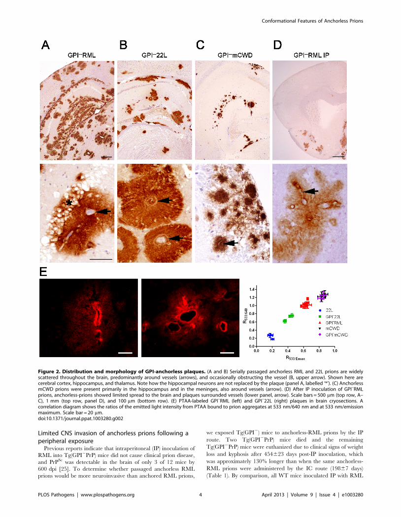
Limited CNS invasion of anchorless prions following aperipheral exposure
Previous reports indicate that intraperitoneal (IP) inoculation of
RML into Tg(GPI2PrP) mice did not cause clinical prion disease,
and PrPSc was detectable in the brain of only 3 of 12 mice by
600 dpi [25]. To determine whether passaged anchorless RML
prions would be more neuroinvasive than anchored RML prions,
we exposed Tg(GPI2) mice to anchorless-RML prions by the IP
route. Two Tg(GPI2PrP) mice died and the remaining
Tg(GPI2PrP) mice were euthanized due to clinical signs of weight
loss and kyphosis after 454623 days post-IP inoculation, which
was approximately 130% longer than when the same anchorless-
RML prions were administered by the IC route (19867 days)
(Table 1). By comparison, all WT mice inoculated IP with RML
Figure 2. Distribution and morphology of GPI-anchorless plaques. (A and B) Serially passaged anchorless RML and 22L prions are widelyscattered throughout the brain, predominantly around vessels (arrows), and occasionally obstructing the vessel (B, upper arrow). Shown here arecerebral cortex, hippocampus, and thalamus. Note how the hippocampal neurons are not replaced by the plaque (panel A, labelled ‘*’). (C) AnchorlessmCWD prions were present primarily in the hippocampus and in the meninges, also around vessels (arrow). (D) After IP inoculation of GPI–RMLprions, anchorless-prions showed limited spread to the brain and plaques surrounded vessels (lower panel, arrow). Scale bars = 500 mm (top row, A–C), 1 mm (top row, panel D), and 100 mm (bottom row). (E) PTAA-labeled GPI–RML (left) and GPI–22L (right) plaques in brain cryosections. Acorrelation diagram shows the ratios of the emitted light intensity from PTAA bound to prion aggregates at 533 nm/640 nm and at 533 nm/emissionmaximum. Scale bar = 20 mm.doi:10.1371/journal.ppat.1003280.g002
Conformational Features of Anchorless Prions
PLOS Pathogens | www.plospathogens.org 4 April 2013 | Volume 9 | Issue 4 | e1003280

developed terminal prion disease after 22761 day, which was only
39% longer than the IC route (16364 days).
In contrast to the widespread distribution of GPI–RML deposits
after IC exposure, IP exposed Tg(GPI2) mice showed only
scattered plaques in the meninges, olfactory bulb, cerebral cortex,
basal ganglia, and cerebellum (Figure 2D) in one mouse and rare
plaques (,10) in 3 other mice. Only one of the five Tg(GPI2PrP)
mice showed PrPSc in brain by ELISA and by western blotting
(Figure S4). However, there was abundant PrPSc in the spleen by
western blot (Figure S4) and in the heart and adipose tissue by
IHC, indicating substantial peripheral PrPSc reservoirs (Figure S3).
Anchorless prions are primarily proteinase K-resistantTo characterize the biochemical properties of the anchorless
strains, we measured the levels of soluble and insoluble PrP in the
brains of mice infected with anchored and anchorless RML and
22L prions. For all samples tested, greater than 70% of the PrP
was insoluble, consistent with previous reports [31,32]. The
anchorless prions showed a slightly higher ratio of insoluble :
soluble PrP as compared to their anchored counterparts
(GPI2RML: 9361% versus RML: 7365% ; GPI222L: 8962%
versus 22L: 7861%; Student’s t-test p,0.05 for RML and 22L)
(Figure 3A).
RML has been previously shown to be composed of primarily
proteinase K (PK)-sensitive PrPSc, with only approximately 20%
PK-resistant PrPSc [33]. To investigate whether the GPI2RML
shows a similar proportion of PK-resistant aggregates, equal
volumes of PK-digested and non-digested brain homogenate
samples were ultracentrifuged. The insoluble fractions were
compared by western blotting, and revealed that anchored RML
contained approximately 40–60% PK-resistant PrP, whereas the
anchorless RML contained approximately 80–100% PK-resistant
PrP (Figure 3B). Similar results were seen with the 22L and
anchorless 22L prions (Figure 3B). PK-sensitivity was also tested by
exposing brain homogenates to PK concentrations ranging from
0–9000 mg/ml for 2 hours at 37uC. The anchorless prions were
significantly more PK-resistant than the anchored prions
(Figure 3C).
Anchorless prions show high stability in chaotropesFibrillar prions were previously found to be more stable in
chaotropes than non-fibrillar prions [26]. To determine the
stability of the anchorless fibrillar prions, we performed standard
conformational stability assays [34] and compared anchored and
anchorless RML and 22L prions. We exposed brain homogenates
to guanidine hydrochloride (GdnHCl) concentrations from 0 to
6 M, diluted the GdnHCl, digested samples with PK, quantified
PrP by direct ELISA, and calculated the concentration at which
half the PrP signal was lost, denoted [GdnHCl]1/2. Interestingly,
lack of a GPI anchor resulted in the formation exceedingly stable
prions, as the [GdnHCl]1/2 values were 5.360.4 for GPI2RML
and 0.9160.1 for RML (p,0.001) (Figure 4A). Additionally, the
Figure 3. Solubility and PK-resistance of anchorless prions. (A) Soluble and insoluble PrP were measured in the anchored and anchorless RMLand 22L infected brain samples by western blot. Graph shows the mean and SE from 3–4 mice per prion. (B) Insoluble PrP fraction: PrP levels weremeasured in undigested and PK digested samples. The graph shows the percentage of PK-resistant PrP over total insoluble PrP (mean and SE from 4mice per prion). (C) PK-resistance of anchored and anchorless RML and 22L prions was assessed after 2 hours of PK digestion at 37uC. Graphs showthe mean and SE from 4 mice per prion.doi:10.1371/journal.ppat.1003280.g003
Conformational Features of Anchorless Prions
PLOS Pathogens | www.plospathogens.org 5 April 2013 | Volume 9 | Issue 4 | e1003280

[GdnHCl]1/2 of anchorless RML decreased with serial passaging
(second passage 5.060.30 and third passage 3.160.25 from n = 4
mice each) (Figure 4A). Similarly, GPI222L showed a higher
[GdnHCl]1/2 than 22L (GPI222L: 2.760.3 and 22L: 0.8260.1,
p,0.001) (Figure 4B).
Thermal denaturation of the anchorless prion strainsWe sought to further assess the aggregate stability differences
between anchored and anchorless prions using a thermal gradient.
PK-digested aliquots of brain homogenate in SDS were heated to
temperatures ranging from 25–99uC and proteins were immedi-
ately resolved by one dimensional denaturing gel electrophoresis.
Monomeric PrP was quantified and plotted at each temperature.
In comparing the temperature at which half of the PrPSc
disassembled into monomers (T1/2), GPI2RML was found to be
only modestly, but significantly more resistant to SDS thermal
disassembly than RML (T1/2 of GPI2RML: 7162uC and RML:
5663uC, p,0.01) (Figure 5A). Similarly the T1/2 of the GPI222L
was significantly greater than 22L (GPI222L: 7162uC and 22L:
5962uC, p,0.01) (Figure 5A).
The modest difference in thermal stability between anchorless
and anchored strains was surprising, yet an underlying assumption
for the thermal stability assays is the complete denaturation of
PrPSc at 99uC for SDS-PAGE. To exclude the possibility of higher
order aggregates not migrating through the gel, we measured the
insoluble PrPSc by ELISA. Brain homogenate samples were heated
in SDS to temperatures from 25 to 99uC and centrifuged. The
insoluble pellet fractions were denatured using 6 M guanidine
isothiocyanate and PrPSc levels were then measured by ELISA.
RML showed low levels of insoluble PrP at low temperatures
which sharply decreased to nearly zero at approximately 55uC(T1/2 of 4461uC). In stark contrast, the GPI2RML (first passage)
showed abundant insoluble PrP with no loss of signal, even at the
highest temperatures (Figure 5B), indicating that there was
substantial PrPSc that remained insoluble, even after heating to
99uC in SDS. Interestingly, in the third passage GPI2RML, PrPSc
became largely soluble at high temperatures (T1/2 of 7464uC),
indicating that the thermostability decreased with passaging,
consistent with results from the GdnHCl denaturation experi-
ments. Similar to RML, the anchored 22L was also significantly
less thermostable than the GPI222L [T1/2 of 5065uC (22L) versus
8162uC (GPI222L); p,0.01], indicating that abundant GPI-
anchorless PrPSc remained insoluble at high temperatures
(Figure 5B).
We reasoned that if the low thermostability values in the initial
experiments (analyzed by western blot) were due higher order
aggregates, these larger aggregates should migrate after complete
denaturation. We PK-digested, heated to 95uC SDS, and
centrifuged two aliquots each of anchored and anchorless RML.
One aliquot of each supernatant and pellet fraction was denatured
with 6 M guanidine isothiocyanate while the other was not
treated, and then all samples were heated to 95uC in SDS prior to
SDS-PAGE. As expected, the soluble anchored and anchorless
PrPSc signals in the supernatant fractions were not altered
following guanidine isothiocyanate denaturation. In contrast, for
the pellet fractions, there was a slight enhancement of the RML
signal and a massive increase in the anchorless RML signal with
denaturation (Figure 5C, last 2 lanes). In sum, the insoluble
fraction of GPI2RML remained highly aggregated after heating in
SDS, requiring the additional denaturation with a strong
chaotrope to fully disassemble aggregates into monomers for
electrophoresis. This result confirms that heating the GPI2RML
in SDS disassembles only half the population of the total
anchorless PrPSc and this property likely underlies the modest
differences in thermal stability initially observed by SDS-PAGE.
Thus the denaturation required for the ELISA enables a much
more accurate measurement of the total fibrillar prion population.
GPI2RML infection of WT miceTo determine whether the anchorless RML would revert back
to an RML disease phenotype in WT mice, we intracerebrally
inoculated anchorless RML prions into WT mice. The anchorless
Figure 4. Conformational stability assay of GPI-anchored and anchorless prions. GPI-anchorless prions were significantly more resistant toGdnHCl-induced denaturation as compared to the GPI-anchored prions. (A) [GdnHCl1/2] of anchored and anchorless RML over three passages in theTg(GPI–PrP) mice was measured by ELISA. (B) [GdnHCl1/2] of anchored and anchorless 22L. Plotted are the averages from n = 4 mice per prion, eachrun in triplicate. Graph (right panel) represents mean 6 SE for all mice from [GdnHCl1/2] values that were independently calculated for each mouse.doi:10.1371/journal.ppat.1003280.g004
Conformational Features of Anchorless Prions
PLOS Pathogens | www.plospathogens.org 6 April 2013 | Volume 9 | Issue 4 | e1003280

RML prions induced disease in WT mice at a time period similar
to RML prions (Figure 6A). The PrPSc aggregate morphology
was diffuse and widespread in the cerebral cortex, basal ganglia
thalamus, hippocampus, cerebellum, and brainstem (Figure 6B),
and there were no differences in the distribution and degree of
the spongiosis, astrogliosis, or PrPSc as seen by a lesion profile
analysis (Figure 6C). RML and GPIRML-WT also showed
indistinguishable glycoform profiles (Figure 6D, NSD between
quantified glycoform ratios), stability in chaotropes ([GdnHCl]1/2
for RML: 0.93 and GPIRML-WT: 0.93) (Figure 6E) and
thermostability (Figure 6F, G) [Fig. 6F: T1/2: 5161uC (RML)
and 5463uC (GPIRML-WT)]. These data suggest anchorless
RML prions are efficiently transmitted to mice expressing GPI-
anchored PrP.
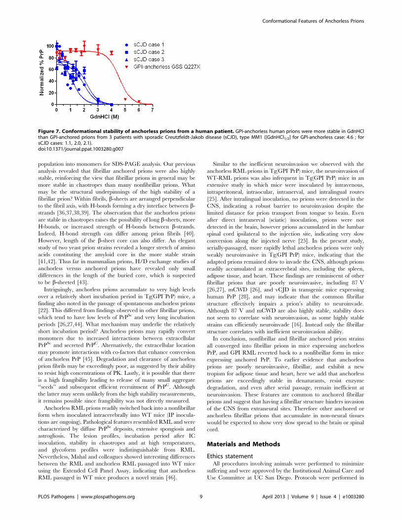
Human full length anchorless prions from a patient arealso exceedingly stable in denaturants
To determine whether the high chaotrope stability of anchorless
prions in mice is also seen with anchorless prions in patients, we
tested brain from a patient expressing full length anchorless PrP
due to a very rare Q227X mutation. This patient died at 45 years
of age and the brain showed extensive plaque-like prion deposits
and a lack of spongiosis. Although full length PrP was expressed,
the PK-resistant core fragment was primarily a 7 kD band that
lacked the amino and carboxy termini [35]. We measured the
chaotrope stability of anchorless prions and found that the
[GdnHCl]1/2 was markedly higher for the anchorless prions than
for three sporadic CJD (MM1) cases (Figure 7).
Figure 5. Thermal denaturation of anchored and anchorless prions. (A) PK-digested anchored and anchorless RML prions were heated totemperatures from 25–99uC and monomers were resolved by SDS-PAGE. Middle graphs show the plot of mean 6 SE of RML and GPI–RML, passage 1(P1) at each temperature (n = 4 mice each). The right graph shows the T1/2 point for RML and serial passages of GPI–RML. There were no significantdifferences among the passages of GPI–RML. Anchored and anchorless 22L were similarly assessed. (B) Samples were heated as in (A) and centrifuged.The remaining insoluble PrPSc was denatured and measured by ELISA. Plotted are the averages from n = 4 mice per prion, each run in triplicate. Datawere normalized to the initial PrPSc intensity. Right panel, T1/2 values were independently calculated for each mouse and plotted (mean and SE). (C)Two aliquots of each of the anchored and anchorless prions were PK-digested, heated in SDS to 95uC and centrifuged, and the supernatants andpellets were collected. One aliquot of each supernatant (S) and pellet (P) was denatured with guanidine isothiocyanate (GdnSCN), and all sampleswere heated to 95uC prior to SDS-PAGE. Graph below shows mean 6 SE from 4 independent experiments using 3 RML and 3 GPI–RML samples (RML:1861, GPI–RML: 5062, p = 0.0001, Students t–test).doi:10.1371/journal.ppat.1003280.g005
Conformational Features of Anchorless Prions
PLOS Pathogens | www.plospathogens.org 7 April 2013 | Volume 9 | Issue 4 | e1003280

Discussion
Prions exhibit a tremendous conformational repertoire, yet the
relationship between their misfolded structure and disease
phenotype is unclear. We have previously found that fibrillar
prions are inefficient at trafficking to the CNS and are highly
stable under denaturing conditions [26]. In the present study, we
perform a comprehensive analysis of the biochemical properties of
a prion in its anchored and anchorless state and correlate results
with the disease pathogenesis. Previous studies have focused on the
first passage of prions in Tg(GPI–PrP) mice. Baron and colleagues
showed that anchored and anchorless 22L exhibit a similar
secondary structure with residues 81–167 highly protected against
deuterium incorporation [31]. Here we demonstrate that serially
passaged anchorless prions have a substantial resistance to
denaturation, a fibrillar morphology, and are poorly neuroinva-
sive, thus differ markedly from their nonfibrillar anchored
counterpart. Additionally, anchorless prions show a high ratio of
insoluble to soluble PrP, suggestive of efficient conversion. Thus
there is a profound biochemical transformation when a non-
fibrillar prion is converted by extracellular anchorless PrPC.
We observed that anchorless prions were exceedingly stable
when exposed to chaotropes and heat. Of note, even heating
anchorless prions to 95uC in SDS solubilized only half the PrPSc
Figure 6. GPI–RML prions inoculated into WT mice (GPIRML-WT). (A) Survival curves of WT mice inoculated with RML or GPI–RML prions(passage 2). (B) PrP and astrocyte immunostaining of brain sections at the level of cerebral cortex and hippocampus show diffuse PrPSc deposits andaccompanying gliosis. (C) Lesion profile analysis. For RML- and GPIRML-infected WT mice, the severity of spongiosis, astrogliosis, and PrPSc depositionwere scored for nine brain regions (see Methods) and were nearly superimposable. Each ring represents 1 point. (D) PK-digested brain samples fromWT mice infected with RML or GPI–RML show no difference in their glycoform profiles. Also shown are Tg(GPI–RML) mice infected with RML, whichshow a shift to a lower molecular weight and only mono-glycosylated PrP. (E) Conformational stability and (F) thermal stability of RML and GPIRML-WT were nearly identical (n = 4 mice each). (G) Thermal denaturation curves of RML- and GPIRML-infected WT mice show the insoluble PrPSc
remaining after heating to various temperatures as measured by ELISA. Right panel, T1/2 values were independently calculated for each mouse andplotted. For (E), plotted are the averages from n = 4 mice per prion, each run in triplicate. For (F), the middle graph shows the mean 6 SE of RML andGPI–RML (n = 4 mice each). Graphs (right panels) represent the mean 6 SE for all mice. Scale bar = 500 mm.doi:10.1371/journal.ppat.1003280.g006
Conformational Features of Anchorless Prions
PLOS Pathogens | www.plospathogens.org 8 April 2013 | Volume 9 | Issue 4 | e1003280
population into monomers for SDS-PAGE analysis. Our previous
analysis revealed that fibrillar anchored prions were also highly
stable, reinforcing the view that fibrillar prions in general may be
more stable in chaotropes than many nonfibrillar prions. What
may be the structural underpinnings of the high stability of a
fibrillar prion? Within fibrils, b-sheets are arranged perpendicular
to the fibril axis, with H-bonds forming a dry interface between b-
strands [36,37,38,39]. The observation that the anchorless prions
are stable in chaotropes raises the possibility of long b-sheets, more
H-bonds, or increased strength of H-bonds between b-strands.
Indeed, H-bond strength can differ among prion fibrils [40].
However, length of the b-sheet core can also differ. An elegant
study of two yeast prion strains revealed a longer stretch of amino
acids constituting the amyloid core in the more stable strain
[41,42]. Thus far in mammalian prions, H/D exchange studies of
anchorless versus anchored prions have revealed only small
differences in the length of the buried core, which is suspected
to be b-sheeted [43].
Intriguingly, anchorless prions accumulate to very high levels
over a relatively short incubation period in Tg(GPI–PrP) mice, a
finding also noted in the passage of spontaneous anchorless prions
[22]. This differed from findings observed in other fibrillar prions,
which tend to have low levels of PrPSc and very long incubation
periods [26,27,44]. What mechanism may underlie the relatively
short incubation period? Anchorless prions may rapidly convert
monomers due to increased interactions between extracellular
PrPSc and secreted PrPC. Alternatively, the extracellular location
may promote interactions with co-factors that enhance conversion
of anchorless PrP [45]. Degradation and clearance of anchorless
prion fibrils may be exceedingly poor, as suggested by their ability
to resist high concentrations of PK. Lastly, it is possible that there
is a high frangibility leading to release of many small aggregate
‘‘seeds’’ and subsequent efficient recruitment of PrPC. Although
the latter may seem unlikely from the high stability measurements,
it remains possible since frangibility was not directly measured.
Anchorless RML prions readily switched back into a nonfibrillar
form when inoculated intracerebrally into WT mice (IP inocula-
tions are ongoing). Pathological features resembled RML and were
characterized by diffuse PrPSc deposits, extensive spongiosis and
astrogliosis. The lesion profiles, incubation period after IC
inoculation, stability in chaotropes and at high temperatures,
and glycoform profiles were indistinguishable from RML.
Nevertheless, Mahal and colleagues showed interesting differences
between the RML and anchorless RML passaged into WT mice
using the Extended Cell Panel Assay, indicating that anchorless
RML passaged in WT mice produces a novel strain [46].
Similar to the inefficient neuroinvasion we observed with the
anchorless RML prions in Tg(GPI–PrP) mice, the neuroinvasion of
WT-RML prions was also infrequent in Tg(GPI–PrP) mice in an
extensive study in which mice were inoculated by intravenous,
intraperitoneal, intraocular, intranerval, and intralingual routes
[25]. After intralingual inoculation, no prions were detected in the
CNS, indicating a robust barrier to neuroinvasion despite the
limited distance for prion transport from tongue to brain. Even
after direct intranerval (sciatic) inoculation, prions were not
detected in the brain, however prions accumulated in the lumbar
spinal cord ipsilateral to the injection site, indicating very slow
conversion along the injected nerve [25]. In the present study,
serially-passaged, more rapidly lethal anchorless prions were only
weakly neuroinvasive in Tg(GPI–PrP) mice, indicating that the
adapted prions remained slow to invade the CNS, although prions
readily accumulated at extracerebral sites, including the spleen,
adipose tissue, and heart. These findings are reminiscent of other
fibrillar prions that are poorly neuroinvasive, including 87 V
[26,27], mCWD [26], and vCJD in transgenic mice expressing
human PrP [28], and may indicate that the common fibrillar
structure effectively impairs a prion’s ability to neuroinvade.
Although 87 V and mCWD are also highly stable, stability does
not seem to correlate with neuroinvasion, as some highly stable
strains can efficiently neuroinvade [16]. Instead only the fibrillar
structure correlates with inefficient neuroinvasion ability.
In conclusion, nonfibrillar and fibrillar anchored prion strains
all converged into fibrillar prions in mice expressing anchorless
PrP, and GPI–RML reverted back to a nonfibrillar form in mice
expressing anchored PrP. To earlier evidence that anchorless
prions are poorly neuroinvasive, fibrillar, and exhibit a new
tropism for adipose tissue and heart, here we add that anchorless
prions are exceedingly stable in denaturants, resist enzyme
degradation, and even after serial passage, remain inefficient at
neuroinvasion. These features are common to anchored fibrillar
prions and suggest that having a fibrillar structure hinders invasion
of the CNS from extraneural sites. Therefore other anchored or
anchorless fibrillar prions that accumulate in non-neural tissues
would be expected to show very slow spread to the brain or spinal
cord.
Materials and Methods
Ethics statementAll procedures involving animals were performed to minimize
suffering and were approved by the Institutional Animal Care and
Use Committee at UC San Diego. Protocols were performed in
Figure 7. Conformational stability of anchorless prions from a human patient. GPI-anchorless human prions were more stable in GdnHClthan GPI-anchored prions from 3 patients with sporadic Creutzfeldt-Jakob disease (sCJD), type MM1 ([GdnHCl1/2] for GPI-anchorless case: 4.6 ; forsCJD cases: 1.1, 2.0, 2.1).doi:10.1371/journal.ppat.1003280.g007
Conformational Features of Anchorless Prions
PLOS Pathogens | www.plospathogens.org 9 April 2013 | Volume 9 | Issue 4 | e1003280

strict accordance with good animal practices, as described in the
Guide for the Use and Care of Laboratory Animals published by the
National Institutes of Health.
Prion inoculationsWT (C57BL/6) or Tg(GPI2PrP) mice (groups of n = 4–5 mice)
were inoculated IC into the left parietal cortex with 30 ml or
inoculated IP with 100 ml of a 0.1% (w/v) prion-infected brain
homogenate prepared from terminally ill mice. Mice were
monitored three times weekly, and TSE was diagnosed according
to clinical criteria including ataxia, kyphosis, stiff tail, hind leg
clasp, and hind leg paresis. Mice were sacrificed at the onset of
terminal disease and incubation period was calculated from the
day of inoculation to the day of terminal clinical disease. Mice
were maintained under specific pathogen-free conditions. All
procedures involving animals were performed to minimize
suffering and were approved by the Institutional Animal Care
and Use Committee at UC San Diego. Protocols were performed
in strict accordance with good animal practices, as described in the
Guide for the Use and Care of Laboratory Animals published by
the National Institutes of Health.
Histopathology and immunohistochemical stainsTwo-mm thick sections were cut onto positively charged
silanized glass slides and stained with hematoxylin and eosin, or
immunostained using antibodies for PrP (SAF84) or astrocytes
(GFAP). For PrP staining, sections were deparaffinized and
incubated for 5 min in 88% formic acid and treated with 5 mg/
ml of proteinase-K to expose epitopes. Sections were then
autoclaved in citrate buffer (pH 6). Immunohistochemical stains
were performed using the TSA Plus DNP kit (PerkinElmer).
Sections were blocked and incubated with anti-PrP SAF-84 (SPI
bio; 1:400) for 45 min followed by anti-mouse HRP (Jackson
Immunolabs; 1:500) for 30 min. Slides were then incubated with
anti-DNP-HRP (PerkinElmer, 1:100) for 30 min, followed by
6 min incubation with DAB. Sections were counterstained with
hematoxylin. GFAP immunohistochemistry for astrocytes (1:500;
DAKO) was similarly performed, however with antigen retrieval
by PK-digestion (20 ug/ml for 10 min at room temperature).
Lesion profileWe selected 9 anatomic brain regions in accordance with
previous strain-typing protocols from 5 mice per group [47,48].
We scored spongiosis, gliosis, and PrP immunological reactivity on
a scale of 0–3 (not detectable, mild, moderate, and severe). A sum
of the three scores resulted in the value obtained for the lesion
profile for the individual animal. The ‘radar plots’ depict the
scores for spongiform changes, gliosis and PrP deposition. The
following brain regions were scored: dorsal medulla, cerebellum,
hypothalamus, medial thalamus, hippocampus, septum, medial
cerebral cortex dorsal to hippocampus, medial cerebral cortex
dorsal to septum, white matter at cerebral peduncles. An
investigator blinded to animal identification performed the
histological analyses.
Western blotting for PrPSc in brain and spleenFor brain and spleen from the IP-inoculated mice, sodium
phosphotungstic acid (NaPTA) PrPSc precipitation was performed
as previously described [49]. Briefly, brain and spleen extracts in
PBS containing 2% sarkosyl were digested with an endonuclease
[Benzonase (Sigma)] followed by treatment with 50 mg/ml PK at
37uC for 30 min. After addition of NaPTA, MgCl2, and protease
inhibitors, extracts were incubated at 37uC for 30 min, and
centrifuged at 18,000 g for 30 min at 37uC. Pellets were
resuspended in 0.1% sarcosyl for electrophoresis and blotting.
For IC-inoculated mice, brain extracts in lysis buffer (10 mM Tris-
HCl pH 7.4, 150 mM NaCl, 2% sarcosyl) were digested with
50 mg/ml PK at 37uC for 40 min.
Samples were electrophoresed through 10% Bis-Tris gels
(Invitrogen) and blotted onto a nitrocellulose membrane. PrP
was detected using monoclonal antibody POM1 (epitope in the
globular domain, amino acids 121–231, a kind gift from Dr.
Adriano Aguzzi) [50] and an HRP-conjugated anti-mouse IgG
secondary antibody. The blots were developed using a chemilu-
minescent substrate (ECL detection Kit, Pierce) and visualized on
a Fuji LAS 4000 imager. Quantification of PrPSc glycoforms was
performed using Multigauge V3 software (Fujifilm).
Conformation stability assayPrion strain stability in GdnHCl was performed as previously
described [34] with minor modifications. Briefly, brain homoge-
nates were denatured in GdnHCl ranging from 0–6 M for 1 hr
and then diluted to 0.15 M GdnHCl. The samples were then
digested with PK at a ratio of 1:500 (1 mg PK : 500 mg total
protein) for 1 hr at 37uC, stopped with PMSF and Complete
protease inhibitor (Roche), and centrifuged at 18,000 g for 1 hr.
The pellets were washed with 500 ml of 0.1 M NaHCO3 (pH 9.6)
and centrifuged for 20 min at 18,000 g. Pellets were denatured in
6 M guanidine isothiocyanate (GdnSCN), diluted 2X with 0.1 M
NaHCO3, and coated passively onto an ELISA plate. PrP was
detected with anti-PrP biotinylated-POM1 antibody, a streptavi-
din HRP-conjugated anti-mouse IgG secondary antibody, and
detected with a chemiluminescent substrate. Each strain was
analyzed in at least 3 separate experiments using 4 mice. Statistical
analysis was performed using a Student’s t test. The human brain
samples were detected using the anti-PrP 3F4 antibody, a
biotinylated anti-mouse secondary antibody, and streptavidin-
HRP followed by the chemiluminescent substrate.
Thermal denaturation assayBrain homogenate in a Tris lysis buffer (10 mM Tris-HCl
pH 7.4, 150 mM NaCl, 2% sarcosyl) was digested with 50 mg/ml
PK for 40 min at 37uC. PK digestion was inactivated with
phenylmethylsulfonyl fluoride (PMSF) (2 mM final concentration)
and Complete protease inhibitor. Aliquots were incubated in 1.6%
SDS (final) and heated to temperatures ranging from 25uC to
99uC (10u intervals) for 6 min with shaking in a thermomixer at
1000 rpm. Western blotting was performed and PrP signals from
monomers were captured and quantified using a Fujifilm LAS-
4000 imager and Multigage software. Each strain was analyzed in
at least 3 separate experiments using 4–11 mice.
Quantification of soluble and insoluble PrPBrain homogenate in a Tris lysis buffer was maintained at 37uC
for 15 min, centrifuged at 150,000 g for 1 hr at 4uC, and
separated into supernatant and pellet fractions. Proteins in the
supernatant were precipitated using cold methanol. Supernatant
and pellet proteins were then analyzed and quantified by western
blotting for PrP. Each strain was analyzed in at least 3 separate
experiments using 3–4 mice.
Quantification of PK resistant and sensitive PrPBrain homogenate in a Tris lysis buffer was maintained at 37uC
for 15 min and split into two aliquots. One aliquot was treated
with PK (50 mg/ml) at 37uC for 30 min, and both aliquots were
centrifuged at 150,000 g for 1 hr at 4uC. PrP in the PK-treated
Conformational Features of Anchorless Prions
PLOS Pathogens | www.plospathogens.org 10 April 2013 | Volume 9 | Issue 4 | e1003280

and untreated pellets were quantified by western blotting. Each
strain was analyzed in at least 3 separate experiments using 4 mice.
Quantification of PK-resistant PrP by ELISAEqual amount of sample in a Tris lysis buffer was maintained at
37uC for 15 min, digested with PK (50 mg/ml) at 37uC for 40 min,
and the PK was inactivated with PMSF (6 mM final). The samples
were denatured with GndHCl (2 M final) at 81uC for 6 min,
diluted 10X with 0.1% TBST and added to an ELISA plate
precoated with POM-2 antibody. PrP was detected using anti-PrP
biotinylated-POM1 antibody, a streptavidin HRP-conjugated
anti-mouse IgG secondary antibody, and 1-Step Ultra TMB-
ELISA substrate (Thermo-Scientific). Each strain was analyzed in
triplicate using 4 mice each. Statistical analysis was performed
using a Student’s t test.
Electron microscopyTissues were post-fixed in osmium tetroxide, embedded in epon
araldite, sectioned with the ultramicrotome, then collected on
grids and post-stained using saturated uranyl acetate solution and
bismuth sub-nitrate. Grids were analyzed with a Zeiss EM10
electron microscope.
PTAA staining of frozen tissue sectionsFrozen sections from mouse brain were dried for 1 hour and
fixed in 100% and 70% ethanol for 10 min each. After washing
with deionized water, sections were equilibrated in 100 mM
sodium carbonate at pH 10.2 for 30 minutes. The PTAA was
diluted in the sodium carbonate buffer (1 mg : 100 ml buffer) and
added to the sections. The sections were incubated with PTAA for
30 min at room temperature and washed with sodium carbonate
buffer. The spectra in the tissue were recorded with a Leica
DM6000 B fluorescence microscope (Leica Microsystems, Wet-
zlar, Germany) fitted with a Spectraview 4.0 (Applied Spectral
Imaging, Migdal, Israel) and a Spectra-Cube (interferometrical
optical head SD 300) module with cooled CCD-camera, through a
436/10 nm (LP 475) bandpass filter in steps of 6 nm. The data
were processed with SpectraView 3.0 EXPO. Spectra were
collected from 8 individual spots within 3–5 plaques from a
minimum of two different cases of each prion-infected brain.
Human patient samplesThe human brain samples were from three patients diagnosed
with sCJD, consisting of one male (age 63) and two females (ages
53 and 60). All had a PRNP genotype encoding 129 MM PrP, a
short disease duration (2–3 months), a type 1 PrPSc pattern on
western blot, and histopathology typical of classic sCJDMM1.
Case typing was performed at the National Prion Disease
Pathology Surveillance Center at Case Western Reserve Univer-
sity in Cleveland, Ohio. The GPI-anchorless Q227X GSS-type
prion case was identified at the Dutch Surveillance Centre for
Prion Diseases, University Medical Centre Utrecht in The
Netherlands.
Supporting Information
Figure S1 PrP ELISA measurements of PK-digestedPrPSc. First through third passages (P1, P2, and P3) of GPI–RML
in Tg(GPI–PrP) mice were assessed (n = 4 mice each). Graph
shows mean 6 SE. Passage 1 was significantly different than
passage 2 (Student’s t-test, p,0.01).
(TIF)
Figure S2 Ultrastructure of brain (cerebral cortex) froma Tg(GPI–PrP) mouse infected with GPI–RML prions. (A)
Low power image of a blood vessel (V) surrounded by
extracellular, loosely arranged fibrils (*). (B) High power image
shows fibrils are short and present in thin bundles that are
haphazardly arranged. (C) Commonly seen were dystrophic
neurites (arrows) containing variably-sized electron dense deposits.
(TIF)
Figure S3 Immunohistochemical stains of heart andadipose tissue (brown fat) for PrP. PrPSc deposits were
observed in heart and adipose tissue of Tg(GPI–PrP) mice for all
strains tested. Scale bars = 100 mm.
(TIF)
Figure S4 PrPSc in brains and spleens of Tg(GPI–PrP)mice inoculated with anchorless RML prions. Brain from
only one of five mice showed detectable PrPSc by (A) NaPTA
precipitation and western blot, or (B) ELISA. (C) In contrast, all
spleens from GPI–RML inoculated Tg(GPI–PrP) mice as well as
RML-inoculated WT mice showed PrPSc detectable by NaPTA
precipitation and western blot. Approximately 7-fold more total
protein was loaded for WT as compared to the Tg(GPI2PrP)
spleen.
(TIF)
Acknowledgments
We thank Carlitos Chen and the animal caretakers at UC San Diego for
providing excellent technical support, and Laurence Cagnon for assistance
in the BSL3 laboratory at The Scripps Research Institute. We thank Dr.
Adriano Aguzzi for providing the anti-PrP antibodies (POM series).
Author Contributions
Conceived and designed the experiments: CB TDK CJS. Performed the
experiments: CB TDK ML MT KPRN. Analyzed the data: CB TDK
KPRN EM MBO CJS. Contributed reagents/materials/analysis tools:
AJR QK MBO. Wrote the paper: CB CJS.
References
1. Colby DW, Prusiner SB (2011) Prions. Cold Spring Harb Perspect Biol 3:a006833.
2. Aguzzi A, Calella AM (2009) Prions: protein aggregation and infectious diseases.
Physiol Rev 89: 1105–1152.3. Kaatz M, Fast C, Ziegler U, Balkema-Buschmann A, Hammerschmidt B, et al.
(2012) Spread of Classic BSE Prions from the Gut via the Peripheral NervousSystem to the Brain. Am J Pathol 181: 515–524.
4. Glatzel M, Abela E, Maissen M, Aguzzi A (2003) Extraneural pathologic prionprotein in sporadic Creutzfeldt-Jakob disease. N Engl J Med 349: 1812–1820.
5. Klein MA, Frigg R, Flechsig E, Raeber AJ, Kalinke U, et al. (1997) A crucial
role for B cells in neuroinvasive scrapie. Nature 390: 687–690.6. Prusiner SB (1982) Novel proteinaceous infectious particles cause scrapie.
Science 216: 136–144.7. Fraser H, Dickinson AG (1973) Scrapie in mice. Agent-strain differences in the
distribution and intensity of grey matter vacuolation. J Comp Pathol 83: 29–40.
8. Bruce ME (2003) TSE strain variation. Br Med Bull 66: 99–108.
9. Collinge J, Clarke AR (2007) A general model of prion strains and theirpathogenicity. Science 318: 930–936.
10. Tixador P, Herzog L, Reine F, Jaumain E, Chapuis J, et al. (2010) The physical
relationship between infectivity and prion protein aggregates is strain-dependent.PLoS Pathog 6: e1000859.
11. Peretz D, Scott MR, Groth D, Williamson RA, Burton DR, et al. (2001) Strain-specified relative conformational stability of the scrapie prion protein. Protein
Sci 10: 854–863.12. Collinge J, Sidle KC, Meads J, Ironside J, Hill AF (1996) Molecular analysis of prion
strain variation and the aetiology of ‘new variant’ CJD. Nature 383: 685–690.
13. Bessen RA, Kocisko DA, Raymond GJ, Nandan S, Lansbury PT, et al. (1995)Non-genetic propagation of strain-specific properties of scrapie prion protein.
Nature 375: 698–700.14. Bessen RA, Marsh RF (1992) Biochemical and physical properties of the prion
protein from two strains of the transmissible mink encephalopathy agent. J Virol
66: 2096–2101.
Conformational Features of Anchorless Prions
PLOS Pathogens | www.plospathogens.org 11 April 2013 | Volume 9 | Issue 4 | e1003280

15. Kuczius T, Groschup MH (1999) Differences in Proteinase K Resistance and
Neuronal Deposition of Abnormal Prion Proteins Characterize BovineSpongiform Encephalopathy (BSE) and Scrapie Strains. Mol Med 5: 406–418.
16. Ayers JI, Schutt CR, Shikiya RA, Aguzzi A, Kincaid AE, et al. (2011) The
strain-encoded relationship between PrP replication, stability and processing inneurons is predictive of the incubation period of disease. PLoS Pathog 7:
e1001317.17. Wiltzius JJ, Landau M, Nelson R, Sawaya MR, Apostol MI, et al. (2009)
Molecular mechanisms for protein-encoded inheritance. Nat Struct Mol Biol 16:
973–978.18. Telling GC, Parchi P, DeArmond SJ, Cortelli P, Montagna P, et al. (1996)
Evidence for the conformation of the pathologic isoform of the prion proteinenciphering and propagating prion diversity. Science 274: 2079–2082.
19. Stahl N, Borchelt DR, Hsiao K, Prusiner SB (1987) Scrapie prion proteincontains a phosphatidylinositol glycolipid. Cell 51: 229–240.
20. Rogers M, Yehiely F, Scott M, Prusiner SB (1993) Conversion of truncated and
elongated prion proteins into the scrapie isoform in cultured cells. Proc NatlAcad Sci U S A 90: 3182–3186.
21. Liberski PP (2012) Gerstmann-Straussler-Scheinker disease. Adv Exp Med Biol724: 128–137.
22. Stohr J, Watts JC, Legname G, Oehler A, Lemus A, et al. (2011) Spontaneous
generation of anchorless prions in transgenic mice. Proc Natl Acad Sci U S A108: 21223–21228.
23. Chesebro B, Trifilo M, Race R, Meade-White K, Teng C, et al. (2005)Anchorless prion protein results in infectious amyloid disease without clinical
scrapie. Science 308: 1435–1439.24. Chesebro B, Race B, Meade-White K, Lacasse R, Race R, et al. (2010) Fatal
transmissible amyloid encephalopathy: a new type of prion disease associated
with lack of prion protein membrane anchoring. PLoS Pathog 6: e1000800.25. Klingeborn M, Race B, Meade-White KD, Rosenke R, Striebel JF, et al. (2011)
Crucial role for prion protein membrane anchoring in the neuroinvasion andneural spread of prion infection. J Virol 85: 1484–1494.
26. Bett C, Joshi-Barr S, Lucero M, Trejo M, Liberski P, et al. (2012) Biochemical
properties of highly neuroinvasive prion strains. PLoS Pathogens 8: e1002522.27. Collis SC, Kimberlin RH (1985) Long-term persistence of scrapie infection in
mouse spleens in the absence of clinical disease. FEMS Microbiology Letters 29:111–114.
28. Beringue V, Le Dur A, Tixador P, Reine F, Lepourry L, et al. (2008) Prominentand persistent extraneural infection in human PrP transgenic mice infected with
variant CJD. PLoS ONE 3: e1419.
29. Trifilo MJ, Yajima T, Gu Y, Dalton N, Peterson KL, et al. (2006) Prion-inducedamyloid heart disease with high blood infectivity in transgenic mice. Science 313:
94–97.30. Race B, Meade-White K, Oldstone MB, Race R, Chesebro B (2008) Detection
of prion infectivity in fat tissues of scrapie-infected mice. PLoS Pathog 4:
e1000232.31. Baron GS, Hughson AG, Raymond GJ, Offerdahl DK, Barton KA, et al. (2011)
Effect of glycans and the glycophosphatidylinositol anchor on strain dependentconformations of scrapie prion protein: improved purifications and infrared
spectra. Biochemistry 50: 4479–4490.32. Sim VL, Caughey B (2008) Ultrastructures and strain comparison of under-
glycosylated scrapie prion fibrils. Neurobiol Aging 30: 2031–42.
33. Cronier S, Gros N, Tattum MH, Jackson GS, Clarke AR, et al. (2008) Detectionand characterization of proteinase K-sensitive disease-related prion protein with
thermolysin. Biochem J 416: 297–305.
34. Peretz D, Williamson RA, Legname G, Matsunaga Y, Vergara J, et al. (2002) A
change in the conformation of prions accompanies the emergence of a new prion
strain. Neuron 34: 921–932.
35. Jansen C, Parchi P, Capellari S, Vermeij AJ, Corrado P, et al. (2010) Prion
protein amyloidosis with divergent phenotype associated with two novel
nonsense mutations in PRNP. Acta Neuropathol 119: 189–197.
36. Diaz-Avalos R, Long C, Fontano E, Balbirnie M, Grothe R, et al. (2003) Cross-
beta order and diversity in nanocrystals of an amyloid-forming peptide. J Mol
Biol 330: 1165–1175.
37. Petkova AT, Leapman RD, Guo Z, Yau WM, Mattson MP, et al. (2005) Self-
propagating, molecular-level polymorphism in Alzheimer’s beta-amyloid fibrils.
Science 307: 262–265.
38. Sawaya MR, Sambashivan S, Nelson R, Ivanova MI, Sievers SA, et al. (2007)
Atomic structures of amyloid cross-beta spines reveal varied steric zippers.
Nature 447: 453–457.
39. Benzinger TL, Gregory DM, Burkoth TS, Miller-Auer H, Lynn DG, et al.
(1998) Propagating structure of Alzheimer’s beta-amyloid(10–35) is parallel beta-
sheet with residues in exact register. Proc Natl Acad Sci U S A 95: 13407–13412.
40. Shashilov V, Xu M, Makarava N, Savtchenko R, Baskakov IV, et al. (2012)
Dissecting Structure of Prion Amyloid Fibrils by Hydrogen-Deuterium
Exchange Ultraviolet Raman Spectroscopy. J Phys Chem B. 2012 Jun 26.
Epub ahead of print.
41. Tanaka M, Collins SR, Toyama BH, Weissman JS (2006) The physical basis of
how prion conformations determine strain phenotypes. Nature 442: 585–589.
42. Toyama BH, Kelly MJ, Gross JD, Weissman JS (2007) The structural basis of
yeast prion strain variants. Nature 449: 233–237.
43. Smirnovas V, Kim JI, Lu X, Atarashi R, Caughey B, et al. (2009) Distinct
structures of scrapie prion protein (PrPSc)-seeded versus spontaneous recombi-
nant prion protein fibrils revealed by hydrogen/deuterium exchange. J Biol
Chem 284: 24233–24241.
44. Tuzi NL, Cancellotti E, Baybutt H, Blackford L, Bradford B, et al. (2008) Host
PrP glycosylation: a major factor determining the outcome of prion infection.
PLoS Biol 6: e100.
45. Piro JR, Wang F, Walsh DJ, Rees JR, Ma J, et al. (2011) Seeding specificity and
ultrastructural characteristics of infectious recombinant prions. Biochemistry 50:
7111–7116.
46. Mahal SP, Jablonski J, Suponitsky-Kroyter I, Oelschlegel AM, Herva ME, et al.
(2012) Propagation of RML Prions in Mice Expressing PrP Devoid of GPI
Anchor Leads to Formation of a Novel, Stable Prion Strain. PLoS Pathog 8:
e1002746.
47. Fraser H, Dickinson AG (1968) The sequential development of the brain lesion
of scrapie in three strains of mice. J Comp Pathol 78: 301–311.
48. Bruce ME, McConnell I, Fraser H, Dickinson AG (1991) The disease
characteristics of different strains of scrapie in Sinc congenic mouse lines:
implications for the nature of the agent and host control of pathogenesis. J Gen
Virol 72: 595–603.
49. Wadsworth JDF, Joiner S., Hill A.F., Campbell T.A., Desbruslais M., Luthert
P.J., Collinge J. (2001) Tissue distribution of protease resistant prion protein in
variant CJD using a highly sensitive immuno-blotting assay. Lancet 358: 171–
180.
50. Polymenidou M, Moos R, Scott M, Sigurdson C, Shi YZ, et al. (2008) The POM
monoclonals: a comprehensive set of antibodies to non-overlapping prion
protein epitopes. PLoS ONE 3: e3872.
Conformational Features of Anchorless Prions
PLOS Pathogens | www.plospathogens.org 12 April 2013 | Volume 9 | Issue 4 | e1003280