Embed Size (px)
Citation preview
Annals of the Rheumatic Diseases, 1984; 43, 258-263
Degenerated annulus fibrosus of the intervertebraldisc contains collagen type 11M. ADAM1 AND Z. DEYL2
From the 'Research Institute for Rheumatic Diseases, Prague, and the 2Physiological Institute of theCzechoslovak Academy ofSciences, Prague, Czechoslovakia
SUMMARY Collagen has been isolated and purified from the pronase digest of the outer annulusfibrosus of degenerated and unaffected intervertebral discs obtained at necropsy. The mixture ofdifferent collagen types has been purified and fractionated by 2 subsequent DEAE-cellulosechromatographic procedures according to established methods. Collagen chain polymers were
then isolated by molecular sieving. Their reduction with 2-mercaptoethanol followed by agarosechromatography resulted in the separation of 3 major peaks corresponding to y, ,/, and a chains.Cyanogen bromide (CNBr) peptides and aminoacid analyses revealed the identity of the isolateda-chain as al(III). Further support for this finding was obtained by immunofluorescence. It issuggested that the presence of collagen type III in the outer annulus fibrosus may be related tointervertebral disc prolapse.
The tnammalian intervertebral disc consists of 3 dif-ferent tissues: the cartilaginous endplates of the ver-tebral-bodies, the nucleus pulposus, and the annulusfibrosus, which is classified as fibrocartilage. Chemi-cal studies showed that the annulus fibrosus consistsmore of noncollagenous material than of tendon andalso contains both types I and II collagen.1 On theother hand the nucleus pulposus, originating from thenotochord, was found to consist of collagen type IIonly.2 However, the boundaries between these sub-tissues are often indistinct, particularly in the adult.The intervertebral disc changes in character withaging. As the developing fetal vertebral bodiesmature to cartilage and afterwards to bone, the por-tions of the notochord within the bodies are reducedto thin remnants and disappear almost completelywithin a few years of birth.3 Pathologists havereported that after 40 years of age all human spineshave degenerative lesions of the discs, particularly inthe cervical and lumbar regions.45
Further, it has been found that not only in inflamedtissues but also in some so-called degenerative pro-cesses-for example, atherosclerosis, osteoarth-rosis6 7-the proportion of individual collagen typesis altered in favour of type III. These findings promp-ted our search for collagen type III in degeneratedannulus fibrosus and intervertebral disc, especially
Accepted for publication 14 February 1983.Correspondence to Dr M. Adam, Rheumatism Research Institute,Na slupi C 4, 128 50 Praha 2, Czechoslovakia.
because the collagenous matrix represents the maintensile element of these tissues. It is easy to imaginethat such alterations, if they do occur, may be ofconsiderable biological importance.
Material and methods
Two intervertebral discs (L4 and L5) were obtainedat necropsy from the same patient, 72 years old. Thedisc spaces were narrowed and large osteophyteswere present. The degenerative process in bothintervertebral discs was verified histologically. Anintervertebral disc from a young male aged 25 yearsand killed in a car accident served as control. Afterdissection the intervertebral discs were immediatelyfrozen and kept at - 25°C. The outer annulus fibrosuswas carefully dissected to prevent contamination bysurrounding tissues, diced, and either pulverized inliquid nitrogen or used for examination by immuno-fluorescence. Immunofluorescence studies weredone on 4 other specimens (patients aged 58, 65, 68,64 years) selected according to the above criteria.The cartilage homogenate was suspended in 15 vol
4 M guanidine HC1 buffered with 50 mM sodiumacetate (pH 5* 8) and rotated for 48 hours at 4°C. Theresidue obtained after centrifugation was washedthoroughly with distilled water, and collagen wasreleased by digestion with pronase (collagen/enzymeratio 100: 1, at 20°C for 40 h) as described else-where.8 The incubation was terminated by centrifug-
258
on April 1, 2022 by guest. P
rotected by copyright.http://ard.bm
j.com/
Ann R
heum D
is: first published as 10.1136/ard.43.2.258 on 1 April 1984. D
ownloaded from
on A
pril 1, 2022 by guest. Protected by copyright.
http://ard.bmj.com
/A
nn Rheum
Dis: first published as 10.1136/ard.43.2.258 on 1 A
pril 1984. Dow
nloaded from
on April 1, 2022 by guest. P
rotected by copyright.http://ard.bm
j.com/
Ann R
heum D
is: first published as 10.1136/ard.43.2.258 on 1 April 1984. D
ownloaded from
Degenerated annulus fibrosus of the intervertebral disc contains collagen type III 259
ation, and the supernatant was dialysed exhaustivelyagainst water to precipitate the solubilised collagen.This material was centrifuged and used for furtherdetermination.DEAE-cellulose chromatography. Proteoglycans
were removed from collagen by diethylaminoethyl(DEAE) cellulose chromatography (WhatmanDE-52) according to the method of Miller.9 In a sec-ond DEAE cellulose chromatography types I and IIIwere purified according to the method of Byers etal. 0 The column was equilibrated and eluted with0-02 M NaCI0 05 M Tris HC1 in 2 M urea (pH 7 5),and when no further ultraviolet (UV)-absorbingmaterial was detected the eluting solvent waschanged to a linear gradient of 0 02 M NaCl to 0 3 MNaCl both in 0 05 M Tris HC1/2 M urea (pH 7 5).The first peak eluted in this separation contained amixture of collagen types I and III and was used forfurther fractionation.
Identification ofcollagen type III by molecularsievechromatography. The identification of collagen typeIII was based on the presence of disulphide bonds inthis collagen type and their absence in collagen type I.The first major peak from the second DEAE cellul-ose chromatography was fractionated on a column ofagarose beads Bio-gel A 1-5 m, 200-400 mesh,Bio-Rad Laboratories) equilibrated in 1 MCaCl2/0 05 Tris HC1, pH 75.511 Samples were heatedto 40°C to ensure denaturation before application tothe column.The fast running fraction from the agarose
-.
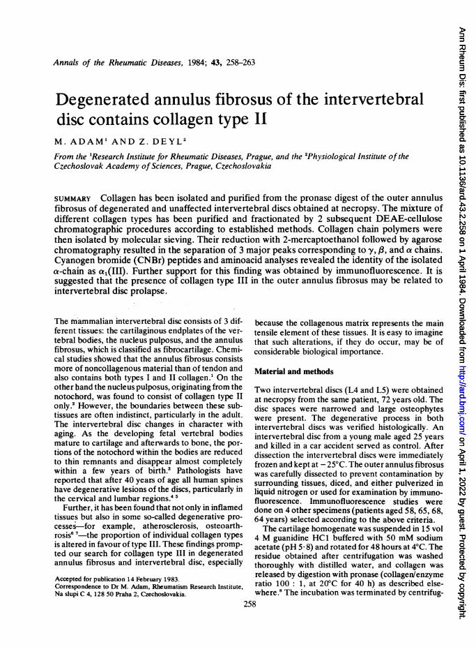
Fig. 1 The annulus fibrosus of degenerated humanintervertebral disc. Frozen section stained with anti-type IIIcollagen antibodies. Clusters of cells are surrounded withcollagen staining positively for collagen type III. Somecollagen fibres are also positively stained.
chromatography containing chain polymers of types Iand III collagen was reduced and alkylated beforebeing subjected to a second agarose run underoperating conditions as above.Reduction and alkylation. Reduction and carboxy-
methylation were performed with 2-mercoapto-ethanol followed by addition of iodoacetate.12The reaction mixture was desalted on Bio-gel P-2
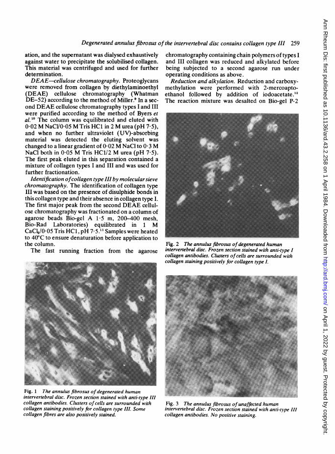
Fig. 2 The annulus fibrosus ofdegenerated humanintervertebral disc. Frozen section stained with anti-type Icollagen antibodies. Clusters ofcells are surrounded withcollagen staining positively for collagen type I.
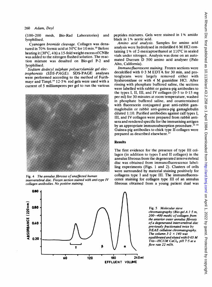
Fig. 3 The annulus fibrosus of unaffected humanintervertebral disc. Frozen section stained with anti-type IIIcollagen antibodies. No positive staining.
on April 1, 2022 by guest. P
rotected by copyright.http://ard.bm
j.com/
Ann R
heum D
is: first published as 10.1136/ard.43.2.258 on 1 April 1984. D
ownloaded from
260 Adam, Deyl
(100-200 mesh, Bio-Rad Laboratories) andlyophilised.Cyanogen bromide cleavage. Collagen was dena-
tured in 70% formic acid at 50°C for 10 min.3 Beforeheating it (30°C, 4 h) a 15-fold weight excess ofCNBrwas added to the nitrogen flushed solution. The reac-tion mixture was desalted on Bio-gel P-2 andlyophilised.Sodium dodecyl sulphate polyacrylamide gel elec-
trophoresis (SDS-PAGE). SDS-PAGE analyseswere performed according to the method of Furth-mayr and Timpl." 12-5% rod gels were used with acurrent of 5 milliamperes per gel to run the various
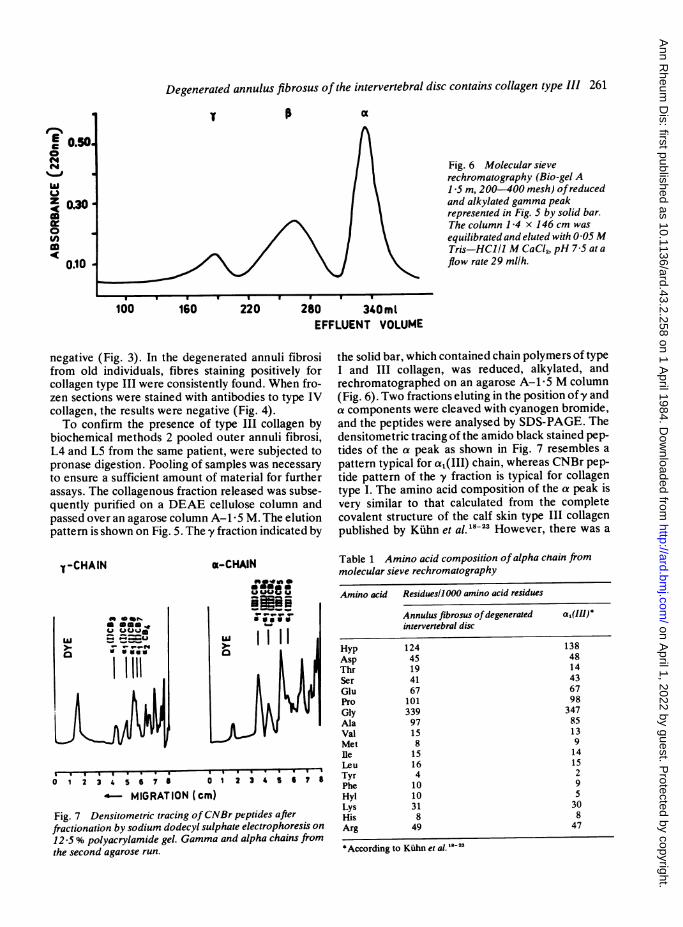
Fig. 4 The annulus fibrosus of unaffected humanintervertebral disc. Frozen section stained with anti-type IVcollagen antibodies. No positive staining.
00
ECo Q6O
z OAO
goIn< 0.20
peptides mixtures. Gels were stained in 1% amidoblack in 1% acetic acid.Amino acid analysis. Samples for amino acid
analysis were hydrolysed in redistilled 6 M HCl con-taining 1% of 2-mercaptoethanol at 11 0°C in sealedvials under nitrogen. Analysis was done on an auto-mated Durrum D 500 amino acid analyser (PaloAlto, California).Immunofluorescent staining. Frozen sections were
decalcified with 0 3 M EDTA for 30 min, and pro-teoglycans were largely removed either withhyaluronidase or with 4 M guanidine HCl. Afterrinsing with phosphate buffered saline, the sectionswere labelled with rabbit or guinea-pig antibodies tothe types I, II, III, and IV collagen (0-5 to 0-15 mgper ml) for 30 minutes at room temperature, washedin phosphate buffered saline, and counterstainedwith fluorescein conjugated goat anti-rabbit gam-maglobulin or rabbit anti-guinea-pig gamaglobulindiluted 1:10. Purified antibodies against calf types I,III, and IV collagen were prepared from rabbit anti-sera and rendered specific for the immunising antigenby an appropriate immunoabsorption procedure.15 16Guinea-pig antibodies to chick type II collagen wereprepared as described elsewhere.17
Results
The first evidence for the presence of type III col-lagen (in addition to types I and II collagen) in theannulus fibrosus from the degenerated intervertebraldisc was obtained from immunofluorescence label-ling experiments (Figs. 1 and 2). Clusters of cellswere surrounded by material staining positively forcollagens type I and type III. The immunofluores-cence staining for collagen type III of an annulusfibrosus obtained from a young patient dual was
Fig. 5 Molecular sievechromatography (Bio-gelA 1 5 m,200-400 mesh) ofcollagen fromthe anterior outer annulus fibrosisofa degenerated intervertebral discpreviously fractionated twice byDEAE cellulose chromatography.The column 3-2 x 140 wasequilibrated and eluted with 0 05MTris-HCI/M CaCl4, pH 7 5 at aflow rate 22 mllh.
120 180 240m1EFFLUENT VOLUME
on April 1, 2022 by guest. P
rotected by copyright.http://ard.bm
j.com/
Ann R
heum D
is: first published as 10.1136/ard.43.2.258 on 1 April 1984. D
ownloaded from
Degenerated annulus fibrosus of the intervertebral disc contains collagen type III 261
Fig. 6 Molecular sieverechromatography (Bio-gel A1 5 m, 200-400 mesh) ofreducedand alkylated gamma peakrepresented in Fig. 5 by solid bar.The column 1 4 x 146 cm was
equilibrated and eluted with 0 05 MTris-HCI11 M CaCl2, pH 7 5 at aflow rate 29 mllh.
100 160 220 280 340m1
EFFLUENT VOLUME
negative (Fig. 3). In the degenerated annuli fibrosifrom old individuals, fibres staining positively forcollagen type III were consistently found. When fro-zen sections were stained with antibodies to type IVcollagen, the results were negative (Fig. 4).To confirm the presence of type III collagen by
biochemical methods 2 pooled outer annuli fibrosi,L4 and L5 from the same patient, were subjected to
pronase digestion. Pooling of samples was necessary
to ensure a sufficient amount of material for furtherassays. The collagenous fraction released was subse-quently purified on a DEAE cellulose column andpassed over an agarose column A-1i 5 M. The elutionpattern is shown on Fig. 5. The -y fraction indicated by
the solid bar, which contained chain polymers of typeI and III collagen, was reduced, alkylated, andrechromatographed on an agarose A-1 5 M column(Fig. 6). Two fractions eluting in the position ofy anda components were cleaved with cyanogen bromide,and the peptides were analysed by SDS-PAGE. Thedensitometric tracing of the amido black stained pep-
tides of the a peak as shown in Fig. 7 resembles a
pattern typical for a,(III) chain, whereas CNBr pep-tide pattern of the y fraction is typical for collagentype I. The amino acid composition of the a peak isvery similar to that calculated from the completecovalent structure of the calf skin type III collagenpublished by Kuhn et al. 18-23 However, there was a
X1-CHAIN x-CHAINS__ _
Ue
liil
01 23567 01 234 6 7
MIGRATION (cm)
Fig. 7 Densitometric tracing ofCNBr peptides afterfractionation by sodium dodecyl sulphate electrophoresis on12 5 % polyacrylamide gel. Gamma and alpha chains fromthe second agarose run.
Table 1 Amino acid composition of alpha chain frommolecular sieve rechromatography
Amino acid Residuesll000 amino acid residues
Annulus fibrosus ofdegenerated al(Ill)*intervertebral disc
Hyp 124Asp 45Thr 19Ser 41Glu 67Pro 101Gly 339Ala 97Val 15Met 8Ile 15Leu 16Tyr 4Phe 10Hyl 10Lys 31His 8Arg 49
According to Kuhn et al. `-2'
A
(44(44
z<
0
(A
0.10
1384814436798
34785139141529530847
on April 1, 2022 by guest. P
rotected by copyright.http://ard.bm
j.com/
Ann R
heum D
is: first published as 10.1136/ard.43.2.258 on 1 April 1984. D
ownloaded from
262 Adam, Deyl
distinct difference in the content of hydroxylysine:the a1(III) chain of the annulus fibrosus contains 10hydroxylysine residues compared with about 5residues in calf skin collagen (Table 1). If the -y peakfrom the first agarose run from the young control wasreduced and alkylated and rechromatographed over
agarose again, and a peaks were absent (data notshown)-that is, collagen type III was not found.
Discussion
During this study the occurrence of collagen type IIIwas revealed in the degenerated outer anteriorannulus fibrosus, while in young unaffected controlsthis collagen type was not identified. Our data arequalitative because of the complex separation pro-cedure. A rough quantitation, however, showed thatthe /8 and a peaks from the second agarosechromatography (containing collagen type III)represent 5% of the total collagen present in thepronase digest. Evidence for the presence of collagentype III is based on the isolation of this protein and itscharacterisation by amino acid analysis and SDS-PAGE profile of cyanogen bromide peptides. Theseresults provide further support for the original evi-dence based on immunofluorescence techniques.
In connection with the finding of collagen type IIIin degenerated annulus fibrosus one may ask thequestion why this particular type of collagen appearsin the tissue and what consequences it may have formechanical function. The type and proportions ofcollagens present within connective tissue vary withage, development, and pathology. Under differentpathological circumstances-for example, acuteinflammation, osteoarthrosis, atherosclerosis-either a significant increase of collagen typeIII occurs or this type of collagen appears denovo.6 24 However, the absence of immunofluores-cence when anti-type IV collagen antibodies wereused on the annulus fibrosus of degenerated interver-tebral disc led us to conclude that the presence ofcollagen type III is not related to the invading capil-laries. One of the many theories why collagen type IIIis formed in the degenerated intervertebral disc isthat mechanical stress alters the metabolism of thedisc.'s tissue. Overloading can cause degenerativelesions, as was shown in experiments on rats. Thepressures involved are sometimes immense: the cal-culated load on disc L3-L4 is over 300 kg on lifting.25
It should be also emphasised that collagen type IIIforms fine reticular networks which are more extens-ible than those formed by collagen type I. This maybe related to prolapse of the intervertebrnl disc. Itshould be noted that Beard et al.,26 using an immuno-fluorescent technique, found collagen type III inscoliotic intervertebral discs. In idiopathic scoliosis
other mechanisms causing enhanced concentrationof collagen type III in annulus fibrosus may operate.
We are indebted to Professor K. Kuhn of the Max Planck Institutefor Biochemistry, Martinsried, Federal Republic of Germany, for hiskind support and useful discussions. We are also grateful to MsMarie Krabcova for her expert technical assistance.
References
1 Eyre D R, Muir H. Collagen polymorphism: two molecularspecies in pig intervertebral disc. FEBS Lett 1974; 42: 192-6.
2 Eyre D R, Muir H. Type I and type II collagen in intervertebraldisk: interchanging radial distribution in annulus fibrosus.Biochem J 1976; 157: 267-72.
3 Eyre D R, Muir H. Quantitative analysis of types I and IIcollagens in human intervertebral discs at various ages. BiochemBiophys Acta 1976; 492: 29-42.
4 De Palma A F, Rothman R H. The intervertebral disc. Philadel-phia: Saunders, 1970.
5 Happey F, Pearson Ch, Naylor A, Turner R L. The aging ofhuman intervertebral disk. Gerontologia 1969; IS: 174-82.
6 Aumailley M, Bricaud H. Collagen synthesis in organ culture ofnormal and atherosclerotic aortas. Atherosclerosis 1981; 39:1-9.
7 Adam M, Deyl Z, Miterova L. In: Deyl Z, Adam M, eds.Connective tissue research, chemistry, biology and physiology.New York: Liss, 1981: 195-207.
8 Adam M, Fietzek P, Kuhn K. Investigations on the reaction ofmetals with collagen in vivo. 2. The formation of cross-links inthe collagen of lathyritic rats after gold treatment in vivo. Eur JBiochem 1968; 3: 411-4.
9 Miller E J. Isolation and characterization of a collagen fromchick cartilage containing three identical chains. Biochemistry1971; 10: 1652-8.
10 Byers P H, McKenney K H, Lichtenstein J A, Martin G R.Preparation of type III procollagen and collagen from rat skin.Biochemistry 1974; 13: 5243-8.
11 Piez K A. Molecular weight determination of random coilpolypeptides from collagen by molecular sieve chromatography.Anal Biochem 1968; 26: 305-12.
12 Dixit S. Type IV collagens: isolation and characterization of twostructurally distinct collagen chains from bovine kidney. Eur JBiochem 1980; 106: 563-70.
13 Bornstein P, Piez K A. The nature of the intermolecular cross-links in collagen. The separation and characterization of pep-tides from the cross-links region of rat-skin collagen. Biochemis-try 1966; 5: 3460-73.
14 Furthmayr H, Timpl R. Characterization of collagen peptides bysodium dodecylsulfate-polyacrylamide electrophoresis. AnalBiochem 1971; 41: 510-6.
15 Nowack H, Gay S, Wick G, Becker U, Timpl R. Preparation anduse in immunohistology of antibodies specific for type I and typeIII collagen and procollagen.J ImmunolMeth 1976; 12: 117-24.
16 Timpl R, Glanville R W, Wick G, Martin G R. Immunochemicalstudies on basement membrane (type IV) collagens. Immunol-ogy 1979; 39: 109-16.
17 von der Mark H, von der Mark K, Gay S. Study of differentialcollagen synthesis during development of the chick embryo byimmunofluorescence I. Preparation ofcollagen type I and type IIspecific antibodies and their application to early stages of thechick embryo. Dev Biol 1976; 48: 237-49.
18 Allmann H, Fietzek P P, Glanville K W, Kuhn K. The covalentstructure of calf skin type III collagen. VI. The amino acidsequence of the carboxy terminal cyanogen bromide peptidea,(III)CB9B (position 928-1028). Hoppe Seylers Z PhysiolChem 1979; 360: 861-8.
on April 1, 2022 by guest. P
rotected by copyright.http://ard.bm
j.com/
Ann R
heum D
is: first published as 10.1136/ard.43.2.258 on 1 April 1984. D
ownloaded from
Degenerated annulus fibrosus of the intevertebral disc contains collagen type III 263
19 Bentz H, Fietzek P P, Kuhn K. Structure of calf skin type IIIcollagen III. The amino acid sequence of the cyanogen bromidepeptide a,(III) CB4 (position 403-551). Hoppe Seylers ZPhysiol Chem 1979; 360: 833-40.
20 Dewes H, Fietzek P P, Kuhn K. Structure of calf skin type IIIcollagen II. The amino acid sequence of the cyanogen bromidepeptidea,(I1I) CB1, 8,10,2 (positions 223-402).HoppeSeylersZ Physiol Chem 1979; 360: 821-32.
21 Dewes H, Fietzek P P, Kuhn K. Structure of calf skin type IIIcollagen V: the amino acid sequence of the cyanogen bromidepeptide a1(III)CB 9A (positions 789-927). Hoppe Seylers ZPhysiol Chem 1979; 360: 851-60.
22 Fietzek P P, Alimann H, Rauterberg J, Henkel W, Wachter E,Kuhn K. Structure of calf skin type III collagen I. The amino acid
sequence of the amino terminal region of the (III) chain (posi-tions 1-222). HoppeSeylersZPhysiol Chem 1979; 360: 807-20.
23 Lang H, Glanville R W, Fietzek P P, Kuhn K. Structure of calfskin type III collagen IV. The amino acid sequence of thecyanogen bromide peptide a,(III) CB5 (positions 552-788).Hoppe Seylers Z Physiol Chem 1979; 360: 841-50.
24 Adam M, Dostal D, Deyl Z. Collagen heterogeneity in systemicscleroderma and other diseases. J Clin Chem Clin Biochem1979; 17: 495-8.
25 Eyre D E. Biochemistry of the intervertebral disc. Int Rev Con-nect Tissue Res 1979; 8: 227-92.
26 Beard H K, Roberts S, O'Brien J P. Immunofluorescent stainingfor collagen and proteoglycan in normal and scoliotic interver-tebral discs. J Bone Joint Surg 1981; 63B: 529-34.
Book reviewClinical Pharmacology and Therapeutics Series. Vol.3. Anti-Rheumatic Drugs. Edited by Edward C. Hus-kisson. Pp. 762. £30-00. Praeger: Eastbourne. 1983.
Fifty-five contributors have prepared the 37 chapters whichmake up this book. To have prevailed upon all members ofthe team to toe the line in writing for a book in whichtopicality is essential represents a considerable feat ofeditorship, although the task was probably eased by the factthat about half of the authors are directly employed by thepharmaceutical industry and some of the others are knownto be closely associated with that influential world in oneway or another. Still, as Dr Huskisson remarks in his pre-face, no apology is really necessary for this: here is wheremuch of the information is readily accessible and where agreat amount of research is today carried out. Certainly aworkmanlike book has been the outcome.
Initial chapters on classification, the mode of action ofarachidonic acid peroxidation inhibitors, and drug interac-tions are followed by accounts of individual non-steroidalanti-inflammatory drugs (NSAIDs), analgesics, cortico-steroids, gold, penicillamine, other thiol compounds,levamisole, azathioprine, chlorambucil, antimalarials, and
synoviorthesis. Among the NSAIDs zomepirac is includedbut methrazone is omitted. The closing chapters consist ofviewpoints, necessarily with a personal bias, on the inte-grated drug treatment of rheumatoid arthritis, SLE, gout,and Paget's disease, with a final section on assessment forclinical trials.The editor has clearly allowed his colleagues considerable
freedom. This has resulted in disproportionate length (orbrevity) of the various chapters. For example, cortico-steroids receive 10 pages, indomethacin 14 pages and dic-lofenac sodium (hardly the last word in treatment) no fewerthan 50. Most of the contents are of course modern in theextreme, although there are one or two nice flashbacks, suchas the recommendation that gastrointestinal distress causedby colchicine (1-0 mg every 2 hours) should be treated with'a gastric sedative, such as tincture of camphorated opium'.The staff of the excellent pharmacy at Charing Cross Hos-pital inform me that this time-honoured remedy is no longerheld in stock and could be prepared only by very specialarrangement; it does, however, remain in the B.P. No doubtwe are the poorer from this loss of pharmacoavailability.The book is recommended as an admirable reference
work.J. T. SCOTT
on April 1, 2022 by guest. P
rotected by copyright.http://ard.bm
j.com/
Ann R
heum D
is: first published as 10.1136/ard.43.2.258 on 1 April 1984. D
ownloaded from
NotesRheumatology ResearchThe 5th EULAR Workshop on Rheumatology Researchwill take place at the Jolly Hotel Milano 2, Segrate, Milan,Italy, on 1-2 March 1985. The official language will beEnglish, and there will be no simultaneous translation.Preselected posters will be discussed in the 5 plenarysessions (4 min + 2 slides per poster). All abstracts shouldbe submitted before 31 December 1984 and will bepublished in Clinical and Experimental Rheumatology.Further information from Professor Raffaele Numo, Cat-tedra di Reumatologia, Policlinico, Piazza Giulio Cesare,70124 Bari, Italy.
Panhellenic CongressThe 9th Panhellenic Congress in Rheumatology will beheld in Athens at the Athenaeum Intercontinental Hotelon 22-24 November 1984. The topic is 'Advances inconnective tissue diseases.' Details from Dr John Lalos,4 Papadiamandopoulou Street, Athens 612, Greece.
Implant surgeryThe 15th Annual International Symposium on ImplantSurgery for the Hand and Upper Extremity will be held on25-27 October 1984 at the Blodgett Memorial MedicalCenter and Amway Grand Plaza Hotel, Grand Rapids,Michigan, USA. Details from Dr Alfred B. Swanson,Blodgett Professional Building, Suite 290, 1900 WealthyStreet, SE, Grand Rapids, Michigan 49506, USA.
Marisa Ara prizeThis prizc of 2 million Italian lire will he awarded to a
woman for research in rheumratology published within thelast five years. Entries must be rcceived by 31 July 1984.Informaition from Professor Carla Caialniello. Via Posillipo102-2, 80123 Naipoli. Italy.
CorrectionsThe title of the paper by M. Adam and Z. Deyl (AnnRheum Dis 1984; 43: 258-63) should have read: 'Degener-ated annulus fibrosus of the intervertebral disc containscollagen type III' (not type II). We regret the error.
In the paper by T. Pullar, J. A. Hunter, and H. A. Capellentitled 'Does second-line therapy affect the radiologicalprogression of rheumatoid arthritis?' Ann Rheum Dis1984; 43: 18-23 we regret that an error occurred on p. 22.The fourth (new) paragraph should read: 'Conversely,with the number in our study we would have required a
difference of over 10 between the change in the "control"and treatment radiograph scores to demonstrate a signifi-cantly beneficial drug effect. We were, however, only ableto demonstrate a difference of 3-87 between gold and"control" groups and only 3.3 between "control" andpenicillamine groups'.


![Adipose stem cells for intervertebral disc regeneration: current … · 2018. 10. 4. · such as nucleus pulposus cells [35, 36], annulus fibrosus cells [37], cartilagenous chondrocytes](https://img.pdfslide.net/doc/110x75/5fe16d83ab12386dd17eecf1/adipose-stem-cells-for-intervertebral-disc-regeneration-current-2018-10-4.jpg)