Embed Size (px)
Citation preview
www.elsevier.com/locate/ynbdi
Review
Degenerative and regenerative mechanisms governing
spinal cord injury
Christos Profyris, Surindar S. Cheema, DaWei Zang, Michael F. Azari,Kristy Boyle, and Steven Petratos*
Motor Neuron Disease and Paralysis Laboratory, Neural Injury and Repair Group, The Howard Florey Institute of Experimental Physiology and Medicine,
University of Melbourne, Parkville, Victoria 3010, Australia
Neurobiology of Disease 15 (2004) 415–436
Received 30 June 2003; revised 3 November 2003; accepted 14 November 2003
Spinal cord injury (SCI) is a major cause of disability, and at present,
there is no universally accepted treatment. The functional decline
following SCI is contributed to both direct mechanical injury and
secondary pathophysiological mechanisms that are induced by the
initial trauma. These mechanisms initially involve widespread haemor-
rhage at the site of injury and necrosis of central nervous system (CNS)
cellular components. At later stages of injury, the cord is observed to
display reactive gliosis. The actions of astrocytes as well as numerous
other cells in this response create an environment that is highly
nonpermissive to axonal regrowth. Also manifesting important effects
is the immune system. The early recruitment of neutrophils and at later
stages, macrophages to the site of insult cause exacerbation of injury.
However, at more chronic stages, macrophages and recruited T helper
cells may potentially be helpful by providing trophic support for
neuronal and non-neuronal components of the injured CNS. Within
this sea of injurious mechanisms, the oligodendrocytes appear to be
highly vulnerable. At chronic stages of SCI, a large number of
oligodendrocytes undergo apoptosis at sites that are distant to the
vicinity of primary injury. This leads to denudement of axons and
deterioration of their conductive abilities, which adds significantly to
functional decline. By indulging into the molecular mechanisms that
cause oligodendrocyte apoptosis and identifying potential targets for
therapeutic intervention, the prevention of this apoptotic wave will be
of tremendous value to individuals living with SCI.
D 2004 Elsevier Inc. All rights reserved.
Keywords: Spinal cord injury; Oligodendrocyte apoptosis; Wallerian
degeneration; Leukaemia inhibitory factor (LIF); Secondary demyelination;
Nogo
Introduction
Spinal cord injury (SCI) occurs in most countries at an annual
rate of 20–40 persons per million (Tator and Fehlings, 1991). In
the United States, this sums up to approximately 10,000 new cases
a year, which continuously adds to the nation’s estimated 200,000
0969-9961/$ - see front matter D 2004 Elsevier Inc. All rights reserved.
doi:10.1016/j.nbd.2003.11.015
* Corresponding author. Fax: +61-3-9348-1707.
E-mail address: [email protected] (S. Petratos).
Available online on ScienceDirect (www.sciencedirect.com.)
quadriplegics (Ropper, 2001; Tyor et al., 2002). The main causes
of trauma to the cord are motor-vehicle accidents, sports and
recreational activities, work-related accidents and falls at home.
More importantly, the majority of SCI victims are young and
otherwise healthy, who in addition to costing society up to
$200,000 each per year, suffer the impingement of life long
disability (Tyor et al., 2002). At present, there is no universally
accepted treatment for this condition (Beattie et al., 2002b).
The greater part of spinal cord injuries in civilian life arise from
fracture or dislocation of the vertebral column. Most commonly
this arises due to compression with flexion in the thoracic cord and
hyperextension or flexion in the cervical cord. Indicators increasing
the risk of SCI as a result of minimal trauma are preexisting
spondylosis, a congenital spinal canal stenosis, hypertrophied
ligamentum flavum and instability of apophyseal joints due to
rheumatoid arthritis (Ropper, 2001).
Manifestation of SCI has varying degrees and is wholly
dependent on the severity and level of injury to the cord. The rule
of thumb is that the higher the level of the lesion, the more severe
the consequences. In the case of high cervical injury, patients
require artificial respiration to stay alive. This type of injury also
leads to tetraplegia, impairment of function in pelvic organs and
loss in motor and sensory function of the arms, trunk and legs.
Injury to lower cord levels, depending again on the exact level,
may leave function in the upper limbs with impairment limited to
lower limbs. This phenomenon of paraplegia is restricted to
injuries of the thoracic or lumbar cord (Maynard et al., 1997;
Ropper, 2001).
Neuropathology of SCI
The pathological sequelae following acute SCI are divided into
two broad chronological events: the primary injury and the
secondary injury (Tator and Fehlings, 1991). The primary injury
encompasses the focal destruction of neural tissue caused by direct
mechanical trauma. This initial insult then instigates a progressive
wave of secondary injury, which via the activation of a barrage of
noxious pathophysiological mechanisms exacerbates the injury to
the spinal cord. As this leads to the destruction of axonal tracts left
C. Profyris et al. / Neurobiology of Disease 15 (2004) 415–436416
intact by the initial trauma it is a major impediment to functional
recovery after SCI (Beattie et al., 2002b; Schwartz and Fehlings,
2002; Tator, 1995; Tator and Fehlings, 1991).
Primary injury
Macroscopic considerations
To mimic the majority of mechanical events that lead to various
forms of human SCI, several experimental models have been
developed (Beattie et al., 2002b). The most commonly used model
is the contusion model (Tator, 1995; Young, 2002). This model
induces instantaneous mechanical deformation of the spinal cord
by dropping either a weight (Noble and Wrathall, 1985), an
impactor rod (Gruner, 1992) or an impounder with computer-
guided assistance (Bresnahan et al., 1987). Another model
employed in SCI research is the compression model; in this model,
injury is induced by applying either a weight or an aneurysm clip
to the spinal cord (Tator and Fehlings, 1991). This model aims to
add to that of the contusion model by replicating the persistence of
cord compression that is commonly observed in human SCI (Tator,
1995).
The shortcoming of both the contusion and compression
models, however, is that the devices they utilise are restricted for
use in large animals such as rats and felines (Young, 2002). As a
result, to utilise the abundance of genetic mutations engineered in
mice, mouse models of SCI are restricted to the use of central
nervous system (CNS) axotomy and extradural clip compression
(Joshi and Fehlings, 2002a,b; Kim et al., 2003; Simonen et al.,
2003; Zheng et al., 2003). The shortcoming of the CNS axotomy
model is that the injury induced is not an accurate portrayal of
mechanical events leading to human SCI.
Microscopic considerations
Mechanical forces applied at the site of primary injury shear
neuronal and endothelial cell membranes. This leads to a haemor-
rhagic zone of necrosis that predominantly localises to the gray
matter due to this region’s soft consistency and highly vascular
nature (Tator, 1995). Central localisation of haemorrhage also
arises due to uneven movement of tissue after injury. Tissue moves
predominantly rostral and caudal to the injury epicentre, with the
greatest movement and therefore shearing of neural cell mem-
branes and connections due to apposing forces is experienced at the
centre of the cord (Blight, 1988). As this leads to relatively little
movement at the surface of the cord, axons localised near the pia
experience minimal disturbance and usually survive to form a
subpial axonal rim. This is in great contrast to axons localised near
the gray matter, which are severely injured after SCI (Young,
2002).
Axonal durability after SCI also depends on the presence of
myelin. A contusion injury brought about by deformation of cells
beyond their capacity through distortion of their natural form can
cause diffuse axonal damage with large myelinated fibres being
most vulnerable (Blight, 1988). Myelinated axons are more vul-
nerable than unmyelinated ones because the longitudinal forces
stretching the fibres are concentrated at the nodes of Ranvier. This
is supported by the observation that axonal microtubule disruption
after SCI tends to be localised at the nodal regions (Maxwell,
1996). In experimental replication of SCI using the transient clip
compression model (15 s per compression), a greater amount of
myelin destruction per mm2 compared with gray matter destruction
was observed with an increasing extent of injury (larger area of
cord compressed, Gruner et al., 1996). This suggests a distinct
vulnerability of the white matter to the extent of compression
injury to the spinal cord. However, it has been clearly demonstrated
that under slow compression injury, there is a distinct sparing of the
large myelinated fibres with small unmyelinated fibres being
vulnerable (Blight and Decrescito, 1986). Furthermore, with the
persistence of compression on the spinal cord, there occur specific
molecular and cellular events that evolve into secondary injury
mechanisms.
Secondary injury
There exist striking pathophysiologic similarities between clin-
ical SCI and experimental models of SCI. As a result, the following
account is based primarily on findings in rat SCI models, which are
justifiably extrapolated to the human form of injury (Tator, 1995).
However, there are striking differences between the regulation of
the secondary events after SCI between animal strains and species
(Hausmann, 2003). Therefore, extrapolation from animal experi-
mental data to human pathophysiology requires caution.
Macroscopic considerations
Apart from gray matter haemorrhage, the primary injury
causes no gross damage to the spinal cord despite the direct
cell death occurring at the site of the lesion (Beattie et al.,
2002b). However, with the advent of secondary injurious mech-
anisms this picture changes drastically. The earliest sign of gross
change (within 2 h) is expansion of the haemorrhagic front at
the site of trauma and appearance of numerous diffuse petechial
haemorrhages (Tator, 1995; Tator and Fehlings, 1991). Shortly
thereafter (6 h), penumbra surrounds the primary lesion and
oedema ensues predominantly in the white matter (Guth et al.,
1999). In the first day following trauma (12–24 h), haemor-
rhagic fronts continue to enlarge and become more confluent.
Furthermore, at the vicinity of injury, the gray and white matter
lose their definition, become softer and swell due to increasing
oedema (Tator, 1995). Haemorrhage is still evident by 3 days;
however, by the eighth day post-injury it is totally absent and
the now much-expanded site of primary injury is affluent in
cellular debris (Beattie et al., 2002b). At 21 days post-injury, the
spinal cord has evolved clearly visible cavitations, which by 14
weeks coalesce to form large cystic regions. These regions are
surrounded by scar tissue originating mainly from glia and to a
lesser extent from the PNS (see The glial scar) (Beattie et al.,
2002b). In 30% of rats with a compression injury and in 10% of
clinical SCI cases (Wallace et al., 1987), the cystic regions
expand for considerable distances rostral and caudal to the
primary injury. This phenomenon, over many years, produces
the long-term syndrome of posttraumatic syringomyelia, which
enhances neurological deficit (Tator, 1995).
At more chronic stages of SCI, evolvement of pathophysiolog-
ical sequale leads to atrophy of the cord at the site of primary
injury. However, due to Wallerian degeneration in ascending and
descending tracts (see Axonal disruption and Wallerian degenera-
tion), cord atrophy can also be seen both rostral and caudal to the
site of initial trauma (Tator, 1995).
Microscopic considerations
A barrage of pathophysiological events that causes both necro-
sis and apoptosis govern the biology of secondary injury after acute
SCI (Beattie et al., 2002b).
C. Profyris et al. / Neurobiology of Disease 15 (2004) 415–436 417
Necrosis. Secondary injury mechanisms initiate a centripetal and
rostro-caudal necrotic wave that originates at the site of primary
injury. This wave, which is irreversible by 8 h, may spread for up
to two vertebral levels both above and below the initial lesion
(Ropper, 2001).
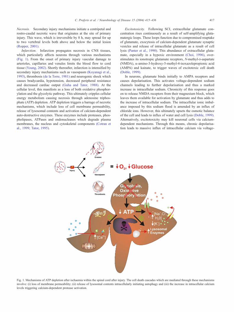
Infarction. Infarction propagates necrosis in CNS tissues,
which particularly affects neurons through various mechanisms
(Fig. 1). From the onset of primary injury vascular damage to
arterioles, capillaries and venules limits the blood flow to cord
tissue (Young, 2002). Shortly thereafter, infarction is intensified by
secondary injury mechanisms such as vasospasm (Koyanagi et al.,
1993), thrombosis (de la Torre, 1981) and neurogenic shock which
causes bradycardia, hypotension, decreased peripheral resistance
and decreased cardiac output (Guha and Tator, 1988). At the
cellular level, this manifests as a loss of both oxidative phosphor-
ylation and the glycolytic pathway. This ultimately cripples cellular
energy metabolism causing necrosis through adenosine triphos-
phate (ATP) depletion. ATP depletion triggers a barrage of necrotic
mechanisms, which include loss of cell membrane permeability,
release of lysosomal contents and activation of calcium-dependent
auto-destructive enzymes. These enzymes include proteases, phos-
pholipases, ATPases and endonucleases which degrade plasma
membranes, the nucleus and cytoskeletal components (Cotran et
al., 1999; Tator, 1995).
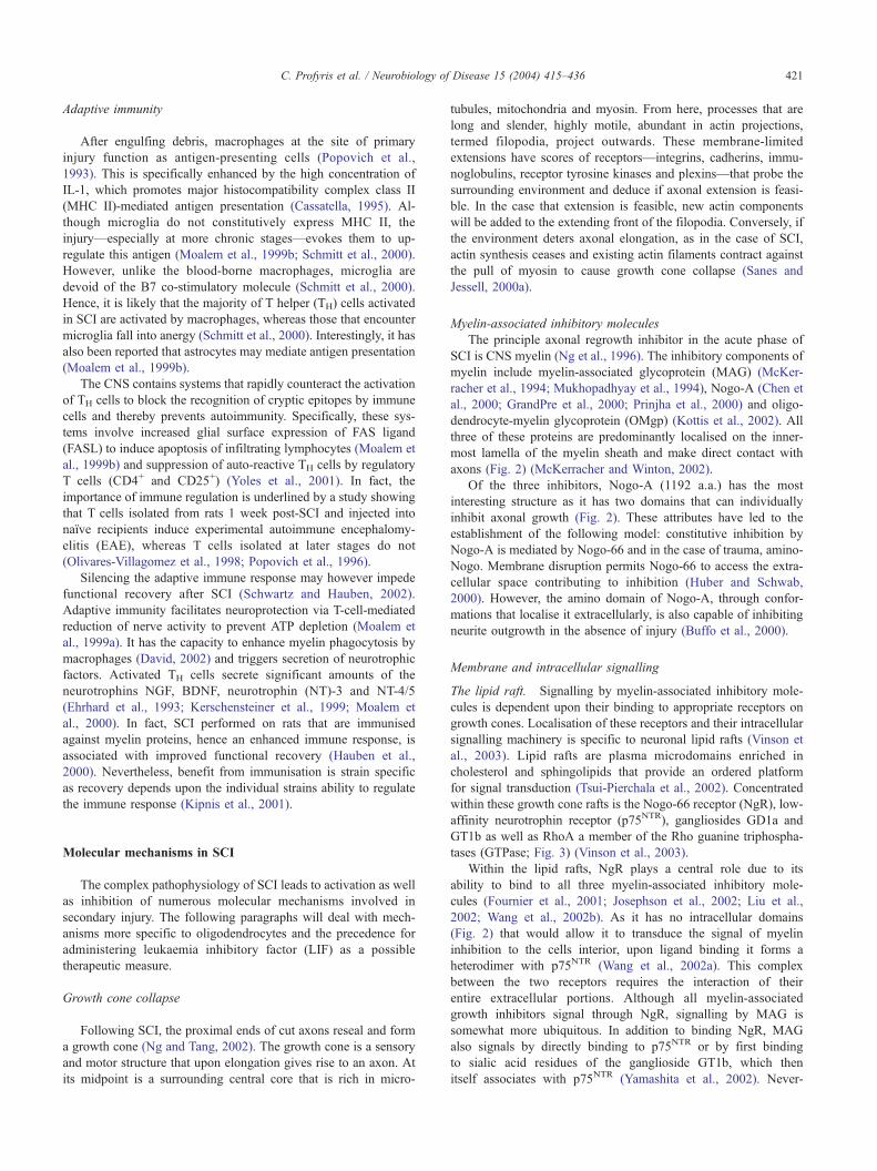
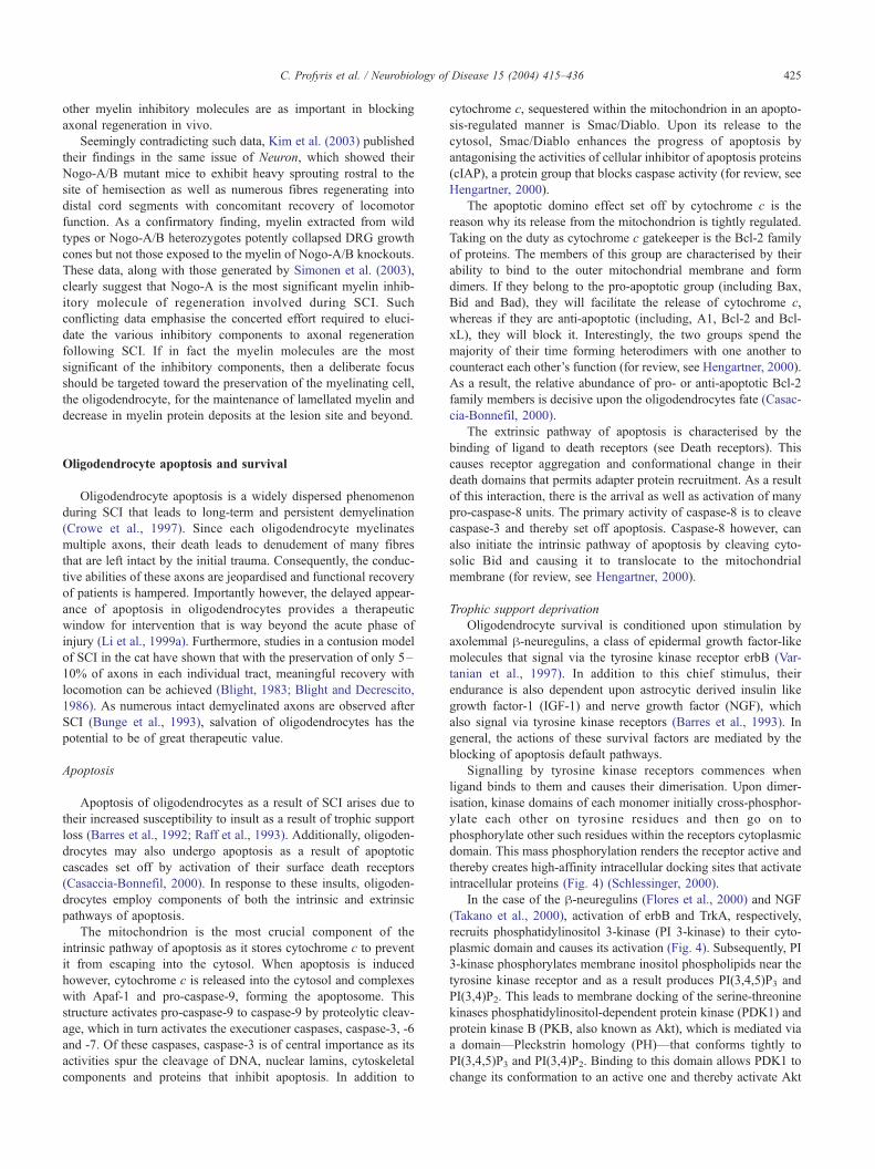
Fig. 1. Mechanisms of ATP depletion after ischaemia within the spinal cord after in
involve: (i) loss of membrane permeability; (ii) release of lysosomal contents intra
levels triggering calcium-dependent protease activation.
Excitotoxicity. Following SCI, extracellular glutamate con-
centration rises continuously as a result of self-amplifying gluta-
matergic loops. These loops function due to compromised reuptake
of glutamate, exocytosis of calcium-dependent glutamate synaptic
vesicles and release of intracellular glutamate as a result of cell
lysis (Panter et al., 1990). This abundance of extracellular gluta-
mate, especially in a hypoxic environment (Choi, 1996), over-
stimulates its ionotropic glutamate receptors, N-methyl-D-aspartate
(NMDA), a-amino-3-hydroxy-5-methyl-4-isoxazolepropionic acid
(AMPA) and kainate, to trigger waves of excitotoxic cell death
(Doble, 1999).
In neurons, glutamate binds initially to AMPA receptors and
causes depolarisation. This activates voltage-dependent sodium
channels leading to further depolarisation and thus a marked
increase in intracellular sodium. Chronicity of this response goes
on to release NMDA receptors from their magnesium block, which
leaves them available for activation by glutamate and thus adds to
the increase of intracellular sodium. The intracellular ionic imbal-
ance imposed by this sodium flood is amended by an influx of
chloride ions. However, this ultimately upsets the osmotic balance
of the cell and leads to influx of water and cell lysis (Doble, 1999).
Alternatively, excitotoxicity may kill neuronal cells via calcium-
dependent mechanisms. Through this means, chronic depolarisa-
tion leads to massive influx of intracellular calcium via voltage-
jury. The cell death cascades which are mediated through these mechanisms
cellularly initiating autophagy and (iii) the increase in intracellular calcium
C. Profyris et al. / Neurobiology of Disease 15 (2004) 415–436418
dependent calcium channels and opening of NMDA receptor
channels. This influx is further fortified by calcium mobilisation
from intracellular stores and reverse operation of the membrane
sodium/calcium exchanger. Consequently, this leads to activation
of auto-destructive calcium-dependent enzymes (see Infarction),
which in due course trigger cell death (Doble, 1999).
Following SCI, excitotoxic cell death also arises in CNS glia, of
which oligodendrocytes are the most vulnerable (McDonald et al.,
1998). As glial cells are void of NMDA receptors (Steinhauser and
Gallo, 1996), excitotoxicity in glia is prompted via AMPA and
kainate receptors. Importantly, both AMPA and kainate receptors in
differentiated oligodendrocytes are more permeable to calcium, than
those of neurons, due to alterations in their subunits (Puchalski et al.,
1994). This, together with the poor capacity of oligodendrocytes to
buffer calcium is the reason why these cells are highly susceptible to
excitotoxic cell death (Mattson et al., 1991; Matute et al., 2001).
Reperfusion injury. Reperfusion of tissue in the first few days
following SCI exacerbates tissue damage within the cord. During
ischaemia, xanthine dehydrogenase within endothelial cells under-
goes limited proteolysis. The resulting modified enzyme, xanthine
oxidase, unlike its original form, transfers electrons to molecular
oxygen. As a result, reexposure of endothelial cells to oxygen leads
to an enzymatic reaction that gives rise to reactive oxygen species
(ROS) (Cotran et al., 1999; Guth et al., 1999). This source of ROS
together with ROS from neutrophils (Carlson et al., 1998) and
necrotic cells (Tator, 1995) creates a potent stimulus for cell death.
ROS can induce damage to CNS cells by modifying their lipids,
proteins or nucleic acids. In plasma and organelle membranes, free
radicals cause peroxidation of lipids by attacking the double bonds
of unsaturated fatty acids. This lipid–radical interaction yields
peroxides, which due to their own reactivity can intensify mem-
brane damage. Protein modification is another means through
which ROS can induce death. This creates havoc within cells by
oxidising amino acid side chains, causing fragmentation of proteins
by backbone oxidation or forming protein–protein cross-linkages.
Finally, ROS can also react with thymine in both nuclear and
mitochondrial deoxyroribonucleic acid (DNA) to produce single
strand breaks (Cotran et al., 1999).
Apoptosis. Apoptosis is an important mediator of secondary
damage after SCI (Beattie et al., 2002b). It incurs its affects through
at least two phases: an initial phase, bywhich apoptosis accompanies
necrosis in the degeneration of multiple cell types and a later phase,
which is predominantly confined to white matter and involves
oligodendrocytes (see Oligodendrocyte apoptosis and survival)
and microglia (Beattie et al., 2000). Chronologically, apoptosis
initially occurs 6 h post-injury at the lesion centre and for several
days thereafter the number of apoptotic cells in this region rises
steadily. However, by 1 week the apoptotic count decreases and
there is now an increase in apoptotic death away from the site of
primary injury. This new apoptotic wave is predominantly localised
in the white matter and can arise at large distances from the lesion
centre (Crowe et al., 1997).
Under certain conditions, such as ischaemia and excitotoxicity,
apoptosis may arise as the emerging cell death pathway at the time
where necrosis is currently the most predominant degenerative
event following SCI (Zipfel et al., 2000).
Axonal disruption and Wallerian degeneration. Axonal patholo-
gy following SCI is profuse. As early as 15 min after injury there
exists periaxonal swelling with the myelin laminations peeling
away from each other. At this time point, there also exists myelin
rupture, and by 24 h, axonal contents can be observed in the
extracellular space. Neuronal axoplasms undergo transition as well;
they display a granular appearance with disarray of their neuro-
filaments and in many axons there is an unusual abundance of
intracellular organelles. As time progresses, other pathologic
features of injured axons, such as those of widespread demyelin-
ation and abortive growth cones, are increasingly observed. In fact,
by 24 h, a common phenomenon is the appearance of giant axons
(greater than 40 Am), which exhibit a combination of all the
aforementioned pathological features (Anthes et al., 1995).
These axonal changes are ultimately accompanied by Wallerian
degeneration, which in rodents lasts for several months and in
humans for years (David, 2002). Wallerian degeneration describes
the withering of axonal segments separated from their neuronal
soma. The process commences with the degeneration of separated
fibres, which is accompanied by fragmentation of their associated
myelin. It continues with the accumulation of resultant debris and
culminates with the phagocytosis of this debris by macrophages
and microglia (Sanes and Jessell, 2000b).
As the primary injury severs both ascending and descending
axonal tracts, Wallerian degeneration is present both rostral and
caudal to the initial lesion (Quencer, 2002). Furthermore, the sites
of degeneration have a strong correlation with areas of delayed
apoptosis. This has important implications, as most of these dying
cells are oligodendrocytes that have lost trophic support from
severed axons with which they were associated (see Trophic
support deprivation) (Warden et al., 2001).
The glial scar. As in any other CNS injury, SCI initiates reactive
gliosis. In this response, glial cells and non-CNS cells invade the
site of primary injury. By invading this site, they clear up debris
and wall it off to prevent secondary pathophysiological injurious
mechanisms from spreading. However, with this comes the unfor-
tunate burden of massive deposition of molecules that inhibits
axonal regrowth (Fawcett and Asher, 1999).
The glial orchestra. The earliest component that makes up the
glial scar is debris from myelin and oligodendrocytes, as well as
oligodendrocytes that survive the primary injury. This initial
component is followed by the activation and migration of micro-
glia (by 48 h), which is also accompanied by invasion of blood
borne macrophages (see Axonal regeneration). Up to this point, the
cellular components of the glial scar with the exception of myelin
debris are permissive to axon regrowth (Fawcett and Asher, 1999).
In fact, phagocytosis by microglia of myelin debris, which is
highly inhibitory to axon regrowth, is postulated to enhance axonal
sprouting (David, 2002).
Following these initial events, the glial scar begins to be
dominated by components that block axonal regrowth (Fawcett
and Asher, 1999). After the intrusion of phagocytes, oligodendro-
cyte precursors (35 days post-SCI) with the surface markers
NG2—an inhibitory chondroitin sulphate proteoglycan (see Axo-
nal regrowth inhibitory molecules)—and platelet-derived growth
factor receptor a (PDGFRa) intrude into the glial scar (Fawcett
and Asher, 1999). As the differentiation of these cells is inhibited
by CNS myelin, they may potentially differentiate into mature
oligodendrocytes at 2 weeks after injury, in areas were axons are
void of myelin (McTigue et al., 2001; Miller, 1999). At this same
time point, meningeal cells from the CNS surface also migrate into
the glial scar. These cells go on to re-form the disrupted glia
limitans that surrounds the CNS by making contact with astrocytes
walling off the primary lesion (Fawcett and Asher, 1999). Shortly
C. Profyris et al. / Neurobiology of Disease 15 (2004) 415–436 419
thereafter, multipotential progenitor cells from the spinal central
canal also invade into the site of primary injury; however, this is
not localised to the primary lesion as these cells can be observed
throughout the cord (Takahashi et al., 2003).
The end point in the evolution of the glial scar is migration and
proliferation of astrocytes. These cells up-regulate their production
of glial fibrillary acidic protein (GFAP), become hypertrophic and
appose many of their processes via gap junctions to the processes
of neighbouring counter partners. Upon their infiltration, astrocytes
isolate the site of primary injury by delineating the necrotic area
and enveloping it with a dense glial lining and basal lamina
(Bignami and Dahl, 1994). As astrocytes also fill the empty space
produced by the primary lesion they eventually form the bulk of
the glial scar (Fawcett and Asher, 1999). More importantly,
astrocytes attempt to reestablish the integrity of the lesioned
microenvironment to retain the function of nondamaged circuits.
They facilitate this by regulating the blood–spinal cord barrier
(BSCB), concentrations of excitatory amino acids (EAA), ionic
concentrations and secreting trophic factors and cytokines (Rab-
chevsky and Smith, 2001). Importantly, these cells are also the
producers of the majority of extracellular inhibitory axonal re-
growth molecules (see Axonal regrowth inhibitory molecules)
(Fawcett and Asher, 1999).
As a result of BSCB disruption, fibroblasts may also migrate
into the glial scar (Grimpe and Silver, 2002). At more chronic
stages (about 3 weeks), another non-CNS cell, the Schwann cell,
may migrate into the lesion as well. Interestingly, Schwann cells
have the ability to remyelinate denuded axons (Li et al., 1999b).
Axonal regrowth inhibitory molecules. The glial scar and
surrounding environment is awash with molecules that are potent
in making growth cones of injured neurons collapse (see Growth
cone collapse). These molecules together with the rigid structure of
the glial scar impose both a molecular and mechanical barrier to
axonal regrowth. In general, the inhibitory molecules that are up-
regulated after SCI can be divided into two categories: (1) myelin-
associated inhibitory molecules (see Myelin-associated inhibitory
molecules) and (2) molecules synthesised by cellular components
of the glial scar, which either remain on the surface of these cells or
are secreted in the extracellular matrix (Fawcett and Asher, 1999;
Morgenstern et al., 2002).
Inhibitory molecules synthesised by cellular components of the
glial scar are dichotomous (Grimpe and Silver, 2002). The first
category involves molecules that are solely inhibitory to neurite
outgrowth. Such molecules are proteoglycans, which are charac-
terised by unbranched repeating disaccharide units termed acid
glycosaminoglycans (AGAGs) connected via ether links to specific
amino acid residues or a protein core (Bannister, 1999). The
chondroitin sulphate proteoglycans (CSPGs), whose AGAGs are
of chondroitin sulphate, is the major inhibitory proteoglycan
subgroup. The members of this subgroup, which include NG2,
neurocan, versican, brevican and phosphocan, are believed to
inhibit neurite outgrowth by binding to the growth-promoting
molecule laminin and thereby preventing it from interacting with
its growth cone receptor integrin (Burg et al., 1996; Morgenstern et
al., 2002; Sanes and Jessell, 2000a). The negative affects of CSPGs
in SCI have been recently confirmed in vivo by Bradbury et al.
(2002). By administering an enzyme that degrades the AGAGs
from CSPGs, they were able to demonstrate both axonal regener-
ation and behavioural improvement in their lesioned animals.
The second category of inhibitory molecules has both inhibitory
and axonal regrowth-promoting effects. Within this category are
the tenascins (tenascin-R, tenascin-C and tenascin-X), large gly-
coproteins consisting of six subunits that makeup a stellate-like
structure. Many of the tenascins, as well as inhibiting neurite
outgrowth, are also able to bind most CSPGs and thereby retain
secreted CSPGs within the vicinity of the glial scar (Fawcett and
Asher, 1999; Grimpe and Silver, 2002). Other members within this
category are the netrins and semaphorins, which are molecules that
contribute to axonal guidance during development (Grimpe and
Silver, 2002).
The immune system in SCI
Inflammatory responses are of central importance in the path-
ogenesis of the acute and chronic phases of SCI. During these
phases, the CNS recruits both the innate and adaptive arms of
immunity (Hauben and Schwartz, 2003).
Innate immunity
The injured environment
The injured environment during the acute phase of SCI is
dominated by the presence of the pro-inflammatory cytokines
tumour necrosis factor a (TNFa), interleukin (IL)-1 and IL-6.
IL-1 is released immediately after injury by microglia (Allan and
Rothwell, 2001). Furthermore, within 15 min, its mRNA levels as
well as those of TNFa and IL-6 are increased in most cellular
components of the CNS (Yan et al., 2001). As both IL-1 and TNFa
can co-stimulate the expression level of each other, as well as that
of IL-6, the levels of all three cytokines increase rapidly (Pan et al.,
2002). Their levels peak after several hours, but by 24 h they are
barely detectable (Klusman and Schwab, 1997). Despite this drop,
the protein levels of TNFa continue to increase during the first
week following SCI (Tyor et al., 2002). This is attributed to
leukocyte infiltration (24 h post-SCI) and secretion of pro-inflam-
matory cytokines at the site of primary injury (Popovich and Jones,
2003). Further fortifying the armada of recruited cytokines is the
up-regulation of a saturable unidirectional transport system in the
blood–brain barrier (BBB) that is specific for TNFa. This allows
TNFa produced by peripheral trauma to enter and thus accumulate
within the CNS (Pan et al., 1999; Pan and Kastin, 2001).
Binding of both TNFa and IL-1 to their receptors causes them
to induce the inflammatory response by signalling through the
nuclear factor nB (NFnB) pathway (Allan and Rothwell, 2001). Anobserved increase of the pathways end product, NFnB, is observedin neurons, microglia and endothelial cells by 30 min post-SCI and
persists for at least 72 h (Bethea et al., 1998). This is crucial as
active NFnB stimulates the production of inflammatory mediators
such as ROS, cytokines, inducible nitric oxide synthase (iNOS),
prostaglandin (synthase-2, arachidonic acid, proteases and endo-
thelial cell adhesion molecules (CAMs) (Allan and Rothwell,
2001; Bethea et al., 1998; Kim et al., 2001). Interestingly, the
anti-inflammatory agent methylprednisolone, which is the only
drug therapy to be approved in SCI treatment, has been shown to
inhibit the NFnB pathway (Xu et al., 1998).
SCI also induces the expression of the anti-inflammatory
cytokine transforming growth factor-h (TGFh) (Tyor et al.,
2002). In contrast to pro-inflammatory cytokines, the expression
of TGFh is delayed. Although it is found at the lesion site within
24 h, its mRNA levels do not peak until 7 days later (Semple-
Rowland et al., 1995). TGF-h counteracts the effects of pro-
C. Profyris et al. / Neurobiology of Disease 15 (2004) 415–436420
inflammatory cytokines by down-regulating iNOS and decreasing
endothelial CAMs (Hamada et al., 1996; Kitamura, 1997).
Neutrophil recruitment
Up-regulation of endothelial CAMs [intracellular (I)-CAM,
vascular (V)-CAM, platelet endothelial (PE)-CAM, P- and E-
selectin] recruits leukocytes to the site of primary injury (Lee et
al., 2000b; McTigue et al., 1998; Schnell et al., 1999). In addition to
these factors, IL-8 and the CXC chemokine, cytokine-induced
neutrophil chemo-attractant 1 (CINC-1), both enhance neutrophil
recruitment and activation even further (Tonai et al., 2001). Con-
sequently, neutrophils adhere to postcapillary venules 6–12 h post-
SCI and by 24 h they migrate into the lesion site to phagocytose
debris (Guth et al., 1999; Taoka et al., 1997). Upon their entry, pro-
inflammatory mediators induce neutrophils to generate their own
cytokines as well as the production of proteases via the NFnBtranslocation pathway (McDonald et al., 1997). Such mediators
include matrix metalloproteinases (MMPs)—in particular MMP-
9—(Noble et al., 2002), and the cytokines TNFa, IL-1, IL-8 and
TGFh (Cassatella, 1995). These mediators loosen the extracellular
matrix to enhance extravasation of leukocytes, stimulate leukocyte
chemotaxis, activate glia and enhance neuronal damage (Carlson et
al., 1998). Furthermore, the activities of the superoxide dismutase
and neutrophil myeloperoxidase enzymes, which mediate the res-
piratory burst, are enhanced. As a result, the attachment of neu-
trophils to endothelium exacerbates reperfusion injury (see
Reperfusion injury) and thereby gives rise to the aforementioned
petechial haemorrhages (see Macroscopic considerations). The
activity of these cells however, is only transient as by 48 h their
recruitment declines (Guth et al., 1999; Taoka et al., 1997).
Macrophage/microglial activation
As neutrophil numbers decline, there is an increasing presence
of monocytes in the injured parenchyma, which by 72 h go on to
differentiate into macrophages. Their chemotaxis is dependent
upon CAMs as well as IL-8 and macrophage inflammatory
proteins (MIP) a and h (Bartholdi and Schwab, 1997; Cassatella,
1995). It has been demonstrated that after CNS injury there exist a
few haematogenously derived macrophages at the site of transec-
tion were the blood–brain barrier is disrupted (George and Griffin,
1994). Following their recruitment, macrophages—and micro-
glia—are activated by TNFa as well as by ligand binding to their
receptors complement receptor 3 (CR3) and mannose receptor
(Fitch et al., 1999). Interestingly, microglial cells are activated at
the site of primary injury at an even earlier time point (1 h) (Dusart
and Schwab, 1994). This results due to an elevated concentration
of extracellular ATP, which via a calcium-dependent mechanism
acutely up-regulates the microglial protein, allograft inflammatory
factor-1 (AIF-1), and thereby causes microglial activation (Schwab
et al., 2001; Tanaka and Koike, 2002). In addition to their acute
activation, microglia are also activated during later stages of SCI (5
days) at sites distant to the primary injury (Koshinaga and
Whittemore, 1995; Watanabe et al., 1999). This activation arises
predominantly in tracts of white matter undergoing Wallerian
degeneration (see Axonal disruption and Wallerian Degeneration)
(Frisen et al., 1994).
The activation of macrophages leads to the secretion of gluta-
mate, the pro-inflammatory cytokines TNFa, IL-1 and IL-6, and
activation of the respiratory burst enzyme inducible nitric oxide
synthase (iNOS) (Leskovar et al., 2000; Satake et al., 2000). In
addition, macrophage activation up-regulates cyclooxygenases,
which are key enzymes in the conversion of arachidonic acid—
plentiful in the injured cord—to prostanoids. Prostanoids have a
wide variety of immunomodulatory properties and have the poten-
tial to augment secondary injury (Schwab et al., 2000).
Upon activation, macrophages and microglia, like neutrophils,
phagocytose necrotic and apoptotic debris (Guth et al., 1999;
Taoka et al., 1997). However, unlike neutrophils, their activity is
sustained for longer periods. In fact, macrophage presence in rat
models of SCI does not decrease until 7 days post-injury and
microglial presence in degenerating tracts plateaus at between 2
and 4 weeks (Popovich et al., 1997). Finally, overactivation of
these cells can lead to their degeneration into ghost cells, lipid
engorged phagocytes, which remain in the spinal cord for months
to years after SCI (Popovich and Jones, 2003).
Due to the lack of specific markers, it has been extremely
difficult to differentiate invading macrophages from activated
microglia within the injured spinal cord. As a consequence, their
individual functional roles remain elusive. Recently, Popovich and
Hickey (2001) have used radiation bone marrow chimeric rats to
directly address this question. These investigators demonstrated
that the initial injury response (onset and plateau) occurring at the
3- and 7-day intervals post-injury, previously thought to be related
to haematogenously derived macrophage infiltration, were in fact
dominated by microglial-derived macrophages. These cells were
prevalent at the injury site and where active Wallerian degeneration
was occurring. Haematogenous macrophages were determined to
infiltrate the injury site slowly, appearing initially in dorsal gray
matter, subpial white matter and around branches of the ventral
spinal artery, eventually occupying nearly all of the gray matter by
7 days. These data signify a predominant association of macro-
phage infiltration and gray matter pathology, as well as a unique
association with Wallerian degeneration, oligodendrocyte death
and microglial activity.
Attenuation of innate immunity
Given the wide array of noxious mediators that are secreted by
neutrophils and macrophages, decreasing their presence has been
widely employed as a therapeutic approach in SCI models. Strat-
egies such as inhibition of MMPs (Noble et al., 2002), decreasing
the availability of CAMs (Taoka et al., 1997), elastase inhibition
(Tonai et al., 2001), inflammatory cytokine down-regulation (Xu et
al., 1998) and hampering the production of ROS and lipid perox-
idation (Hall, 1992) have all been employed with some success.
Despite this success, decreasing the presence of macrophages and
microglia at the locus of primary injury may potentially be harmful.
In addition to ridding the injured environment of myelin derived
axon-growth inhibitory molecules (see Myelin-associated inhibito-
ry molecules), macrophages and microglia can potentiate the
release of mediators that promote CNS repair (Nguyen et al.,
2002). These mediators, which include ciliary neurotrophic factor
(CNTF) (Herx et al., 2000), insulin-like growth factor 1 (IGF-1)
(Mason et al., 2001) and the neurotrophins, nerve growth factor
(NGF) (Heese et al., 1998) and brain-derived growth factor
(BDNF) (Kerschensteiner et al., 1999) are mainly released by
astrocytes in response to macrophage and microglial stimulation
by TNFa and IL-1 (Nguyen et al., 2002). Hence, after contributing
to the acute phase of injury, these phagocytes may potentially aid
regeneration at more chronic stages by secreting neuronal survival
and regeneration factors. Furthermore, TNFa via binding to TNF
receptor 2 (TNFR2) has been shown to promote re-myelination of
demyelinated axons (Arnett et al., 2001).
ogy of Disease 15 (2004) 415–436 421
Adaptive immunity
After engulfing debris, macrophages at the site of primary
injury function as antigen-presenting cells (Popovich et al.,
1993). This is specifically enhanced by the high concentration of
IL-1, which promotes major histocompatibility complex class II
(MHC II)-mediated antigen presentation (Cassatella, 1995). Al-
though microglia do not constitutively express MHC II, the
injury—especially at more chronic stages—evokes them to up-
regulate this antigen (Moalem et al., 1999b; Schmitt et al., 2000).
However, unlike the blood-borne macrophages, microglia are
devoid of the B7 co-stimulatory molecule (Schmitt et al., 2000).
Hence, it is likely that the majority of T helper (TH) cells activated
in SCI are activated by macrophages, whereas those that encounter
microglia fall into anergy (Schmitt et al., 2000). Interestingly, it has
also been reported that astrocytes may mediate antigen presentation
(Moalem et al., 1999b).
The CNS contains systems that rapidly counteract the activation
of TH cells to block the recognition of cryptic epitopes by immune
cells and thereby prevents autoimmunity. Specifically, these sys-
tems involve increased glial surface expression of FAS ligand
(FASL) to induce apoptosis of infiltrating lymphocytes (Moalem et
al., 1999b) and suppression of auto-reactive TH cells by regulatory
T cells (CD4+ and CD25+) (Yoles et al., 2001). In fact, the
importance of immune regulation is underlined by a study showing
that T cells isolated from rats 1 week post-SCI and injected into
naıve recipients induce experimental autoimmune encephalomy-
elitis (EAE), whereas T cells isolated at later stages do not
(Olivares-Villagomez et al., 1998; Popovich et al., 1996).
Silencing the adaptive immune response may however impede
functional recovery after SCI (Schwartz and Hauben, 2002).
Adaptive immunity facilitates neuroprotection via T-cell-mediated
reduction of nerve activity to prevent ATP depletion (Moalem et
al., 1999a). It has the capacity to enhance myelin phagocytosis by
macrophages (David, 2002) and triggers secretion of neurotrophic
factors. Activated TH cells secrete significant amounts of the
neurotrophins NGF, BDNF, neurotrophin (NT)-3 and NT-4/5
(Ehrhard et al., 1993; Kerschensteiner et al., 1999; Moalem et
al., 2000). In fact, SCI performed on rats that are immunised
against myelin proteins, hence an enhanced immune response, is
associated with improved functional recovery (Hauben et al.,
2000). Nevertheless, benefit from immunisation is strain specific
as recovery depends upon the individual strains ability to regulate
the immune response (Kipnis et al., 2001).
C. Profyris et al. / Neurobiol
Molecular mechanisms in SCI
The complex pathophysiology of SCI leads to activation as well
as inhibition of numerous molecular mechanisms involved in
secondary injury. The following paragraphs will deal with mech-
anisms more specific to oligodendrocytes and the precedence for
administering leukaemia inhibitory factor (LIF) as a possible
therapeutic measure.
Growth cone collapse
Following SCI, the proximal ends of cut axons reseal and form
a growth cone (Ng and Tang, 2002). The growth cone is a sensory
and motor structure that upon elongation gives rise to an axon. At
its midpoint is a surrounding central core that is rich in micro-
tubules, mitochondria and myosin. From here, processes that are
long and slender, highly motile, abundant in actin projections,
termed filopodia, project outwards. These membrane-limited
extensions have scores of receptors—integrins, cadherins, immu-
noglobulins, receptor tyrosine kinases and plexins—that probe the
surrounding environment and deduce if axonal extension is feasi-
ble. In the case that extension is feasible, new actin components
will be added to the extending front of the filopodia. Conversely, if
the environment deters axonal elongation, as in the case of SCI,
actin synthesis ceases and existing actin filaments contract against
the pull of myosin to cause growth cone collapse (Sanes and
Jessell, 2000a).
Myelin-associated inhibitory molecules
The principle axonal regrowth inhibitor in the acute phase of
SCI is CNS myelin (Ng et al., 1996). The inhibitory components of
myelin include myelin-associated glycoprotein (MAG) (McKer-
racher et al., 1994; Mukhopadhyay et al., 1994), Nogo-A (Chen et
al., 2000; GrandPre et al., 2000; Prinjha et al., 2000) and oligo-
dendrocyte-myelin glycoprotein (OMgp) (Kottis et al., 2002). All
three of these proteins are predominantly localised on the inner-
most lamella of the myelin sheath and make direct contact with
axons (Fig. 2) (McKerracher and Winton, 2002).
Of the three inhibitors, Nogo-A (1192 a.a.) has the most
interesting structure as it has two domains that can individually
inhibit axonal growth (Fig. 2). These attributes have led to the
establishment of the following model: constitutive inhibition by
Nogo-A is mediated by Nogo-66 and in the case of trauma, amino-
Nogo. Membrane disruption permits Nogo-66 to access the extra-
cellular space contributing to inhibition (Huber and Schwab,
2000). However, the amino domain of Nogo-A, through confor-
mations that localise it extracellularly, is also capable of inhibiting
neurite outgrowth in the absence of injury (Buffo et al., 2000).
Membrane and intracellular signalling
The lipid raft. Signalling by myelin-associated inhibitory mole-
cules is dependent upon their binding to appropriate receptors on
growth cones. Localisation of these receptors and their intracellular
signalling machinery is specific to neuronal lipid rafts (Vinson et
al., 2003). Lipid rafts are plasma microdomains enriched in
cholesterol and sphingolipids that provide an ordered platform
for signal transduction (Tsui-Pierchala et al., 2002). Concentrated
within these growth cone rafts is the Nogo-66 receptor (NgR), low-
affinity neurotrophin receptor (p75NTR), gangliosides GD1a and
GT1b as well as RhoA a member of the Rho guanine triphospha-
tases (GTPase; Fig. 3) (Vinson et al., 2003).
Within the lipid rafts, NgR plays a central role due to its
ability to bind to all three myelin-associated inhibitory mole-
cules (Fournier et al., 2001; Josephson et al., 2002; Liu et al.,
2002; Wang et al., 2002b). As it has no intracellular domains
(Fig. 2) that would allow it to transduce the signal of myelin
inhibition to the cells interior, upon ligand binding it forms a
heterodimer with p75NTR (Wang et al., 2002a). This complex
between the two receptors requires the interaction of their
entire extracellular portions. Although all myelin-associated
growth inhibitors signal through NgR, signalling by MAG is
somewhat more ubiquitous. In addition to binding NgR, MAG
also signals by directly binding to p75NTR or by first binding
to sialic acid residues of the ganglioside GT1b, which then
itself associates with p75NTR (Yamashita et al., 2002). Never-
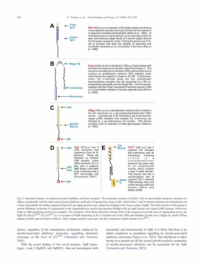
Fig. 2. Structural features of myelin-associated inhibitors and their receptors. The molecular structure of MAG, with its extracellular interactive domain (A)
differs considerably with the other major myelin inhibitory molecule of regeneration, Nogo-A (B), which both C- and N-terminus domains are internalised, yet
a short extracellular 66-residue segment (Nogo-66) can signal growth cone collapse by binding to the Nogo receptor (NgR). The third member of this group of
myelin inhibitory molecules of regeneration is the oligodendrocyte myelin glycoprotein (OMgp) with an eight leucine-rich repeat (LRR) domain, which also
binds to NgR producing growth cone collapse. The receptors, which these molecules interact with on the progressive growth cone of regenerating axons, are
NgR (D) and p75NTR (E). p75NTR is a co-receptor of NgR interacting at the C-terminal end of the LRR and mediates growth cone collapse by small GTPase
adapter proteins and activation of RhoA. These adaptor proteins associate with the cytoplasmic death domain of p75NTR.
C. Profyris et al. / Neurobiology of Disease 15 (2004) 415–436422
theless, regardless of the transduction mechanism employed by
myelin-associated inhibitory molecules, signalling ultimately
converges at the level of p75NTR (Yamashita and Tohyama,
2003).
With the recent finding of two novel proteins—NgR homo-
logue 1 and 2 (NgRH1 and NgRH2)—that are homologous both
structurally and biochemically to NgR, it is likely that there is an
added complexity to membrane signalling by myelin-associated
inhibitory molecules (Pignot et al., 2003). This likelihood is rather
strong as at present not all the axonal growth restrictive properties
of myelin-associated inhibitors can be accounted for by NgR
(Yamashita and Tohyama, 2003).
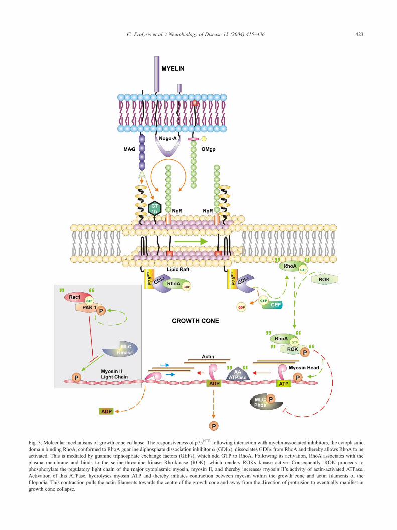
Fig. 3. Molecular mechanisms of growth cone collapse. The responsiveness of p75NTR following interaction with myelin-associated inhibitors, the cytoplasmic
domain binding RhoA, conformed to RhoA guanine diphosphate dissociation inhibitor a (GDIa), dissociates GDIa from RhoA and thereby allows RhoA to be
activated. This is mediated by guanine triphosphate exchange factors (GEFs), which add GTP to RhoA. Following its activation, RhoA associates with the
plasma membrane and binds to the serine-threonine kinase Rho-kinase (ROK), which renders ROKs kinase active. Consequently, ROK proceeds to
phosphorylate the regulatory light chain of the major cytoplasmic myosin, myosin II, and thereby increases myosin II’s activity of actin-activated ATPase.
Activation of this ATPase, hydrolyses myosin ATP and thereby initiates contraction between myosin within the growth cone and actin filaments of the
filopodia. This contraction pulls the actin filaments towards the centre of the growth cone and away from the direction of protrusion to eventually manifest in
growth cone collapse.
C. Profyris et al. / Neurobiology of Disease 15 (2004) 415–436 423
gy of Disease 15 (2004) 415–436
Intracellular signalling
Retraction via contractility. The responsiveness of p75NTR fol-
lowing interaction with myelin-associated inhibitors induces con-
formational change of its intracellular domains fifth a helical
loop (Yamashita and Tohyama, 2003). In the absence of inhib-
itory signalling, this domain binds RhoA that is conformed to
RhoA guanine diphosphate dissociation inhibitor a (GDIa) (Fig.
3). GDIa-bound RhoA is inactive as GDIa prevents the release
of GDP from RhoA and thereby binding of GTP, which is
required for RhoAs activation. Once MAG, Nogo or OMgp bind
to their targets, however, p75NTR dissociates GDIa from RhoA
and thereby allows RhoA to be activated. This is mediated by
guanine triphosphate exchange factors (GEFs), which add GTP
to RhoA (Yamashita and Tohyama, 2003). Following its activa-
tion, RhoA associates with the plasma membrane and binds to
the serine-threonine kinase Rho-kinase (ROK), which renders
ROKs kinase active (Leung et al., 1996). Consequently, ROK
proceeds to phosphorylate the regulatory light chain of the major
cytoplasmic myosin, myosin II, and thereby increases myosin
II’s activity of actin-activated ATPase. Activation of this ATPase
hydrolyses myosin ATP and thereby initiates contraction be-
tween myosin within the growth cone and actin filaments of the
filopodia. This contraction pulls the actin filaments towards the
centre of the growth cone and away from the direction of
protrusion to eventually manifest in growth cone collapse
(Amano et al., 1996). Interestingly, ROK also phosphorylates
the myosin light chain phosphatase and as a result inhibits its
activity. Since this phosphatase is unable to de-phosphorylate the
ATPase that ROK phosphorylated in the first place, the likeli-
hood of growth cone collapse is enhanced even further (Kimura
et al., 1996).
Whilst active RhoA GTPase is up-regulated during myelin
contact-mediated growth cone collapse, another member in the
Rho family of GTPases, Rac1, is down-regulated (Niederost et al.,
2002). Rac1 is down-regulated as its activities mediate growth
cone extension (Fig. 3, gray inset). It accomplishes this by binding
to the serine-threonine kinase p21-activated kinase 1 (PAK1),
which auto-phosphorylates upon their interaction (Manser et al.,
1995). Once activated, PAK1 inhibits the myosin light chain
kinase, resulting in decreased phosphorylation of the myosin light
chain, whose phosphorylation is a necessity if contractility between
myosin and actin is to ensue. Hence, by down-regulation of Rac1,
the mechanisms leading to contraction by RhoA remain unopposed
(Sanders et al., 1999).
Retraction via aberrant actin polymerisation. Apart from affect-
ing the dynamics of growth cone contractility, the Rho GTPases
also influence the kinetics of actin filament assembly. In myelin-
mediated growth cone collapse, this assembly is drastically altered
by both RhoA and Rac1 (Niederost et al., 2002).
Via the RhoA pathway, Lin-11, Isl-1 and Mec-3 kinase (LIMK)
is phosphorylated and activated by ROK (Maekawa et al., 1999).
Once active, LIMK has the ability to inactivate, by phosphoryla-
tion, the actin de-polymerising protein cofilin. This leads to a
shortage of actin monomers and thereby halts the extension of the
growth cone (Arber et al., 1998). Furthermore, it has been
hypothesised that the initial phosphorylation of cofilin leads to
activation via another pathway of a phosphatase that restores
cofilin activity. This reactivation however is unremitting and leads
to excessive actin depolymerisation causing growth cone collapse
(Niederost et al., 2002).
C. Profyris et al. / Neurobiolo424
Inhibition of Rac1 activity is instrumental in modulating the
kinetics of actin assembly in a manner that causes abortive
axon sprouting. Normally, Rac1 enhances the activity of its
effector phosphatidylinositol-4-phosphate 5-kinase (PI4P5K),
which generates phosphatidylinositol-4,5,-bisphosphate (PIP2)
(Tolias et al., 1995, 1998). Once produced, PIP2 localises at
lamellipodia and binds the control region of Neural Wiskott
Aldrich syndrome protein (N-WASP) to stimulate its association
with the Arp 2/3 complex. Once associated, this seven-protein
complex nucleates the formation of new actin filaments and is
thereby responsible for growth cone elongation (Rohatgi et al.,
2000). Remarkably, PIP2 further promotes actin assembly by
inhibiting capping protein, which caps filament ends to prevent
their elongation. Hence, inhibition of this molecular cascade by
down-regulation of the original effector, Rac1, enhances the
probability of growth cone collapse (Janmey and Stossel, 1987;
Meyer and Feldman, 2002).
Axonal regeneration
Elucidation of the dynamics involved in abortive axonal sprout-
ing has brought about exciting progress in finding new ways to
stimulate axonal regeneration. Strategies that block myelin-associ-
ated inhibitory molecules and their receptors have been employed
with some success. For instance, rats having undergone SCI and
then administered antibodies against myelin inhibitors (IN-1: an
antibody against Nogo-A, -B and -C) have enhanced functional
recovery and considerable axonal regeneration at the lesion site
(Thallmair et al., 1998). Other strategies, which aim to promote
axonal regeneration by blocking intracellular targets promoting
abortive sprouting, have also been employed. In this case, success
has come about by blocking the activity of ROK with the analogue
Y27632 (Dergham et al., 2002). Although axonal regeneration will
not restore the initial neuroanatomical wiring before SCI, it does
lead to restoration of lost function possibly by enhancing rewiring
of local circuitry (Schwab, 2002).
Nogo mutants and SCI
Considering the success of the antibody studies described above
in the enhancement of the regenerative capacities of severed axons
with subsequent functional improvements in these animals (Thall-
mair et al., 1998), it was of some surprise that three recent
independent studies generated conflicting data on the regenerative
capacities in Nogo-mutant mice following SCI (Kim et al., 2003;
Simonen et al., 2003; Zheng et al., 2003). One of these groups
generated two lines of mutant mice, one lacking the isoforms
Nogo-A and -B but not -C, and the other lacking all three isoforms
(Nogo-A/B/C mutants) (Zheng et al., 2003). Despite the Nogo-A/B
mutant showing decreased inhibitory activity in neuritic outgrowth
assays in culture, anterograde labelling experiments of descending
corticospinal fibres in vivo following dorsal hemisection in mouse
spinal cords revealed no real increase in regeneration or sprouting
(Zheng et al., 2003). Furthermore, CNS myelin from Nogo-A/B
mutant mice was still inhibitory in culture. However, when the C
isoform of Nogo was deleted from these animals as well, the Nogo-
A/B/C mutants on average exhibited slightly better locomotor
function without statistical significance among the groups. These
data collectively would suggest that animals without expression of
Nogo-A and -B do not have an increased ability to regenerate
axons following the induction of Wallerian degeneration and that
C. Profyris et al. / Neurobiology of Disease 15 (2004) 415–436 425
other myelin inhibitory molecules are as important in blocking
axonal regeneration in vivo.
Seemingly contradicting such data, Kim et al. (2003) published
their findings in the same issue of Neuron, which showed their
Nogo-A/B mutant mice to exhibit heavy sprouting rostral to the
site of hemisection as well as numerous fibres regenerating into
distal cord segments with concomitant recovery of locomotor
function. As a confirmatory finding, myelin extracted from wild
types or Nogo-A/B heterozygotes potently collapsed DRG growth
cones but not those exposed to the myelin of Nogo-A/B knockouts.
These data, along with those generated by Simonen et al. (2003),
clearly suggest that Nogo-A is the most significant myelin inhib-
itory molecule of regeneration involved during SCI. Such
conflicting data emphasise the concerted effort required to eluci-
date the various inhibitory components to axonal regeneration
following SCI. If in fact the myelin molecules are the most
significant of the inhibitory components, then a deliberate focus
should be targeted toward the preservation of the myelinating cell,
the oligodendrocyte, for the maintenance of lamellated myelin and
decrease in myelin protein deposits at the lesion site and beyond.
Oligodendrocyte apoptosis and survival
Oligodendrocyte apoptosis is a widely dispersed phenomenon
during SCI that leads to long-term and persistent demyelination
(Crowe et al., 1997). Since each oligodendrocyte myelinates
multiple axons, their death leads to denudement of many fibres
that are left intact by the initial trauma. Consequently, the conduc-
tive abilities of these axons are jeopardised and functional recovery
of patients is hampered. Importantly however, the delayed appear-
ance of apoptosis in oligodendrocytes provides a therapeutic
window for intervention that is way beyond the acute phase of
injury (Li et al., 1999a). Furthermore, studies in a contusion model
of SCI in the cat have shown that with the preservation of only 5–
10% of axons in each individual tract, meaningful recovery with
locomotion can be achieved (Blight, 1983; Blight and Decrescito,
1986). As numerous intact demyelinated axons are observed after
SCI (Bunge et al., 1993), salvation of oligodendrocytes has the
potential to be of great therapeutic value.
Apoptosis
Apoptosis of oligodendrocytes as a result of SCI arises due to
their increased susceptibility to insult as a result of trophic support
loss (Barres et al., 1992; Raff et al., 1993). Additionally, oligoden-
drocytes may also undergo apoptosis as a result of apoptotic
cascades set off by activation of their surface death receptors
(Casaccia-Bonnefil, 2000). In response to these insults, oligoden-
drocytes employ components of both the intrinsic and extrinsic
pathways of apoptosis.
The mitochondrion is the most crucial component of the
intrinsic pathway of apoptosis as it stores cytochrome c to prevent
it from escaping into the cytosol. When apoptosis is induced
however, cytochrome c is released into the cytosol and complexes
with Apaf-1 and pro-caspase-9, forming the apoptosome. This
structure activates pro-caspase-9 to caspase-9 by proteolytic cleav-
age, which in turn activates the executioner caspases, caspase-3, -6
and -7. Of these caspases, caspase-3 is of central importance as its
activities spur the cleavage of DNA, nuclear lamins, cytoskeletal
components and proteins that inhibit apoptosis. In addition to
cytochrome c, sequestered within the mitochondrion in an apopto-
sis-regulated manner is Smac/Diablo. Upon its release to the
cytosol, Smac/Diablo enhances the progress of apoptosis by
antagonising the activities of cellular inhibitor of apoptosis proteins
(cIAP), a protein group that blocks caspase activity (for review, see
Hengartner, 2000).
The apoptotic domino effect set off by cytochrome c is the
reason why its release from the mitochondrion is tightly regulated.
Taking on the duty as cytochrome c gatekeeper is the Bcl-2 family
of proteins. The members of this group are characterised by their
ability to bind to the outer mitochondrial membrane and form
dimers. If they belong to the pro-apoptotic group (including Bax,
Bid and Bad), they will facilitate the release of cytochrome c,
whereas if they are anti-apoptotic (including, A1, Bcl-2 and Bcl-
xL), they will block it. Interestingly, the two groups spend the
majority of their time forming heterodimers with one another to
counteract each other’s function (for review, see Hengartner, 2000).
As a result, the relative abundance of pro- or anti-apoptotic Bcl-2
family members is decisive upon the oligodendrocytes fate (Casac-
cia-Bonnefil, 2000).
The extrinsic pathway of apoptosis is characterised by the
binding of ligand to death receptors (see Death receptors). This
causes receptor aggregation and conformational change in their
death domains that permits adapter protein recruitment. As a result
of this interaction, there is the arrival as well as activation of many
pro-caspase-8 units. The primary activity of caspase-8 is to cleave
caspase-3 and thereby set off apoptosis. Caspase-8 however, can
also initiate the intrinsic pathway of apoptosis by cleaving cyto-
solic Bid and causing it to translocate to the mitochondrial
membrane (for review, see Hengartner, 2000).
Trophic support deprivation
Oligodendrocyte survival is conditioned upon stimulation by
axolemmal h-neuregulins, a class of epidermal growth factor-like
molecules that signal via the tyrosine kinase receptor erbB (Var-
tanian et al., 1997). In addition to this chief stimulus, their
endurance is also dependent upon astrocytic derived insulin like
growth factor-1 (IGF-1) and nerve growth factor (NGF), which
also signal via tyrosine kinase receptors (Barres et al., 1993). In
general, the actions of these survival factors are mediated by the
blocking of apoptosis default pathways.
Signalling by tyrosine kinase receptors commences when
ligand binds to them and causes their dimerisation. Upon dimer-
isation, kinase domains of each monomer initially cross-phosphor-
ylate each other on tyrosine residues and then go on to
phosphorylate other such residues within the receptors cytoplasmic
domain. This mass phosphorylation renders the receptor active and
thereby creates high-affinity intracellular docking sites that activate
intracellular proteins (Fig. 4) (Schlessinger, 2000).
In the case of the h-neuregulins (Flores et al., 2000) and NGF
(Takano et al., 2000), activation of erbB and TrkA, respectively,
recruits phosphatidylinositol 3-kinase (PI 3-kinase) to their cyto-
plasmic domain and causes its activation (Fig. 4). Subsequently, PI
3-kinase phosphorylates membrane inositol phospholipids near the
tyrosine kinase receptor and as a result produces PI(3,4,5)P3 and
PI(3,4)P2. This leads to membrane docking of the serine-threonine
kinases phosphatidylinositol-dependent protein kinase (PDK1) and
protein kinase B (PKB, also known as Akt), which is mediated via
a domain—Pleckstrin homology (PH)—that conforms tightly to
PI(3,4,5)P3 and PI(3,4)P2. Binding to this domain allows PDK1 to
change its conformation to an active one and thereby activate Akt
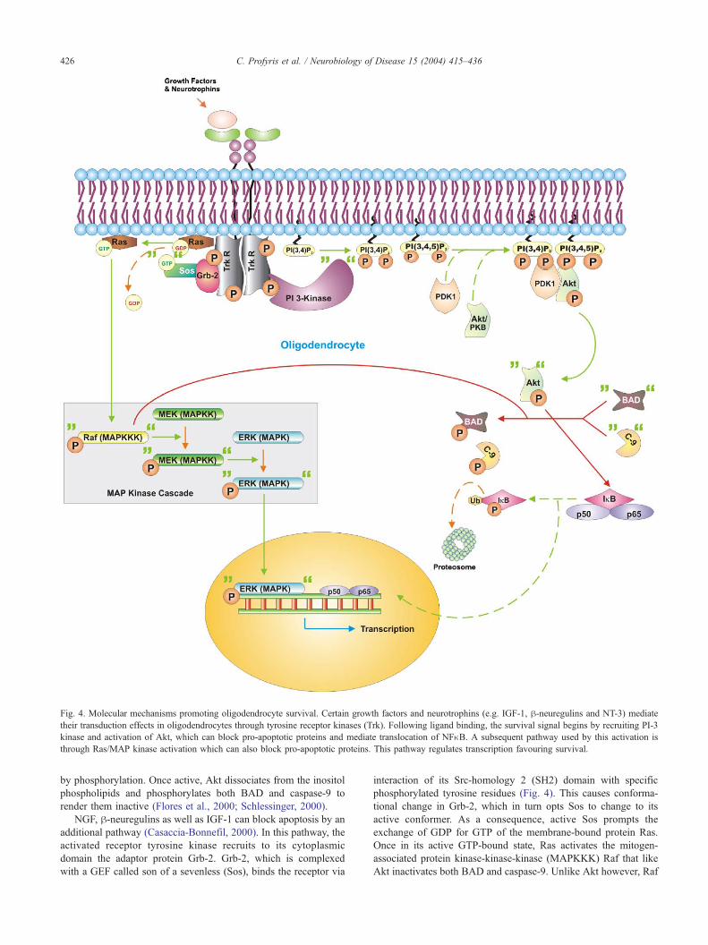
Fig. 4. Molecular mechanisms promoting oligodendrocyte survival. Certain growth factors and neurotrophins (e.g. IGF-1, h-neuregulins and NT-3) mediate
their transduction effects in oligodendrocytes through tyrosine receptor kinases (Trk). Following ligand binding, the survival signal begins by recruiting PI-3
kinase and activation of Akt, which can block pro-apoptotic proteins and mediate translocation of NFnB. A subsequent pathway used by this activation is
through Ras/MAP kinase activation which can also block pro-apoptotic proteins. This pathway regulates transcription favouring survival.
C. Profyris et al. / Neurobiology of Disease 15 (2004) 415–436426
by phosphorylation. Once active, Akt dissociates from the inositol
phospholipids and phosphorylates both BAD and caspase-9 to
render them inactive (Flores et al., 2000; Schlessinger, 2000).
NGF, h-neuregulins as well as IGF-1 can block apoptosis by an
additional pathway (Casaccia-Bonnefil, 2000). In this pathway, the
activated receptor tyrosine kinase recruits to its cytoplasmic
domain the adaptor protein Grb-2. Grb-2, which is complexed
with a GEF called son of a sevenless (Sos), binds the receptor via
interaction of its Src-homology 2 (SH2) domain with specific
phosphorylated tyrosine residues (Fig. 4). This causes conforma-
tional change in Grb-2, which in turn opts Sos to change to its
active conformer. As a consequence, active Sos prompts the
exchange of GDP for GTP of the membrane-bound protein Ras.
Once in its active GTP-bound state, Ras activates the mitogen-
associated protein kinase-kinase-kinase (MAPKKK) Raf that like
Akt inactivates both BAD and caspase-9. Unlike Akt however, Raf
C. Profyris et al. / Neurobiology of Disease 15 (2004) 415–436 427
is also able to activate the survival-orientated extracellular signal-
regulated kinase (ERK) pathway (Casaccia-Bonnefil, 2000). It
accomplishes this by activating the MAP-kinase-kinase (MAPKK),
MEK, through phosphorylation of a key residue in its activation
loop. As a consequence, this MAPKK then phosphorylates the
activation loop of the mitogen-activated protein kinase (MAPK)
ERK (Schlessinger, 2000). Upon its activation, ERK quickly
translocates to the nucleus where it alters transcription in favour
of survival. This is mediated by counteracting the pro-apoptotic
actions of other stress-activated MAPKs such as c-Jun NH(2)-
terminal kinase (JNK) and p38 (Nakahara et al., 1999; Xia et al.,
1995).
Death receptors
TNF receptors. The high concentration of TNFa in the CNS after
SCI, together with its ability to induce apoptosis make it a likely
instigator of oligodendrocyte apoptosis (Lee et al., 2000a; Yune et
al., 2003). TNFa mediates its effects by binding to its cognate
receptors TNF receptor 1 and 2 (TNFR1 and 2), of which both
belong to the TNFR superfamily. TNFR1 mediates the majority of
apoptotic effects as well as cell survival signals, whereas TNFR2
predominantly signals survival (Gupta, 2002).
Binding of trimeric TNFa to TNFR activates the receptor by
inducing it to form trimers (Fig. 5). TNFR1 trimerisation induces
the release from its death domain of the inhibitory protein, silencer
of death domains (SODD), which allows for recruitment of the
adapter protein TNFR-associated death domain (TRADD).
TRADD then recruits the Fas-associated death domain (FADD)
adaptor molecule that in turn causes activation of caspase-8 (see
Apoptosis) (Chen and Goeddel, 2002; Gupta, 2002).
TNFa can set off yet another apoptotic pathway following SCI,
which can be mediated by both TNFR1 and 2 (Nakahara et al.,
1999). The pathway commences with the binding of the adapter
protein TNFR-associated factor 2 (TRAF2) to TRADD (Fig. 5).
Once TRAF2 binds it recruits and subsequently activates the
MAPKKK, apoptosis-stimulated kinase 1 (ASK1). In turn,
ASK1, via MAPKs pathways that are similar to the one recruited
by Ras (see Trophic support deprivation; Fig. 4, gray inset)
activates the pro-apoptotic MAP kinases JNK and p38 (Chen
and Goeddel, 2002; Gupta, 2002; Nakahara et al., 1999; Xia et
al., 1995). JNK in particular, up-regulates the expression of pro-
apoptotic members of the Bcl-2 family and thereby causes cell
death (Harris and Johnson, 2001).
Despite the pro-apoptotic potential of TNFa (Lee et al., 2000a;
Yune et al., 2003), more compelling evidence by Kim et al. (2001)
indicates that TNFRs mediate anti-apoptotic affects following SCI.
This group showed that mice with deletions in TNFR1 or 2
presented with a worse functional outcome after SCI than their
wild-type counterparts. They correlated this outcome to the ability
of both TNFRs to induce anti-apoptotic signals (Kim et al., 2001).
In the case of TNFR1, TRADD has the ability to bind
additional adaptor proteins other than FADD, which include the
aforementioned TRAF2 and receptor interacting protein (RIP) (Fig.
5). Both TRAF2 and RIP promote cell survival by activating the
NFnB pathway. TRAF2 and RIP accomplish this by recruiting a
protein kinase complex composed of NFnB essential modulator
(NEMO), InB kinase (IKK) a and IKKh. Activation of this
complex activates the cytoplasmic NFnB heterotrimer composed
of p50, p65 and inhibitor nB (InB). Specifically, the complex
phosphorylates InB and renders it for ubiquitination and thereby
proteosomal degradation. As a result, the remaining NFnB dimer is
activated and translocates to the nucleus to transcribe numerous
genes of which a subset inhibits apoptosis (Chen and Goeddel,
2002; Gupta, 2002). Such gene products include cIAP1, cIAP2,
A1, Bcl-xL, Flice-inhibitory protein (FLIP), TRAF1 and TRAF2.
The cIAP1 and 2 block activated caspase-3, -7 and -9; A1 and Bcl-
xL prevent the release of cytochrome c; FLIP restricts the activa-
tion of caspase-8; and finally the TRAF proteins potentiate the
activation of the NFnB pathway and thereby survival. Importantly,
the cIAP proteins may also be activated by direct interaction with
TRAF2 (Gupta, 2002).
TNFR2 initiates most of TNFR1s signalling pathways, albeit
that of caspase-8 activation, by directly binding both TRAF1 and
TRAF2 to its intracellular domain (Gupta, 2002). Interestingly, in
addition to having a reduced capacity to promote apoptosis, it
seems that TNFR2 also has a reduced capacity to signal oligoden-
drocyte survival. This is suggested by the fact that the TNFR2
knockout mice recover better after SCI than their TNFR1 knockout
counterparts (Kim et al., 2001).
NGF/pro-NGF/P75NTR. Following SCI, there is acute up-regula-
tion in the expression of both the immature (pro-) and mature
forms of NGF as well as oligodendrocyte expression of p75NTR, a
member of the TNFR superfamily (Beattie et al., 2002a; Casha et
al., 2001). As spinal cord oligodendrocytes are void of the receptor
that NGF uses for pro-survival signalling, tropomyosin-related
kinase A (TrkA) (Beattie et al., 2002a), NGF signalling in this
oligodendrocyte population can only induce apoptosis through its
interaction with p75NTR (Casaccia-Bonnefil et al., 1996; Dechant
and Barde, 1997; Yoon et al., 1998). Furthermore, the immature
form of NGF, which binds p75NTR with an even higher affinity, is a
potent inducer of oligodendrocyte apoptosis regardless of TrkA
expression (Lee et al., 2001). Consequently, signalling of pro-NGF
and NGF via p75NTR is a powerful stimulus for oligodendrocyte
apoptosis in SCI. This has been recently confirmed by Beattie et al.
(2002a) who showed that p75NTR knockout mice show enhanced
recovery after experimental SCI.
The intracellular mechanisms leading to apoptosis after the
binding of pro-NGF or NGF to p75NTR have not been fully
described (Roux and Barker, 2002). Nevertheless, it is unequivocal
that oligodendrocyte apoptosis caused by p75NTR causes activation
of the injury-specific JNK3 MAPK and that this is activated by the
Rho GTPase Rac (Harrington et al., 2002). Although the link
between p75NTR and Rac is unclear, it is possible that Rac may be
activated by the protein 14-3-3q. 14-3-3q has been shown to
associate with the adaptor protein p75NTR-associated cell death
executor (NADE) (Kimura et al., 2001), which in turn binds the
intracellular domain of p75NTR (Mukai et al., 2000). The basis for
this link is supported by the fact that members of the 14-3-3 group
can bind Rho GTPases (Kimura et al., 1996). Furthermore, the
interactions between p75NTR and NADE, as well as NADE and 14-
3-3q are NGF dependent and stimulate the production of active
caspase-3 (Kimura et al., 2001; Mukai et al., 2000).
Remarkably, although p75NTR has a death domain, it does not
appear to activate caspase-8 in oligodendrocyte apoptosis. Instead,
the caspases that are activated are caspases-1, -2 and -3 (Gu et al.,
1999). Activation of these caspases is most likely the result of
JNK3-mediated transcriptional activation of pro-apoptotic mem-
bers of the Bcl-2 family (Gu et al., 1999).
An additional pathway by which p75NTR may activate oligo-
dendrocyte apoptosis is the ceramide pathway. This pathway is
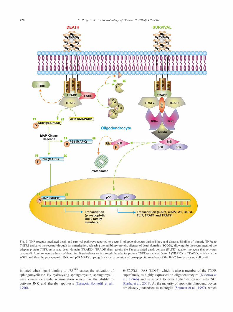
Fig. 5. TNF receptor mediated death and survival pathways reported to occur in oligodendrocytes during injury and disease. Binding of trimeric TNFa to
TNFR1 activates the receptor through its trimerisation, releasing the inhibitory protein, silencer of death domains (SODD), allowing for the recruitment of the
adapter protein TNFR-associated death domain (TRADD). TRADD then recruits the Fas-associated death domain (FADD) adapter molecule that activates
caspase-8. A subsequent pathway of death in oligodendrocytes is through the adapter protein TNFR-associated factor 2 (TRAF2) to TRADD, which via the
ASK1 and then the pro-apoptotic JNK and p38 MAPK, up-regulates the expression of pro-apoptotic members of the Bcl-2 family causing cell death.
C. Profyris et al. / Neurobiology of Disease 15 (2004) 415–436428
initiated when ligand binding to p75NTR causes the activation of
sphingomyelinase. By hydrolysing sphingomyelin, sphingomyeli-
nase causes ceramide accumulation which has the ability to
activate JNK and thereby apoptosis (Casaccia-Bonnefil et al.,
1996).
FASL/FAS. FAS (CD95), which is also a member of the TNFR
superfamily, is highly expressed on oligodendrocytes (D’Souza et
al., 1996b) and is subject to even higher expression after SCI
(Casha et al., 2001). As the majority of apoptotic oligodendrocytes
are closely juxtaposed to microglia (Shuman et al., 1997), which
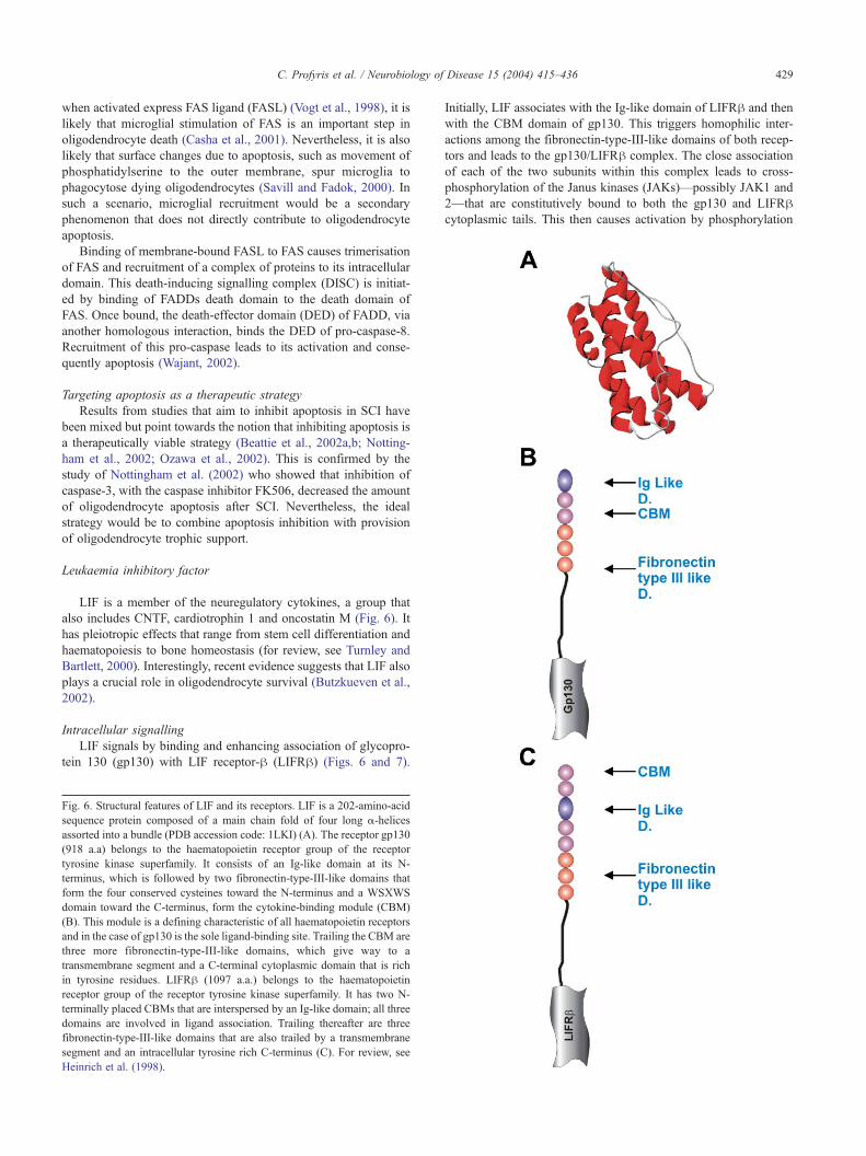
C. Profyris et al. / Neurobiology of Disease 15 (2004) 415–436 429
when activated express FAS ligand (FASL) (Vogt et al., 1998), it is
likely that microglial stimulation of FAS is an important step in
oligodendrocyte death (Casha et al., 2001). Nevertheless, it is also
likely that surface changes due to apoptosis, such as movement of
phosphatidylserine to the outer membrane, spur microglia to
phagocytose dying oligodendrocytes (Savill and Fadok, 2000). In
such a scenario, microglial recruitment would be a secondary
phenomenon that does not directly contribute to oligodendrocyte
apoptosis.
Binding of membrane-bound FASL to FAS causes trimerisation
of FAS and recruitment of a complex of proteins to its intracellular
domain. This death-inducing signalling complex (DISC) is initiat-
ed by binding of FADDs death domain to the death domain of
FAS. Once bound, the death-effector domain (DED) of FADD, via
another homologous interaction, binds the DED of pro-caspase-8.
Recruitment of this pro-caspase leads to its activation and conse-
quently apoptosis (Wajant, 2002).
Targeting apoptosis as a therapeutic strategy
Results from studies that aim to inhibit apoptosis in SCI have
been mixed but point towards the notion that inhibiting apoptosis is
a therapeutically viable strategy (Beattie et al., 2002a,b; Notting-
ham et al., 2002; Ozawa et al., 2002). This is confirmed by the
study of Nottingham et al. (2002) who showed that inhibition of
caspase-3, with the caspase inhibitor FK506, decreased the amount
of oligodendrocyte apoptosis after SCI. Nevertheless, the ideal
strategy would be to combine apoptosis inhibition with provision
of oligodendrocyte trophic support.
Leukaemia inhibitory factor
LIF is a member of the neuregulatory cytokines, a group that
also includes CNTF, cardiotrophin 1 and oncostatin M (Fig. 6). It
has pleiotropic effects that range from stem cell differentiation and
haematopoiesis to bone homeostasis (for review, see Turnley and
Bartlett, 2000). Interestingly, recent evidence suggests that LIF also
plays a crucial role in oligodendrocyte survival (Butzkueven et al.,
2002).
Intracellular signalling
LIF signals by binding and enhancing association of glycopro-
tein 130 (gp130) with LIF receptor-h (LIFRh) (Figs. 6 and 7).
Fig. 6. Structural features of LIF and its receptors. LIF is a 202-amino-acid
sequence protein composed of a main chain fold of four long a-helices
assorted into a bundle (PDB accession code: 1LKI) (A). The receptor gp130
(918 a.a) belongs to the haematopoietin receptor group of the receptor
tyrosine kinase superfamily. It consists of an Ig-like domain at its N-
terminus, which is followed by two fibronectin-type-III-like domains that
form the four conserved cysteines toward the N-terminus and a WSXWS
domain toward the C-terminus, form the cytokine-binding module (CBM)
(B). This module is a defining characteristic of all haematopoietin receptors
and in the case of gp130 is the sole ligand-binding site. Trailing the CBM are
three more fibronectin-type-III-like domains, which give way to a
transmembrane segment and a C-terminal cytoplasmic domain that is rich
in tyrosine residues. LIFRh (1097 a.a.) belongs to the haematopoietin
receptor group of the receptor tyrosine kinase superfamily. It has two N-
terminally placed CBMs that are interspersed by an Ig-like domain; all three
domains are involved in ligand association. Trailing thereafter are three
fibronectin-type-III-like domains that are also trailed by a transmembrane
segment and an intracellular tyrosine rich C-terminus (C). For review, see
Heinrich et al. (1998).
Initially, LIF associates with the Ig-like domain of LIFRh and then
with the CBM domain of gp130. This triggers homophilic inter-
actions among the fibronectin-type-III-like domains of both recep-
tors and leads to the gp130/LIFRh complex. The close association
of each of the two subunits within this complex leads to cross-
phosphorylation of the Janus kinases (JAKs)—possibly JAK1 and
2—that are constitutively bound to both the gp130 and LIFRhcytoplasmic tails. This then causes activation by phosphorylation
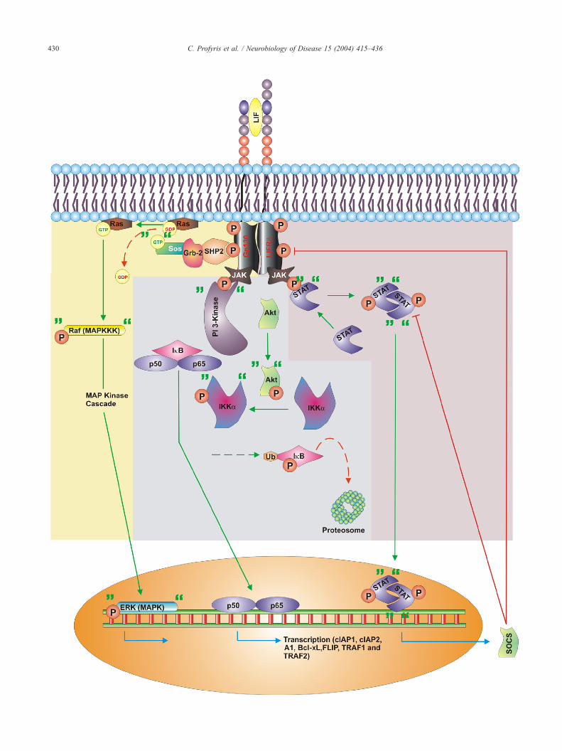
C. Profyris et al. / Neurobiology of Disease 15 (2004) 415–436430

C. Profyris et al. / Neurobiology of Disease 15 (2004) 415–436 431
of their tyrosine kinase domains, which leads to phosphorylation of
tyrosine residues of the receptor complex. Consequently, this
creates docking sites for the SH2 domain of signal transducers
and activators of transcription (STAT)—STAT1 and 3—and there-
by causes the recruitment of these molecules and their phosphor-
ylation by JAK. Upon activation, STAT is released by JAK, but
now its SH2 domain causes it to associate with an SH2 counterpart
of another activated STAT. This homophilic dimerisation permits
nuclear translocation of STAT where it modulates transcription by
binding specific DNA response elements with other regulatory
proteins—CREB-binding protein and p300 (for review, see Hein-
rich et al., 1998; Turnley and Bartlett, 2000). Interestingly, in the
context of oligodendrocyte survival, the transcriptional products of
the STAT3 pathway include anti-apoptotic members of the Bcl-2
family, which includes Bcl-2 and Bcl-xL (Bowman et al., 2000).
LIF may potentiate oligodendrocyte survival via additional
pathways (Fig. 7). A likely pathway is that of the ERK MAP
kinase (Segal and Greenberg, 1996). This pathway is activated by
association of SHP2, a tyrosine phosphatase, to phosphotyrosine
residues on gp130. SHP2 has been shown to associate with Grb2,
thus, providing a link between LIF signalling and the ERK MAP
kinase pathway (Segal and Greenberg, 1996). LIF may also
promote oligodendrocyte survival via the NFnB pathway (Mid-
dleton et al., 2000). This pathway may commence when PI 3-
kinase associates with the gp130/LIFRh complex and thereby
causes activation of Akt. Activated Akt goes on to activate IKKa
by phosphorylation, which in turn phosphorylates InB to render
NFnB active (Middleton et al., 2000).
Signalling via LIF is tightly regulated and subject to feedback
inhibition (Fig. 7). The phosphorylation of components in the LIF
pathway is only transient. Furthermore, the transcriptional products
of JAK/STAT signalling include the protein suppressor-of-cyto-
kine-signalling (SOCS). SOCS inhibits the JAK/STAT pathway by
inhibiting tyrosine phosphorylation of gp130, STAT1 and 3.
Interestingly, SOCS-1 can also protect fibroblasts from apoptosis
induced by TNF-a (Morita et al., 2000). Hence, it is possible that
LIF, via SOCS, may increase the durability of oligodendrocytes to
the armada of pro-inflammatory cytokines released in SCI. Finally,
an extra level of feedback inhibition is accomplished by receptor-
mediated endocytosis followed by lysosomal degradation of the
LIF-bound gp130/LIFRh complex (for review, see Heinrich et al.,
1998; Turnley and Bartlett, 2000).
Precedent for administering LIF in SCI
There exists a precedent for the pharmacological administration
of LIF following SCI, which has been set by work in multiple
sclerosis (MS) research by Butzkueven et al. (2002). Based on
evidence that oligodendrocyte apoptosis is implicated in certain
categories of MS, this group examined the efficacy of LIF in
attenuating oligodendrocyte death in the animal model of MS,
EAE. They showed that LIF reduced EAE symptoms, reduced
oligodendrocyte apoptosis via a JAK/STAT mechanism and that
Fig. 7. Transduction mechanisms of LIF signalling. High-affinity interaction with t
cascade of survival events within the neural cell. JAK tyrosine kinases bound to
phosphorylation which leads to the phosphorylation of tyrosine residues of the
transducers and activators of transcription (STAT)—STAT 1 and 3—and in tu
dimerisation of two phospho-STAT molecules and their translocation to the nucleu
response elements. This predominantly involves the suppression of cytokine signal
STAT pathway. Transcriptional products of STAT3 include anti-apoptotic members
through the phosphorylation of SHP2 binding the adaptor molecule Grb-2, activa
the therapeutic benefits of LIF were negated when it was admin-
istered to mice that were double heterzygous for the LIFRh-nullmutation and a gp130 intracellular truncation. In fact, the LIFRhand gp130 mutant mice displayed an even worse EAE phenotype
than control animals receiving vehicle (Butzkueven et al., 2002).
Corroborative evidence implicating a protective role of LIF in
oligodendrocyte survival arises from in vitro data showing that
LIF increases the ability of oligodendrocytes to survive in
cultured media (Barres et al., 1993; Mayer et al., 1994). It has
also been shown that LIF completely abrogates interferon-g-
induced death in rat oligodendrocytes (Vartanian et al., 1995).
Interestingly, ciliary neurotrophic factor (CNTF), another neure-
gulatory cytokine, protects human oligodendrocytes from death
by either TNFa or growth factor withdrawal (D’Souza et al.,
1996a). These findings, together with the Butzkueven study,
imply that LIF signalling is a mechanism that naturally inhibits
oligodendrocyte apoptosis and upon enhancement makes these
cells more robust to insult. Consequently, it is essential that the
role of LIF as a therapeutic measure in experimental SCI be
investigated.
Acknowledgments
This study was supported by the National Health and Medical
Research Council of Australia Program Grant and the Trish-MS
Research Foundation.
References
Allan, S.M., Rothwell, N.J., 2001. Cytokines and acute neurodegeneration.
Nat. Rev., Neurosci. 2, 734–744.
Amano, M., Ito, M., Kimura, K., Fukata, Y., Chihara, K., Nakano, T.,
Matsuura, Y., Kaibuchi, K., 1996. Phosphorylation and activation of
myosin by Rho-associated kinase (Rho-kinase). J. Biol. Chem. 271,
20246–20249.
Anthes, D.L., Theriault, E., Tator, C.H., 1995. Characterization of axonal
ultrastructural pathology following experimental spinal cord compres-
sion injury. Brain Res. 702, 1–16.
Arber, S., Barbayannis, F.A., Hanser, H., Schneider, C., Stanyon, C.A.,
Bernard, O., Caroni, P., 1998. Regulation of actin dynamics through
phosphorylation of cofilin by LIM-kinase. Nature 393, 805–809.
Arnett, H.A., Mason, J., Marino, M., Suzuki, K., Matsushima, G.K., Ting,
J.P., 2001. TNF alpha promotes proliferation of oligodendrocyte pro-
genitors and remyelination. Nat. Neurosci. 4, 1116–1122.
Bannister, L.H., 1999. Connective tissues. In: Williams, P.L., Bannister,
M.M., Berry, M.M., Collins, P., Dyson, M., Dussek, J.E., Ferguson,
M.W.J. (Eds.), Gray’s Anatomy, thirty-eight ed. Churchill Livingstone,
Edinburgh, pp. 75–90.
Barres, B.A., Hart, I.K., Coles, H.S., Burne, J.F., Voyvodic, J.T., Richard-
son, W.D., Raff, M.C., 1992. Cell death and control of cell survival in
the oligodendrocyte lineage. Cell 70, 31–46.
Barres, B.A., Schmid, R., Sendnter, M., Raff, M.C., 1993. Multiple extrac-
he heterodimeric receptor complex of gp130 and LIFRh sets off a secondary
the cytoplasmic domains of gp130 and LIFRh are activated through their
receptor complex, providing docking sites for the SH2 domain of signal
rn producing their activation by phosphorylation. This ultimately causes
s in which they mediate transcriptional events after binding to specific DNA
ling (SOCS) proteins, which negatively regulate this cytokine-induced JAK/
of the Bcl-2 family (Bcl-2 and Bcl-xL). Other survival signals are triggered
ting the Ras/MAP kinase pathway of survival.
C. Profyris et al. / Neurobiology of Disease 15 (2004) 415–436432
ellular signals are required for long-term oligodendrocyte survival. De-
velopment 118, 283–295.
Bartholdi, D., Schwab, M.E., 1997. Expression of pro-inflammatory cyto-
kine and chemokine mRNA upon experimental spinal cord injury in
mouse: an in situ hybridization study. Eur. J. Neurosci. 9, 1422–1438.
Beattie, M.S., Farooqui, A.A., Bresnahan, J.C., 2000. Review of current
evidence for apoptosis after spinal cord injury. J. Neurotrauma 17,
915–925.
Beattie, M.S., Harrington, A.W., Lee, R., Kim, J.Y., Boyce, S.L., Longo,
J.C., Bresnahan, J.C., Hempstead, B.L., Yoon, S.O., 2002a. ProNGF
induces p75-mediated death of oligodendrocytes following spinal cord
injury. Neuron 36, 375–386.
Beattie, M.S., Hermann, G.E., Rogers, R.C., Bresnahan, J.C., 2002b. Cell
death in models of spinal cord injury. Prog. Brain Res. 137, 37–47.
Bethea, J.R., Castro, M., Keane, R.W., Lee, T.T., Dietrich, W.D., Yezierski,
R.P., 1998. Traumatic spinal cord injury induces nuclear factor-kappaB
activation. J. Neurosci. 18, 3251–3260.
Bignami, A., Dahl, D., 1994. General Pathology. Medical Intelligence Unit:
Glial Cells In The Central Nervous System And Their Reaction To
Injury, first ed. R.G. Landes Company, Austin, pp. 77–92.
Blight, A.R., 1983. Cellular morphology of chronic spinal cord injury in
the cat: analysis of myelinated axons by line-sampling. Neuroscience
10, 521–543.
Blight, A., 1988. Mechanical factors in experimental spinal cord injury. J.
Am. Parapleg. Soc. 11, 26–34.
Blight, A.R., Decrescito, V., 1986. Morphometric analysis of experimental
spinal cord injury in the cat: the relation of injury intensity to survival of
myelinated axons. Neuroscience 19, 321–341.
Bowman, T., Garcia, R., Turkson, J., Jove, R., 2000. STATs in oncogenesis.
Oncogene 19, 2474–2488.
Bradbury, E.J., Moon, L.D., Popat, R.J., King, V.R., Bennett, G.S., Patel,
P.N., Fawcett, J.W., McMahon, S.B., 2002. Chondroitinase ABC pro-
motes functional recovery after spinal cord injury. Nature 416, 636–640.
Bresnahan, J.C., Beattie, M.S. , Todd III, F.D., Noyes, D.H. 1987. A
behavioral and anatomical analysis of spinal cord injury produced by
a feedback-controlled impaction device. Exp. Neurol. 95, 548–570.
Buffo, A., Zagrebelsky, M., Huber, A.B., Skerra, A., Schwab, M.E., Strata,
F., Rossi, F., 2000. Application of neutralizing antibodies against NI-35/
250 myelin-associated neurite growth inhibitory proteins to the adult rat
cerebellum induces sprouting of uninjured Purkinje cell axons. J. Neu-
rosci. 20, 2275–2286.
Bunge, R.P., Puckett, W.R., Becerra, J.L., Marcillo, A., Quencer, R.M.,
1993. Observations on the pathology of human spinal cord injury. A
review and classification of 22 new cases with details from a case of
chronic cord compression with extensive focal demyelination. Adv.
Neurol. 59, 75–89.
Burg, M.A., Tillet, E., Timpl, R., Stallcup, W.B., 1996. Binding of the NG2
proteoglycan to type VI collagen and other extracellular matrix mole-
cules. J. Biol. Chem. 271, 26110–26116.
Butzkueven, H., Zhang, J.G., Soilu-Hanninen, M., Hochrein, H., Chionh,
K.A., Shipham, K.A., Emery, B., Turnley, A.M., Petratos, S., Ernst, M.,
Bartlett, P.F., Kilpatrick, T.J., 2002. LIF receptor signaling limits im-
mune-mediated demyelination by enhancing oligodendrocyte survival.
Nat. Med. 8, 613–619.
Carlson, S.L., Parrish, M.E., Springer, J.E., Doty, K., Dossett, L., 1998.
Acute inflammatory response in spinal cord following impact injury.
Exp. Neurol. 151, 77–88.
Casaccia-Bonnefil, P., 2000. Cell death in the oligodendrocyte lineage: a
molecular perspective of life/death decisions in development and dis-
ease. Glia 29, 124–135.
Casaccia-Bonnefil, P., Carter, B.D., Dobrowsky, R.T., Chao, M.V., 1996.
Death of oligodendrocytes mediated by the interaction of nerve growth
factor with its receptor p75. Nature 383, 716–719.
Casha, S., Yu, W.R., Fehlings, M.G., 2001. Oligodendroglial apoptosis
occurs along degenerating axons and is associated with FAS and p75
expression following spinal cord injury in the rat. Neuroscience 103,
203–218.
Cassatella, M.A., 1995. The production of cytokines by polymorphonu-
clear neutrophils. Immunol. Today 16, 21–26.
Chen, G., Goeddel, D.V., 2002. TNF-R1 signaling: a beautiful pathway.
Science 296, 1634–1635.
Chen, M.S., Huber, A.B., van der Haar, M.E., Frank, M., Schnell, L.,
Spillmann, A.A., Christ, F., Schwab, M.E., 2000. Nogo-A is a mye-
lin-associated neurite outgrowth inhibitor and an antigen for monoclo-
nal antibody IN-1. Nature 403, 434–439.
Choi, D.W., 1996. Ischemia-induced neuronal apoptosis. Curr. Opin. Neu-
robiol. 6, 667–672.
Cotran, R.S., Kumar, V., Collins, T., 1999. Cellular pathology I: cell injury
and cell death. In: Venkatachalam, M. (Ed.), Robbins Pathologic Basis
of Disease, sixth ed. W.B. Saunders Company, Philadelphia, pp. 5–18.
Crowe, M.J., Bresnahan, J.C., Shuman, S.L., Masters, J.N., Beattie, M.S.,
1997. Apoptosis and delayed degeneration after spinal cord injury in
rats and monkeys. Nat. Med. 3, 73–76.
David, S., 2002. Recruiting the immune response to promote long dis-
tance axon regeneration after spinal cord injury. Prog. Brain Res. 137,
407–414.
Dechant, G., Barde, Y.A., 1997. Signalling through the neurotrophin re-
ceptor p75NTR. Curr. Opin. Neurobiol. 7, 413–418.
de la Torre, J.C., 1981. Spinal cord injury. Review of basic and applied
research. Spine 6, 315–335.
Dergham, P., Ellezam, B., Essagian, C., Avedissian, H., Lubell, W.D.,
McKerracher, L., 2002. Rho signaling pathway targeted to promote
spinal cord repair. J. Neurosci. 22, 6570–6577.
Doble, A., 1999. The role of excitotoxicity in neurodegenerative disease:
implications for therapy. Pharmacol. Ther. 81, 163–221.
D’Souza, S.D., Alinauskas, K.A., Antel, J.P., 1996a. Ciliary neurotrophic
factor selectively protects human oligodendrocytes from tumor necrosis
factor-mediated injury. J. Neurosci. Res. 43, 289–298.
D’Souza, S.D., Bonetti, B., Balasingam, V., Cashman, N.R., Barker, P.A.,
Troutt, A.B., Raine, C.S., Antel, J.P., 1996b. Multiple sclerosis: Fas
signaling in oligodendrocyte cell death. J. Exp. Med. 184, 2361–2370.
Dusart, I., Schwab, M.E., 1994. Secondary cell death and the inflammatory
reaction after dorsal hemisection of the rat spinal cord. Eur. J. Neurosci.
6, 712–724.
Ehrhard, P.B., Erb, P., Graumann, U., Otten, U., 1993. Expression of nerve
growth factor and nerve growth factor receptor tyrosine kinase Trk in
activated CD4-positive T-cell clones. Proc. Natl. Acad. Sci. U. S. A. 90,
10984–10988.
Fawcett, J.W., Asher, R.A., 1999. The glial scar and central nervous system
repair. Brain Res. Bull. 49, 377–391.
Fitch, M.T., Doller, C., Combs, C.K., Landreth, G.E., Silver, J., 1999.
Cellular and molecular mechanisms of glial scarring and progressive
cavitation: in vivo and in vitro analysis of inflammation-induced sec-
ondary injury after CNS trauma. J. Neurosci. 19, 8182–8198.
Flores, A.I., Mallon, B.S., Matsui, T., Ogawa, W., Rosenzweig, A., Oka-
moto, T., Macklin, W.B., 2000. Akt-mediated survival of oligodendro-
cytes induced by neuregulins. J. Neurosci. 20, 7622–7630.
Fournier, A.E., GrandPre, T., Strittmatter, S.M., 2001. Identification of a
receptor mediating Nogo-66 inhibition of axonal regeneration. Nature
409, 341–346.
Frisen, J., Haegerstrand, A., Fried, K., Piehl, F., Cullheim, S., Risling, M.,
1994. Adhesive/repulsive properties in the injured spinal cord: relation
to myelin phagocytosis by invading macrophages. Exp. Neurol. 129,
183–193.
George, R., Griffin, J.W., 1994. The proximo-distal spread of axonal de-
generation in the dorsal columns of the rat. J. Neurocytol. 23, 657–667.
GrandPre, T., Nakamura, F., Vartanian, T., Strittmatter, S.M., 2000. Iden-
tification of the Nogo inhibitor of axon regeneration as a Reticulon
protein. Nature 403, 439–444.
Grimpe, B., Silver, J., 2002. The extracellular matrix in axon regeneration.
Prog. Brain Res. 137, 333–349.
Gruner, J.A., 1992. A monitored contusion model of spinal cord injury in
the rat. J. Neurotrauma 9, 123–126 ((discussion 126-128)) .
Gruner, J.A., Yee, A., Blight, A.R., 1996. Histological and functional eval-
C. Profyris et al. / Neurobiology of Disease 15 (2004) 415–436 433
uation of experimental spinal cord injury: evidence of a stepwise re-
sponse to graded compression. Brain Res. 729, 90–101.
Gu, C., Casaccia-Bonnefil, P., Srinivasan, A., Chao, M.V., 1999. Oligoden-
drocyte apoptosis mediated by caspase activation. J. Neurosci. 19,
3043–3049.
Guha, A., Tator, C.H., 1988. Acute cardiovascular effects of experimental
spinal cord injury. J. Trauma 28, 481–490.
Gupta, S., 2002. A decision between life and death during TNF-alpha-
induced signaling. J. Clin. Immunol. 22, 185–194.
Guth, L., Zhang, Z., Steward, O., 1999. The unique histopathological
responses of the injured spinal cord. Implications for neuroprotective
therapy. Ann. N.Y. Acad. Sci. 890, 366–384.
Hall, E.D., 1992. The neuroprotective pharmacology of methylpredniso-
lone. J. Neurosurg. 76, 13–22.
Hamada, Y., Ikata, T., Katoh, S., Katoh, K., Niwa, M., Tsutsumishita, Y.,
Fukuzawa, K., 1996. Effects of exogenous transforming growth factor-
beta 1 on spinal cord injury in rats. Neurosci. Lett. 203, 97–100.
Harrington, A.W., Kim, J.Y., Yoon, S.O., 2002. Activation of Rac GTPase
by p75 is necessary for c-jun N-terminal kinase-mediated apoptosis. J.
Neurosci. 22, 156–166.
Harris, C.A. , Johnson Jr., E.M. 2001. BH3-only Bcl-2 family members are
coordinately regulated by the JNK pathway and require Bax to induce
apoptosis in neurons. J. Biol. Chem. 276, 37754–37760.
Hauben, E., Schwartz, M., 2003. Therapeutic vaccination for spinal cord
injury: helping the body to cure itself. Trends Pharmacol. Sci. 24, 7–12.
Hauben, E., Butovsky, O., Nevo, U., Yoles, E., Moalem, G., Agranov, E.,
Mor, F., Leibowitz-Amit, R., Pevsner, E., Akselrod, S., Neeman, M.,
Cohen, I.R., Schwartz, M., 2000. Passive or active immunization with
myelin basic protein promotes recovery from spinal cord contusion. J.
Neurosci. 20, 6421–6430.
Hausmann, O.N., 2003. Post-traumatic inflammation following spinal cord
injury. Spinal Cord 41, 369–378.
Heese, K., Hock, C., Otten, U., 1998. Inflammatory signals induce
neurotrophin expression in human microglial cells. J. Neurochem.
70, 699–707.
Heinrich, P.C., Behrmann, I., Muller-Newen, G., Schaper, F., Graeve, L.,
1998. Interleukin-6-type cytokine signalling through the gp130/Jak/
STAT pathway. Biochem. J. 334, 297–314.
Hengartner, M.O., 2000. The biochemistry of apoptosis. Nature 407,
770–776.
Herx, L.M., Rivest, S., Yong, V.W., 2000. Central nervous system-initiated
inflammation and neurotrophism in trauma: IL-1 beta is required for the
production of ciliary neurotrophic factor. J. Immunol. 165, 2232–2239.
Huber, A.B., Schwab, M.E., 2000. Nogo-A, a potent inhibitor of neurite
outgrowth and regeneration. Biol. Chem. 381, 407–419.
Janmey, P.A., Stossel, T.P., 1987. Modulation of gelsolin function by phos-
phatidylinositol 4,5-bisphosphate. Nature 325, 362–364.
Josephson, A., Trifunovski, A., Widmer, H.R., Widenfalk, J., Olson, L.,
Spenger, C., 2002. Nogo-receptor gene activity: cellular localization
and developmental regulation of mRNA in mice and humans. J. Comp.
Neurol. 453, 292–304.
Joshi, M., Fehlings, M.G., 2002a. Development and characterization of a
novel, graded model of clip compressive spinal cord injury in the
mouse: Part 1. Clip design, behavioral outcomes, and histopathology.
J. Neurotrauma 19, 175–190.
Joshi, M., Fehlings, M.G., 2002b. Development and characterization of a
novel, graded model of clip compressive spinal cord injury in the
mouse: Part 2. Quantitative neuroanatomical assessment and analysis
of the relationships between axonal tracts, residual tissue, and locomo-
tor recovery. J. Neurotrauma 19, 191–203.
Kerschensteiner, M., Gallmeier, E., Behrens, L., Leal, V.V., Misgeld, T.,
Klinkert, W.E., Kolbeck, R., Hoppe, E., Oropeza-Wekerle, R.L., Bartke,
C., Stadelmann, C., Lassmann, H., Wekerle, H., Hohlfeld, R., 1999.
Activated human T cells, B cells, and monocytes produce brain-derived
neurotrophic factor in vitro and in inflammatory brain lesions: a neuro-
protective role of inflammation? J. Exp. Med. 189, 865–870.
Kim, G.M., Xu, J., Song, S.K., Yan, P., Ku, G., Xu, X.M., Hsu, C.Y., 2001.
Tumor necrosis factor receptor deletion reduces nuclear factor-kappaB
activation, cellular inhibitor of apoptosis protein 2 expression, and func-
tional recovery after traumatic spinal cord injury. J. Neurosci. 21,
6617–6625.
Kim, J.E., Li, S., Grandpre, T., Qiu, D., Strittmatter, S.M., 2003. Axon
regeneration in young adult mice lacking Nogo-A/B. Neuron 38,
187–199.
Kimura, K., Ito, M., Amano, M., Chihara, K., Fukata, Y., Nakafuku, M.,
Yamamori, B., Feng, J., Nakano, T., Okawa, K., Iwamatsu, A., Kaibu-
chi, K., 1996. Regulation of myosin phosphatase by Rho and Rho-
associated kinase (Rho-kinase). Science 273, 245–248.
Kimura, M.T., Irie, S., Shoji-Hoshino, S., Mukai, J., Nadano, D.,
Oshimura, M., Sato, T.A., 2001. 14-3-3 is involved in p75 neuro-
trophin receptor-mediated signal transduction. J. Biol. Chem. 276,
17291–17300.
Kipnis, J., Yoles, E., Schori, H., Hauben, E., Shaked, I., Schwartz, M.,
2001. Neuronal survival after CNS insult is determined by a genetically
encoded autoimmune response. J. Neurosci. 21, 4564–4571.
Kitamura, M., 1997. Identification of an inhibitor targeting macrophage
production of monocyte chemoattractant protein-1 as TGF-beta 1. J.
Immunol. 159, 1404–1411.
Klusman, I., Schwab, M.E., 1997. Effects of pro-inflammatory cytokines in
experimental spinal cord injury. Brain Res. 762, 173–184.
Koshinaga, M., Whittemore, S.R., 1995. The temporal and spatial acti-
vation of microglia in fiber tracts undergoing anterograde and retro-
grade degeneration following spinal cord lesion. J. Neurotrauma 12,
209–222.
Kottis, V., Thibault, P., Mikol, D., Xiao, Z.C., Zhang, R., Dergham, P.,
Braun, P.E., 2002. Oligodendrocyte-myelin glycoprotein (OMgp) is an
inhibitor of neurite outgrowth. J. Neurochem. 82, 1566–1569.
Koyanagi, I., Tator, C.H., Lea, P.J., 1993. Three-dimensional analysis of the
vascular system in the rat spinal cord with scanning electron micro-
scopy of vascular corrosion casts. Part 2: acute spinal cord injury.
Neurosurgery 33, 285–291 ((discussion 292)) .
Lee, Y.B., Yune, T.Y., Baik, S.Y., Shin, Y.H., Du, S., Rhim, H., Lee, E.B.,
Kim, Y.C., Shin, M.L., Markelonis, G.J., Oh, T.H., 2000a. Role of
tumor necrosis factor-alpha in neuronal and glial apoptosis after spinal
cord injury. Exp. Neurol. 166, 190–195.
Lee, Y.L., Shih, K., Bao, P., Ghirnikar, R.S., Eng, L.F., 2000b. Cytokine
chemokine expression in contused rat spinal cord. Neurochem. Int. 36,
417–425.
Lee, R., Kermani, P., Teng, K.K., Hempstead, B.L., 2001. Regulation of
cell survival by secreted proneurotrophins. Science 294, 1945–1948.
Leskovar, A., Moriarty, L.J., Turek, J.J., Schoenlein, I.A., Borgens, R.B.,
2000. The macrophage in acute neural injury: changes in cell numbers
over time and levels of cytokine production in mammalian central and
peripheral nervous systems. J. Exp. Biol. 203, 1783–1795.
Leung, T., Chen, X.Q., Manser, E., Lim, L., 1996. The p160 RhoA-binding
kinase ROK alpha is a member of a kinase family and is involved in the
reorganization of the cytoskeleton. Mol. Cell. Biol. 16, 5313–5327.
Li, G.L., Farooque, M., Holtz, A., Olsson, Y., 1999a. Apoptosis of oligo-
dendrocytes occurs for long distances away from the primary injury
after compression trauma to rat spinal cord. Acta Neuropathol. (Berl)
98, 473–480.
Li, Y., Field, P.M., Raisman, G., 1999b. Death of oligodendrocytes and
microglial phagocytosis of myelin precede immigration of Schwann
cells into the spinal cord. J. Neurocytol. 28, 417–427.
Liu, B.P., Fournier, A., GrandPre, T., Strittmatter, S.M., 2002. Myelin-
associated glycoprotein as a functional ligand for the Nogo-66 receptor.
Science 297, 1190–1193.
Maekawa, M., Ishizaki, T., Boku, S., Watanabe, N., Fujita, A., Iwamatsu,
T., Obinata, T., Ohashi, K., Mizuno, K., Narumiya, S., 1999. Signaling
from Rho to the actin cytoskeleton through protein kinases ROCK and
LIM-kinase. Science 285, 895–898.
Manser, E., Chong, C., Zhao, Z.S., Leung, T., Michael, G., Hall, C., Lim,
L., 1995. Molecular cloning of a new member of the p21-Cdc42/Rac-
activated kinase (PAK) family. J. Biol. Chem. 270, 25070–25078.
C. Profyris et al. / Neurobiology of Disease 15 (2004) 415–436434
Mason, J.L., Suzuki, K., Chaplin, D.D., Matsushima, G.K., 2001. Interleu-
kin-1beta promotes repair of the CNS. J. Neurosci. 21, 7046–7052.
Mattson, M.P., Rychlik, B., Chu, C., Christakos, S., 1991. Evidence for
calcium-reducing and excito-protective roles for the calcium-binding
protein calbindin-D28k in cultured hippocampal neurons. Neuron 6,
41–51.
Matute, C., Alberdi, E., Domercq, M., Perez-Cerda, F., Perez-Samartin,
M.V., Sanchez-Gomez, M.V., 2001. The link between excitotoxic
oligodendroglial death and demyelinating diseases. Trends Neurosci.
24, 224–230.
Maxwell, W.L., 1996. Histopathological changes at central nodes of Ranv-
ier after stretch-injury. Microsc. Res. Tech. 34, 522–535.
Mayer, M., Bhakoo, K., Noble, M., 1994. Ciliary neurotrophic factor and
leukemia inhibitory factor promote the generation, maturation and sur-
vival of oligodendrocytes in vitro. Development 120, 143–153.
Maynard Jr., F.M., Bracken, M.B., Creasey, G., Ditunno Jr., J.F., Donovan,
W.H., Ducker, T.B., Garber, S.L., Marino, R.J., Stover, S.L., Tator,
C.H., Waters, R.L., Wilberger, J.E., Young, W., 1997. International
standards for neurological and functional classification of spinal cord
injury. American spinal injury association. Spinal Cord 35, 266–274.
McDonald, P.P., Bald, A., Cassatella, M.A., 1997. Activation of the NF-
kappaB pathway by inflammatory stimuli in human neutrophils. Blood
89, 3421–3433.
McDonald, J.W., Althomsons, S.P., Hyrc, K.L., Choi, D.W., Goldberg,
M.P., 1998. Oligodendrocytes from forebrain are highly vulnerable
to AMPA/kainate receptor-mediated excitotoxicity. Nat. Med. 4,
291–297.
McKerracher, L., Winton, M.J., 2002. Nogo on the go. Neuron 36,
345–348.
McKerracher, L., David, S., Jackson, D.L., Kottis, V., Dunn, R.J., Braun,
P.E., 1994. Identification of myelin-associated glycoprotein as a major
myelin-derived inhibitor of neurite growth. Neuron 13, 805–811.
McTigue, D.M., Tani, M., Krivacic, K., Chernosky, A., Kelner, G.S., Ma-
ciejewski, D., Maki, R., Ransohoff, R.M., Stokes, B.T., 1998. Selective
chemokine mRNA accumulation in the rat spinal cord after contusion
injury. J. Neurosci. Res. 53, 368–376.
McTigue, D.M., Wei, P., Stokes, B.T., 2001. Proliferation of NG2-positive
cells and altered oligodendrocyte numbers in the contused rat spinal
cord. J. Neurosci. 21, 3392–3400.
Meyer, G., Feldman, E.L., 2002. Signaling mechanisms that regulate actin-
based motility processes in the nervous system. J. Neurochem. 83,
490–503.
Middleton, G., Hamanoue, M., Enokido, Y., Wyatt, S., Pennica, D., Jaffray,
E., Hay, R.T., Davies, A.M., 2000. Cytokine-induced nuclear factor
kappa B activation promotes the survival of developing neurons. J. Cell
Biol. 148, 325–332.
Miller, R.H., 1999. Contact with central nervous system myelin inhibits
oligodendrocyte progenitor maturation. Dev. Biol. 216, 359–368.
Moalem, G., Leibowitz-Amit, R., Yoles, E., Mor, F., Cohen, I.R., Schwartz,
M., 1999a. Autoimmune T cells protect neurons from secondary degen-
eration after central nervous system axotomy. Nat. Med. 5, 49–55.
Moalem, G., Monsonego, A., Shani, Y., Cohen, I.R., Schwartz, M., 1999b.
Differential T cell response in central and peripheral nerve injury: con-
nection with immune privilege. FASEB J. 13, 1207–1217.
Moalem, G., Gdalyahu, A., Shani, Y., Otten, U., Lazarovici, P., Cohen, I.R.,
Schwartz, M., 2000. Production of neurotrophins by activated T cells:
implications for neuroprotective autoimmunity. J. Autoimmun. 15,
331–345.
Morgenstern, D.A., Asher, R.A., Fawcett, J.W., 2002. Chondroitin sul-
phate proteoglycans in the CNS injury response. Prog. Brain Res.
137, 313–332.
Morita, Y., Naka, T., Kawazoe, Y., Fujimoto, M., Narazaki, M., Nakagawa,
H., Fukuyama, H., Nagata, S., Kishimoto, T., 2000. Signals transducers
and activators of transcription (STAT)-induced STAT inhibitor-1 (SSI-
1)/suppressor of cytokine signaling-1 (SOCS-1) suppresses tumor ne-
crosis factor alpha-induced cell death in fibroblasts. Proc. Natl. Acad.
Sci. U. S. A. 97, 5405–5410.
Mukai, J., Hachiya, T., Shoji-Hoshino, S., Kimura, M.T., Nadano, D.,
Suvanto, P., Hanaoka, T., Li, Y., Irie, S., Greene, L.A., Sato, T.A.,
2000. NADE, a p75NTR-associated cell death executor, is involved
in signal transduction mediated by the common neurotrophin receptor
p75NTR. J. Biol. Chem. 275, 17566–17570.
Mukhopadhyay, G., Doherty, P., Walsh, F.S., Crocker, P.R., Filbin, M.T.,
1994. A novel role for myelin-associated glycoprotein as an inhibitor of
axonal regeneration. Neuron 13, 757–767.
Nakahara, S., Yone, K., Sakou, T., Wada, S., Nagamine, T., Niiyama, T.,
Ichijo, H., 1999. Induction of apoptosis signal regulating kinase 1
(ASK1) after spinal cord injury in rats: possible involvement of
ASK1-JNK and -p38 pathways in neuronal apoptosis. J. Neuropathol.
Exp. Neurol. 58, 442–450.
Ng, C.E., Tang, B.L., 2002. Nogos and the Nogo-66 receptor: factors
inhibiting CNS neuron regeneration. J. Neurosci. Res. 67, 559–565.
Ng, W.P., Cartel, N., Roder, J., Roach, A., Lozano, A., 1996. Human
central nervous system myelin inhibits neurite outgrowth. Brain Res.
720, 17–24.
Nguyen, M.D., Julien, J.P., Rivest, S., 2002. Innate immunity: the missing
link in neuroprotection and neurodegeneration? Nat. Rev., Neurosci. 3,
216–227.
Niederost, B., Oertle, T., Fritsche, J., McKinney, R.A., Bandtlow, C.E.,
2002. Nogo-A and myelin-associated glycoprotein mediate neurite
growth inhibition by antagonistic regulation of RhoA and Rac1. J.
Neurosci. 22, 10368–10376.
Noble, L.J., Wrathall, J.R., 1985. Spinal cord contusion in the rat:
morphometric analyses of alterations in the spinal cord. Exp. Neurol.
88, 135–149.
Noble, L.J., Donovan, F., Igarashi, T., Goussev, S., Werb, Z., 2002. Matrix
metalloproteinases limit functional recovery after spinal cord injury by
modulation of early vascular events. J. Neurosci. 22, 7526–7535.
Nottingham, S., Knapp, P., Springer, J., 2002. FK506 treatment inhibits
caspase-3 activation and promotes oligodendroglial survival following
traumatic spinal cord injury. Exp. Neurol. 177, 242–251.
Olivares-Villagomez, D., Wang, Y., Lafaille, J.J., 1998. Regulatory CD4(+)
T cells expressing endogenous T cell receptor chains protect myelin
basic protein-specific transgenic mice from spontaneous autoimmune
encephalomyelitis. J. Exp. Med. 188, 1883–1894.
Ozawa, H., Keane, R.W., Marcillo, A.E., Diaz, P.H., Dietrich, W.D., 2002.
Therapeutic strategies targeting caspase inhibition following spinal cord
injury in rats. Exp. Neurol. 177, 306–313.
Pan, W., Kastin, A.J., 2001. Increase in TNFalpha transport after SCI
is specific for time, region, and type of lesion. Exp. Neurol. 170,
357–363.
Pan, W., Kastin, A.J., Bell, R.L., Olson, R.D., 1999. Upregulation of tumor
necrosis factor alpha transport across the blood–brain barrier after acute
compressive spinal cord injury. J. Neurosci. 19, 3649–3655.
Pan, J.Z., Ni, L., Sodhi, A., Aguanno, A., Young, W., Hart, R.P., 2002.
Cytokine activity contributes to induction of inflammatory cytokine
mRNAs in spinal cord following contusion. J. Neurosci. Res. 68,
315–322.
Panter, S.S., Yum, S.W., Faden, A.I., 1990. Alteration in extracellular
amino acids after traumatic spinal cord injury. Ann. Neurol. 27, 96–99.
Pignot, V., Hein, A.E., Barske, C., Wiessner, C., Walmsley, A.R., Kaup-
mann, K., Mayeur, H., Sommer, B., Mir, A.K., Frentzel, S., 2003.
Characterization of two novel proteins, NgRH1 and NgRH2, structur-
ally and biochemically homologous to the Nogo-66 receptor. J. Neuro-
chem. 85, 717–728.
Popovich, P.G., Hickey, W.F., 2001. Bone marrow chimeric rats reveal the
unique distribution of resident and recruited macrophages in the con-
tused rat spinal cord. J. Neuropathol. Exp. Neurol. 60, 676–685.
Popovich, P.G., Jones, T.B., 2003. Manipulating neuroinflammatory reac-
tions in the injured spinal cord: back to basics. Trends Pharmacol. Sci.
24, 13–17.
Popovich, P.G., Streit, W.J., Stokes, B.T., 1993. Differential expression of
MHC class II antigen in the contused rat spinal cord. J. Neurotrauma 10,
37–46.
C. Profyris et al. / Neurobiology of Disease 15 (2004) 415–436 435
Popovich, P.G., Stokes, B.T., Whitacre, C.C., 1996. Concept of autoim-
munity following spinal cord injury: possible roles for T lymphocytes in
the traumatized central nervous system. J. Neurosci. Res. 45, 349–363.
Popovich, P.G., Wei, P., Stokes, B.T., 1997. Cellular inflammatory response
after spinal cord injury in Sprague–Dawley and Lewis rats. J. Comp.
Neurol. 377, 443–464.
Prinjha, R., Moore, S.E., Vinson, M., Blake, S., Morrow, R., Christie, G.,
Michalovich, D., Simmons, D.L., Walsh, F.S., 2000. Inhibitor of neurite
outgrowth in humans. Nature 403, 383–384.
Puchalski, R.B., Louis, J.C., Brose, N., Traynelis, S.F., Egebjerg, J., Ku-
kekov, V., Wenthold, R.J., Rogers, S.W., Lin, F., Moran, T., et al., 1994.
Selective RNA editing and subunit assembly of native glutamate recep-
tors. Neuron 13, 131–147.
Quencer, R.M., 2002. Advances in imaging of spinal cord injury: implica-
tions for treatment and patient evaluation. Prog. Brain Res. 137, 3–8.
Rabchevsky, A.G., Smith, G.M., 2001. Therapeutic interventions following
mammalian spinal cord injury. Arch. Neurol. 58, 721–726.
Raff, M.C., Barres, B.A., Burne, J.F., Coles, H.S., Ishizaki, Y., Jacobson,
M.D., 1993. Programmed cell death and the control of cell survival:
lessons from the nervous system. Science 262, 695–700.
Rohatgi, R., Ho, H.Y., Kirschner, M.W., 2000. Mechanism of N-WASP
activation by CDC42 and phosphatidylinositol 4,5-bisphosphate. J. Cell
Biol. 150, 1299–1310.
Ropper, A.H., 2001. Traumatic injuries of the head and spine. In: Braun-
wald, E., Fauci, A.S., Kasper, D.L., Hauser, S.L., Longo, D.L., James-
on, J.L. (Eds.), Harrison’s Principles of Internal Medicine, fifteenth ed.
McGraw-Hill, New York, pp. 2441–2442.
Roux, P.P., Barker, P.A., 2002. Neurotrophin signaling through the p75
neurotrophin receptor. Prog. Neurobiol. 67, 203–233.
Sanders, L.C., Matsumura, F., Bokoch, G.M., de Lanerolle, P., 1999. In-
hibition of myosin light chain kinase by p21-activated kinase. Science
283, 2083–2085.
Sanes, J.R., Jessell, T.M., 2000a. The guidance of axons to their targets. In:
Kandel, E.R., Schwartz, J.H., Jessell, T.M. (Eds.), Principles of neural
science, fourth ed. McGraw-Hill, New York, pp. 1063–1086.
Sanes, J.R., Jessell, T.M., 2000b. The formation and regeneration of syn-
apses. In: Kandel, E.R., Schwartz, J.H., Jessell, T.M. (Eds.), Principles
of neural science, fourth ed. McGraw-Hill, New York, pp. 1087–1114.
Satake, K., Matsuyama, Y., Kamiya, M., Kawakami, H., Iwata, H., Adachi,
K., Kiuchi, K., 2000. Nitric oxide via macrophage iNOS induces apop-
tosis following traumatic spinal cord injury. Brain Res., Mol. Brain Res.
85, 114–122.
Savill, J., Fadok, V., 2000. Corpse clearance defines the meaning of cell
death. Nature 407, 784–788.
Schlessinger, J., 2000. Cell signaling by receptor tyrosine kinases. Cell 103,
211–225.
Schmitt, A.B., Buss, A., Breuer, S., Brook, G.A., Pech, K., Martin, D.,
Schoenen, J., Noth, J., Love, S., Schroder, J.M., Kreutzberg, G.W.,
Nacimiento, W., 2000. Major histocompatibility complex class II ex-
pression by activated microglia caudal to lesions of descending tracts in
the human spinal cord is not associated with a T cell response. Acta
Neuropathol. (Berl) 100, 528–536.
Schnell, L., Fearn, S., Klassen, H., Schwab, M.E., Perry, V.H., 1999. Acute
inflammatory responses to mechanical lesions in the CNS: differences
between brain and spinal cord. Eur. J. Neurosci. 11, 3648–3658.
Schwab, M.E., 2002. Increasing plasticity and functional recovery of the
lesioned spinal cord. Prog. Brain Res. 137, 351–359.
Schwab, J.M., Brechtel, K., Nguyen, T.D., Schluesener, H.J., 2000. Persis-
tent accumulation of cyclooxygenase-1 (COX-1) expressing microglia/
macrophages and upregulation by endothelium following spinal cord
injury. J. Neuroimmunol. 111, 122–130.
Schwab, J.M., Frei, E., Klusman, I., Schnell, L., Schwab, M.E., Schlues-
ener, H.J., 2001. AIF-1 expression defines a proliferating and alert
microglial/macrophage phenotype following spinal cord injury in rats.
J. Neuroimmunol. 119, 214–222.
Schwartz, G., Fehlings, M.G., 2002. Secondary injury mechanisms of spi-
nal cord trauma: a novel therapeutic approach for the management of
secondary pathophysiology with the sodium channel blocker riluzole.
Prog. Brain Res. 137, 177–190.
Schwartz, M., Hauben, E., 2002. T cell-based therapeutic vaccination for
spinal cord injury. Prog. Brain Res. 137, 401–406.
Segal, R.A., Greenberg, M.E., 1996. Intracellular signaling pathways acti-
vated by neurotrophic factors. Annu. Rev. Neurosci. 19, 463–489.
Semple-Rowland, S.L., Mahatme, A., Popovich, P.G., Green, D.A., Hassler
Jr., G., Stokes, B.T., Streit, W.J., 1995. Analysis of TGF-beta 1 gene
expression in contused rat spinal cord using quantitative RT-PCR. J.
Neurotrauma 12, 1003–1014.
Shuman, S.L., Bresnahan, J.C., Beattie, M.S., 1997. Apoptosis of microglia
and oligodendrocytes after spinal cord contusion in rats. J. Neurosci.
Res. 50, 798–808.
Simonen, M., Pedersen, V., Weinmann, O., Schnell, L., Buss, A., Leder-
mann, B., Christ, F., Sansig, G., van der Putten, H., Schwab, M.E.,
2003. Systemic deletion of the myelin-associated outgrowth inhibitor
Nogo-A improves regenerative and plastic responses after spinal cord
injury. Neuron 38, 201–211.
Steinhauser, C., Gallo, V., 1996. News on glutamate receptors in glial cells.
Trends Neurosci. 19, 339–345.
Takahashi, M., Arai, Y., Kurosawa, H., Sueyoshi, N., Shirai, S., 2003.
Ependymal cell reactions in spinal cord segments after compression
injury in adult rat. J. Neuropathol. Exp. Neurol. 62, 185–194.
Takano, R., Hisahara, S., Namikawa, K., Kiyama, H., Okano, H., Miura,
M., 2000. Nerve growth factor protects oligodendrocytes from tumor
necrosis factor-alpha-induced injury through Akt-mediated signaling
mechanisms. J. Biol. Chem. 275, 16360–16365.
Tanaka, S., Koike, T., 2002. Selective inflammatory stimulations enhance
release of microglial response factor (MRF)-1 from cultured microglia.
Glia 40, 360–371.
Taoka, Y., Okajima, K., Uchiba, M., Murakami, K., Kushimoto, S., Johno,
M., Naruo, M., Okabe, H., Takatsuki, K., 1997. Role of neutrophils in
spinal cord injury in the rat. Neuroscience 79, 1177–1182.
Tator, C.H., 1995. Update on the pathophysiology and pathology of acute
spinal cord injury. Brain Pathol. 5, 407–413.
Tator, C.H., Fehlings, M.G., 1991. Review of the secondary injury theory
of acute spinal cord trauma with emphasis on vascular mechanisms. J.
Neurosurg. 75, 15–26.
Thallmair, M., Metz, G.A., Z’Graggen, W.J., Raineteau, O., Kartje, G.L.,
Schwab, M.E., 1998. Neurite growth inhibitors restrict plasticity and
functional recovery following corticospinal tract lesions. Nat. Neurosci.
1, 124–131.
Tolias, K.F., Cantley, L.C., Carpenter, C.L., 1995. Rho family GTPases
bind to phosphoinositide kinases. J. Biol. Chem. 270, 17656–17659.
Tolias, K.F., Rameh, L.E., Ishihara, H., Shibasaki, Y., Chen, J., Prestwich,
G.D., Cantley, L.C., Carpenter, C.L., 1998. Type I phosphatidylinositol-
4-phosphate 5-kinases synthesize the novel lipids phosphatidylinositol
3,5-bisphosphate and phosphatidylinositol 5-phosphate. J. Biol. Chem.
273, 18040–18046.
Tonai, T., Shiba, K., Taketani, Y., Ohmoto, Y., Murata, K., Muraguchi, M.,
Ohsaki, H., Takeda, E., Nishisho, T., 2001. A neutrophil elastase inhib-
itor (ONO-5046) reduces neurologic damage after spinal cord injury in
rats. J. Neurochem. 78, 1064–1072.
Tsui-Pierchala, B.A., Encinas, M., Milbrandt, J. , Johnson Jr., E.M. 2002.
Lipid rafts in neuronal signaling and function. Trends Neurosci. 25,
412–417.
Turnley, A.M., Bartlett, P.F., 2000. Cytokines that signal through the leu-
kemia inhibitory factor receptor-beta complex in the nervous system. J.
Neurochem. 74, 889–899.
Tyor, W.R., Avgeropoulos, N., Ohlandt, G., Hogan, E.L., 2002. Treatment
of spinal cord impact injury in the rat with transforming growth factor-
beta. J. Neurol. Sci. 200, 33–41.
Vartanian, T., Li, Y., Zhao, M., Stefansson, K., 1995. Interferon-gamma-
induced oligodendrocyte cell death: implications for the pathogenesis of
multiple sclerosis. Mol. Med. 1, 732–743.
Vartanian, T., Goodearl, A., Viehover, A., Fischbach, G., 1997. Axonal
neuregulin signals cells of the oligodendrocyte lineage through activa-
C. Profyris et al. / Neurobiology of Disease 15 (2004) 415–436436
tion of HER4 and Schwann cells through HER2 and HER3. J. Cell Biol.
137, 211–220.
Vinson, M., Rausch, O., Maycox, P.R., Prinjha, R.K., Chapman, D., Mor-
row, R., Harper, A.J., Dingwall, C., Walsh, F.S., Burbidge, S.A., Rid-
dell, D.R., 2003. Lipid rafts mediate the interaction between myelin-
associated glycoprotein (MAG) on myelin and MAG-receptors on neu-
rons. Mol. Cell. Neurosci. 22, 344–352.
Vogt, M., Bauer, M.K., Ferrari, D., Schulze-Osthoff, K., 1998. Oxidative
stress and hypoxia/reoxygenation trigger CD95 (APO-1/Fas) ligand
expression in microglial cells. FEBS Lett. 429, 67–72.
Wajant, H., 2002. The Fas signaling pathway: more than a paradigm.
Science 296, 1635–1636.
Wallace, M.C., Tator, C.H., Lewis, A.J., 1987. Chronic regenerative
changes in the spinal cord after cord compression injury in rats. Surg.
Neurol. 27, 209–219.
Wang, K.C., Kim, J.A., Sivasankaran, R., Segal, R., He, Z., 2002a. P75
interacts with the Nogo receptor as a co-receptor for Nogo, MAG and
OMgp. Nature 420, 74–78.
Wang, K.C., Koprivica, V., Kim, J.A., Sivasankaran, R., Guo, Y., Neve, R.L.,
He, Z., 2002b. Oligodendrocyte-myelin glycoprotein is a Nogo receptor
ligand that inhibits neurite outgrowth. Nature 417, 941–944.
Warden, P., Bamber, N.I., Li, H., Esposito, A., Ahmad, K.A., Hsu, C.Y.,
Xu, X.M., 2001. Delayed glial cell death following wallerian degener-
ation in white matter tracts after spinal cord dorsal column cordotomy in
adult rats. Exp. Neurol. 168, 213–224.
Watanabe, T., Yamamoto, T., Abe, Y., Saito, N., Kumagai, T., Kayama, H.,
1999. Differential activation of microglia after experimental spinal cord
injury. J. Neurotrauma 16, 255–265.
Xia, Z., Dickens, M., Raingeaud, J., Davis, R.J., Greenberg, M.E., 1995.
Opposing effects of ERK and JNK-p38 MAP kinases on apoptosis.
Science 270, 1326–1331.
Xu, J., Fan, G., Chen, S., Wu, Y., Xu, X.M., Hsu, C.Y., 1998. Meth-
ylprednisolone inhibition of TNF-alpha expression and NF-kB acti-
vation after spinal cord injury in rats. Brain Res., Mol. Brain Res.
59, 135–142.
Yamashita, T., Tohyama, M., 2003. The p75 receptor acts as a displacement
factor that releases Rho from Rho-GDI. Nat. Neurosci. 14, 14.
Yamashita, T., Higuchi, H., Tohyama, M., 2002. The p75 receptor trans-
duces the signal from myelin-associated glycoprotein to Rho. J. Cell
Biol. 157, 565–570.
Yan, P., Li, Q., Kim, G.M., Xu, J., Hsu, C.Y., Xu, X.M., 2001. Cellular
localization of tumor necrosis factor-alpha following acute spinal cord
injury in adult rats. J. Neurotrauma 18, 563–568.
Yoles, E., Hauben, E., Palgi, O., Agranov, E., Gothilf, A., Cohen, A.,
Kuchroo, V., Cohen, I.R., Weiner, H., Schwartz, M., 2001. Protective
autoimmunity is a physiological response to CNS trauma. J. Neurosci.
21, 3740–3748.
Yoon, S.O., Casaccia-Bonnefil, P., Carter, B., Chao, M.V., 1998. Compet-
itive signaling between TrkA and p75 nerve growth factor receptors
determines cell survival. J. Neurosci. 18, 3273–3281.
Young, W., 2002. Spinal cord contusion models. Prog. Brain Res. 137,
231–255.
Yune, T.Y., Chang, M.J., Kim, S.J., Lee, Y.B., Shin, S.W., Rhim, H., Kim,
Y.C., Shin, M.L., Oh, Y.J., Han, C.T., Markelonis, G.J., Oh, T.H., 2003.
Increased production of tumor necrosis factor-alpha induces apoptosis
after traumatic spinal cord injury in rats. J. Neurotrauma 20, 207–219.
Zheng, B., Ho, C., Li, S., Keirstead, H., Steward, O., Tessier-Lavigne, M.,
2003. Lack of enhanced spinal regeneration in Nogo-deficient mice.
Neuron 38, 213–224.
Zipfel, G.J., Babcock, D.J., Lee, J.M., Choi, D.W., 2000. Neuronal apop-
tosis after CNS injury: the roles of glutamate and calcium. J. Neuro-
trauma 17, 857–869.