Embed Size (px)
Citation preview
1
1 Deoxynivalenol Promotes Porcine Epidemic Diarrhea Virus Infection and
2 Aggravates Gut Barrier Injury
3 Dandan Liu1,2,3¶ • Lei Ge1,2,3& • Qing Wang1,2,3& • Jiarui Su1,2,3& • Xingxiang Chen1,2,3&
4 • Chunfeng Wang4* • Kehe Huang1,2,3*
5 1. College of Veterinary Medicine, Nanjing Agricultural University, Nanjing 210095,
6 Jiangsu Province, China
7 2. Institute of Nutritional and Metabolic Disorders in Domestic Animals and Fowls,
8 Nanjing Agricultural University, Nanjing 210095, Jiangsu Province, China
9 3. MOE Joint International Research Laboratory of Animal Health and Food Safety,
10 College of Veterinary Medicine, Nanjing Agricultural University, Nanjing 210095,
11 Jiangsu Province, China
12 4. College of Animal Science and Technology, Jilin Agricultural University,
13 Changchun, Jilin Province, China.
14
15 * Corresponding authors:
16 Dr. Kehe Huang, email: [email protected].
17 Dr. Chunfeng Wang, email: [email protected].
18
19 ¶These authors contributed equally to this work.
20 &These authors also contributed equally to this work.
21
.CC-BY 4.0 International licensewas not certified by peer review) is the author/funder. It is made available under aThe copyright holder for this preprint (whichthis version posted November 22, 2019. . https://doi.org/10.1101/852608doi: bioRxiv preprint
2
22 Abstract
23 Porcine epidemic diarrhea virus (PEDV) is a highly contagious pathogenic virus that
24 causes severe diarrhea and dehydration in pigs of all ages. Deoxynivalenol (DON), the
25 most abundant trichothecene in food and feed, causes vomit and diarrhea in animals
26 and human. However, whether DON exposure could affect PEDV infection remains
27 unknown. Herein, we investigated the impacts of DON on entry and replication of
28 PEDV, morbidity situation of piglets and the mechanisms involved. In vivo, twenty-
29 seven piglets infected naturally with PEDV were randomly divided into three groups,
30 receiving the basal diet containing 0, 750 and 1500 μg/kg DON, respectively. We
31 observed significant increases in the diarrhea rates, the villous injury of jejunums and
32 the PEDV proliferation of duodenum, jejunum, ileum and mesenterium of piglets in
33 experimental groups compared with control. Additionally, the autophagosome-like
34 vesicles and the autophagy-related protein expressions were also increased in
35 experimental groups. In vitro, we observed that, approximately 2 hrs post-infection,
36 0.1, 0.5 and 1.0 μM DON promoted PEDV entry (P < 0.05) in IPEC-J2s and resulted
37 in tight junction protein occludin internalization. Knockdown of occludin and
38 CRISPR‐Cas9‐mediated knockout of LC3B indicated a vital role of autophagy-induced
39 occludin internalization in DON-promoted PEDV entry. We also observed that, 24 hrs
40 post-infection, a significant increase in PEDV replication after 0.1, 0.5 and 1.0 μM
41 DON treatment, along with the induction of a complete autophagy. Specifically,
42 deletion of LC3B indicated a crucial role of autophagy in DON-promoted PEDV
.CC-BY 4.0 International licensewas not certified by peer review) is the author/funder. It is made available under aThe copyright holder for this preprint (whichthis version posted November 22, 2019. . https://doi.org/10.1101/852608doi: bioRxiv preprint
3
43 replication. Pretreatment with SB202190, a p38 signaling inhibitor, abolished the
44 induction of autophagy. Furthermore, downregulation of type I interferon revealed that
45 DON contributed PEDV to escape innate immune. Mechanistically, DON-caused
46 innate immune escape was related to the upregulation of LC3B, which further inhibited
47 STING phosphorylation. Taken together, DON could promote PEDV infection by
48 inducing occludin internalization and innate immune escape via triggering p38-
49 mediated autophagy.
50 Keywords: Deoxynivalenol, PEDV, LC3B, Occludin Internalization, Type I
51 Interferon, STING
52 Author summary
53 Porcine epidemic diarrhea (PED), a devastating enteric disease, leads to catastrophic
54 economic loss to the global pig industry. Its primary pathogen is the coronavirus PED
55 virus (PEDV). Growing evidence indicates that pathogen infection is not the only factor
56 of PED outbreaks, other non-infectious factors is also related to this disease. We
57 guessed some ubiquitous substances, such as deoxynivalenol (DON), that lead to pig
58 intestinal epithelial cell stress might encourage the progress and spread of PED. In the
59 present study, the weaning piglets infected naturally with PEDV and the IPEC-J2 cell
60 line were selected as models to explore the effects of DON on PEDV infection,
61 morbidity and gut barrier. Our results showed that DON exposure can promote PEDV
62 infection in vitro and in vivo, and the underlying mechanism might be related to LC3B-
63 mediated autophagy. Our findings reveal new pathways for developing potential novel
.CC-BY 4.0 International licensewas not certified by peer review) is the author/funder. It is made available under aThe copyright holder for this preprint (whichthis version posted November 22, 2019. . https://doi.org/10.1101/852608doi: bioRxiv preprint
4
64 antiviral strategies against PEDV infection.
65 Introduction
66 Porcine epidemic diarrhea (PED) is a devastating enteric disease characterized by
67 vomiting, diarrhea and dehydration in pigs of all ages, with up to 90% mortality in
68 suckling piglets, leading to catastrophic economic loss to the global pig industry [1-3].
69 PED has occurred in China, the United States, Canada, and Vietnam, but its outbreak
70 has a large difference in scale among countries [4-7]. The primary pathogen of PED is
71 PED virus (PEDV), a member of the Coronaviridae family. Growing evidence
72 indicates that pathogen infection is not the only cause of PED outbreaks, other non-
73 infectious factors, including stress, management, host intestinal barrier function and
74 immune stimulation, have been suggested to be related to this disease [8-10].
75 Deoxynivalenol (DON) is a trichothecene mycotoxin that could impair intestinal barrier
76 dysfunction. It is produced by Fusarium species and occurs frequently in cereals and
77 animal forages throughout the world, resulting in regular animals and human exposure
78 [11, 12]. Among the farm animals, pigs are the most sensitive species to DON. It has
79 been reported that DON is a major threat to pig health, welfare and performance [13,
80 14]. However, whether DON contributes to the progress and spread of PED remains
81 unknown.
82 Autophagy is the major intracellular degradation system that is essential for
83 survival, differentiation and homeostasis [15, 16]. MAP1LC3B/LC3B (microtubule-
84 associated protein 1 light chain 3 β), a marker of autophagic activity, is present during
.CC-BY 4.0 International licensewas not certified by peer review) is the author/funder. It is made available under aThe copyright holder for this preprint (whichthis version posted November 22, 2019. . https://doi.org/10.1101/852608doi: bioRxiv preprint
5
85 the entirety of this autophagic process and is regulated by lots of signaling [17, 18].
86 Autophagy principally serves a regulatory mechanism to control the innate immune
87 response against intracellular pathogens [19-21]. On the contrary, in certain viral
88 infection settings, the self-cannibalistic or, paradoxically, even the pro-survival
89 functions of autophagy may be deleterious. Evidence suggest that some viruses,
90 including PEDV, may induce autophagy in order to utilize it for their replication when
91 they infect a target cell [22-24]. However, the impacts of DON on PEDV infection are
92 equivocal, and questions about whether DON induces autophagy in target cells remain
93 unanswered.
94 In this study, we evaluated the effects of DON on PEDV infection in vitro and in
95 vivo and found that DON promotes autophagosomes formation, thereby facilitating
96 PEDV replication. Unexpectedly, we also found that DON is required for PEDV entry
97 into the infected cells. Our findings reveal new pathways for developing potential novel
98 antiviral strategies against PEDV infection.
99 Results
100 Low doses exposure of DON could aggravate intestinal injury and facilitate PEDV
101 infection in weaning piglets
102 To evaluate the effects of DON exposure on PEDV-infected piglets, we performed
103 animal experiments. Twenty-seven piglets infected naturally with PEDV were
104 randomly divided into three groups: group I received a basal diet, group II received the
105 basal diet containing 750 μg/kg DON; group III received the basal diet containing 1500
.CC-BY 4.0 International licensewas not certified by peer review) is the author/funder. It is made available under aThe copyright holder for this preprint (whichthis version posted November 22, 2019. . https://doi.org/10.1101/852608doi: bioRxiv preprint
6
106 μg/kg DON. After 14 days, we observed that the average daily gain (Fig 1A, left) and
107 small intestine weight (Fig 1A, right) of piglets in experiment groups were lower than
108 that in control group (P < 0.05). The diarrhea rates (Fig 1B, left) and diarrhea index
109 (Fig 1B, right) of piglets in experiment groups were increased compared with that in
110 control group (P < 0.05). The pathological results showed that DON exposure
111 aggravates gut barrier injury of PEDV-infected piglets, as demonstrated by the
112 decreases in villus length (Fig 1D, left) and villus/crypt ratio (Fig 1D, right) of jejunums.
113 It's worth noting that this effect is not caused by DON, as 1500 μg/kg DON exposure
114 alone did not significantly damage the intestinal tract [25]. It also confirmed that 750
115 and 1500 μg/kg DON were low doses exposure.
116 Next, we evaluated that whether low doses exposure of DON could affect PEDV
117 proliferation using transmission electron microscopy (TEM), immunohistochemistry,
118 RT-PCR and immunoblotting. Compared with control group, the virus particles (black
119 arrowheads) observed under TEM were increased in jejunum of piglets in experiment
120 groups (Fig 1E). The immunohistochemistry results showed that the PEDV antigens
121 represented by brown signals in enterocytes of piglets fed with the DON contamination
122 diet were enhanced (Fig 1F). In addition, we found that both the mRNA levels of
123 PEDV-N gene (Fig 1G) and the protein levels of PEDV-N (Fig 1H and 1G) of piglets
124 in experiment groups were increased significantly compared with that in control group.
125 These data suggested that low doses exposure of DON could aggravate intestinal injury
126 of PEDV-infected piglets and facilitate virus proliferation.
.CC-BY 4.0 International licensewas not certified by peer review) is the author/funder. It is made available under aThe copyright holder for this preprint (whichthis version posted November 22, 2019. . https://doi.org/10.1101/852608doi: bioRxiv preprint
7
127 Low doses exposure of DON could trigger autophagy in the intestinal tissues of
128 weaning piglets.
129 To explore whether autophagy can be induced in piglets exposed to DON, the
130 intestinal autophagy levels of piglets in experiment groups were measured by testing
131 the autophagosome-like vesicles formation using TEM and the LC3-II/LC3-I ratio
132 using immunoblotting. As shown in Figure 1E, a larger number of double- or single-
133 membrane vesicles (black asterisk) were observed in jejunum of piglets with the basal
134 diet containing 750 and 1500 μg/kg DON compared with piglets received a basal diet.
135 The LC3-II/LC3-I ratios were also significantly increased in the duodenum, jejunum,
136 ileum and mesenterium of piglets in II and III groups compared with that of piglets in
137 I group (Fig 1I and 1J). These data indicated that DON could trigger autophagy in
138 PEDV-infected piglets, which might be related to DON-promoted PEDV infection as
139 autophagy can facilitate to PEDV proliferation [22].
140 Low concentrations of DON could facilitate PEDV entry in IPEC-J2 cells.
141 To eliminate the effects of DON on cytotoxicity, the viability of IPEC-J2 cells treated
142 with different concentrations of DON was analyzed by enzymatic reduction of MTT.
143 As shown in Fig 2A (left), the viability of IPEC-J2 cells was decreased at
144 concentrations of 1.5 to 4.0 μM (P < 0.05). The release of LDH in the supernatant was
145 quantified by detection of LDH enzymatic activity to evaluate the effect of increasing
146 concentrations of DON on the permeabilization of IPEC-J2 cell membrane. Significant
147 increases were observed in the release of LDH after treatment with 1.5 to 4.0 μM DON
.CC-BY 4.0 International licensewas not certified by peer review) is the author/funder. It is made available under aThe copyright holder for this preprint (whichthis version posted November 22, 2019. . https://doi.org/10.1101/852608doi: bioRxiv preprint
8
148 (Fig 2A, right). Therefore, 0.01, 0.1, 0.5 and 1.0 μM DON were regarded as low
149 concentrations and used in subsequent experiments.
150 To explore whether DON can also affect PEDV infection in vitro, we surveyed the
151 relationship between DON exposure and virus attachment, entry and release in IPEC-
152 J2 cells. As determined by immunoblotting, PEDV entry were increased in IPEC-J2
153 cells exposed to 0.1 - 1.0 μM DON, but there was little change in PEDV attachment
154 and release (Fig 2B). Of note, virus attachment, entry and release revealed no
155 remarkable change following above 1.0 μM DON exposure (Data not shown). These
156 data indicate that low concentrations of DON could facilitate PEDV entry into and
157 release from IPEC-J2 cells.
158 Alteration of occludin protein distribution induced by DON contributed to PEDV
159 entry.
160 To explore the mechanism that low concentrations exposure of DON could facilitate
161 PEDV entry, we analyzed the protein levels of the tight junction proteins (ZO-1,
162 occludin and claudin-1) in PEDV-infected IPEC-J2 cells exposed to DON. As
163 determined by immunoblotting, the protein levels of claudin-1 were significantly
164 decreased by 0.5 and 1.0 μM DON and that of ZO-1 had changed little, however, that
165 of occludin were significantly increased by 0.1, 0.5 and 1.0 μM DON in PEDV-infected
166 IPEC-J2 cells (Fig 3A). The cellular expression and distribution of occludin and
167 claudin-1 were measured to further explore the relationship between tight junction
168 proteins and DON-promoted PEDV infection in IPEC-J2 cells. Immunofluorescence
.CC-BY 4.0 International licensewas not certified by peer review) is the author/funder. It is made available under aThe copyright holder for this preprint (whichthis version posted November 22, 2019. . https://doi.org/10.1101/852608doi: bioRxiv preprint
9
169 analysis (IFA) showed that tight junction formation in mock cells; PEDV infection
170 induced the slight internalization of occludin, not claudin-1, indicating that occludin
171 staining in the junctional area was decreased, and that in cytoplasm was increased;
172 meaning that DON aggravated the internalization of occludin (Fig 3B).
173 Subsequently, small interfering RNA (siRNA) duplexes targeting the occludin gene
174 was used to further determine whether occludin internalization is required for PEDV
175 infection promoted by DON in IPEC-J2 cells. As expected, immunoblotting showed
176 that occludin siRNA-transfected-cells exposed to DON at 0.5 μM were exhibited very
177 low levels of PEDV entry compared with DON+PEDV group (Fig 3C). IFA results
178 validated that occludin knockdown induced a reduction of PEDV-N protein expression
179 (Fig 3D). These data indicate that DON facilitated PEDV entry via altering the cell
180 junctional localization of the occludin.
181 CRISPR‐Cas9‐mediated knockout of the LC3B in IPEC-J2 cells abolished the
182 contribution of DON to occludin-mediated PEDV entry.
183 To explore how DON altered the localization of occludin, we constructed the LC3B-
184 IPEC-J2 cells by CRISPR‐Cas9 system to verify whether the LC3B, a hallmark for
185 assessing autophagy, contributed to the occludin localization (Fig 4A). The expression
186 of autophagy-related proteins, occludin and PEDV-N were then measured. The results
187 showed that the expression of LC3-II and the degradation of SQSTM1 were increased
188 significantly in LC3B+/+ IPEC-J2 cells treated with DON, which were consistent with
189 the changes of occludin expression. However, the increase in occludin expression and
.CC-BY 4.0 International licensewas not certified by peer review) is the author/funder. It is made available under aThe copyright holder for this preprint (whichthis version posted November 22, 2019. . https://doi.org/10.1101/852608doi: bioRxiv preprint
10
190 virus proliferation by DON was abolished in LC3B-/- IPEC-J2 cells (Fig 4B). Next, we
191 performed pEGFP transfection assays and observed that the colocalization of LC3B,
192 occludin and virus induced by DON were arrested in LC3B- IPEC-J2 cells (Fig 4C),
193 suggesting that DON induced occludin internalization upon a canonical autophagy.
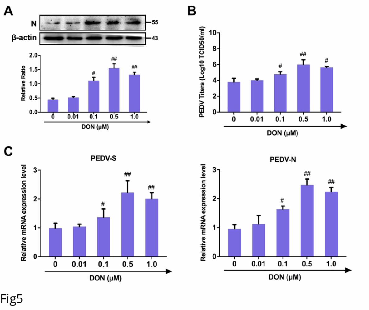
194 Low concentrations of DON facilitate PEDV replication in IPEC-J2 cells.
195 To determine the effects of DON on PEDV replication, IPEC-J2 cell monolayers were
196 infected with 1 MOI PEDV for 2 h, and cultured with DON at concentrations between
197 0.01 and 1 μM for an additional 24 h. The data showed that, compared with the PEDV
198 group, the protein level of PEDV-N (Fig 5A), the viral titer (Fig 5B) and the mRNA
199 levels of PEDV-N and -S genes (Fig 5C) were increased in PEDV-infected cells treated
200 with 0.1, 0.5 or 1 μM DON for 24 h. The maximal effects were observed at 0.5 μM
201 DON. Exposure of cells to DMSO, the solvent control, and 1.5 - 4 μM DON had no
202 effect on PEDV replication (Data not shown). Taken together, these results suggest that
203 low concentrations of DON contributed to PEDV replication in IPEC-J2 cells.
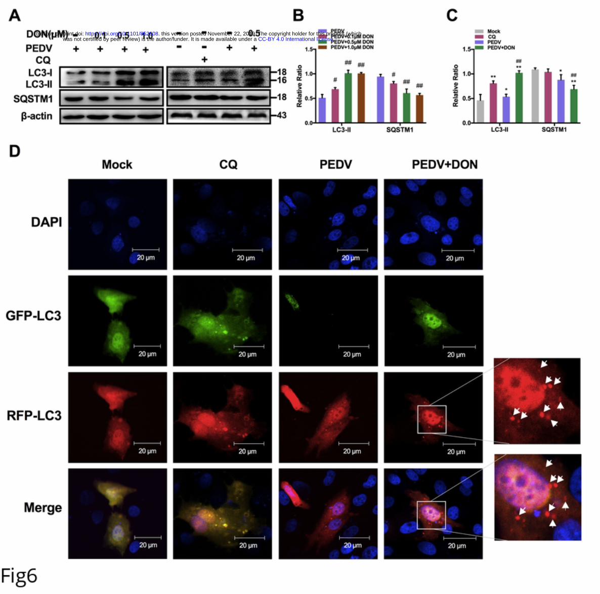
204 DON triggers a complete autophagic flux in PEDV-infected IPEC-J2 cells.
205 To determine whether autophagy could also play a role in DON-promoted PEDV
206 replication, the level of LC3B was examined and the results showed that DON
207 treatment led to a significant upregulation of LC3-II expression (Fig 6A and 6B). The
208 expression of SQSTM1 was examined to further determine whether a complete
209 autophagic flux was occurred by DON. We found that the protein level of SQSTM1 in
210 PEDV-infected IPEC-J2 cells decreased after DON treatment (Fig 6A and 6B, P < 0.05).
.CC-BY 4.0 International licensewas not certified by peer review) is the author/funder. It is made available under aThe copyright holder for this preprint (whichthis version posted November 22, 2019. . https://doi.org/10.1101/852608doi: bioRxiv preprint
11
211 The maximal effects of DON on the expression of autophagic markers were observed
212 at an DON concentration of 0.5 μM, which is consistent with that in the virus replication
213 result. Moreover, the monomeric red fluorescent protein (mRFP)-Green fluorescent
214 protein (GFP-LC3) tandem reporter construct was used to further measure DON-
215 induced autophagic flux. In the acidic pH of the lysosome, lysosomal hydrolysis can
216 attenuate the green fluorescence of this tandem autophagosome reporter, whereas it has
217 no effect on red fluorescence. Therefore, autophagosomes have both GFP and mRFP
218 signals, whereas autolysosomes have only mRFP signals [26]. As shown in Fig 5D,
219 treatment with CQ, which inhibits the fusion of autophagosomes and lysosomes,
220 resulted in yellow color-labeled autophagosomes, and RFP-LC3-labeled puncta
221 structures were detected in PEDV-infected IPEC-J2 cells expressing the mRFP-GFP-
222 LC3 reporter after incubation with 0.5 μM DON. The similar results could be observed
223 in the immunoblotting experiment (Fig 6A and 6C). These observations indicated that
224 DON induced a complete autophagic flux in PEDV-infected IPEC-J2 cells, which
225 might be responsible for DON-promoted PEDV infection.
226 CRISPR‐Cas9‐mediated knockout of LC3B in IPEC-J2 cells suppressed the
227 promotion of DON to PEDV replication.
228 To further confirm the role of autophagy in DON-promoted PEDV replication, we
229 compared the viral yield in LC3B+/+ and LC3B-/- IPEC-J2 cells exposed to DON. A
230 significant decrease was observed in the protein expression of LC3B in LC3B-/- IPEC-
231 J2 cells compared with that in LC3B-/- cells. Moreover, PEDV viral yield exhibited the
.CC-BY 4.0 International licensewas not certified by peer review) is the author/funder. It is made available under aThe copyright holder for this preprint (whichthis version posted November 22, 2019. . https://doi.org/10.1101/852608doi: bioRxiv preprint
12
232 same decrease, as demonstrated by the down-regulation of PEDV-N protein level (Fig
233 7B), PEDV viral titers (Fig 7C), and PEDV-N / -S mRNA levels (Fig 7D), indicating
234 that the decreased viral yield was due to the inhibition of autophagy. Collectively, these
235 results suggest that the LC3B-medicated autophagy machinery was required for DON-
236 promoted PEDV replication in IPEC-J2 cells.
237 Activation of p38/MTORC1 signaling pathway was required for the upregulation
238 of LC3B by DON in PEDV-infected IPEC-J2 cells.
239 To explore how DON induced autophagy, JAKs, PI3K and MAPKs signaling related-
240 proteins were detected. The immunoblotting results revealed that there was no
241 significance in JAK1, PI3K, JNK/p-JNK and ERK/p-ERK proteins expression after
242 DON treatment. But, a significant increase in p-p38 was observed (Fig 8A). In addition,
243 p-MTORC1 was significantly downregulated by DON. So, we supposed the activation
244 of p-p38 might induce autophagy. To determine our hypothesis, the inhibitor of p-p38,
245 SB202190, was supplied. The data showed that SB202190 inhibited the LC3II
246 activation and SQSTM1 degradation induced by DON, upregulated the p-MTORC1
247 expression (Fig 8B) and blocked the formation of autophagosomes (Fig 8C). Therefore,
248 DON induced autophagy via upregulating the p38/MTORC1 signaling pathway in
249 PEDV-infected IPEC-J2 cells.
250 Low concentrations of DON facilitate PEDV to escape innate immune by
251 activating autophagy in IPEC-J2 cells.
252 To explore how autophagy affected virus replication, the effects of DON on interferon
.CC-BY 4.0 International licensewas not certified by peer review) is the author/funder. It is made available under aThe copyright holder for this preprint (whichthis version posted November 22, 2019. . https://doi.org/10.1101/852608doi: bioRxiv preprint
13
253 (IFN-α, IFN-β, IFN-γ, and IFN-λ) expression in PEDV-infected IPEC-J2 cells were
254 measured as autophagy is an important component of both innate and acquired
255 immunity to pathogens [27]. Following poly (I:C) transfection, 0.1, 0.5 and 1 μM DON
256 treatment specifically inhibited the expression of IFN-α and IFN-β compared with
257 PEDV-infected cells (Fig 9A and 9B). However, there was no significance in the
258 mRNA level of IFN-γ and IFN-λ (Fig 9C and 9D) after DON treatment. The shLC3B
259 was used to confirm whether autophagy played a role in downregulation of type I
260 interfere. The results showed that DON-downregulated type I interfere could be
261 blocked by shLC3B (Fig 9E and 9F, P < 0.05). These data indicated that autophagy
262 played a key role in the suppression of antiviral innate immune by DON.
263 Autophagy-mediated STING pathway was required for PEDV escape innate
264 immune in IPEC-J2 cells treated with DON.
265 To explore how autophagy participated in the regulation of innate immune, STING
266 signaling was detected in PEDV-infected IPEC-J2 cells treated with DON. The
267 immunoblotting results revealed that DON treatment inhibited the phosphorylation of
268 STING (Fig 10A, P < 0.01), which was consistent with the changes in expression of
269 IFN-α and IFN-β. So, we supposed that autophagy downregulated the expression of
270 type I interfere via inhibiting STING signaling pathway. The scrambled and shLC3B
271 plasmids were constructed and used to determine our hypothesis. The data showed that
272 shLC3B significantly blocked the inhibitory effects of DON on STING
273 phosphorylation (Fig 10B). In addition, the inhibition of type I interfere expression
.CC-BY 4.0 International licensewas not certified by peer review) is the author/funder. It is made available under aThe copyright holder for this preprint (whichthis version posted November 22, 2019. . https://doi.org/10.1101/852608doi: bioRxiv preprint
14
274 were also significantly blocked (Fig 10C and 10D), suggesting that STING signaling
275 was required to control the replication promotion of PEDV by DON and its antiviral
276 activities were mediated by autophagy.
277 Discussion
278 The contamination of foods and feeds with DON is a significantly serious problem
279 worldwide [28-30]. Pigs are considered to be one of the most sensitive species. They
280 are frequently exposed to DON owing to grains account for a large proportion in their
281 feedstuffs. PED outbreaks caused by PEDV is now distributed all over the world [31].
282 Thus, the co-existence of DON and PEDV occurs frequently in global pig farms.
283 However, whether DON exposure may increase the susceptibility to PEDV remains
284 unknown. In this study, we provide the first strong evidence that DON exposure can
285 promote PEDV infection in vitro and in vivo, and the underlying mechanism might be
286 related to LC3B-mediated autophagy.
287 Intestinal mucosal, the first barrier to food contaminants, chemicals, and pathogens,
288 plays an important role in regulating the immune response to these stressors [32, 33].
289 Since the ability of DON to efficiently cross biological barriers, fast dividing cells such
290 as intestinal epithelial cells will be more susceptible to the detrimental effects of DON
291 [11, 34]. And PEDV mainly infects pig small intestinal epithelial cells. We then
292 hypothesized that some substances, such as DON, that lead to pig intestinal epithelial
293 cell stress might encourage the progress and spread of PED. Therefore, the intestinal
294 porcine epithelial cell line IPEC-J2 was used as an in vitro model of swine small
.CC-BY 4.0 International licensewas not certified by peer review) is the author/funder. It is made available under aThe copyright holder for this preprint (whichthis version posted November 22, 2019. . https://doi.org/10.1101/852608doi: bioRxiv preprint
15
295 intestine epithelium. Experiments in vivo were performed on twenty-seven weaning
296 piglets received the basal diet containing DON. In the present study, we found, for the
297 first time, that low concentrations of DON could facilitate PEDV infection in vivo and
298 in vitro.
299 What drives DON to promote virus infection? PEDV crosses the porcine intestinal
300 mucosa to cause intestinal infection, and then results in an acute viral enteric disease,
301 which means that PEDV must gain access to the tight junctions. As noted earlier, the
302 alteration of tight junction proteins distribution might participate in virus entry, for
303 example, occludin internalization contributes to PEDV entry [35]. In the experiments
304 reported here, we demonstrated that DON could aggravate occludin internalization in
305 PEDV-infected cells. When the occludin gene was silenced by siRNA, the promotion
306 of DON to PEDV entry in IPEC-J2 cells was disappeared simultaneously, indicating
307 that occludin internalization contributes to the DON-induced PEDV entry. And what
308 was responsible for occludin internalization was further confirmed.
309 Autophagy is a selective degradation process of various subcellular structures,
310 including protein aggregates. Increasing evidence indicated that autophagy is related to
311 cell membrane integrity and membrane proteins distribution [36]. It can serve dual roles
312 in virus infection with either pro- or anti- viral functions depending on the virus and the
313 stage of the viral replication cycle [37]. It not only is required for an antiviral response
314 against some virus infection [38], but also take an active part in the viral life cycle by,
315 eg, facilitating its entry into and release from cells [39]. In PEDV-infected cells,
.CC-BY 4.0 International licensewas not certified by peer review) is the author/funder. It is made available under aThe copyright holder for this preprint (whichthis version posted November 22, 2019. . https://doi.org/10.1101/852608doi: bioRxiv preprint
16
316 autophagy is often hijacked by viruses and manipulated to their own advantage [22].
317 Our data in this study have showed that the autophagy levels were significantly
318 increased by DON in vitro and in vivo, which are consistent with the viral infection
319 levels. CRISPR‐Cas9‐mediated knockout of the LC3B blocked the promotion of DON
320 to PEDV viral yield. Therefore, we concluded that autophagy is required for DON-
321 promoted PEDV infection, including virus entry and replication. In addition, previous
322 studies indicated that induction of autophagy is associated with enhanced JAK1 [40],
323 PI3K and MAPKs signalings [41-43]. Our study confirmed that MAPK p38 signaling
324 was enhanced in PEDV-infected IPEC-J2 cells treated with DON, accompanied by a
325 decrease in mTORC1 levels.
326 But, how did DON-activated autophagy promote PEDV replication? The primary
327 role of autophagy in innate immune is regulating the IFN-I expression [44, 45]. It is
328 well known that IFN-I is an important antiviral defense cytokine in innate immunity.
329 Upregulation of IFN-I can inhibit viral proliferation, whereas downregulation of it
330 contributes to virus infection [46]. PEDV infection is one of the main mechanisms for
331 inhibiting IFN-I signaling during continuous infection [47]. However, questions that
332 whether DON exposure could facilitate down-regulation of IFN-I in PEDV-infected
333 cells remain unanswered. We investigated the effects of DON to IFN-I expression and
334 confirmed that low concentrations of DON facilitated PEDV to escape innate immune
335 by activating autophagy in IPEC-J2 cells. Stimulator of interferon genes (STING,
336 TMEM173, MITA) is a critical component of the cellular innate immune response to
.CC-BY 4.0 International licensewas not certified by peer review) is the author/funder. It is made available under aThe copyright holder for this preprint (whichthis version posted November 22, 2019. . https://doi.org/10.1101/852608doi: bioRxiv preprint
17
337 pathogenic cytoplasmic DNA and expressed predominantly in the endoplasmic
338 reticulum (ER) [48]. It can be activated by the enzyme cGAMP synthase (cGAS) and
339 then activates interferon regulatory factors (IRFs) and NF-κB, which leads to the
340 induction of type I interferon and other immune response genes. We then investigated
341 the contribution of STING to autophagy-inhibited IFN expression for that attenuation
342 of the STING signaling can occur through autophagy [49] and confirmed that
343 autophagy-mediated STING pathway played a crucial role in the innate immune escape
344 of PEDV in IPEC-J2 cells treated with DON.
345 Current PEDV pathogenesis target primarily virus infection, little attention focus
346 on non-infectious factors. This study provides evidence for the first time that low
347 concentrations of DON can promote PEDV infection in vitro and in vivo, documenting
348 that autophagy activated by DON modulates the promotion. The present study also
349 provides new insight into developing potential novel antiviral strategies against PEDV
350 infection.
351 Materials and methods
352 Animal experiments. All experiments were conducted according to the standards of
353 the European Guidelines for Animal Welfare and were approved by the Committee for
354 the Care and Use of Experimental Animals of the Nanjing Agricultural University
355 (Animal Ethics Number: SYXK (Su) 2011-0036). Animal experiments were carried
356 out at a 1400-weaning piglets farm. Eighty weaning piglets (age 3 weeks) were selected.
357 These piglets were positive for PEDV naturally and negative for transmissible
.CC-BY 4.0 International licensewas not certified by peer review) is the author/funder. It is made available under aThe copyright holder for this preprint (whichthis version posted November 22, 2019. . https://doi.org/10.1101/852608doi: bioRxiv preprint
18
358 gastroenteritis virus and porcine rotavirus as determined by fecal and blood diagnostics.
359 Twenty-seven out of eighty piglets (BW= 5.5 ± 0.5 kg) were selected and randomly
360 divided into three groups, with 3 replicates per group and 3 piglets per replicate. Piglets
361 were assigned to 3 groups (I: control, received a basal diet, II: received the basal diet
362 containing 750 μg/kg DON, and III: received the basal diet containing 1500 μg/kg
363 DON), for 14 days. All piglets were housed in the same facility but different rooms
364 under biosafety conditions and allowed free access to water and feed during the
365 experiment. Body weight and feed intake were recorded to determine the growth
366 performance of piglets. On dpi 14, the piglets were euthanized and tissue samples of
367 duodenum, jejunum, ileum and mesenterium were collected.
368 Diarrhea rate and diarrhea index evaluation. Diarrhea in each pig was recorded and
369 scored daily according to the state of feces. Piglets with dry and cylindrical feces are
370 scored 0 point. Piglets with soft and tangible feces are scored 1 point. Piglets with sticky
371 and semi-solid feces are scored 2 points. Piglets with liquid and unformed feces are
372 scored 3 points. Diarrhea rate = [number of diarrhea piglets per replicate / (number of
373 piglets per replicate * days)] * 100%. Diarrhea index = diarrhea scores sum of piglets
374 of per replicate / (number of piglets per replicate * days).
375 Reagents and antibodies. Deoxynivalenol (DON, purity≥98%, for experiments in
376 vitro), 3-methlyadenine (3-MA), chloroquine (CQ) and rabbit anti-LC3B antibody were
377 purchased from Sigma-Aldrich (St. Louis, USA). Deoxynivalenol (DON, purity≥98%,
378 for experiments in vivo) was purchased from Pribolab (Immunos, Singapore). Rabbit
.CC-BY 4.0 International licensewas not certified by peer review) is the author/funder. It is made available under aThe copyright holder for this preprint (whichthis version posted November 22, 2019. . https://doi.org/10.1101/852608doi: bioRxiv preprint
19
379 anti-SQSTM1, anti-MAPKs, anti-JAK1, anti-pSTING/STING, anti-PI3K, anti-β-actin
380 antibodies and horseradish peroxidase (HRP)-conjugated goat anti-rabbit secondary
381 antibody were purchased from Cell Signaling Technology (Boston, USA). Rabbit anti-
382 claudin1, anti-occludin, anti-ZO-1 and anti-p-MTORC1/MTORC1 antibodies were
383 purchased from Abcam (Cambridge, UK). Porcine epidemic diarrhea virus (PEDV)
384 strain CV777 was obtained from Jiangsu Academy of Agricultural Sciences (Nanjing,
385 China). Rabbit anti-PEDV-N antibody was prepared by our lab. Poly (I:C) (LMW) /
386 LyoVecTM was purchased from InvivoGen (San Diego, USA). SB203580 was
387 purchased from MCE (New Jersey, USA).
388 Histological analysis. Jejunum tissues samples were fixed in 4% paraformaldehyde,
389 embedded in paraffin and sectioned at a thickness of 4 μm. For histopathological
390 examination, tissue slices were stained with hematoxylin and eosin, and observed under
391 the microscope. For immunohistochemistry examination, tissue slices were incubated
392 with antibodies against PEDV N protein, followed by incubation with the second
393 antibody and streptavidin-peroxidase complex. The peroxidase conjugates were
394 visualized using DAB solution.
395 Quantitative Real-Time PCR (RT-qPCR) Analysis. RT-qPCR was performed using
396 the StepOnePlus Real-Time PCR System (Applied Biosciences) as previously
397 described [50]. Total RNA was isolated from the cells using an RNA Extraction Kit
398 (Takara, Japan) and then reverse-transcribed to cDNA using the PrimeScript RT Master
399 Mix Kit (Takara, Japan). All primers are listed in Table 1. The relative expression was
.CC-BY 4.0 International licensewas not certified by peer review) is the author/funder. It is made available under aThe copyright holder for this preprint (whichthis version posted November 22, 2019. . https://doi.org/10.1101/852608doi: bioRxiv preprint
20
400 determined using the Δcycle threshold (ΔCt) method with GAPDH serving as a
401 reference gene.
402 Table 1 Primers used in this study.
Primers Sequence (5’ - 3’) Purpose
GAPDHF: TGGGCGTGAACCATGAGAA
R: CCTCCACGATGCCGAAGT
RT-PCR for detection of
GAPDH
PEDV-NF: GTCTGA-CAACAGCGGCAAAA
R: TTTCGCCCTTGGGAATTCTC
RT-PCR for detection of
PEDV-N
PEDV-SF: TGTTTATTCTGTCACGCCATGTT
R: CCAGGCAACTCCCTAGTATTGCT
RT-PCR for detection of
PEDV-S
IFN-αF: ACTTCCACAGACTCACCCTCTATC
R: ATGACTTCTGCCCTGATGATCT
RT-PCR for detection of
IFN-α
IFN-βF: TGCATCCTCCAAATCGCTCT
R: ATTGAGGAGTCCCAGGCAAC
RT-PCR for detection of
IFN-β
IFN-γF: TTTTGTCACTCTCCTCTTTCCA
R: GACTTTGTGTTTTTCTGGCTCTTAC
RT-PCR for detection of
IFN-γ
IFN-λF: GGTGCTGGCGACTGTGATG
R: GATTGGAACTGGCCCATGTG
RT-PCR for detection of
IFN-λ
shLC3BGCTTGCAGCTCAATGCTAACCCTCGA
GGGTTAGCATTGAGCTGCAAGCKnockdown of LC3B
.CC-BY 4.0 International licensewas not certified by peer review) is the author/funder. It is made available under aThe copyright holder for this preprint (whichthis version posted November 22, 2019. . https://doi.org/10.1101/852608doi: bioRxiv preprint
21
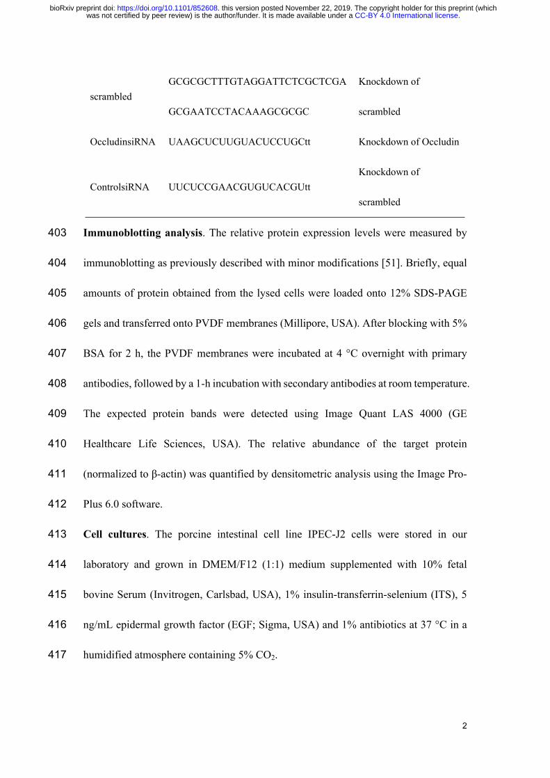
scrambledGCGCGCTTTGTAGGATTCTCGCTCGA
GCGAATCCTACAAAGCGCGC
Knockdown of
scrambled
OccludinsiRNA UAAGCUCUUGUACUCCUGCtt Knockdown of Occludin
ControlsiRNA UUCUCCGAACGUGUCACGUttKnockdown of
scrambled
403 Immunoblotting analysis. The relative protein expression levels were measured by
404 immunoblotting as previously described with minor modifications [51]. Briefly, equal
405 amounts of protein obtained from the lysed cells were loaded onto 12% SDS-PAGE
406 gels and transferred onto PVDF membranes (Millipore, USA). After blocking with 5%
407 BSA for 2 h, the PVDF membranes were incubated at 4 °C overnight with primary
408 antibodies, followed by a 1-h incubation with secondary antibodies at room temperature.
409 The expected protein bands were detected using Image Quant LAS 4000 (GE
410 Healthcare Life Sciences, USA). The relative abundance of the target protein
411 (normalized to β-actin) was quantified by densitometric analysis using the Image Pro-
412 Plus 6.0 software.
413 Cell cultures. The porcine intestinal cell line IPEC-J2 cells were stored in our
414 laboratory and grown in DMEM/F12 (1:1) medium supplemented with 10% fetal
415 bovine Serum (Invitrogen, Carlsbad, USA), 1% insulin-transferrin-selenium (ITS), 5
416 ng/mL epidermal growth factor (EGF; Sigma, USA) and 1% antibiotics at 37 °C in a
417 humidified atmosphere containing 5% CO2.
.CC-BY 4.0 International licensewas not certified by peer review) is the author/funder. It is made available under aThe copyright holder for this preprint (whichthis version posted November 22, 2019. . https://doi.org/10.1101/852608doi: bioRxiv preprint
22
418 Cell viability assay. Cell viability was monitored by 3-(4,5-dimethyl-2-thiazolyl)-2,5-
419 diphenyl-2-H-tetrazolium bromide (MTT; Sigma, USA) assay as previously described
420 [52]. Briefly, IPEC-J2 cells were cultured in 96-well plates at a density of 5 × 10³
421 cells/well with corresponding treatments. Then, each well was added with 15 μl of MTT
422 (5 mg/ml) for another 4 h at 37 °C. The supernatants were discarded and incubated with
423 150 μl DMSO to dissolve the precipitate. Absorbance was measured at 490 nm with a
424 reference wavelength of 595 nm. All tests were performed three times.
425 LC3B−/− IPEC-J2 cell production by CRISPR/ Cas9 system
426 The small guide RNAs (sgRNAs) were designed using Breaking‐Cas (http://
427 bioinfogp.cnb.csic.es/tools/breakingcas/) online tool and synthesized (Invitrogen). The
428 sgRNA was cloned pCas-Puro-U6 plasmid and the pCas-Puro-U6 plasmid Linear was
429 obtained using the BbsI restriction enzyme (Thermo Fisher Scientific). The plasmids
430 containing sgRNA were transfected into IPEC-J2 cells with GeneTran III (Biomiga)
431 for 48 h, and then the transfected cells were selected using 5 μg/mL of puromycin. The
432 selected cells were subjected to serial dilutions in 96‐well plate to obtain a single cell
433 colony. After 14 days of colony formation, each single colony was picked and expanded.
434 Genomic DNA was extracted from individual clones and sequenced to confirm the
435 specificity of targeting.
436 Fluorescence Microscopy. Cells grown on coverslips were fixed with 4%
437 paraformaldehyde for 20 min at 4 °C. After washing three times, cells were blocked
438 with 1% BSA at room temperature and incubated with primary rabbit anti-occludin
.CC-BY 4.0 International licensewas not certified by peer review) is the author/funder. It is made available under aThe copyright holder for this preprint (whichthis version posted November 22, 2019. . https://doi.org/10.1101/852608doi: bioRxiv preprint
23
439 antibody and secondary FITC-conjugated goat anti-rabbit antibody (Invitrogen, USA),
440 respectively. For the analysis of LC3B expression, cells grown on coverslips to 60-70%
441 confluence were transfected with the pLVX-mRFP-EGFP-LC3B plasmid (provided by
442 Prof. Qian Yang, Nanjing Agriculture University, Nanjing, China) using jetPRIME
443 transfection reagent (Plolyplus-transfection, Illkirch, France) according to the
444 manufacturer’s protocols. Nuclei were stained with DAPI (Blue, Beyotime
445 Biotechnology, China). Fluorescence microscopy was performed using a Zeiss
446 LSM710 confocal microscope (Zeiss, Oberkochen, Germany).
447 RNA interference. Occludin-specific siRNA and control siRNA were designed and
448 synthesized by Invitrogen (Thermo Fisher, USA). All primers are listed in Table 1.
449 Cells were transfected with 100 nM occludin-specific or control siRNA duplexes by
450 use of jetPRIME transfection reagent according to the manufacturer’s guidelines.
451 Twenty-four hours after transfection, cells were washed with DMEM/F12 and cultured
452 in DMEM/F12 with 4% FBS until further treatments.
453 Quantification of virus titer. Viral titers were determined by 50% endpoint dilution
454 (50% tissue culture infective dose [TCID50]) assays on IPEC-J2 cells as previously
455 described [52]. Briefly, IPEC-J2 cells cultured in 96-well plates were inoculated with
456 10-fold dilutions of the harvested culture supernatants for indicated time. Microscope
457 was used to detect the viral antigen according to the cell damage. Viral titers were
458 expressed as TCID50/ml by using the Reed-Muench method.
.CC-BY 4.0 International licensewas not certified by peer review) is the author/funder. It is made available under aThe copyright holder for this preprint (whichthis version posted November 22, 2019. . https://doi.org/10.1101/852608doi: bioRxiv preprint
24
459 Flow Cytometry. Apoptotic cell death was measured by Annexin V/propidium iodide
460 (PI) staining assay (BD Pharmingen, USA) using flow cytometry (FACS Calibur, BD
461 Biosciences, USA) according to manufacturer’s instructions. In a word, the harvested
462 cells were resuspended in 100 μl binding buffer followed by incubation with 10 μl
463 Annexin V per test for 10 min, and 10 μl PI per test was added for 5 min. Cells were
464 then suspended in 500 μl of binding buffer and immediately analyzed by FACS.
465 Statistical Analysis. Statistical analyses were performed using Graph Pad Prism 7.0 by
466 one-way analysis of variance (ANOVA), and the data were expressed as the means ±
467 standard deviation (SD). P < 0.05 was regarded as significant.
468 Acknowledgements
469 This study was financially supported by the National Key Research and Development
470 Program (2017YFD0501001), the National Natural Science Foundation of China
471 (31772811 and 31602123) and the Priority Academic Program Development of Jiangsu
472 Higher Education Institutions (Jiangsu, China).
473 Conflict of interest
474 The authors have no conflicts of interest to disclose.
475 Reference
476 1. Song D, Park B. Porcine epidemic diarrhoea virus: a comprehensive review of
477 molecular epidemiology, diagnosis, and vaccines. Virus genes. 2012;44(2):167-75.
478 Epub 2012/01/25. doi: 10.1007/s11262-012-0713-1. PubMed PMID: 22270324.
479 2. Madson DM, Magstadt DR, Arruda PH, Hoang H, Sun D, Bower LP, et al.
.CC-BY 4.0 International licensewas not certified by peer review) is the author/funder. It is made available under aThe copyright holder for this preprint (whichthis version posted November 22, 2019. . https://doi.org/10.1101/852608doi: bioRxiv preprint
25
480 Pathogenesis of porcine epidemic diarrhea virus isolate (US/Iowa/18984/2013) in 3-
481 week-old weaned pigs. Vet Microbiol. 2014;174(1-2):60-8. doi:
482 10.1016/j.vetmic.2014.09.002. PubMed PMID: 25278366.
483 3. Li W, Li H, Liu Y, Pan Y, Deng F, Song Y, et al. New variants of porcine epidemic
484 diarrhea virus, China, 2011. Emerging infectious diseases. 2012;18(8):1350-3. Epub
485 2012/07/31. doi: 10.3201/eid1808.120002. PubMed PMID: 22840964; PubMed
486 Central PMCID: PMCPMC3414035.
487 4. Lee YH, Su SB, Huang CC, Sheu HM, Tsai JC, Lin CH, et al. N-acetylcysteine
488 attenuates hexavalent chromium-induced hypersensitivity through inhibition of cell
489 death, ROS-related signaling and cytokine expression. PloS one. 2014;9(9):e108317.
490 doi: 10.1371/journal.pone.0108317. PubMed PMID: 25248126; PubMed Central
491 PMCID: PMCPMC4172727.
492 5. Tian Y, Yu Z, Cheng K, Liu Y, Huang J, Xin Y, et al. Molecular characterization
493 and phylogenetic analysis of new variants of the porcine epidemic diarrhea virus in
494 Gansu, China in 2012. Viruses. 2013;5(8):1991-2004. doi: 10.3390/v5081991. PubMed
495 PMID: 23955500; PubMed Central PMCID: PMCPMC3761238.
496 6. Kim YK, Lim SI, Lim JA, Cho IS, Park EH, Le VP, et al. A novel strain of porcine
497 epidemic diarrhea virus in Vietnamese pigs. Archives of virology. 2015;160(6):1573-
498 7. Epub 2015/04/13. doi: 10.1007/s00705-015-2411-5. PubMed PMID: 25864174.
499 7. Ojkic D, Hazlett M, Fairles J, Marom A, Slavic D, Maxie G, et al. The first case of
500 porcine epidemic diarrhea in Canada. The Canadian veterinary journal = La revue
.CC-BY 4.0 International licensewas not certified by peer review) is the author/funder. It is made available under aThe copyright holder for this preprint (whichthis version posted November 22, 2019. . https://doi.org/10.1101/852608doi: bioRxiv preprint
26
501 veterinaire canadienne. 2015;56(2):149-52. Epub 2015/02/20. PubMed PMID:
502 25694663; PubMed Central PMCID: PMCPMC4298265.
503 8. Annamalai T, Saif LJ, Lu Z, Jung K. Age-dependent variation in innate immune
504 responses to porcine epidemic diarrhea virus infection in suckling versus weaned pigs.
505 Vet Immunol Immunopathol. 2015;168(3-4):193-202. Epub 2015/10/05. doi:
506 10.1016/j.vetimm.2015.09.006. PubMed PMID: 26433606.
507 9. Kim Y, Yang M, Goyal SM, Cheeran MC, Torremorell M. Evaluation of
508 biosecurity measures to prevent indirect transmission of porcine epidemic diarrhea
509 virus. BMC veterinary research. 2017;13(1):89. Epub 2017/04/07. doi:
510 10.1186/s12917-017-1017-4. PubMed PMID: 28381304; PubMed Central PMCID:
511 PMCPMC5382501.
512 10. Lowe J, Gauger P, Harmon K, Zhang J, Connor J, Yeske P, et al. Role of
513 transportation in spread of porcine epidemic diarrhea virus infection, United States.
514 Emerging infectious diseases. 2014;20(5):872-4. Epub 2014/04/23. doi:
515 10.3201/eid2005.131628. PubMed PMID: 24750785; PubMed Central PMCID:
516 PMCPMC4012813.
517 11. Danicke S, Hegewald AK, Kahlert S, Kluess J, Rothkotter HJ, Breves G, et al.
518 Studies on the toxicity of deoxynivalenol (DON), sodium metabisulfite, DON-sulfonate
519 (DONS) and de-epoxy-DON for porcine peripheral blood mononuclear cells and the
520 Intestinal Porcine Epithelial Cell lines IPEC-1 and IPEC-J2, and on effects of DON and
521 DONS on piglets. Food Chem Toxicol. 2010;48(8-9):2154-62. doi:
.CC-BY 4.0 International licensewas not certified by peer review) is the author/funder. It is made available under aThe copyright holder for this preprint (whichthis version posted November 22, 2019. . https://doi.org/10.1101/852608doi: bioRxiv preprint
27
522 10.1016/j.fct.2010.05.022. PubMed PMID: 20478350.
523 12. Pestka JJ. Deoxynivalenol: mechanisms of action, human exposure, and
524 toxicological relevance. Archives of Toxicology. 2010;84(9):663-79. doi:
525 10.1007/s00204-010-0579-8.
526 13. Pierron A, Alassane-Kpembi I, Oswald IP. Impact of two mycotoxins
527 deoxynivalenol and fumonisin on pig intestinal health. Porcine Health Management.
528 2016;2(1):21. doi: 10.1186/s40813-016-0041-2.
529 14. Broekaert N, Devreese M, van Bergen T, Schauvliege S, De Boevre M, De Saeger
530 S, et al. In vivo contribution of deoxynivalenol-3-β-d-glucoside to deoxynivalenol
531 exposure in broiler chickens and pigs: oral bioavailability, hydrolysis and
532 toxicokinetics. Archives of Toxicology. 2017;91(2):699-712. doi: 10.1007/s00204-
533 016-1710-2.
534 15. Pott J, Maloy KJ. Epithelial autophagy controls chronic colitis by reducing TNF-
535 induced apoptosis. Autophagy. 2018;14(8):1460-1. Epub 2018/05/26. doi:
536 10.1080/15548627.2018.1450021. PubMed PMID: 29799774; PubMed Central
537 PMCID: PMCPMC6103553.
538 16. Matsuda J, Namba T, Takabatake Y, Kimura T, Takahashi A, Yamamoto T, et al.
539 Antioxidant role of autophagy in maintaining the integrity of glomerular capillaries.
540 Autophagy. 2018;14(1):53-65. Epub 2017/11/14. doi:
541 10.1080/15548627.2017.1391428. PubMed PMID: 29130363; PubMed Central
542 PMCID: PMCPMC5846506.
.CC-BY 4.0 International licensewas not certified by peer review) is the author/funder. It is made available under aThe copyright holder for this preprint (whichthis version posted November 22, 2019. . https://doi.org/10.1101/852608doi: bioRxiv preprint
28
543 17. Ramkumar A, Murthy D, Raja DA, Singh A, Krishnan A, Khanna S, et al. Classical
544 autophagy proteins LC3B and ATG4B facilitate melanosome movement on
545 cytoskeletal tracks. Autophagy. 2017;13(8):1331-47. Epub 2017/06/10. doi:
546 10.1080/15548627.2017.1327509. PubMed PMID: 28598240; PubMed Central
547 PMCID: PMCPMC5584859.
548 18. Liu Z, Sin KWT, Ding H, Doan HA, Gao S, Miao H, et al. p38beta MAPK mediates
549 ULK1-dependent induction of autophagy in skeletal muscle of tumor-bearing mice.
550 Cell stress. 2018;2(11):311-24. Epub 2019/06/22. doi: 10.15698/cst2018.11.163.
551 PubMed PMID: 31225455; PubMed Central PMCID: PMCPMC6551802.
552 19. Riffelmacher T, Richter FC, Simon AK. Autophagy dictates metabolism and
553 differentiation of inflammatory immune cells. Autophagy. 2018;14(2):199-206. Epub
554 2017/08/15. doi: 10.1080/15548627.2017.1362525. PubMed PMID: 28806133;
555 PubMed Central PMCID: PMCPMC5902226.
556 20. McEwan DG. Host-pathogen interactions and subversion of autophagy. Essays in
557 biochemistry. 2017;61(6):687-97. Epub 2017/12/14. doi: 10.1042/ebc20170058.
558 PubMed PMID: 29233878; PubMed Central PMCID: PMCPMC5869863.
559 21. Yang Q, Liu TT, Lin H, Zhang M, Wei J, Luo WW, et al. TRIM32-TAX1BP1-
560 dependent selective autophagic degradation of TRIF negatively regulates TLR3/4-
561 mediated innate immune responses. PLoS Pathog. 2017;13(9):e1006600. Epub
562 2017/09/13. doi: 10.1371/journal.ppat.1006600. PubMed PMID: 28898289; PubMed
563 Central PMCID: PMCPMC5595311.
.CC-BY 4.0 International licensewas not certified by peer review) is the author/funder. It is made available under aThe copyright holder for this preprint (whichthis version posted November 22, 2019. . https://doi.org/10.1101/852608doi: bioRxiv preprint
29
564 22. Guo X, Zhang M, Zhang X, Tan X, Guo H, Zeng W, et al. Porcine Epidemic
565 Diarrhea Virus Induces Autophagy to Benefit Its Replication. Viruses. 2017;9(3):53.
566 doi: 10.3390/v9030053.
567 23. Harris KG, Morosky SA, Drummond CG, Patel M, Kim C, Stolz DB, et al. RIP3
568 Regulates Autophagy and Promotes Coxsackievirus B3 Infection of Intestinal
569 Epithelial Cells. Cell Host Microbe. 2015;18(2):221-32. doi:
570 10.1016/j.chom.2015.07.007. PubMed PMID: 26269957; PubMed Central PMCID:
571 PMCPMC4562276.
572 24. Chen Y, Chen Q, Li M, Mao Q, Chen H, Wu W, et al. Autophagy pathway induced
573 by a plant virus facilitates viral spread and transmission by its insect vector. PLoS
574 Pathog. 2017;13(11):e1006727. Epub 2017/11/11. doi: 10.1371/journal.ppat.1006727.
575 PubMed PMID: 29125860; PubMed Central PMCID: PMCPMC5708841.
576 25. Pasternak JA, Aiyer VIA, Hamonic G, Beaulieu AD, Columbus DA, Wilson HL.
577 Molecular and Physiological Effects on the Small Intestine of Weaner Pigs Following
578 Feeding with Deoxynivalenol-Contaminated Feed. Toxins (Basel). 2018;10(1). Epub
579 2018/01/13. doi: 10.3390/toxins10010040. PubMed PMID: 29329218; PubMed
580 Central PMCID: PMCPMC5793127.
581 26. Xing Y, Liqi Z, Jian L, Qinghua Y, Qian Y. Doxycycline Induces Mitophagy and
582 Suppresses Production of Interferon-beta in IPEC-J2 Cells. Frontiers in cellular and
583 infection microbiology. 2017;7:21. Epub 2017/02/17. doi: 10.3389/fcimb.2017.00021.
584 PubMed PMID: 28203548; PubMed Central PMCID: PMCPMC5285722.
.CC-BY 4.0 International licensewas not certified by peer review) is the author/funder. It is made available under aThe copyright holder for this preprint (whichthis version posted November 22, 2019. . https://doi.org/10.1101/852608doi: bioRxiv preprint
30
585 27. Sumpter R, Jr., Levine B. Autophagy and innate immunity: triggering, targeting
586 and tuning. Seminars in cell & developmental biology. 2010;21(7):699-711. Epub
587 2010/04/21. doi: 10.1016/j.semcdb.2010.04.003. PubMed PMID: 20403453; PubMed
588 Central PMCID: PMCPMC2930105.
589 28. Yu M, Chen L, Peng Z, Nussler AK, Wu Q, Liu L, et al. Mechanism of
590 deoxynivalenol effects on the reproductive system and fetus malformation: Current
591 status and future challenges. Toxicology in vitro : an international journal published in
592 association with BIBRA. 2017;41:150-8. Epub 2017/03/14. doi:
593 10.1016/j.tiv.2017.02.011. PubMed PMID: 28286114.
594 29. Liao Y, Peng Z, Chen L, Nussler AK, Liu L, Yang W. Deoxynivalenol, gut
595 microbiota and immunotoxicity: A potential approach? Food Chem Toxicol.
596 2018;112:342-54. Epub 2018/01/15. doi: 10.1016/j.fct.2018.01.013. PubMed PMID:
597 29331731.
598 30. Escriva L, Font G, Manyes L. In vivo toxicity studies of fusarium mycotoxins in
599 the last decade: a review. Food Chem Toxicol. 2015;78:185-206. Epub 2015/02/15.
600 doi: 10.1016/j.fct.2015.02.005. PubMed PMID: 25680507.
601 31. Lee C. Porcine epidemic diarrhea virus: An emerging and re-emerging epizootic
602 swine virus. Virology journal. 2015;12:193. Epub 2015/12/23. doi: 10.1186/s12985-
603 015-0421-2. PubMed PMID: 26689811; PubMed Central PMCID: PMCPMC4687282.
604 32. Kagnoff MF. The intestinal epithelium is an integral component of a
605 communications network. The Journal of clinical investigation. 2014;124(7):2841-3.
.CC-BY 4.0 International licensewas not certified by peer review) is the author/funder. It is made available under aThe copyright holder for this preprint (whichthis version posted November 22, 2019. . https://doi.org/10.1101/852608doi: bioRxiv preprint
31
606 Epub 2014/07/02. doi: 10.1172/jci75225. PubMed PMID: 24983425; PubMed Central
607 PMCID: PMCPMC4071395.
608 33. Perez-Lopez A, Behnsen J, Nuccio SP, Raffatellu M. Mucosal immunity to
609 pathogenic intestinal bacteria. Nature reviews Immunology. 2016;16(3):135-48. Epub
610 2016/02/24. doi: 10.1038/nri.2015.17. PubMed PMID: 26898110.
611 34. Maresca M. From the gut to the brain: journey and pathophysiological effects of
612 the food-associated trichothecene mycotoxin deoxynivalenol. Toxins (Basel).
613 2013;5(4):784-820. doi: 10.3390/toxins5040784. PubMed PMID: 23612752; PubMed
614 Central PMCID: PMCPMC3705292.
615 35. Luo X, Guo L, Zhang J, Xu Y, Gu W, Feng L, et al. Tight Junction Protein Occludin
616 Is a Porcine Epidemic Diarrhea Virus Entry Factor. Journal of virology. 2017;91(10).
617 doi: 10.1128/JVI.00202-17. PubMed PMID: 28275187; PubMed Central PMCID:
618 PMCPMC5411586.
619 36. Mansilla Pareja ME, Bongiovanni A, Lafont F, Colombo MI. Alterations of the
620 Coxiella burnetii Replicative Vacuole Membrane Integrity and Interplay with the
621 Autophagy Pathway. Frontiers in cellular and infection microbiology. 2017;7:112.
622 Epub 2017/05/10. doi: 10.3389/fcimb.2017.00112. PubMed PMID: 28484683;
623 PubMed Central PMCID: PMCPMC5401879.
624 37. Paul P, Münz C. Chapter Four - Autophagy and Mammalian Viruses: Roles in
625 Immune Response, Viral Replication, and Beyond. In: Kielian M, Maramorosch K,
626 Mettenleiter TC, editors. Advances in Virus Research. 95: Academic Press; 2016. p.
.CC-BY 4.0 International licensewas not certified by peer review) is the author/funder. It is made available under aThe copyright holder for this preprint (whichthis version posted November 22, 2019. . https://doi.org/10.1101/852608doi: bioRxiv preprint
32
627 149-95.
628 38. Moy RH, Gold B, Molleston JM, Schad V, Yanger K, Salzano MV, et al. Antiviral
629 autophagy restrictsRift Valley fever virus infection and is conserved from flies to
630 mammals. Immunity. 2014;40(1):51-65. Epub 2014/01/01. doi:
631 10.1016/j.immuni.2013.10.020. PubMed PMID: 24374193; PubMed Central PMCID:
632 PMCPMC3951734.
633 39. Montespan C, Marvin SA, Austin S, Burrage AM, Roger B, Rayne F, et al. Multi-
634 layered control of Galectin-8 mediated autophagy during adenovirus cell entry through
635 a conserved PPxY motif in the viral capsid. PLoS Pathog. 2017;13(2):e1006217. Epub
636 2017/02/14. doi: 10.1371/journal.ppat.1006217. PubMed PMID: 28192531; PubMed
637 Central PMCID: PMCPMC5325606.
638 40. Fleming SB. Viral Inhibition of the IFN-Induced JAK/STAT Signalling Pathway:
639 Development of Live Attenuated Vaccines by Mutation of Viral-Encoded IFN-
640 Antagonists. Vaccines. 2016;4(3). Epub 2016/07/02. doi: 10.3390/vaccines4030023.
641 PubMed PMID: 27367734; PubMed Central PMCID: PMCPMC5041017.
642 41. Schmeisser H, Fey SB, Horowitz J, Fischer ER, Balinsky CA, Miyake K, et al.
643 Type I interferons induce autophagy in certain human cancer cell lines. Autophagy.
644 2013;9(5):683-96. doi: 10.4161/auto.23921. PubMed PMID: 23419269; PubMed
645 Central PMCID: PMCPMC3669179.
646 42. Schmeisser H, Bekisz J, Zoon KC. New function of type I IFN: induction of
647 autophagy. Journal of interferon & cytokine research : the official journal of the
.CC-BY 4.0 International licensewas not certified by peer review) is the author/funder. It is made available under aThe copyright holder for this preprint (whichthis version posted November 22, 2019. . https://doi.org/10.1101/852608doi: bioRxiv preprint
33
648 International Society for Interferon and Cytokine Research. 2014;34(2):71-8. Epub
649 2014/01/17. doi: 10.1089/jir.2013.0128. PubMed PMID: 24428799; PubMed Central
650 PMCID: PMCPMC3924851.
651 43. Xu D, Zhang T, Xiao J, Zhu K, Wei R, Wu Z, et al. Modification of BECN1 by
652 ISG15 plays a crucial role in autophagy regulation by type I IFN/interferon. Autophagy.
653 2015;11(4):617-28. doi: 10.1080/15548627.2015.1023982. PubMed PMID: 25906440;
654 PubMed Central PMCID: PMCPMC4502663.
655 44. Tian Y, Wang ML, Zhao J. Crosstalk between Autophagy and Type I Interferon
656 Responses in Innate Antiviral Immunity. Viruses. 2019;11(2). Epub 2019/02/06. doi:
657 10.3390/v11020132. PubMed PMID: 30717138.
658 45. Martin PK, Marchiando A, Xu R, Rudensky E, Yeung F, Schuster SL, et al.
659 Autophagy proteins suppress protective type I interferon signalling in response to the
660 murine gut microbiota. Nature microbiology. 2018;3(10):1131-41. Epub 2018/09/12.
661 doi: 10.1038/s41564-018-0229-0. PubMed PMID: 30202015; PubMed Central PMCID:
662 PMCPMC6179362.
663 46. Song J, Hu Y, Li J, Zheng H, Wang J, Guo L, et al. Suppression of the toll-like
664 receptor 7-dependent type I interferon production pathway by autophagy resulting from
665 enterovirus 71 and coxsackievirus A16 infections facilitates their replication. Archives
666 of virology. 2018;163(1):135-44. Epub 2017/10/21. doi: 10.1007/s00705-017-3592-x.
667 PubMed PMID: 29052054; PubMed Central PMCID: PMCPMC5756282.
668 47. Deng X, van Geelen A, Buckley AC, O'Brien A, Pillatzki A, Lager KM, et al.
.CC-BY 4.0 International licensewas not certified by peer review) is the author/funder. It is made available under aThe copyright holder for this preprint (whichthis version posted November 22, 2019. . https://doi.org/10.1101/852608doi: bioRxiv preprint
34
669 Coronavirus endoribonuclease activity in porcine epidemic diarrhea virus suppresses
670 type I and type III interferon responses. Journal of virology. 2019. Epub 2019/02/08.
671 doi: 10.1128/jvi.02000-18. PubMed PMID: 30728254.
672 48. Garcia-Belmonte R, Perez-Nunez D, Pittau M, Richt JA, Revilla Y. African Swine
673 Fever Virus Armenia/07 Virulent Strain Controls Interferon Beta Production through
674 the cGAS-STING Pathway. Journal of virology. 2019;93(12). Epub 2019/03/29. doi:
675 10.1128/jvi.02298-18. PubMed PMID: 30918080; PubMed Central PMCID:
676 PMCPMC6613762.
677 49. Prabakaran T, Bodda C, Krapp C, Zhang BC, Christensen MH, Sun C, et al.
678 Attenuation of cGAS-STING signaling is mediated by a p62/SQSTM1-dependent
679 autophagy pathway activated by TBK1. The EMBO journal. 2018;37(8). Epub
680 2018/03/03. doi: 10.15252/embj.201797858. PubMed PMID: 29496741; PubMed
681 Central PMCID: PMCPMC5897779.
682 50. Liu D, Su J, Lin J, Qian G, Chen X, Song S, et al. Activation of AMPK-dependent
683 SIRT-1 by astragalus polysaccharide protects against ochratoxin A-induced immune
684 stress in vitro and in vivo. International journal of biological macromolecules.
685 2018;120(Pt A):683-92. Epub 2018/09/01. doi: 10.1016/j.ijbiomac.2018.08.156.
686 PubMed PMID: 30170064.
687 51. Qian G, Liu D, Hou L, Hamid M, Chen X, Gan F, et al. Ochratoxin A induces
688 cytoprotective autophagy via blocking AKT/mTOR signaling pathway in PK-15cells.
689 Food and chemical toxicology : an international journal published for the British
.CC-BY 4.0 International licensewas not certified by peer review) is the author/funder. It is made available under aThe copyright holder for this preprint (whichthis version posted November 22, 2019. . https://doi.org/10.1101/852608doi: bioRxiv preprint
35
690 Industrial Biological Research Association. 2018;122:120-31. Epub 2018/10/06. doi:
691 10.1016/j.fct.2018.09.070. PubMed PMID: 30287338.
692 52. Liu D, Lin J, Su J, Chen X, Jiang P, Huang K. Glutamine Deficiency Promotes
693 PCV2 Infection through Induction of Autophagy via Activation of ROS-Mediated
694 JAK2/STAT3 Signaling Pathway. J Agric Food Chem. 2018;66(44):11757-66. doi:
695 10.1021/acs.jafc.8b04704. PubMed PMID: 30343565.
696 Figure Captions
697 Fig 1. Low concentrations of DON exposure could aggravate intestinal injury and
698 facilitates PEDV infection in weaning piglets. Piglets infected naturally with PEDV
699 were fed with a basal diet containing 0, 750 or 1500 μg/kg DON. (A) Effects of DON
700 on average daily gain and small intestine weight of piglets. (B) Effects of DON on
701 diarrhea rate and diarrhea index of piglets. (C) Histopathological examination.
702 (magnification, × 200) (D) The villus length and villus/crypt ratio of jejunums were
703 quantified. (E) TEM observation. The virus particles (black arrowheads) and the fine
704 ultrastructure of autophagosomes (black asterisk) were observed. The scale bar
705 indicates 1.0 μm. (F) Immunohistochemistry examination. The PEDV-N (brown
706 signals) expression in jejunums of piglets was measured. (magnification, × 200) (G)
707 Effects of DON on the mRNA levels of PEDV-N gene in duodenum, jejunum, ileum
708 and mesenterium. (H, I and J) Effects of DON on the protein levels of PEDV-N and
709 LC3B in duodenum, jejunum, ileum and mesenterium. The data are expressed as mean
710 ± SD (n=3). # P < 0.05, ## P < 0.01 vs. PEDV. TEM: transmission electron microscopy.
.CC-BY 4.0 International licensewas not certified by peer review) is the author/funder. It is made available under aThe copyright holder for this preprint (whichthis version posted November 22, 2019. . https://doi.org/10.1101/852608doi: bioRxiv preprint
36
711 Fig 2. Low concentrations of DON promoted the entry of PEDV in IPEC-J2 cells.
712 (A) Effects of DON on the cell viability and LDH release of IPEC-J2 cells. IPEC-J2
713 cell monolayers were cultured with various concentrations of DON for 48 h, cell
714 viability and LDH release were assayed as described in Materials and Methods. (B)
715 Effects of low concentrations DON on the PEDV attachment, entry and release. IPEC-
716 J2 cell monolayers were infected with 2 MOI PEDV and further cultured as indicated.
717 Cell lysates were subjected to immunoblotting with antibodies to PEDV nucleocapsid
718 (N) protein or β-actin (loading control). The data are expressed as mean ± SD (n=3). *
719 P < 0.05, ** P < 0.01 vs. control (mock); # P < 0.05, ## P < 0.01 vs. PEDV. DON:
720 deoxynivalenol. LDH: lactate dehydrogenase.
721 Fig 3. Occludin internalization was required for DON-promoted PEDV entry in
722 IPEC-J2 cells. (A, B) Effects of various concentrations DON on the expression and
723 distribution of tight junction proteins in PEDV-infected IPEC-J2 cells. (C, D) Effects
724 of occludin konckdown on PEDV entry in IPEC-J2 cells exposed to 0.5 μM DON.
725 IPEC-J2 cell monolayers were infected with 2 MOI PEDV and further cultured as
726 indicated. Cell lysates were subjected to immunoblotting (A, C) with antibodies to ZO-
727 1, occludin, claudin-1, PEDV-N protein or β-actin (loading control) and subjected to
728 IFA (B, D) with antibodies to occludin (green), claudin-1 (green) and PEDV-N protein
729 (red). Cell nuclei were stained with DAPI (blue). The scale bar indicates 20 μm. The
730 data are expressed as mean ± SD (n=3). # P < 0.05, ## P < 0.01 vs. PEDV; $ P < 0.05,
731 $$ P < 0.01 vs. PEDV+DON. IFA: Immunofluorescence analysis.
.CC-BY 4.0 International licensewas not certified by peer review) is the author/funder. It is made available under aThe copyright holder for this preprint (whichthis version posted November 22, 2019. . https://doi.org/10.1101/852608doi: bioRxiv preprint
37
732 Fig 4. LC3B was required for occludin internalization-induced PEDV entry in
733 IPEC-J2 cells exposed to DON. (A) Generation of LC3B‐knockout IPEC-J2 cells. (B,
734 C) Effects of autophagy deficiency on the occludin expression and PEDV entry. IPEC-
735 J2 cell monolayers were infected with 2 MOI PEDV and further cultured as indicated.
736 Cell lysates were subjected to immunoblotting (B) with antibodies to occludin, PEDV-
737 N protein, LC3B or β-actin (loading control). Cell lysates were subjected to IFA (C)
738 with antibody to occludin (red) and plasmid to LC3B (green, white arrowheads). Cell
739 nuclei were stained with DAPI (blue). The scale bar indicates 20 μm. The data are
740 expressed as mean ± SD (n=3). # P < 0.05, ## P < 0.01 vs. scrambled PEDV; $ P <
741 0.05, $$ P < 0.01 vs. scrambled PEDV+DON.
742 Fig 5. Low concentrations of DON could promote PEDV replication in IPEC-J2
743 cells. (A) Cell lysates were subjected to immunoblotting with antibodies to PEDV-N
744 protein or β-actin (loading control). (B) Cells were assayed for PEDV viral titers. (C)
745 RT-qPCR were performed to analyze the mRNA levels of PEDV-S and -N genes. The
746 data are expressed as mean ± SD (n=3). # P < 0.05, ## P < 0.01 vs. PEDV.
747 Fig 6. Low concentrations of DON could promote autophagosomes formation in
748 PEDV-infected IPEC-J2 cells and piglets. Cell monolayers were infected with 1 MOI
749 PEDV for 2 h, and cultured with or without DON for another 24 h. (A, B and C) Cell
750 lysates were subjected to immunoblotting with antibodies to autophagy-related proteins
751 (LC3B and SQSTM1) or β-actin (loading control). (D) Cell lysates were subjected to
752 IFA with plasmid to autophagosomes (green spots) and autophagolysosomes (red spots,
.CC-BY 4.0 International licensewas not certified by peer review) is the author/funder. It is made available under aThe copyright holder for this preprint (whichthis version posted November 22, 2019. . https://doi.org/10.1101/852608doi: bioRxiv preprint
38
753 white arrowheads). Cell nuclei were stained with DAPI (blue). The scale bar indicates
754 20 μm. The data are expressed as mean ± SD (n=3). * P < 0.05, ** P < 0.01 vs. control
755 (mock); # P < 0.05, ## P < 0.01 vs. PEDV. CQ: chloroquine.
756 Fig 7. Inhibition of autophagy could decrease DON-promoted viral yield in IPEC-
757 J2 cells. Cell monolayers were infected with 1 MOI PEDV for 2 h, and cultured with
758 or without DON or shLC3B for another 24 h. (A) Cell lysates were subjected to
759 immunoblotting with antibodies to LC3B, SQSTM1, PEDV-N or β-actin (loading
760 control). (B) Cells were assayed for PEDV viral titers. RT-qPCR were performed to
761 analyze the mRNA levels of PEDV-N (C) and -S (D) genes. The data are expressed as
762 mean ± SD (n=3). * P < 0.05, ** P < 0.01 vs. control (mock); # P < 0.05, ## P < 0.01
763 vs. PEDV; $ P < 0.05, $$ P < 0.01 vs. PEDV+DON.
764 Fig 8. Activation of p38/MTORC1 signaling pathway was required for the
765 activation of LC3B-mediated autophagy by DON in PEDV-infected IPEC-J2 cells.
766 Cell monolayers were infected with 1 MOI PEDV for 2 h, and cultured with or without
767 DON for another 24 h. (A) Cell lysates were subjected to immunoblotting with
768 antibodies to JAK1, PI3K, MAPKs, p-MTORC1/MTORC1 or β-actin (loading control).
769 (B) Cell lysates were subjected to immunoblotting with antibodies to LC3B, SQSTM1,
770 p-MTORC1/MTORC1 or β-actin (loading control) at the present of p-p38 inhibitor,
771 SB202190. (C) Cell lysates were subjected to confocal with plasmid to
772 autophagosomes (red spots, white arrowheads). The scale bar indicates 20 μm. The data
773 are expressed as mean ± SD (n=3). # P < 0.05, ## P < 0.01 vs. PEDV; $ P < 0.05, $$
.CC-BY 4.0 International licensewas not certified by peer review) is the author/funder. It is made available under aThe copyright holder for this preprint (whichthis version posted November 22, 2019. . https://doi.org/10.1101/852608doi: bioRxiv preprint
39
774 P < 0.01 vs. PEDV+DON.
775 Fig 9. Low concentrations of DON could mitigate the antiviral innate immune
776 response by activating autophagy in PEDV-infected IPEC-J2 cells. Cell
777 monolayers were infected with 1 MOI PEDV for 2 h, and cultured with or without DON
778 for another 24 h. RT-qPCR were performed to analyze the mRNA levels of IFN-α (A),
779 IFN-β (B), IFN-γ (C) and IFN-λ (D). RT-qPCR were performed to analyze the mRNA
780 levels of IFN-α (E) and IFN-β (F) after treating with autophagy inhibitor, 3-MA. The
781 data are expressed as mean ± SD (n=3). * P < 0.05, ** P < 0.01 vs. control (mock); #
782 P < 0.05, ## P < 0.01 vs. PEDV.
783 Fig 10. Activation of autophagy by DON suppressed the antiviral innate immune
784 response via inhibiting STING signaling phosphorylation in PEDV-infected
785 IPEC-J2 cells. Cell monolayers were infected with 1 MOI PEDV for 2 h, and cultured
786 with or without DON for another 24 h. (A) Cell lysates were subjected to
787 immunoblotting with antibodies to p-STING and STING. (B) Cell lysates were
788 subjected to immunoblotting with antibodies to LC3B, p-STING/STING or β-actin
789 (loading control) at the present of scrambled or LC3B. RT-qPCR were performed to
790 analyze the mRNA levels of IFN-α (C) and IFN-β (D) at the present of scrambled or
791 LC3B. The data are expressed as mean ± SD (n=3). # P < 0.05, ## P < 0.01 vs. PEDV.
792 Fig 11. Schematic depicting role of LC3B during PEDV infection. DON exposure
793 activates p38 signaling and triggers a complete autophagy in PEDV-infected IPEC-J2
794 cells. Approximately 2 hrs post-infection, LC3B induces occludin internalization to
.CC-BY 4.0 International licensewas not certified by peer review) is the author/funder. It is made available under aThe copyright holder for this preprint (whichthis version posted November 22, 2019. . https://doi.org/10.1101/852608doi: bioRxiv preprint
40
795 promote PEDV entry through its role as a positive regulator of autophagy. Later in
796 infection (24 hpi.), the up-regulation of LC3B by DON contributes PEDV to escape
797 innate immune via inhibiting the STING signaling phosphorylation, leading to
798 production of large amounts of virus.
.CC-BY 4.0 International licensewas not certified by peer review) is the author/funder. It is made available under aThe copyright holder for this preprint (whichthis version posted November 22, 2019. . https://doi.org/10.1101/852608doi: bioRxiv preprint
.CC-BY 4.0 International licensewas not certified by peer review) is the author/funder. It is made available under aThe copyright holder for this preprint (whichthis version posted November 22, 2019. . https://doi.org/10.1101/852608doi: bioRxiv preprint
.CC-BY 4.0 International licensewas not certified by peer review) is the author/funder. It is made available under aThe copyright holder for this preprint (whichthis version posted November 22, 2019. . https://doi.org/10.1101/852608doi: bioRxiv preprint
.CC-BY 4.0 International licensewas not certified by peer review) is the author/funder. It is made available under aThe copyright holder for this preprint (whichthis version posted November 22, 2019. . https://doi.org/10.1101/852608doi: bioRxiv preprint
.CC-BY 4.0 International licensewas not certified by peer review) is the author/funder. It is made available under aThe copyright holder for this preprint (whichthis version posted November 22, 2019. . https://doi.org/10.1101/852608doi: bioRxiv preprint
.CC-BY 4.0 International licensewas not certified by peer review) is the author/funder. It is made available under aThe copyright holder for this preprint (whichthis version posted November 22, 2019. . https://doi.org/10.1101/852608doi: bioRxiv preprint
.CC-BY 4.0 International licensewas not certified by peer review) is the author/funder. It is made available under aThe copyright holder for this preprint (whichthis version posted November 22, 2019. . https://doi.org/10.1101/852608doi: bioRxiv preprint
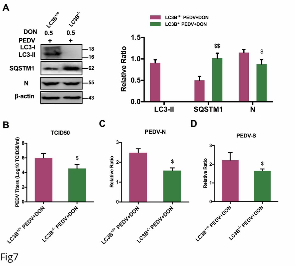
.CC-BY 4.0 International licensewas not certified by peer review) is the author/funder. It is made available under aThe copyright holder for this preprint (whichthis version posted November 22, 2019. . https://doi.org/10.1101/852608doi: bioRxiv preprint
.CC-BY 4.0 International licensewas not certified by peer review) is the author/funder. It is made available under aThe copyright holder for this preprint (whichthis version posted November 22, 2019. . https://doi.org/10.1101/852608doi: bioRxiv preprint
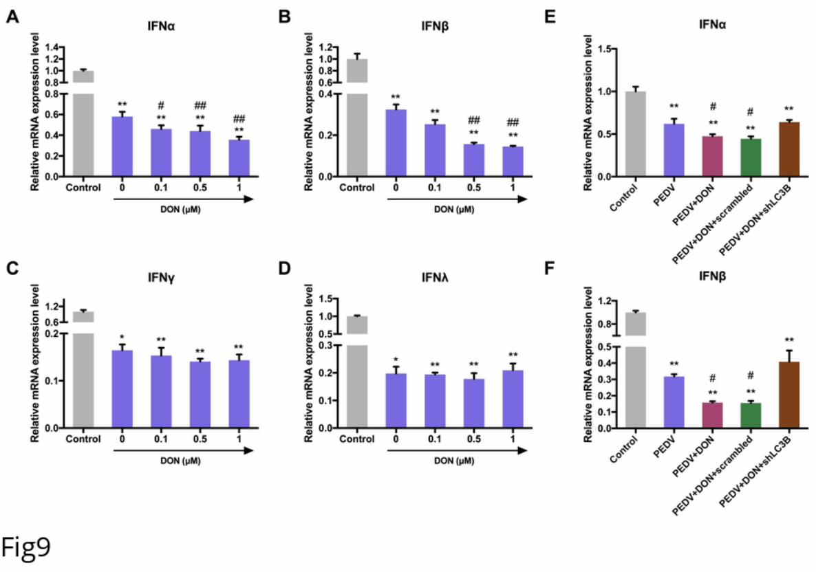
.CC-BY 4.0 International licensewas not certified by peer review) is the author/funder. It is made available under aThe copyright holder for this preprint (whichthis version posted November 22, 2019. . https://doi.org/10.1101/852608doi: bioRxiv preprint
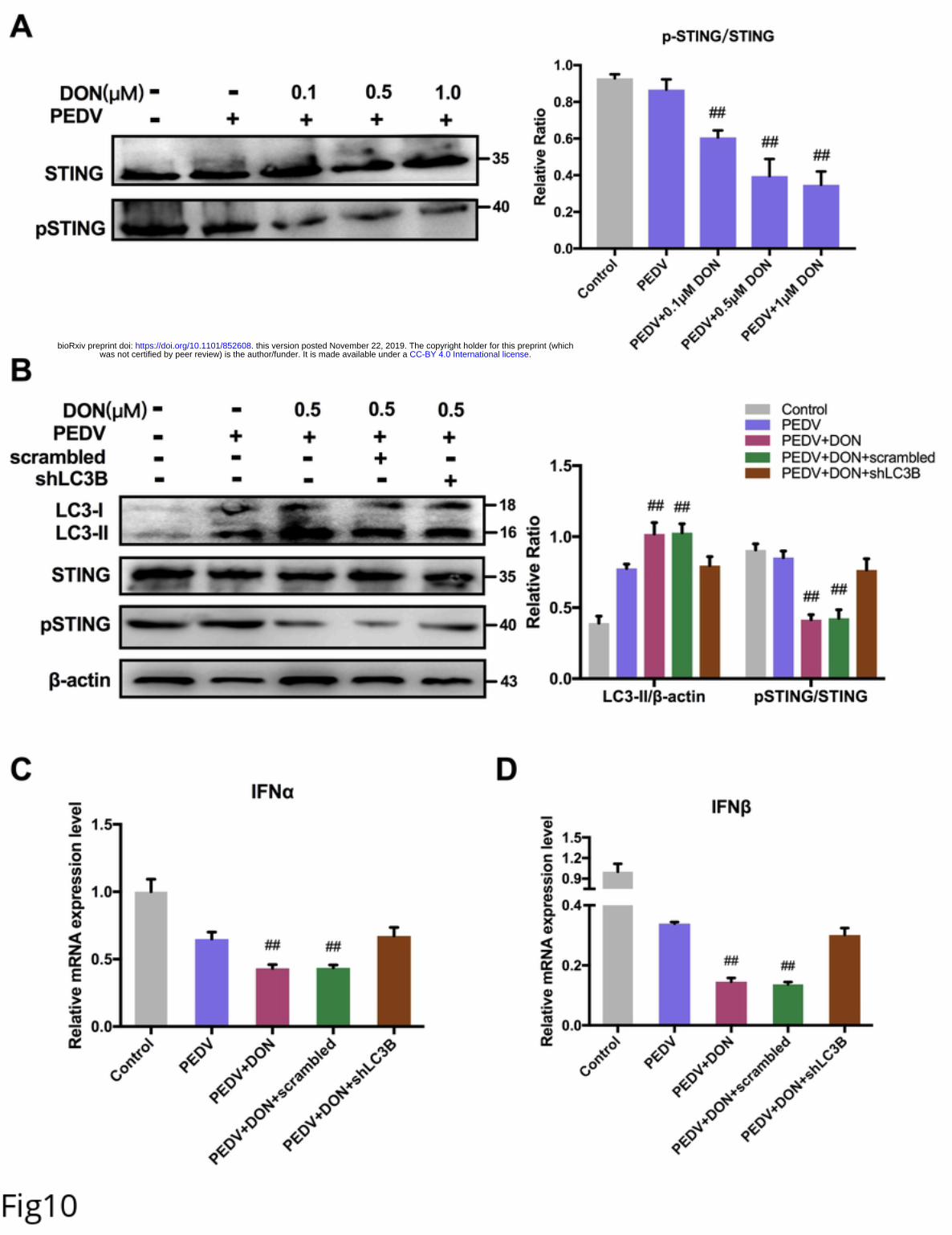
.CC-BY 4.0 International licensewas not certified by peer review) is the author/funder. It is made available under aThe copyright holder for this preprint (whichthis version posted November 22, 2019. . https://doi.org/10.1101/852608doi: bioRxiv preprint
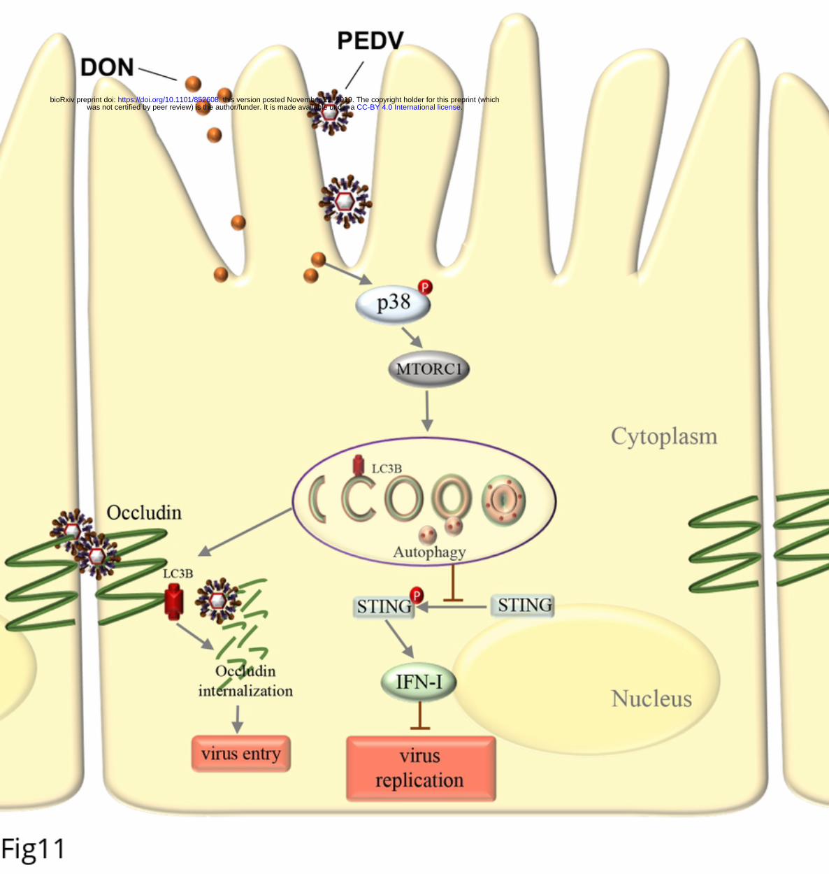
.CC-BY 4.0 International licensewas not certified by peer review) is the author/funder. It is made available under aThe copyright holder for this preprint (whichthis version posted November 22, 2019. . https://doi.org/10.1101/852608doi: bioRxiv preprint


![Deoxynivalenol (Vomitoxin) Nivalenol - FAMIC · Deoxynivalenol (Vomitoxin) Nivalenol [Methods listed in the Feed Analysis Standards] 1 Simultaneous analysis of mycotoxins by liquid](https://img.pdfslide.net/doc/110x75/5e2972e79268725bcf18f42b/deoxynivalenol-vomitoxin-nivalenol-deoxynivalenol-vomitoxin-nivalenol-methods.jpg)