Embed Size (px)
Citation preview
Department of Kinesiology and Applied Physiology WCR
Chapter 3: Cells• Overview• Plasma membrane: structure• Plasma membrane: transport• Resting membrane potential• Cell-environment interactions• Cytoplasm• Nucleus• Cell growth & reproduction• Extracellular materials• Developmental aspects
Department of Kinesiology and Applied Physiology 2
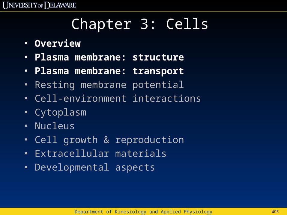
Do exercise scientists need to think about cells?Exercise in a Pill
•“AMPK and PPARδ Agonists Are Exercise Mimetics”•AICAR activates intracellular pathways that are also activated by exercise. Mice taking AICAR look like mice on exercise. Mice on AICAR plus exercise are supermice.
Narkar et al., Cell 2008.
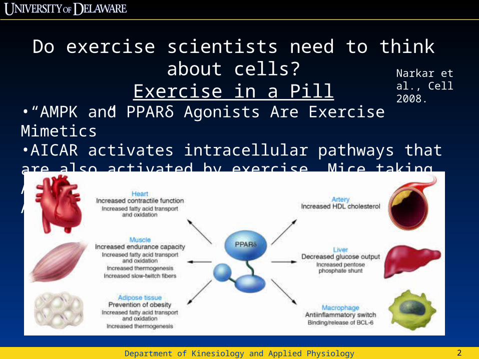
Fibroblasts
Erythrocytes
Epithelial cells
(d) Cell that fights disease
Nerve cell
Fat cell
Sperm
(a) Cells that connect body parts, form linings, or transport gases
(c) Cell that storesnutrients
(b) Cells that move organs and body parts
(e) Cell that gathers information and control body functions
(f) Cell of reproduction
SkeletalMusclecell
Smoothmuscle cells
Macrophage
Figure 3.1
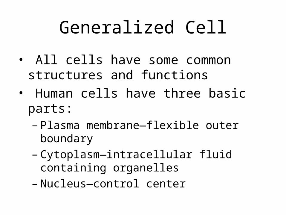
Generalized Cell
• All cells have some common structures and functions
• Human cells have three basic parts:– Plasma membrane—flexible outer boundary– Cytoplasm—intracellular fluid containing
organelles– Nucleus—control center

Copyright © 2010 Pearson Education, Inc. Figure 3.2
Secretion beingreleased from cellby exocytosis
Peroxisome
Ribosomes
Roughendoplasmicreticulum
Nucleus
Nuclear envelopeChromatin
Golgi apparatus
Nucleolus
Smooth endoplasmicreticulum
Cytosol
Lysosome
Mitochondrion
CentriolesCentrosomematrix
Cytoskeletalelements• Microtubule• Intermediate filaments
Plasmamembrane
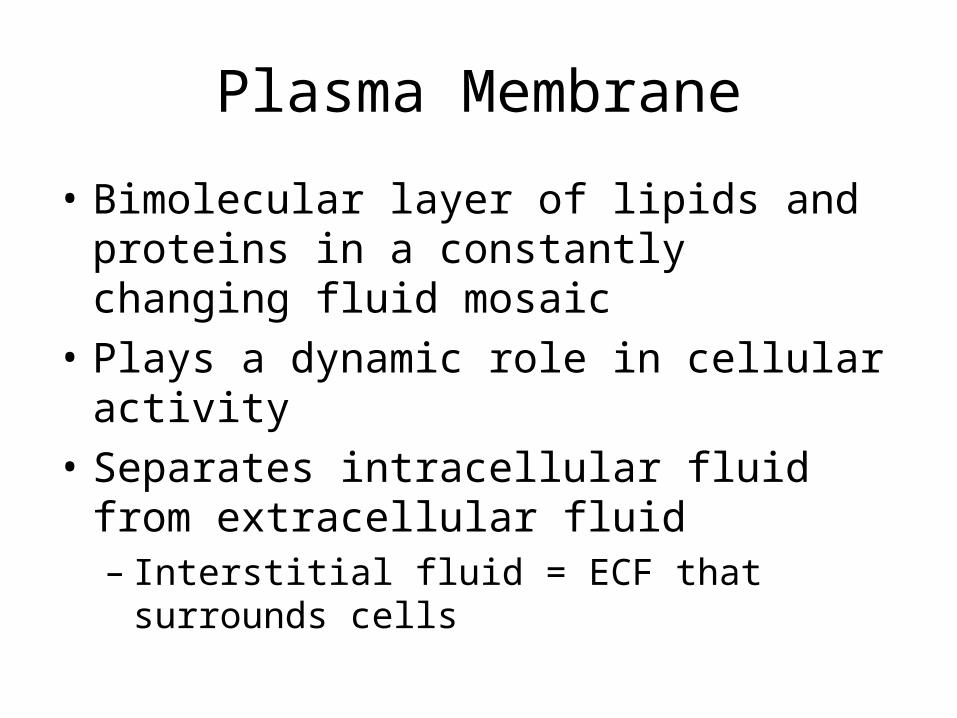
Plasma Membrane
• Bimolecular layer of lipids and proteins in a constantly changing fluid mosaic
• Plays a dynamic role in cellular activity• Separates intracellular fluid from extracellular
fluid– Interstitial fluid = ECF that surrounds cells
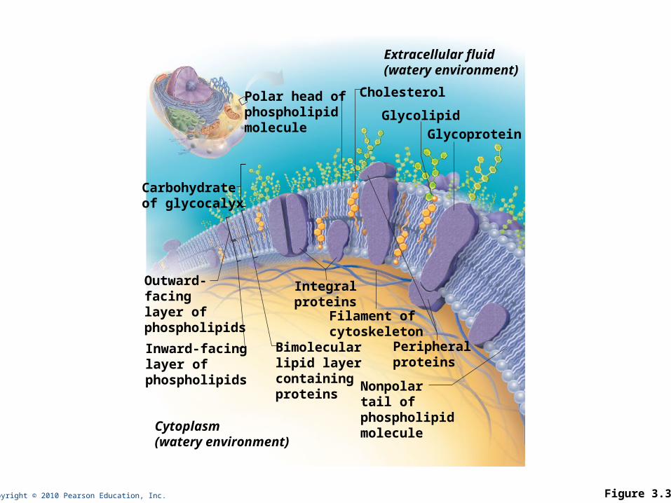
Copyright © 2010 Pearson Education, Inc. Figure 3.3
Integralproteins
Extracellular fluid(watery environment)
Cytoplasm(watery environment)
Polar head ofphospholipid molecule
Glycolipid
Cholesterol
Peripheralproteins
Bimolecularlipid layercontainingproteins
Inward-facinglayer ofphospholipids
Outward-facinglayer ofphospholipids
Carbohydrate of glycocalyx
Glycoprotein
Filament of cytoskeleton
Nonpolar tail of phospholipid molecule
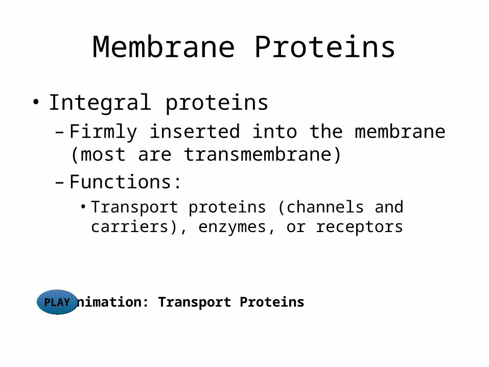
Membrane Proteins
• Integral proteins– Firmly inserted into the membrane (most are
transmembrane)– Functions:
• Transport proteins (channels and carriers), enzymes, or receptors
Animation: Transport ProteinsPLAYPLAY
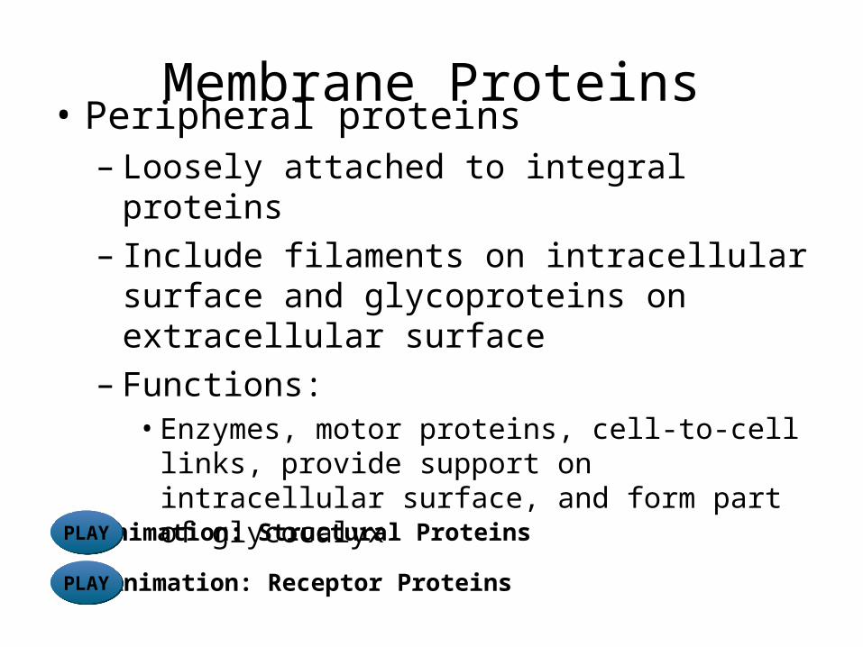
Membrane Proteins• Peripheral proteins
– Loosely attached to integral proteins – Include filaments on intracellular surface and
glycoproteins on extracellular surface– Functions:
• Enzymes, motor proteins, cell-to-cell links, provide support on intracellular surface, and form part of glycocalyx
Animation: Structural ProteinsPLAYPLAY
Animation: Receptor ProteinsPLAYPLAY
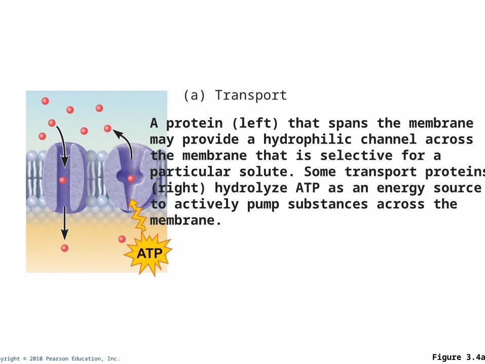
Copyright © 2010 Pearson Education, Inc. Figure 3.4a
A protein (left) that spans the membrane may provide a hydrophilic channel across the membrane that is selective for a particular solute. Some transport proteins (right) hydrolyze ATP as an energy source to actively pump substances across the membrane.
(a) Transport
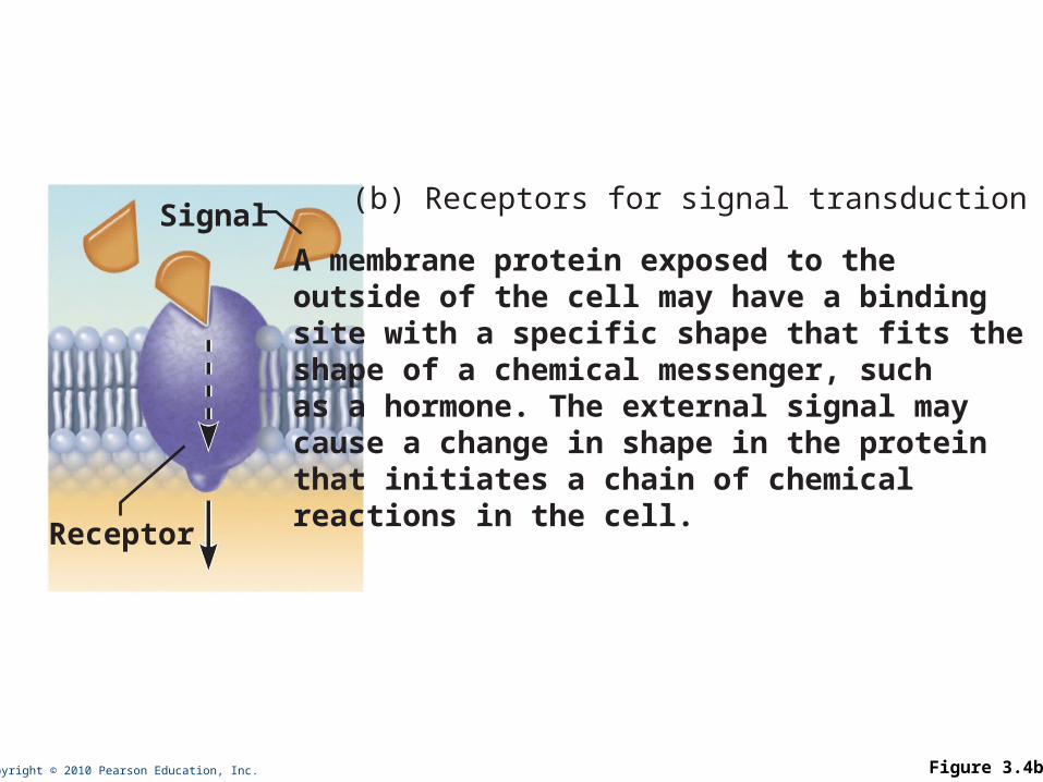
Copyright © 2010 Pearson Education, Inc. Figure 3.4b
A membrane protein exposed to the outside of the cell may have a binding site with a specific shape that fits the shape of a chemical messenger, such as a hormone. The external signal may cause a change in shape in the protein that initiates a chain of chemical reactions in the cell.
(b) Receptors for signal transductionSignal
Receptor
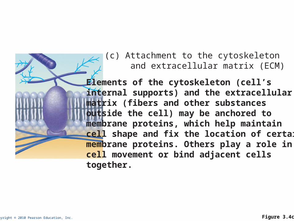
Copyright © 2010 Pearson Education, Inc. Figure 3.4c
Elements of the cytoskeleton (cell’s internal supports) and the extracellular matrix (fibers and other substances outside the cell) may be anchored to membrane proteins, which help maintain cell shape and fix the location of certain membrane proteins. Others play a role in cell movement or bind adjacent cells together.
(c) Attachment to the cytoskeleton and extracellular matrix (ECM)
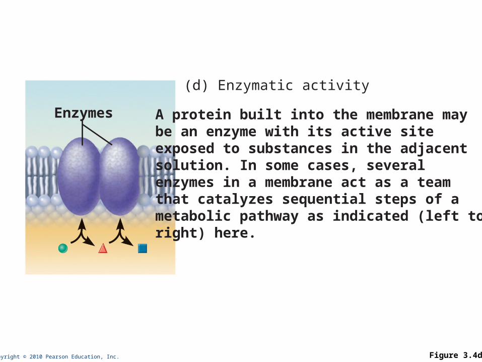
Copyright © 2010 Pearson Education, Inc. Figure 3.4d
A protein built into the membrane may be an enzyme with its active site exposed to substances in the adjacent solution. In some cases, several enzymes in a membrane act as a team that catalyzes sequential steps of a metabolic pathway as indicated (left to right) here.
(d) Enzymatic activity
Enzymes
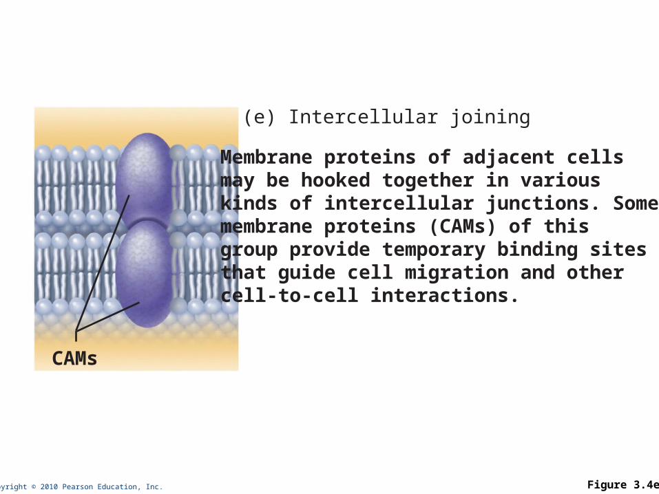
Copyright © 2010 Pearson Education, Inc. Figure 3.4e
Membrane proteins of adjacent cells may be hooked together in various kinds of intercellular junctions. Some membrane proteins (CAMs) of this group provide temporary binding sites that guide cell migration and other cell-to-cell interactions.
CAMs
(e) Intercellular joining
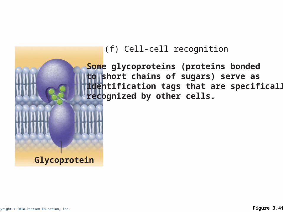
Copyright © 2010 Pearson Education, Inc. Figure 3.4f
Some glycoproteins (proteins bonded to short chains of sugars) serve as identification tags that are specifically recognized by other cells.
(f) Cell-cell recognition
Glycoprotein
Membrane Junctions
• Three types:– Tight junction – Desmosome – Gap junction
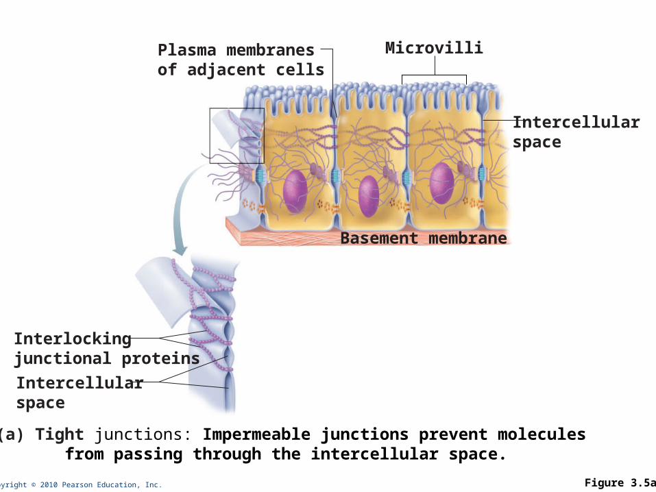
Copyright © 2010 Pearson Education, Inc. Figure 3.5a
Interlockingjunctional proteins
Intercellularspace
Plasma membranesof adjacent cells
Microvilli
Intercellularspace
Basement membrane
(a) Tight junctions: Impermeable junctions prevent molecules from passing through the intercellular space.
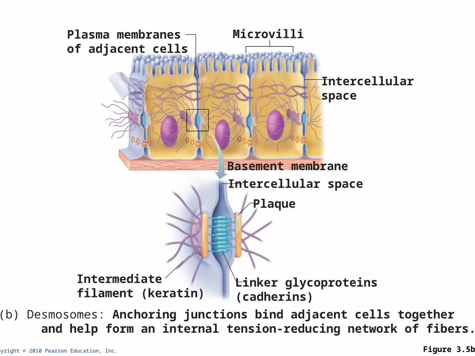
Copyright © 2010 Pearson Education, Inc. Figure 3.5b
Intercellular space
Plasma membranesof adjacent cells
Microvilli
Intercellularspace
Plaque
Linker glycoproteins(cadherins)
Intermediatefilament (keratin)
(b) Desmosomes: Anchoring junctions bind adjacent cells together and help form an internal tension-reducing network of fibers.
Basement membrane
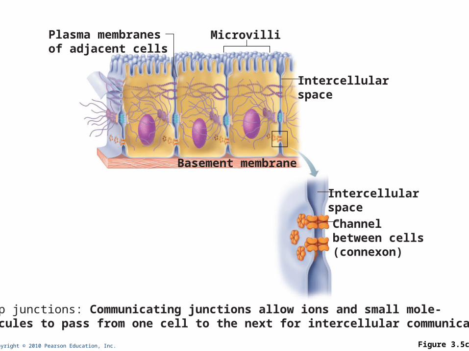
Copyright © 2010 Pearson Education, Inc. Figure 3.5c
Plasma membranesof adjacent cells
Microvilli
Intercellularspace
Intercellularspace
Channelbetween cells(connexon)
(c) Gap junctions: Communicating junctions allow ions and small mole- cules to pass from one cell to the next for intercellular communication.
Basement membrane
Membrane Transport: How things get in and out of cells
Plasma membranes are selectively permeable: some molecules easily pass through the membrane; others do not
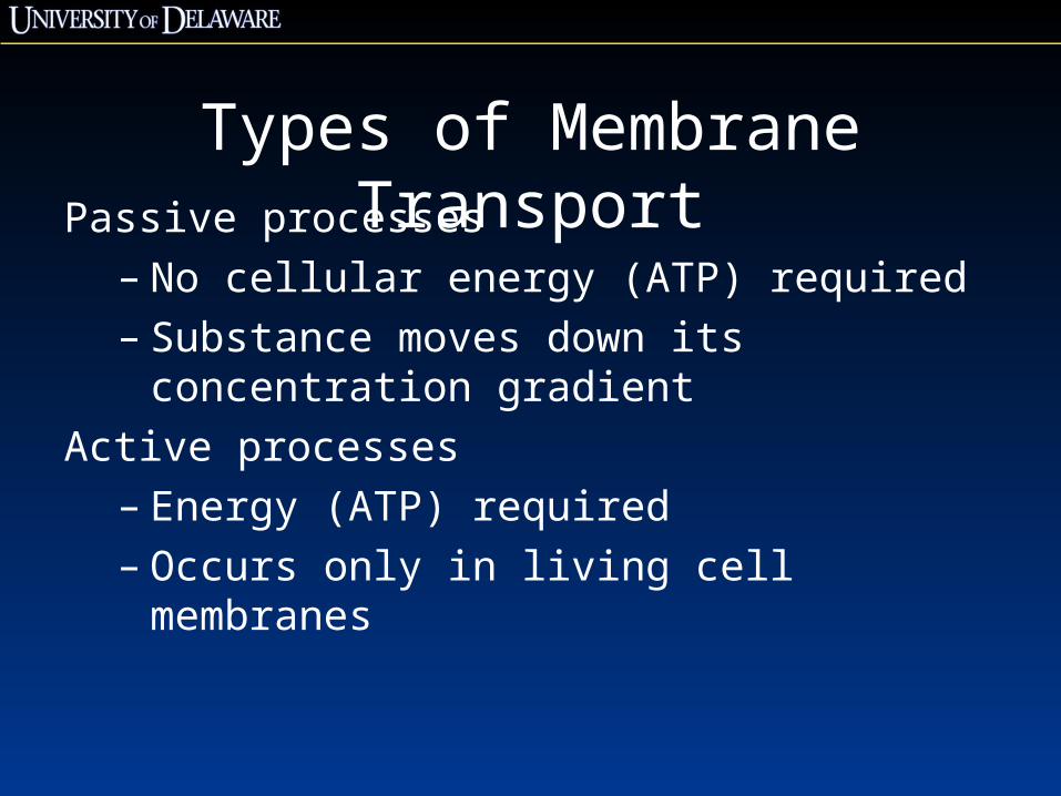
Types of Membrane TransportPassive processes
– No cellular energy (ATP) required– Substance moves down its concentration gradient
Active processes– Energy (ATP) required– Occurs only in living cell membranes
Copyright © 2010 Pearson Education, Inc.
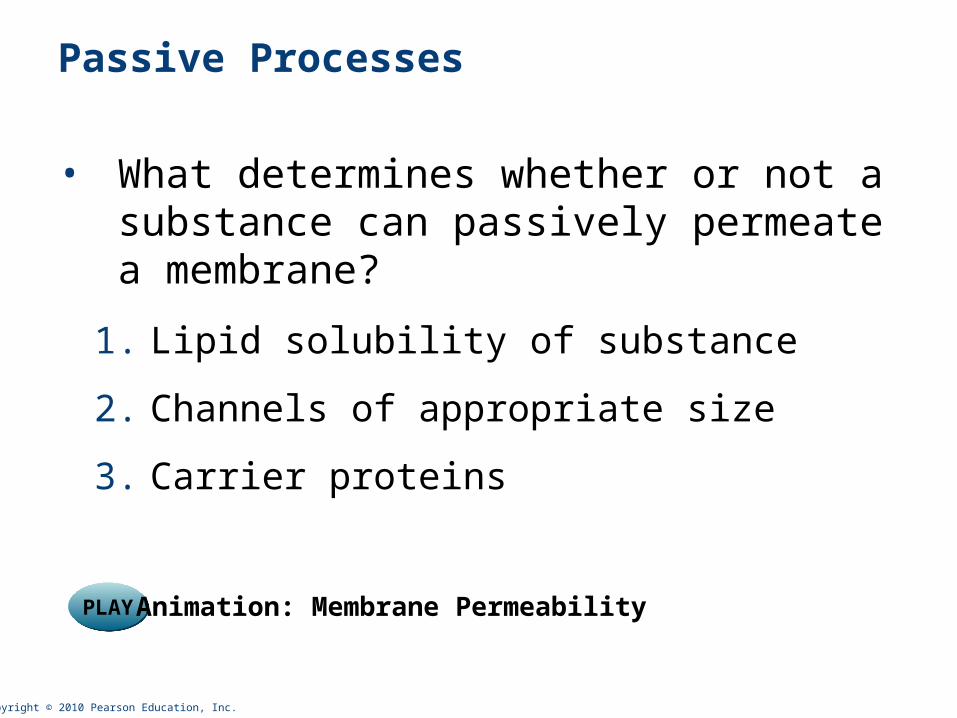
Passive Processes
• What determines whether or not a substance can passively permeate a membrane?
1. Lipid solubility of substance
2. Channels of appropriate size
3. Carrier proteins
PLAYPLAY Animation: Membrane Permeability
Copyright © 2010 Pearson Education, Inc.
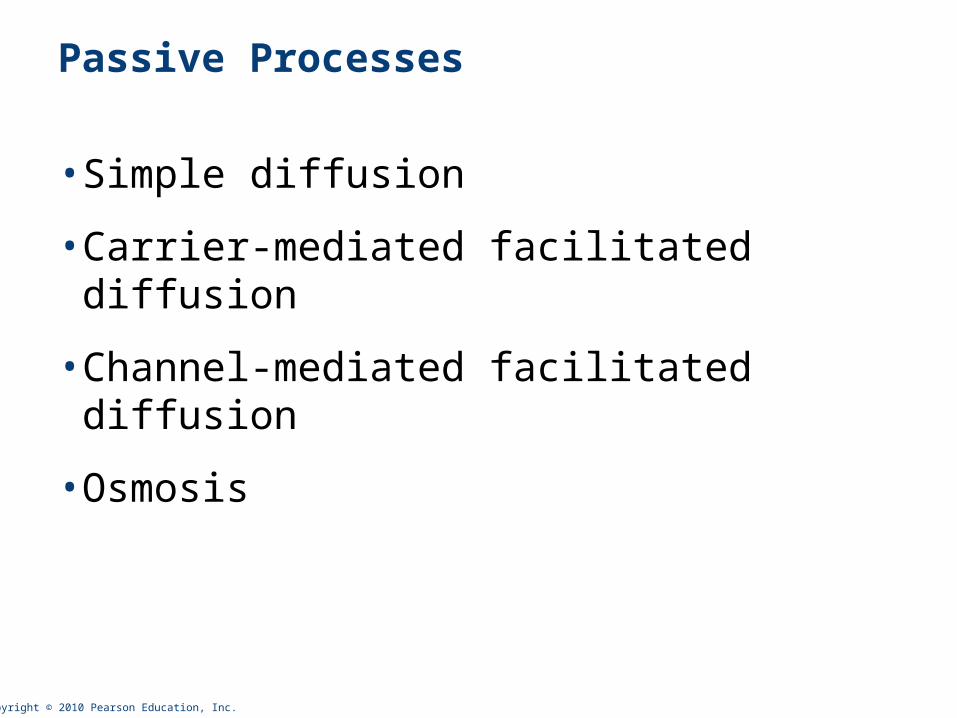
Passive Processes
• Simple diffusion
• Carrier-mediated facilitated diffusion
• Channel-mediated facilitated diffusion
• Osmosis
Copyright © 2010 Pearson Education, Inc.
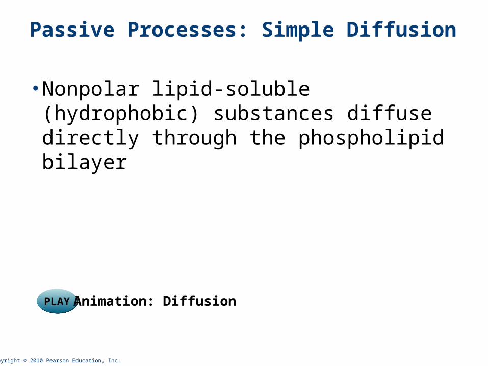
Passive Processes: Simple Diffusion
• Nonpolar lipid-soluble (hydrophobic) substances diffuse directly through the phospholipid bilayer
PLAYPLAY Animation: Diffusion
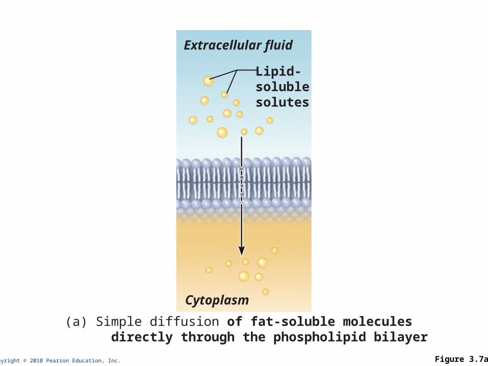
Copyright © 2010 Pearson Education, Inc. Figure 3.7a
Extracellular fluid
Lipid-solublesolutes
Cytoplasm
(a) Simple diffusion of fat-soluble molecules directly through the phospholipid bilayer
Copyright © 2010 Pearson Education, Inc.
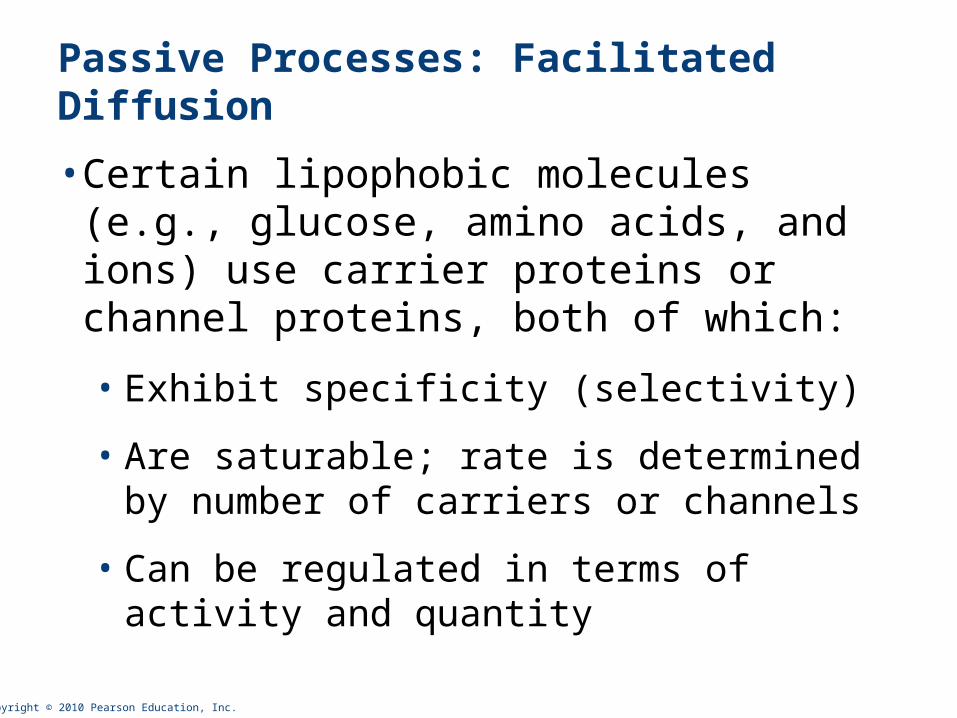
Passive Processes: Facilitated Diffusion
• Certain lipophobic molecules (e.g., glucose, amino acids, and ions) use carrier proteins or channel proteins, both of which:
• Exhibit specificity (selectivity)
• Are saturable; rate is determined by number of carriers or channels
• Can be regulated in terms of activity and quantity
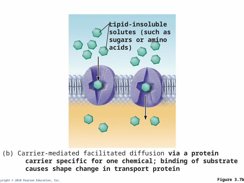
Copyright © 2010 Pearson Education, Inc. Figure 3.7b
Lipid-insoluble solutes (such as sugars or amino acids)
(b) Carrier-mediated facilitated diffusion via a protein carrier specific for one chemical; binding of substrate causes shape change in transport protein
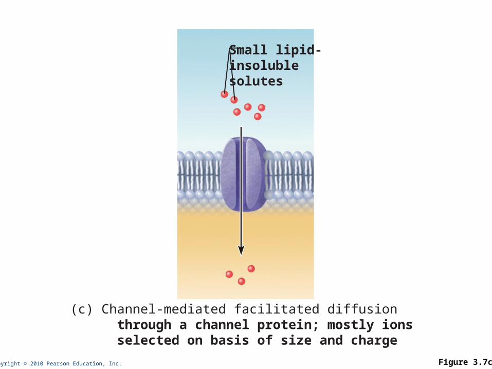
Copyright © 2010 Pearson Education, Inc. Figure 3.7c
Small lipid-insoluble solutes
(c) Channel-mediated facilitated diffusion through a channel protein; mostly ions selected on basis of size and charge
Copyright © 2010 Pearson Education, Inc.
Passive Processes: Osmosis
• Movement of solvent (water) across a selectively permeable membrane
•Water diffuses through plasma membranes:
• Through the lipid bilayer
• Through water channels called aquaporins
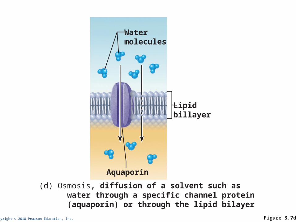
Copyright © 2010 Pearson Education, Inc. Figure 3.7d
Watermolecules
Lipidbillayer
Aquaporin
(d) Osmosis, diffusion of a solvent such as water through a specific channel protein (aquaporin) or through the lipid bilayer
Copyright © 2010 Pearson Education, Inc.
Importance of Osmosis
•When osmosis occurs, water enters or leaves a cell
• Change in cell volume disrupts cell function
PLAYPLAY Animation: Osmosis
Copyright © 2010 Pearson Education, Inc.
Tonicity
• Tonicity: How much dissolved material there is in a solution. Tonicity determines whether a solution will make cells shrink or swell.
• Isotonic: A solution with the same solute concentration as the inside of a normal cell
• Hypertonic: A solution with a greater solute concentration than than a normal cell
• Hypotonic: A solution with a lesser solute concentration than a normal cell
Copyright © 2010 Pearson Education, Inc.
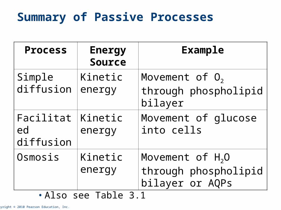
Summary of Passive Processes
• Also see Table 3.1
Process Energy Source
Example
Simple diffusion
Kinetic energy
Movement of O2 through phospholipid bilayer
Facilitated diffusion
Kinetic energy
Movement of glucose into cells
Osmosis Kinetic energy
Movement of H2O through phospholipid bilayer or AQPs
Copyright © 2010 Pearson Education, Inc.
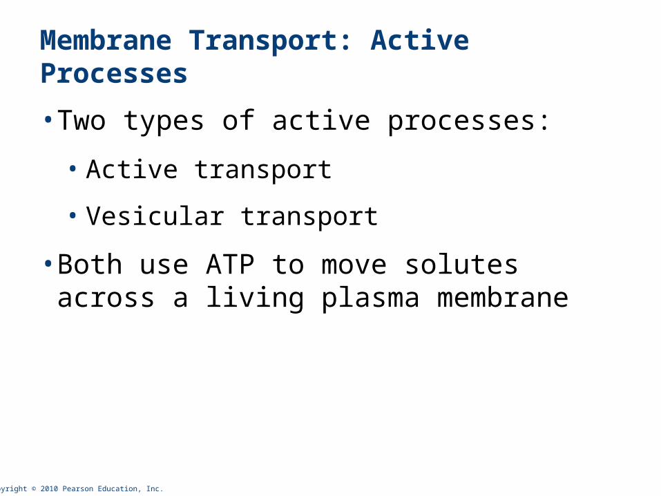
Membrane Transport: Active Processes
• Two types of active processes:
• Active transport
• Vesicular transport
• Both use ATP to move solutes across a living plasma membrane
Copyright © 2010 Pearson Education, Inc.
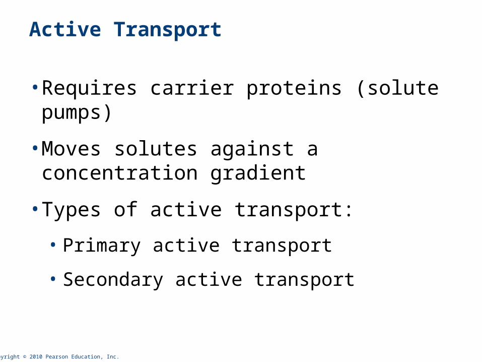
Active Transport
• Requires carrier proteins (solute pumps)
• Moves solutes against a concentration gradient
• Types of active transport:
• Primary active transport
• Secondary active transport
Copyright © 2010 Pearson Education, Inc.
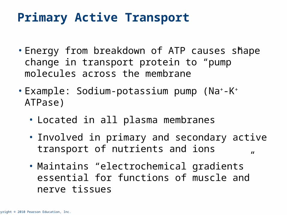
Primary Active Transport
• Energy from breakdown of ATP causes shape change in transport protein to “pump” molecules across the membrane
• Example: Sodium-potassium pump (Na+-K+ ATPase)
• Located in all plasma membranes
• Involved in primary and secondary active transport of nutrients and ions
• Maintains “electrochemical gradients” essential for functions of muscle and nerve tissues
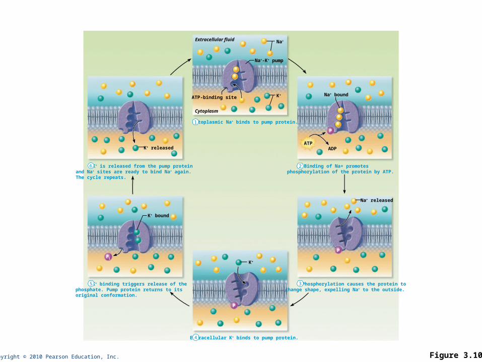
Copyright © 2010 Pearson Education, Inc. Figure 3.10
Extracellular fluid
K+ is released from the pump proteinand Na+ sites are ready to bind Na+ again.The cycle repeats.
Binding of Na+ promotesphosphorylation of the protein by ATP.
Cytoplasmic Na+ binds to pump protein.
Na+
Na+-K+ pump
K+ released
ATP-binding siteNa+ bound
Cytoplasm
ATPADP
P
K+
K+ binding triggers release of thephosphate. Pump protein returns to itsoriginal conformation.
Phosphorylation causes the protein tochange shape, expelling Na+ to the outside.
Extracellular K+ binds to pump protein.
Na+ released
K+ bound
P
K+
PPi
1
2
3
4
5
6

Copyright © 2010 Pearson Education, Inc.
Secondary Active Transport
• Energy stored in ionic gradients is used indirectly to drive transport of other solutes
• Always involves cotransport – transport of more than one substance at a time
• Two substances transported in same direction (Na+, glucose)
• Two substances transported in opposite directions (Na+, H+)
Mod WCR

Copyright © 2010 Pearson Education, Inc. Figure 3.11
The ATP-driven Na+-K+ pump stores energy by creating a steep concentration gradient for Na+ entry into the cell.
As Na+ diffuses back across the membrane through a membrane cotransporter protein, it drives glucose against its concentration gradientinto the cell. (ECF = extracellular fluid)
Na+-glucosesymporttransporterloadingglucose fromECF
Na+-glucosesymport transporterreleasing glucoseinto the cytoplasm
Glucose
Na+-K+
pump
Cytoplasm
Extracellular fluid
1 2
Copyright © 2010 Pearson Education, Inc.
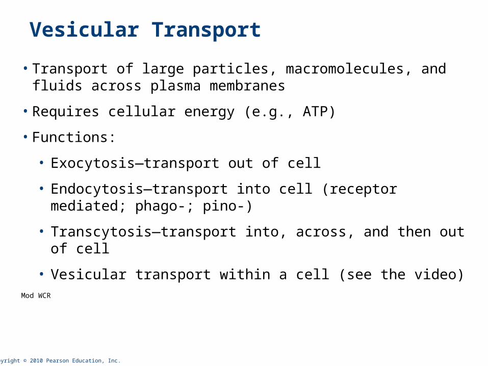
Vesicular Transport
• Transport of large particles, macromolecules, and fluids across plasma membranes
• Requires cellular energy (e.g., ATP)
• Functions:
• Exocytosis—transport out of cell
• Endocytosis—transport into cell (receptor mediated; phago-; pino-)
• Transcytosis—transport into, across, and then out of cell
• Vesicular transport within a cell (see the video)Mod WCR
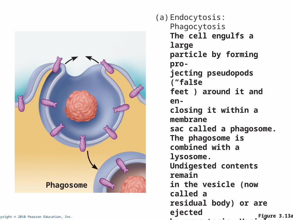
Copyright © 2010 Pearson Education, Inc. Figure 3.13a
Phagosome
(a) Endocytosis:PhagocytosisThe cell engulfs a large particle by forming pro-jecting pseudopods (“false feet”) around it and en-closing it within a membrane sac called a phagosome. The phagosome is combined with a lysosome. Undigested contents remain in the vesicle (now called a residual body) or are ejected by exocytosis. Vesicle may or may not be protein-coated but has receptors capable of binding to microorganisms or solid particles.
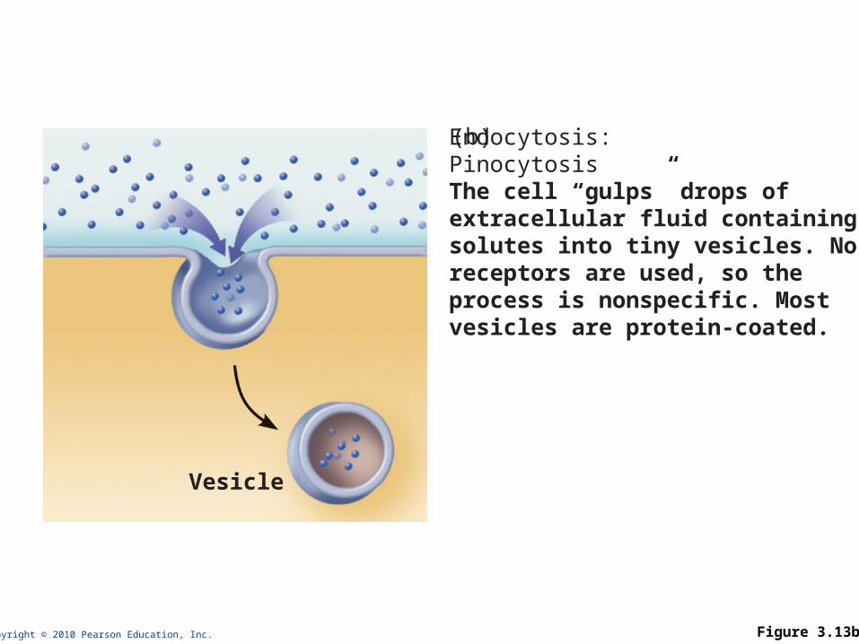
Copyright © 2010 Pearson Education, Inc. Figure 3.13b
Vesicle
(b) Endocytosis:PinocytosisThe cell “gulps” drops of extracellular fluid containing solutes into tiny vesicles. No receptors are used, so the process is nonspecific. Most vesicles are protein-coated.
Copyright © 2010 Pearson Education, Inc. Figure 3.13c
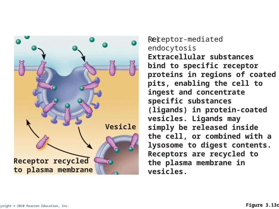
Vesicle
Receptor recycledto plasma membrane
(c) Receptor-mediatedendocytosisExtracellular substances bind to specific receptor proteins in regions of coated pits, enabling the cell to ingest and concentrate specific substances (ligands) in protein-coated vesicles. Ligands may simply be released inside the cell, or combined with a lysosome to digest contents. Receptors are recycled to the plasma membrane in vesicles.
Copyright © 2010 Pearson Education, Inc. Figure 3.14a
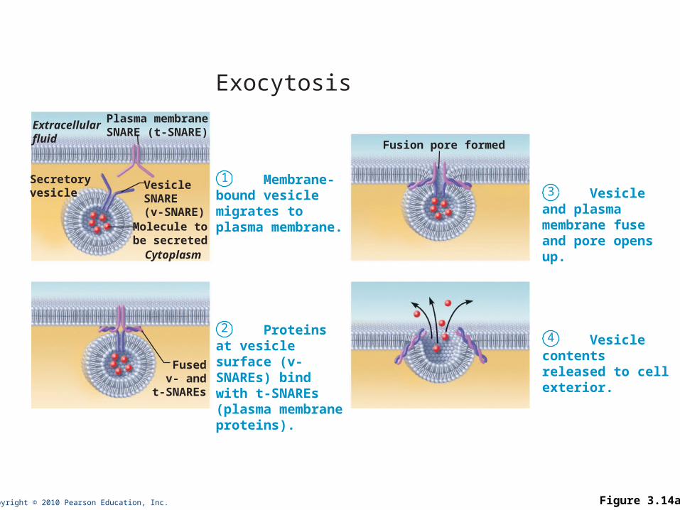
1 Membrane-bound vesicle migrates toplasma membrane.
2 Proteins at vesicle surface (v-SNAREs) bind with t-SNAREs (plasma membrane proteins).
Exocytosis
Extracellularfluid
Plasma membraneSNARE (t-SNARE)
Secretoryvesicle
VesicleSNARE(v-SNARE)
Molecule tobe secretedCytoplasm
Fusedv- and
t-SNAREs
3 Vesicle and plasma membrane fuse and pore opens up.
4 Vesicle contentsreleased to cell exterior.
Fusion pore formed
Copyright © 2010 Pearson Education, Inc.
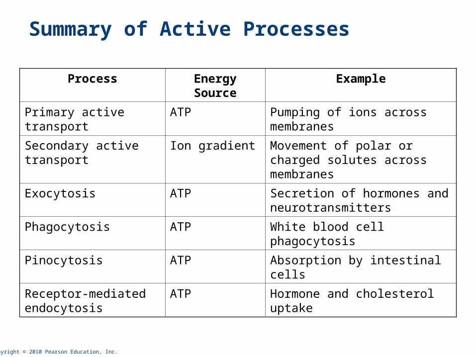
Summary of Active Processes
Process Energy Source Example
Primary active transport ATP Pumping of ions across membranes
Secondary active transport
Ion gradient Movement of polar or charged solutes across membranes
Exocytosis ATP Secretion of hormones and neurotransmitters
Phagocytosis ATP White blood cell phagocytosis
Pinocytosis ATP Absorption by intestinal cells
Receptor-mediated endocytosis
ATP Hormone and cholesterol uptake


![Plasma Membrane [7.2] Goals: Understand the concept of homeostasis in relation to the plasma membrane Demonstrate and understand how the plasma membrane](https://img.pdfslide.net/doc/110x75/5697c01d1a28abf838cd0a9a/plasma-membrane-72-goals-understand-the-concept-of-homeostasis-in-relation.jpg)