Embed Size (px)
Citation preview
Proc. Natl. Acad. Sci. USAVol. 76, No. 8, pp. '3665-3669, August 1979Biochemistry
An adenovirus type 5 early gene function regulates expression ofother early viral genes
(host range deletion mutants/early viral RNAs)
NICHOLAS JONES* AND THOMAS SHENKDepartment of Microbiology, University of Connecticut Health Center, Farmington, Connecticut 06032
Communicated by M. J. Osborn, May 7,1979
ABSTRACT We have identified an adenovirus type 5 (Ad5)early gene function located in early region 1 which is requirefor the production of early cytoplasmic mRNAs correspondingto early regions 2, 3, and 4. Mutant d1312 (lacks the segmentbetween 1.5 and 4.5 map units) grows as well as wild-type virusin 293 cells (AdS-transformed human embryonic kidney cells),but its growth is severely restricted in HeLa cells. We detect noviral RNAs in the cytoplasm of d1312-infected HeLa cells. ViralRNA sequences are present, however, in dl312-infected HeLacell nuclei.
At least four segments of the adenovirus type 5 (Ad5) genomeare represented in cytoplasmic transcripts synthesized at earlytimes after infection. Two of these segments [region 1 (1.5-11.5map units) and region 3 (76-86 map units)] are located on therightward-reading viral DNA strand, and two are on the left-ward-reading DNA strand [region 2 (62-76 map units) andregion 4 (91-99 map units)] (1-4). Each region contains at leastone promoter (5, 6).
Recently, we isolated a group of deletion mutants that lackportions of early region 1 (unpublished data). These mutantswere isolated by selecting viral DNAs that lack the Xba I re-striction endonuclease cleavage site-at 4 map units by the pro-cedure of Jones and Shenk (7). These mutants are propagatedin 293 cells (an Ad5-transformed human embryonic kidney cellline; ref. 8); they do not replicate in HeLa cells.
In this report we show that the deletions in these mutantsmake predictable alterations in the cytoplasmic mRNAs en-coded by early region 1. Further, we find that one of thesemutants, d1312, defines a region 1 gene function that is requiredfor the production of early cytoplasmic mRNAs correspondingto early regions 2, 3, and 4.
MATERIALS AND METHODSCells and Viruses. The 293 cells were provided by F.
Graham and have been described (8). The cells were main-tained in Dulbecco's modified minimal essential mediumcontaining 10% fetal calf serum. HeLa cells were obtained fromJ. Williams. They were grown in Dulbecco's modified mediumcontaining 5% fetal calf serum. Wild-type Ad5 (H5wt300) isa plaque-purified derivative of a virus stock originally obtainedfrom H. Ginsberg. Mutant HSts36 was from J. Williams (9) andH5ts125 was from H. Ginsberg (10). Mutants H5dl31,H5dl312, and H5dl313 were selected for the loss of the Xba Iendonuclease cleavage site at 4 map units (unpublisheddata).
Enzymes, DNAs, and RNAs. Restriction endonucleases werepurchased from Bethesda Research (Rockville, MD), and S1endonuclease and electrophoretically purified deoxyribonu-clease I were from Sigma. Viral DNA was prepared from virionsas described (7). 32P-Labeled AdS DNAs, labeled in vivo, were
prepared by the method of Tibbetts and Pettersson (11) exceptthat 293 cells were substituted for HeLa cells. The specific ac-tivity of these DNAs ranged from 1 to 5 X 10⁶ cpm/,gg. Ad5DNA fragments were labeled with 32P in titro by the nicktranslation method of Rigby et al. (12). The specific activityof these probe DNAs ranged from 8 X 10⁷ to 2 X 10⁸ cpm/11g.RNA was isolated from cells (treated with 20,ug of cytosine
arabinoside per ml) 8 hr after infection at a multiplicity of 50plaque-forming units (PFU)/cell. Infected cells were dividedinto nuclear and cytoplasmic fractions. Cells were suspendedin isotonic buffer (10mM Tris1HCl, pH 7.8/1.5mM MgCl2/150mM NaCI), and Nonidet P-40 was added to 0.6% after themixture was cooled to 40C. The mixture was held on ice for 10mi. then mixed on a Vortex for 10 sec; the nuclei were pelletedby centrifugation. The supernatant was the cytoplasmic fractionand the pellet was the nuclear fraction after two additionalwashes in isotonic buffer.We prepared nuclear RNA by dissolving the nuclear pellet
in a low pH sodium dodecyl sulfate buffer (50mM NaOAc, pH5.2/100 mM NaCI/10 mM EDTA/0.5% sodium dodecyl sul-fate), extracting the RNA twice at room temperature with anequal volume of phenol (equilibrated with 50mM NaOAc, pH5.2), extracting it once with chloroform/isoamyl alcohol, 49:1(vol/vol), and precipitating the RNA by addition of 2 vol ofethanol. The RNA was resuspended in 10 mM Tris-HCl, pH7.4/10 mM MgCI2, electrophoretically purified DNase I wasadded to 50 ,ug/ml, and the solution was incubated at 37°C for30 min. Finally, the RNA was reextracted with phenol and withchloroform/isoamyl alcohol, and precipitated with ethanol.Cytoplasmic RNA was prepared by mixing the cytoplasmiccellular fraction with 3 vol of 100 mM Tris-HCl (pH 9), ex-tracting twice at room temperature with phenol (pH 9)/chlo-roform/isoamyl alcohol, 500:100:1 (vol/vol) and once withchloroform/isoamyl alcohol, 49:1 (vol/vol), and precipitatingwith 2 vol of ethanol.
SI Mapping of Cytoplasmic mRNAs. The protocol of Berkand Sharp (1) was used. Hybridizations in 80% formamide (13)were in 20,l with cytoplasmic RNA (5 mg/ml) and a restrictionendonuclease-generated, 32P-labeled DNA fragment (3 ,ugequivalents per ml). Hybridization was for 3 hr at 59°C. TheRNA-DNA hybrids were treated with SI endonuclease [100units (1) in 200 jil of 30 mM NaOAc/250 mM NaCl/1 mMZnCl2/5% (wt/vol) glycerol for 30 min at room temperature],and the digestion products were analyzed by electrophoresisin 3.8% polyacrylamide slab-gels (1.5 mm thick, 20 cm long)containing 8 M urea and Tris borate buffer (50 mM Trisbase/50 mM boric acid/i mM EDTA).
Liquid Hybridizations. A 32P-labeled DNA probe (ap-proximately 400 nucleotides in size) and either cytoplasmic or
The publication costs of this article were defrayed in part by pagecharge payment. This article must therefore be hereby marked "ad-vertisement" in accordance with 18 U. S. C. §1734 solely to indicatethis fact.
3665
Abbreviations: AdS, adenovirus type 5; PFU, plaque-forming units.* Present address: Department of Biological Sciences, Purdue Uni-versity, West Lafayette, IN 47907.
3666 Biochemistry: Jones and Shenk
nuclear RNA (1 mg/ml) were mixed in distilled water and theDNA was denatured by boiling for 3 min. Hybridization was
in 10 mM Tris-HCl, pH 7.5/1 M NaCl at 680C. Aliquots werewithdrawn and treated with Si endonuclease (same buffer andenzyme concentration as above) and acid-precipitable radio-activity was determined.
RESULTSThe physical and phenotypic characteristics of the early region1 deletion mutants studied in this report are summarized inTable 1. The deletions range in size between 150 and 2350 basepairs and they are located between 1.5 and 10.5 map units onthe AdS chromosome. They all lack the Xba I endonucleasecleavage site at 4 map units. The deletion mutants all grow as
well as wild-type virus in 293 cells, but their growth is severelyrestricted in HeLa cells. Their defect in HeLa cells occurs atan early stage of the viral growth cycle since none of the mu-tants synthesize viral DNA in these cells. Finally, although d1311is able to transform rat embryo cells as efficiently as the wild-type, mutants d1312 and d1313 are defective for transforma-tion.
Deletion Mutations Alter Early Region 1 Viral mRNAs.Berk and Sharp (14) have defined the structure of the majorAd2 cytoplasmic RNAs produced during the early phase ofinfection. To map mRNAs, they digested hybrids formed be-tween cytoplasmic RNA and 32P-labeled restriction endonu-clease-generated DNA fragments of the viral genome with S1endonuclease or exonuclease VII and analyzed the products bygel electrophoresis. Si endonuclease digests both single-strandedends and loops (corresponding to intervening sequences) inRNA-DNA hybrids to generate 32P-labeled DNA fragments thesize of coding sequences. Exonuclease VII digests only thesingle-stranded ends, producing 32P-labeled DNA fragmentsthe size of coding plus intervening sequences. This procedureidentified three major mRNAs encoded by Ad2 early region1. The two left-most mRNAs (region la, Fig. 1) contain infor-mation from the same segment of the viral chromosome(1.5-4.5 map units) and differ only in the size of their inter-vening sequences. The third mRNA is coded by the regionbetween 4.5 and 11.5 map units (region lb, Fig. 1), and it lacksa small intervening sequence at 10 map units.
Because our region 1 deletion mutants were not in an Ad2background, but derived from the closely related Ad5, it wasnecessary to confirm that wild-type Ad5 synthesized the sameregion 1 RNAs as Ad2. Accordingly, cytoplasmic RNA fromwt300 Ad5-infected HeLa cells was hybridized to a 32P-labeledAd5 DNA fragment (Xho I-C fragment, 0-15 map units) andthe hybrids were digested with SI endonuclease. Then the di-gestion products were analyzed by polyacrylamide gel elec-trophoresis (denaturing gel containing 8 M urea) (Fig. 2, region1). Fragments were identified that corresponded in size to eachcoding sequence previously described for Ad2: 660-, 485-, and375-nucleotide fragments composing the region la mRNAs;and 1850- and 485-nucleotide fragments corresponding to theregion lb mRNA. The locations of the AdS coding sequences
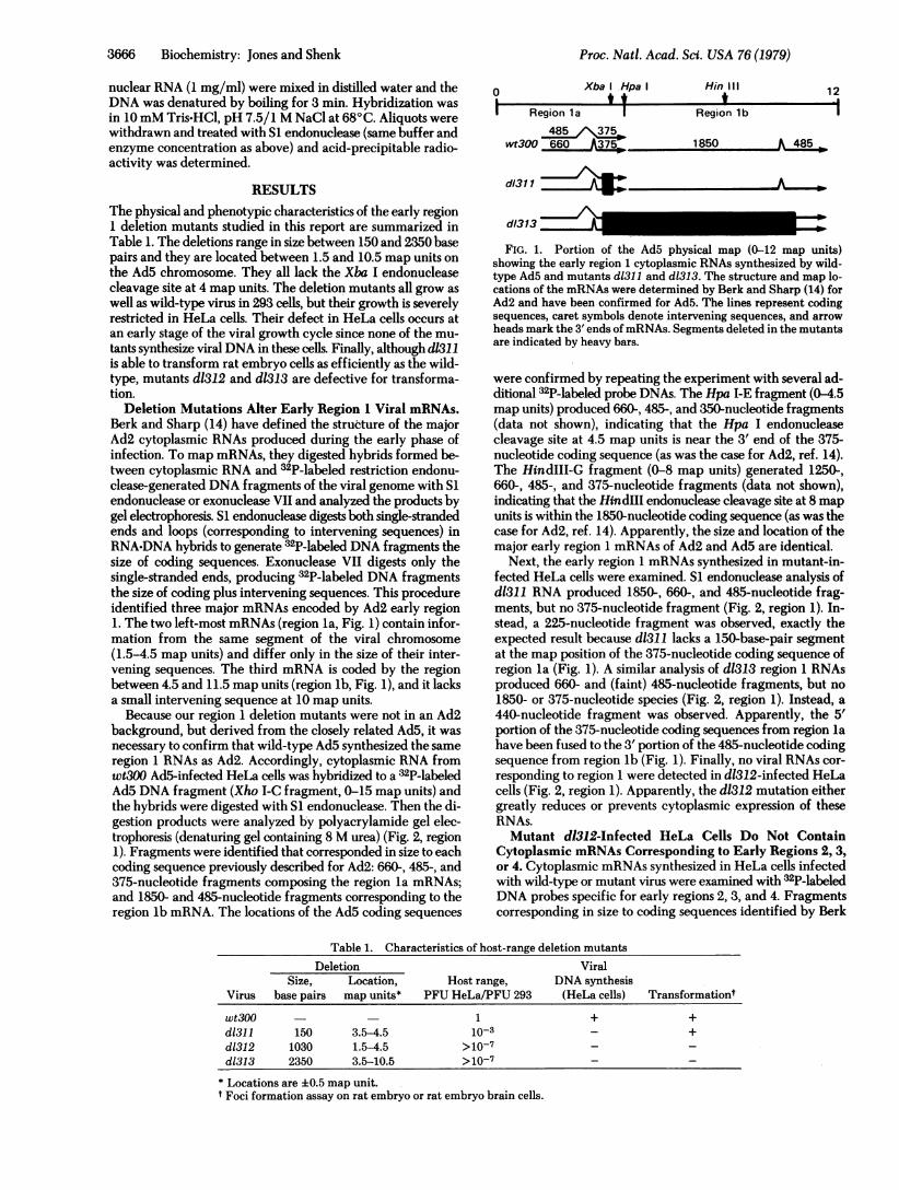
0Xba I Hpa I
I Region 1a 1
wt300 660
d131 1
d/313
Hin III
Region lb
1850 A 485
A-Z3
iN
FIG. 1. Portion of the Ad5 physical map (0-12 map units)showing the early region 1 cytoplasmic RNAs synthesized by wild-type Ad5 and mutants d1311 and d1313. The structure and map lo-cations of the mRNAs were determined by Berk and Sharp (14) forAd2 and have been confirmed for Ad5. The lines represent codingsequences, caret symbols denote intervening sequences, and arrowheads mark the 3' ends ofmRNAs. Segments deleted in the mutantsare indicated by heavy bars.
were confirmed by repeating the experiment with several ad-ditional 32P-labeled probe DNAs. The Hpa I-E fragment (0-4.5map units) produced 660-, 485-, and 350-nucleotide fragments(data not shown), indicating that the Hpa I endonucleasecleavage site at 4.5 map units is near the 3' end of the 375-nucleotide coding sequence (as was the case for Ad2, ref. 14).The HindIII-G fragment (0-8 map units) generated 1250-,660-, 485-, and 375-nucleotide fragments (data not shown),indicating that the HindIII endonuclease cleavage site at 8 mapunits is within the 1850-nucleotide coding sequence (as was thecase for Ad2, ref. 14). Apparently, the size and location of themajor early region 1 mRNAs of Ad2 and Ad5 are identical.
Next, the early region 1 mRNAs synthesized in mutant-in-fected HeLa cells were examined. SI endonuclease analysis ofd1311 RNA produced 1850-, 660-, and 485-nucleotide frag-ments, but no 375-nucleotide fragment (Fig. 2, region 1). In-stead, a 225-nucleotide fragment was observed, exactly theexpected result because d1311 lacks a 150-base-pair segmentat the map position of the 375-nucleotide coding sequence ofregion la (Fig. 1). A similar analysis of d1313 region 1 RNAsproduced 660- and (faint) 485-nucleotide fragments, but no
1850- or 375-nucleotide species (Fig. 2, region 1). Instead, a
440-nucleotide fragment was observed. Apparently, the 5'portion of the 375-nucleotide coding sequences from region lahave been fused to the 3' portion of the 485-nucleotide codingsequence from region lb (Fig. 1). Finally, no viral RNAs cor-
responding to region 1 were detected in d1312-infected HeLacells (Fig. 2, region 1). Apparently, the d1312 mutation eithergreatly reduces or prevents cytoplasmic expression of theseRNAs.Mutant dl312-Infected HeLa Cells Do Not Contain
Cytoplasmic mRNAs Corresponding to Early Regions 2, 3,or 4. Cytoplasmic mRNAs synthesized in HeLa cells infectedwith wild-type or mutant virus were examined with 32P-labeledDNA probes specific for early regions 2, 3, and 4. Fragmentscorresponding in size to coding sequences identified by Berk
Table 1. Characteristics of host-range deletion mutantsDeletion Viral
Size, Location, Host range, DNA synthesisVirus base pairs map units* PFU HeLa/PFU 293 (HeLa cells) Transformationt
wt300 1 + +d1311 150 3.5-4.5 10-3 - +d1312 1030 1.5-4.5 >10- - -d1313 2350 3.5-10.5 >10-7-
* Locations are ±0.5 map unit.t Foci formation assay on rat embryo or rat embryo brain cells.
Proc. Natl. Acad. Sci. USA 76 (1979)
Proc. Natl. Acad. Sci. USA 76 (1979) 3667
1.5 E-1 I 1.5
Region El
WT 311 312 3131850 0
76.5 -E3 - 8661.5* ---755 91 -- 99
Region E2 Region E3 Region E4
WT 311 312 313 WT 311 312 313 WT 311 312 313
1700 ^ Yi1* '-IP1900 'I* !kw w w *150G0 x
660 -
485
340 * *375
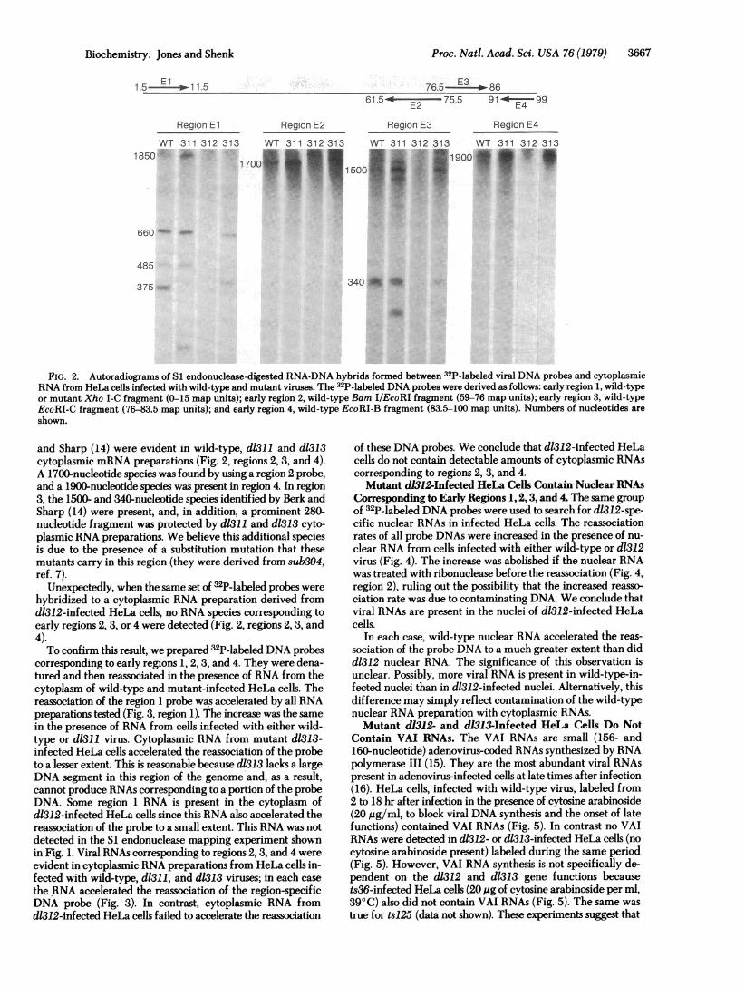
FIG. 2. Autoradiograms of S1 endonuclease-digested RNA-DNA hybrids formed between 32P-labeled viral DNA probes and cytoplasmicRNA from HeLa cells, infected with wild-type and mutant viruses. The 32P-labeled DNA probes were derived as follows: early region 1, wild-typeor mutant Xho I-C fragment (0-15 map units); early region 2, wild-type Bam I/EcoRI fragment (59-76 map units); early region 3, wild-typeEcoRI-C fragment (76-83.5 map units); and early region 4, wild-type EcoRI-B fragment (83.5-100 map units). Numbers of nucleotides areshown.
and Sharp (14) were evident in wild-type, d1311 and d1313cytoplasmic mRNA preparations (Fig. 2, regions 2, 3, and 4).A 1700-nucleotide species was found by using a region 2 probe,and a 1900-nucleotide species was present in region 4. In region3, the 1500- and 340-nucleotide species identified by Berk andSharp (14) were present, and, in addition, a prominent 280-nucleotide fragment was protected by d1311 and d1313 cyto-plasmic RNA preparations. We believe this additional speciesis due to the presence of a substitution mutation that thesemutants carry in this region (they were derived from sub3O4,ref. 7).
Unexpectedly, when the same set of 32P-labeled probes werehybridized to a cytoplasmic RNA preparation derived fromd1312-infected HeLa cells, no RNA species corresponding toearly regions 2, 3, or 4 were detected (Fig. 2, regions 2, 3, and4).To confirm this result, we prepared 32P-labeled DNA probes
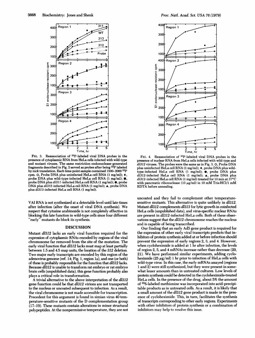
corresponding to early regions 1, 2, 3, and 4. They were dena-tured and then reassociated in the presence of RNA from thecytoplasm of wild-type and mutant-infected HeLa cells. Thereassociation of the region 1 probe was accelerated by all RNApreparations tested (Fig. 3, region 1). The increase was the samein the presence of RNA from cells infected with either wild-type or d1311 virus. Cytoplasmic RNA from mutant d1313-infected HeLa cells accelerated the reassociation of the probeto a lesser extent. This is reasonable because dl313 lacks a largeDNA segment in this region of the genome and, as a result,cannot produce RNAs corresponding to a portion of the probeDNA. Some region 1 RNA is present in the cytoplasm ofd1812-infected HeLa cells since this RNA also accelerated thereassociation of the probe to a small extent. This RNA was notdetected in the S1 endonuclease mapping experiment shownin Fig. 1. Viral RNAs corresponding to regions 2, 3, and 4 wereevident in cytoplasmic RNA preparations from HeLa cells in-fected with wild-type, d1311, and d1313 viruses; in each casethe RNA accelerated the reassociation of the region-specificDNA probe (Fig. 3). In contrast, cytoplasmic RNA fromd1312-infected HeLa cells failed to accelerate the reassociation
of these DNA probes. We conclude that d1312-infected HeLacells do not contain detectable amounts of cytoplasmic RNAscorresponding to regions 2, 3, and 4.Mutant d1312-Infected HeLa Cells Contain Nuclear RNAs
Corresponding to Early Regions 1, 2,3, and 4. The same groupof 32P-labeled DNA probes were used to search for d1312-spe-cific nuclear RNAs in infected HeLa cells. The reassociationrates of all probe DNAs were increased in the presence of nu-clear RNA from cells infected with either wild-type or d1312virus (Fig. 4). The increase was abolished if the nuclear RNAwas treated with ribonuclease before the reassociation (Fig. 4,region 2), ruling out the possibility that the increased reasso-ciation rate was due to contaminating DNA. We conclude thatviral RNAs are present in the nuclei of d1312-infected HeLacells.
In each case, wild-type nuclear RNA accelerated the reas-sociation of the probe DNA to a much greater extent than didd1312 nuclear RNA. The significance of this observation isunclear. Possibly, more viral RNA is present in wild-type-in-fected nuclei than in d1312-infected nuclei. Alternatively, thisdifference may simply reflect contamination of the wild-typenuclear RNA preparation with cytoplasmic RNAs.Mutant d1312- and d1313-Infected HeLa Cells Do Not
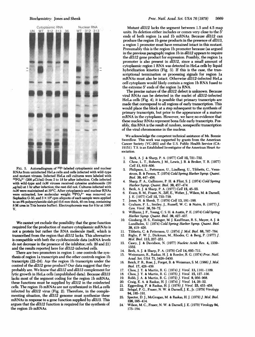
Contain VAI RNAs. The VAI RNAs are small (156- and160-nucleotide) adenovirus-coded RNAs synthesized by RNApolymerase III (15). They are the most abundant viral RNAspresent in adenovirus-infected cells at late times after infection(16). HeLa cells, infected with wild-type virus, labeled from2 to 18 hr after infection in the presence of cytosine arabinoside(20 ,g/ml, to block viral DNA synthesis and the onset of latefunctions) contained VAI RNAs (Fig. 5). In contrast no VAIRNAs were detected in d1312- or dl313-infected HeLa cells (nocytosine arabinoside present) labeled during the same period(Fig. 5). However, VAI RNA synthesis is not specifically de-pendent on the d1312 and d1313 gene functions becausets36-infected HeLa cells (20 ttg of cytosine arabinoside per ml,39°C) also did not contain VAI RNAs (Fig. 5). The same wastrue for ts125 (data not shown). These experiments suggest that
Biochemistry: Jones and Shenk
-
3668 Biochemistry: Jones and Shenk
E Region 2a 300 -
- 200-
00 -------@ - -----°-- °-0- Region 3C)0 300 *_
200-~ ~ ~~-00 Q= ----.--e ~-- -;-8.0 0. .......Region 4
300-~ ~ ~~-200- ,-
1 00 $,2,[email protected]., --*----^-s''0 1 2 3 4 5
Time, hr
FIG. 3. Reassociation of 32P-labeled viral DNA probes in thepresence of cytoplasmic RNA from HeLa cells infected with wild-typeand mutant viruses. The same restriction endonuclease-generatedfragments described in Fig. 2 served as probes after being 32p labeledby nick translation. Each time point sample contained 1500-2000 32pcpm. 0, Probe DNA plus uninfected HeLa cell RNA (1 mg/ml); *,probe DNA plus wild-type-infected HeLa cell RNA (1 mg/ml); *,probe DNA plus d1311-infected HeLa cell RNA (1 mg/ml); *, probeDNA plus d1312-infected HeLa cell RNA (1 mg/ml); A, probe DNAplus d1313-infected HeLa cell RNA (1 mg/ml).
VAI RNA is not synthesized at a detectable level until late timesafter infection (after the onset of viral DNA synthesis). Wesuspect that cytosine arabinoside is not completely effective inblocking this late function in wild-type cells since four different"early" mutants do block its synthesis.
DISCUSSIONMutant d1312 lacks an early viral function required for theexpression of cytoplasmic RNAs encoded by regions of the viralchromosome far removed from the site of the mutation. Theearly viral function that d1312 lacks must map at least partiallybetween 1.5 and 4.5 map units, the location of the 312 deletion.Two major early transcripts are encoded by this region of theadenovirus genome (ref. 14; Fig. 1, region la), and one (or both)of these is probably responsible for the function that d1312 lacks.Because d1312 is unable to transform rat embryo or rat embryobrain cells (unpublished data), this gene function probably alsoplays a critical role in transformation.A trivial alternative to the above interpretation of the d1312
gene function could be that d1312 virions are not transportedto the nucleus or uncoated subsequent to infection. As a result,the viral chromosome is not made accessible for transcription.Precedent for this argument is found in simian virus 40 tem-perature-sensitive mutants of the D complementation group(17-19). These mutants contain alterations in minor structuralpolypeptides. At the nonpermissive temperature, they are not
EC 300 .C
C4Z 200 -
~100K' 9- A
° 300Region33
C 200 - 1o ------...~..~..D.0..... . ..Do
300 Region 4 . . - .
200 -
1001 ........0.0 1 2 3 4 5
Time, hr
FIG. 4. Reassociation of 32P-labeled viral DNA probes in thepresence of nuclear RNA from HeLa cells infected with wild-type andd1312 viruses. The probes were the same as in Fig. 3. 0, Probe DNAplus uninfected HeLa cell RNA (1 mg/ml); *, probe DNA plus wild-type-infected HeLa cell RNA (1 mg/ml); *, probe DNA plusd1312-infected HeLa cell RNA (1 mg/ml); A, probe DNA plusd1312-infected HeLa cell RNA (1 mg/ml) treated for 10 min at 37°Cwith pancreatic ribonuclease (10 ,ug/ml) in 10 mM Tris-HCl/l mMEDTA before annealing.
uncoated and they fail to complement other temperature-sensitive mutants. This alternative is quite unlikely in d1312.Mutant d1312 complements d1313 for lytic growth in coinfectedHeLa cells (unpublished data), and virus-specific nuclear RNAsare present in d1312-infected HeLa cells. Both of these obser-vations suggest that the d1312 chromosome reaches the nucleusand is capable of being transcribed.Our finding that an early AdS gene product is required for
the expression of other early viral transcripts predicts that in-hibitors of protein synthesis added at or before infection shouldprevent the expression of early regions 2, 3, and 4. However,when cycloheximide is added at 1 hr after infection, the levelsof region 2, 3, and 4 mRNAs increase rather than decrease (20,21). We have performed similar experiments, adding cyclo-heximide (25 ,ug/ml) 1 hr prior to infection of HeLa cells withwild-type virus. In this case, the early mRNAs assayed (regions1 and 2) were still synthesized, but they were present in some-what lesser amounts than in untreated cultures. Low levels ofprotein synthesis could be detected in the cycloheximide-treatedHeLa cells. In the presence of the drug, about 5% the amountof 35S-labeled methionine was incorporated into acid-precipi-table products as in untreated cells. As a result, it is likely thata small amount of the d1312 gene product is made in the pres-ence of cycloheximide. This, in turn, facilitates the synthesisof transcripts corresponding to other early regions. Experimentswith other inhibitors of protein synthesis or a combination ofinhibitors may help to resolve this issue.
Proc. Natl. Acad. Sci. USA 76 (1979)
Proc. Natl. Acad. Sci. USA 76 (1979) 3669
Cytoplasmic RNA Nuclear RNAUN WT 312 313 36 WT 312 313
111111 111:3 U~~~~~~~~~~~~~~~~~~~~~~~~~~~~~~~~~~~~~~~~~~~~~~~~~~~~~~~~~~~~~~~~~~~~.Ow ~ q
_o_
*OIDON gasF
. A-
:ts.es.-K
a
2 9
FIG. 5. Autoradiogram of 32P-labeled cytoplasmic and nuclearRNAs from uninfected HeLa cells and cells infected with wild-typeand mutant viruses. Infected HeLa cell cultures were labeled with32po42- (200 ACi/ml) from 2 to 18 hr after infection. Cells infectedwith wild-type and ts36 viruses received cytosine arabinoside (20,ug/ml) at 1 hr after infection; the rest did not. Cultures infected withts36 were maintained at 390C. After cytoplasmic and nuclear RNAswere extracted, low molecular weight 32pO42- was removed on
Sephadex G-50, and 3 X 10⁵ cpm aliquots of each sample were appliedto an 8%o polyacrylamide slab gel (0.6 mm thick, 40 cm long, containing8 M urea in Tris borate buffer). Electrophoresis was for 8 hr at 1000V.
We cannot yet exclude the possibility that the gene functionrequired for the production of mature cytoplasmic mRNAs isnot a protein but rather the RNA molecule itself, which istranscribed from the region that d1312 lacks. This alternativeis compatible with both the cycloheximide data (mRNA levelsdo not decrease in the presence of the inhibitor; refs. 20 and 21)and the results reported here for d1312-infected cells.
There .are two promoters in region 1: one controls the syn-thesis of region la transcripts and the other controls region lbtranscripts.(22-24). Are the region lb transcripts under thecontrol of the dl312 gene product? Our data suggest that theyprobably are. We know that d1312 and d1313 complement forlytic growth in HeLa cells (unpublished data). Because d1313lacks most of the segment coding for the region lb mRNAs,these functions must be supplied by d1312 in the coinfectedcells. The region lb mRNAs are not synthesized in HeLa cellsinfected by d1312 virus (Fig. 2). Therefore, in the comple-menting situation, the d1312 genome must synthesize thesemRNAs in response to a gene function supplied by d1313. Thisargues that the d1312 function is required for the synthesis ofthe region lb mRNAs.
Mutant d1312 lacks the segment between 1.5 and 4.5 mapunits. Its deletion either includes or comes very close to the 5'ends of both region la and lb mRNAs. Because d1312 canproduce the region lb gene products in the presence of d1313,a region I.promoter must have remained intact in this mutant.Presumably this is the region lb promoter because (as arguedin the previous paragraph) region lb in d1312 appears to requirethe d1312 gene product for expression. Possibly, the region lapromoter is also present in d1312, since a small amount ofcytoplasmic region 1 RNA was detected in HeLa cells by liquidhybridization kinetics (Fig. 3). If this is the case, the tran-scriptional termination or processing signals for region lamRNAs must also be intact. Otherwise d1312-infected HeLacell cytoplasm would likely contain a region lb RNA fused tothe extreme 5' ends of the region la RNA.The precise nature of the d1312 defect is unknown. Because
viral RNAs can be detected in the nuclei of d1312-infectedHeLa cells (Fig. 4), it is possible that primary transcripts aremade that correspond to all regions of early transcription. Thiswould place the block at a step subsequent to the synthesis ofprimary transcripts, but prior to the appearance of a maturemRNA in the cytoplasm. However, we have no evidence thatthese nuclear RNAs represent bona fide early transcripts. Pos-sibly, this RNA is the result of random, nonspecific transcriptionof the viral chromosome in the nucleus.We acknowledge the competent technical assistance of Ms. Bonnie
Swerdlow. This work was supported by grants from the AmericanCancer Society (VC-263) and the U.S. Public Health Service (CA-19151). T.S. is an Established Investigator of the American Heart As-sociation.
1. Berk, A. J. & Sharp, P. A. (1977) Cell 12, 721-732.2. Chow, L. T., Roberts, J. M., Lewis, J. B. & Broker, T. R. (1977)
Cell 11, 819-836.3. Philipson, L., Pettersson, U., Lindberg, U., Tibbetts, C., Venn-
strom, B. & Person, T. (1974) Cold Spring Harbor Symp. Quant.Biol. 39, 447-456.
4. Sharp, P. A., Gallimore, P. H. & Flint, S. J. (1974) Cold SpringHarbor Symp. Quant. Biol. 39,457-474.
5. Berk, A. J. & Sharp, P. A. (1977) Cell 12, 45-55.6. Evans, R. M., Fraser, N., Ziff, E., Weber, J., Wilson, M. & Darnell,
J. E. (1977) Cell 12, 733-739.7. Jones, N. & Shenk, T. (1978) Cell 13, 181-188.8. Graham, F. L., Smiley, J., Russell, W. C. & Nairn, R. (1977) J.
Gen. Virol. 36,59-72.9. Williams, J. F., Young, C. S. H. & Austin, P. E. (1974) Cold Spring
Harbor Symp. Quant. Biol. 39,427-437.10. Ginsberg, H. S., Ensinger, M. J. Kauffman, R. S., Mayer, A. J. &
Lundholm, U. (1974) Cold Spring Harbor Symp. Quant. Biol.39,419-426.
11. Tibbetts, C. & Pettersson, U. (1974) J. Mol. Biol. 88, 767-784.12. Rigby, P. W. J., Dickman, M., Rhodes, C. & Berg, P. (1977) J.
Mol. Biol. 113,237-251.13. Casey, J. & Davidson, N. (1977) Nucleic Acids Res. 4, 1539-
1552.14. Berk, A. J. & Sharp, P. A. (1978) Cell 14,695-711.15. Weinmann, R., Raskas, H. J. & Roeder, R. G. (1974) Proc. Natl.
Acad. Sci. USA 71, 3426-3430.16. Reich, P. R., Rose, J., Forget, B. & Weissman, S. M. (1966) J. Mol.
Biol. 17, 428-439.17. Chou, J. Y. & Martin, R. G. (1974) J. Virol. 13, 1101-1109.18. Chou, J. Y. & Martin, R. G. (1975) J. Virol. 15, 127-136.19. Robb, J. A. & Martin, R. G. (1972) J. Virol. 9, 956-968.20. Craig, E. A. & Raskas, H. J. (1974) J. Virol. 14, 26-32.21. Eggerding, F. & Raskas, H. J. (1978) J. Virol. 25,453-458.22. Sehgal, P. G., Fraser, N. W. & Darnell, J. E., Jr. (1979) Virology
94, 185-191.23. Spector, D. J., McGrogan, M. & Raskas, H. J. (1979) J. Mol. Biol.
126,395-414.24. Wilson, M. C., Fraser, N. W. & Darnell, J. E. (1979) Virology 94,
175-184.
Biochemistry: Jones and Shenk