Embed Size (px)
Citation preview
Proc. Natl. Acad. Sci. USAVol. 77, No. 8, pp. 4779-4783, August 1980Cell Biology
Similarities and dissimilarities between calmodulin and aChlamydomonas flagellar protein
(motility/phenothiazines/immunochemistry/calcium modulated proteins/phosphodiesterase activation)
LINDA J. VAN ELDIK, GIANNI PIPERNO, AND D. MARTIN WATTERSONThe Rockefeller University, 1230 York Avenue, New York, New York 10021
Communicated by Philip Siekevitz, May 8,1980
ABSTRACT A protein that resembles vertebrate calmodu-lins and troponin C has been isolated from Chlamydomonasflagella by using a calmodulin purification protocol that in-cluded calcium-dependent affinity-based adsorption chroma-tography on phenothiazine-Sepharose conjugates. The flagellarprotein resembled calmodulin in elution from reverse-phasecolumns, had a peptide map similar to that of calmodulin, andcompeted with vertebrate calmodulin in a radioimmunoassayusing antisera against vertebrate calmodulin. However, thisflagellar protein did not activate phosphodiesterase, lackedN'-trimethyllysine, and had an isoelectric point approximately0.3 pH unit higher than that of vertebrate calmodulin. Whenanalyzed by polyacrylamide gel electrophoresis under variousconditions, the Chiamydomonas protein migrated betweenvertebrate calmodulins and rabbit skeletal muscle troponin Cand did not manifest a large calcium-dependent mobility shift.This calmodulin-like protein was identified as one of the ap-proximately 200 35S-labeled components in Chlamydomonasflagella resolved by two-dimensional gel electrophoresis. Thesestudies indicate that calmodulin and a structurally and func-tionally homologous protein are present in the same cell. Thesestudies also demonstrate that caution is necessary: (i) in iden-tifying a protein as a calmodulin, (ii) in using phenothiazinesor antisera directed against vertebrate calmodulins as specificprobes for calmodulin, and (Mi) in the interpretation of experi-ments on biological systems in which calmodulin is substitutedfor the homologous calmodulin-like protein.
Calcium has been implicated in the regulation of metabolic andmechanochemical processes in a number of biological systems(for a review, see ref. 1). Thermodynamic, kinetic, and crys-tallographic data strongly suggest that the targets of calciumacting as a biological signal transducer in the cytoplasm arecalcium-modulated proteins (2). Two examples of this class ofcalcium-binding proteins are troponin C and calmodulin (3-6).The most extensively studied calcium-dependent mechano-chemical system is vertebrate striated muscle and its regulationby troponin C (7). Other mechanochemical systems in whichcalcium regulation has been implicated are eukaryotic cilia andflagella (8-10). However, in these structures the moleculartargets of calcium have not been described.
In Chlamydomonas reinhardtll the effect of calcium onflagellar movement has been observed in isolated flagellarapparatus and in whole cells subjected to photostimulation (11,12). Both studies indicated that above a calcium concentrationof 1 MM, swimming changes from a forward to a backwardmotion. Besides the physiological dependence of motility oncalcium, use of C. reinhardtll provides some unique advantagesfor the study of regulation of flagellar motility by calcium.Polypeptide components can be labeled to a high specific ac-tivity by growth in a medium containing [3aSisulfuric acid (13).
This allows for the reproducible resolution of complex mixturesof flagellar polypeptides by two-dimensional gel electrophoresis(13). In addition, procedures for the isolation of motility mutantshave been described (14). The combined use of mutant analysis,differential extraction, two-dimensional polyacrylamide gelanalysis, and electron microscopic analysis has resulted in theidentification of several axonemal polypeptides as componentsof different flagellar substructures (13, 15, 16). Recently, anactin-like protein has been isolated from Chlamydomonasflagella and shown to be one of the polypeptides assembled inthe axoneme (17). The isolation of calcium-modulated proteinsfrom C. reinhardtll flagella and the correlation of these proteinswith specific sets of flagellar components analyzed by two-dimensional gel electrophoresis would enhance our knowledgeof flagellar substructures and provide a firm basis for futurestudies of calcium regulation.We describe here the isolation of a protein from Chlamy-
domonas flagella that resembles calmodulin and troponin C."Calmodulin" is the name proposed (4) for a brain phospho-diesterase activator protein. This activator protein has beenshown (5, 18) to be indistinguishable from the unique chemicalstructure termed "modulator protein" (5, 6). Bovine braincalmodulin is a multifunctional, calcium-modulated proteinthat has a calculated molecular mass of 16,680 g/mol and isstructurally homologous to troponin C (6). As previously dis-cussed (5, 6, 19, 20), calcium-modulated proteins have ex-tremely.similar physical, chemical, and sometimes functionalproperties. The flagellar protein described in this report clearlypossesses many of the characteristic properties of this class ofproteins, especially those features previously thought to beunique features of calmodulin (3, 4). However, the flagellarprotein does not appear to be a calmodulin.
MATERIALS AND METHODSCulture of cells, labeling with [a5S]sulfuric acid, preparationof flagella, and two-dimensional electrophoresis of polypeptideswere performed as described (15, 16). Silver staining of unla-beled flagellar polypeptides in two-dimensional gels was asdescribed elsewhere (21). Calmodulin was isolated from bovinebrain and chicken gizzard by using described procedures (20)and' phenothiazine-Sepharose [2-chloro-10-(3-aminopro-pyl)phenothiazine-Sepharose] chromatography (22). Parval-bumin and S100b were generous gifts of R. Kretsinger (Char-lottesville, VA). Flagellar preparations were sonicated (Bransonmodel W185 with microtip) for 30 sec at 40C (approximately25 W) in 2 vol of buffer I (0.1 M Tris-HC1/2 mM [ethylene-bis(oxyethylenenitrilo)]tetraacetic acid (EGTA)/2 mM 2-mercaptoethanol, pH 8.0), and centrifuged at 10,000 X g for
Abbreviation: EGTA, [ethylenebis(oxyethylenenitrilo)]tetraaceticacid.
4779
The publication costs of this article were defrayed in part by pagecharge payment. This article must therefore be hereby marked "ad-vertisement" in accordance with 18 U. S. C. §1734 solely to indicatethis fact.
4780 Cell Biology: Van Eldik et al.
30 min. The supernatant fraction was prepared for chroma-tography on phenothiazine-Sepharose conjugates as described(22).
Troponin was isolated from rabbit skeletal muscle by theprocedure of Ebashi et al. (23), and troponin C was subse-quently purified by the method of Greaser and Gergely (24).Trypsin digestions were performed in the presence of EGTAas described (20). Column peptide maps of trypsin digests wereobtained on a Whatman Partisil M9 ODS-2 column (9.4 X 250mm) with a modified (25) Hewlett-Packard 1084B liquidchromatography system. Solvent A was 0.1% hydrochloric acid(Baker, Ultrex) and solvent B was acetonitrile (Burdick &Jackson, Muskegon, MI, UV grade). The microprocessor-con-trolled elution gradient consisted of the following time program:0 min, %B = 5; 5.0 minm %B = 5; 5.5 min, %B = 10; 10.0 min,%B = 10; 40.0 min, %B = 50; 45.0 min, %B = 50. Amino acidand protein analyses were done as described (20, 22). Fluor-ography was done according to the procedure of Bonner andLaskey (26). Polyacrylamide gel electrophoresis in the presenceand absence of sodium dodecyl sulfate was performed essen-tially as described (20, 22); details are given where appropriatein the text. Phosphodiesterase was prepared and activator assayswere done as described (25). Antibody against calmodulin wasprepared and radioimmunoassays were performed as describedelsewhere (27).
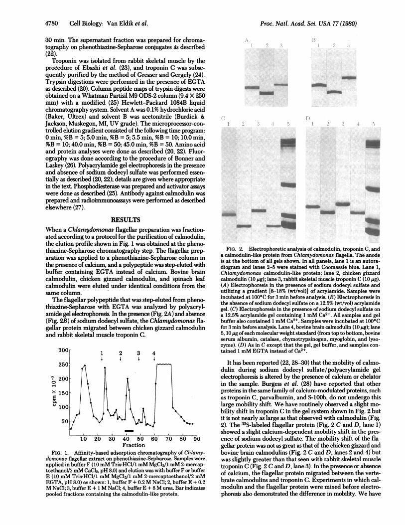
RESULTSWhen a Chlamydomonas flagellar preparation was fraction-ated according to a protocol for the purification of calmodulin,the elution profile shown in Fig. 1 was obtained at the pheno-thiazine-Sepharose chromatography step. The flagellar prep-aration was applied to a phenothiazine-Sepharose column inthe presence of calcium, and a polypeptide was step-eluted withbuffer containing EGTA instead of calcium. Bovine braincalmodulin, chicken gizzard calmodulin, and spinach leafcalmodulin were eluted under identical conditions from thesame column.The flagellar polypeptide that was step-eluted from pheno-
thiazine-Sepharose with EGTA was analyzed by polyacryl-amide gel electrophoresis. In the presence (Fig. 2A) and absence(Fig. 2B) of sodium dodecyl sulfate, the Chlamydomonas fla-gellar protein migrated between chicken gizzard calmodulinand rabbit skeletal muscle troponin C.
3001 1 2 3 4
250
200
X150
100
50
10 20 30 40 50 60 70 80 90Fraction
FIG. 1. Affinity-based adsorption chromatography of Chlamy-domonas flagellar extract on phenothiazine-Sepharose. Samples wereapplied in buffer F (10mM Tris-HCl/l mM MgCl2/1 mM 2-mercap-toethanol/2 mM CaCl2, pH 8.0) and elution was with buffer F or bufferE (10 mM Tris-HCl/1 mM MgCl2/1 mM 2-mercaptoethanol/2 mMEGTA, pH 8.0) as shown: 1, buffer F + 0.2M NaCl; 2, buffer E + 0.2M NaCl; 3, buffer E + 1 M NaCl; 4, buffer E + 8M urea. Bar indicatespooled fractions containing the calmodulin-like protein.
__m
-a_
)
m.
I_
__mb__ ~ a
FIG. 2. Electrophoretic analysis of calmodulin, troponin C, anda calmodulin-like protein from Chlamydomonas flagella. The anodeis at the bottom of all gels shown. In all panels, lane 1 is an autora-diogram and lanes 2-5 were stained with Coomassie blue. Lane 1,Chlamydomonas calmodulin-like protein; lane 2, chicken gizzardcalmodulin (10 ,g); lane 3, rabbit skeletal muscle troponin C (10 ,g).(A) Electrophoresis in the presence of sodium dodecyl sulfate andutilizing a gradient [8-18% (wt/vol)] of acrylamide. Samples wereincubated at 100°C for 3 min before analysis. (B) Electrophoresis inthe absence of sodium dodecyl sulfate on a 12.5% (wt/vol) acrylamidegel. (C) Electrophoresis in the presence of sodium dodecyl sulfate ona 12.5% acrylamide gel containing 1 mM Ca2+. All samples and gelbuffer also contained 1 mM Ca2+. Samples were incubated at 1000Cfor 3 min before analysis. Lane 4, bovine brain calmodulin (10 tig); lane5, 10 Mg ofeach molecular weight standard (from top to bottom, bovineserum albumin, catalase, chymotrypsinogen, myoglobin, and lyso-zyme). (D) As in C except that the gel, gel buffer, and samples con-tained 1 mM EGTA instead of Ca2 .
It has been reported (22, 28-30) that the mobility of calmo-dulin during sodium dodecyl sulfate/polyacrylamide gelelectrophoresis is altered by the presence of calcium or chelatorin the sample. Burgess et al. (28) have reported that otherproteins in the same family of calcium-modulated proteins, suchas troponin C, parvalbumin, and S-100b, do not undergo thislarge mobility shift. We have routinely observed a slight mo-bility shift in troponin C in the gel system shown in Fig. 2 butit is not nearly as large as that observed with calmodulin (Fig.2). The 35S-labeled flagellar protein (Fig. 2 C and D, lane 1)showed a slight calcium-dependent mobility shift in the pres-ence of sodium dodecyl sulfate. The mobility shift of the fla-gellar protein was not as great as that of the chicken gizzard andbovine brain calmodulins (Fig. 2 C and D, lanes 2 and 4) butwas slightly greater than that seen with rabbit skeletal muscletroponin C (Fig. 2 C and D, lane 3). In the presence or absenceof calcium, the flagellar protein migrated between the verte-brate calmodulins and troponin C. Experiments in which cal-modulin and the flagellar protein were mixed before electro-phoresis also demonstrated the difference in mobility. We have
Proc. Nati. Acad. Sci. USA 77 (1980)
Proc. Natl. Acad. Sci. USA 77 (1980) 4781
previously reported (22) that plant calmodulin and calmodulinlacking NE-trimethyllysine also undergo this shift.
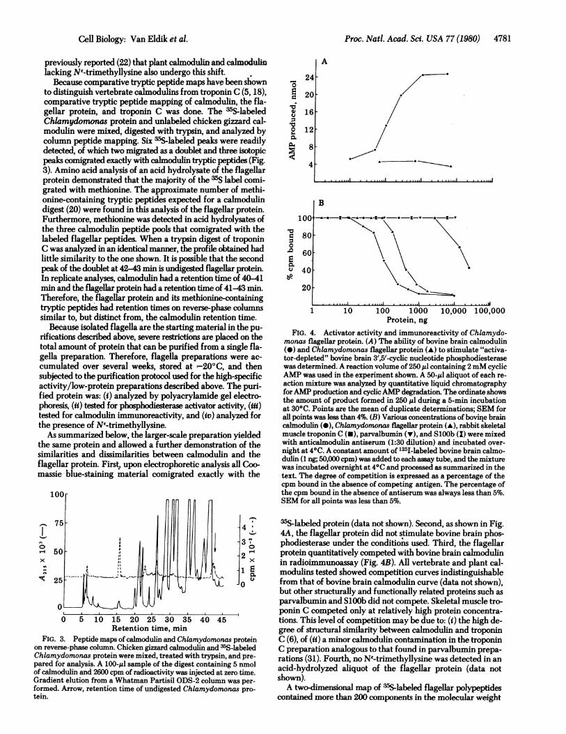
Because comparative tryptic peptide maps have been shownto distinguish vertebrate calmodulins from troponin C (5, 18),comparative tryptic peptide mapping of calmodulin, the fla-gellar protein, and troponin C was done. The 35S-labeledChlamydomonas protein and unlabeled chicken gizzard cal-modulin were mixed, digested with trypsin, and analyzed bycolumn peptide mapping. Six 35Slabeled peaks were readilydetected, of which two migrated as a doublet and three isotopicpeaks comigrated exactly with calmodulin tryptic peptides (Fig.3). Amino acid analysis of an acid hydrolysate of the flagellarprotein demonstrated that the majority of the 35S label comi-grated with methionine. The approximate number of methi-onine-containing tryptic peptides expected for a calmodulindigest (20) were found in this analysis of the flagellar protein.Furthermore, methionine was detected in acid hydrolysates ofthe three calmodulin peptide pools that comigrated with thelabeled flagellar peptides. When a trypsin digest of troponinC was analyzed in an identical manner, the profile obtained hadlittle similarity to the one shown. It is possible that the secondpeak of the doublet at 42-43 min is undigested flagellar protein.In replicate analyses, calmodulin had a retention time of 40-41min and the flagellar protein had a retention time of 41-43 min.Therefore, the flagellar protein and its methionine-containingtryptic peptides had retention times on reverse-phase columnssimilar to, but distinct from, the calmodulin retention time.
Because isolated flagella are the starting material in the pu-rifications described above, severe restrictions are placed on thetotal amount of protein that can be purified from a single fla-gella preparation. Therefore, flagella preparations were ac-cumulated over several weeks, stored at -200C, and thensubjected to the purification protocol used for the high-specificactivity/low-protein preparations described above. The puri-fied protein was: (W) analyzed by polyacrylamide gel electro-phoresis, (ii) tested for phosphodiesterase activator activity, (mi)tested for calmodulin immunoreactivity, and (iv) analyzed forthe presence of NE-trimethyllysine.
As summarized below, the larger-scale preparation yieldedthe same protein and allowed a further demonstration of thesimilarities and dissimilarities between calmodulin and theflagellar protein. First,, upon electrophoretic analysis all Coo-massie blue-staining material comigrated exactly with the
14
0
x
C1
-4
-3
2x
E00Q
-0
0 5 10 15 20 25 30 35 40 45Retention time, min
FIG. 3. Peptide maps of calmodulin and Chlamydomonas proteinon reverse-phase column. Chicken gizzard calmodulin and m5S-labeledChlamydomonas protein were mixed, treated with trypsin, and pre-pared for analysis. A 100-ul sample of the digest containing 5 nmolof calmodulin and 2600 cpm of radioactivity was injected at zero time.Gradient elution from a Whatman Partisil ODS-2 column was per-formed. Arrow, retention time of undigested Chlamydomonas pro-tein.
'0
0)
10
'00.0
a0v
24
20
16
12[
8
4
A
B
100',.__ , _ _,
80-
60
40 -
20-
1 10 100 1000Protein, ng
10,000 100,000
FIG. 4. Activator activity and immunoreactivity of Chlamydo-monas flagellar protein. (A) The ability of bovine brain calmodulin(-) and Chlamydomonas flagellar protein (-) to stimulate "activa-tor-depleted" bovine brain 3',5'-cycic nucleotide phosphodiesterasewas determined. A reaction volume of 250,gl containing 2mM cyclicAMP was used in the experiment shown. A 50-$l aliquot of each re-action mixture was analyzed by quantitative liquid chromatographyforAMP production and cyclicAMP degradation. The ordinate showsthe amount of product formed in 250 ,l during a 5-min incubationat 300C. Points are the mean of duplicate determinations; SEM forall points was less than 4%. (B) Various concentrations of bovine braincalmodulin (@), Chlamydomonas flagellar protein (-), rabbit skeletalmuscle troponin C (U), parvalbumin (v), and SlOOb (X) were mixedwith anticalmodulin antiserum (1:30 dilution) and incubated over-night at 40C. A constant amount of 12I-labeled bovine brain calmo-dulin (1 ng; 50,000 cpm) was added to each assay tube, and the mixturewas incubated overnight at 40C and processed as summarized in thetext. The degree of competition is expressed as a percentage of thecpm bound in the absence of competing antigen. The percentage ofthe cpm bound in the absence of antiserum was always less than 5%.SEM for all points was less than 5%.
35S-labeled protein (data not shown). Second, as shown in Fig.4A, the flagellar protein did not stimulate bovine brain phos-phodiesterase under the conditions used. Third, the flagellarprotein quantitatively competed with bovine brain calmodulinin radioimmunoassay (Fig. 4B). All vertebrate and plant cal-modulins tested showed competition curves indistinguishablefrom that of bovine brain calmodulin curve (data not shown),but other structurally and functionally related proteins such asparvalbumin and SlOOb did not compete. Skeletal muscle tro-ponin C competed only at relatively high protein concentra-tions. This level of competition may be due to: (i) the high de-gree of structural similarity between calmodulin and troponinC (6), of (ii) a minor calmodulin contamination in the troponinC preparation analogous to that found in parvalbumin prepa-rations (31). Fourth, no NI-trimethyllysine was detected in anacid-hydrolyzed aliquot of the flagellar protein (data notshown).A two-dimensional map of 3sS-labeled flagellar polypeptides
contained more than 200 components in the molecular weight
Cell Biology: Van Eldik et al.
0 0
0
4782 Cell Biology: Van Eldik et al.
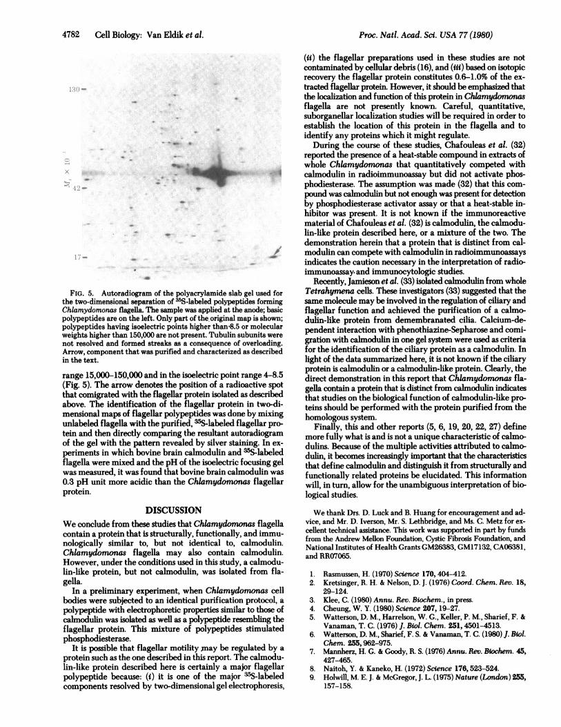
FIG. 5. Autoradiogram of the polyacrylamide slab gel used for
the two-dimensional separation of -",S-labeled polypeptides forming
Chiamydomonas flagella. The sample was applied at the anode; basic
polypeptides are on the left. Only part of the.original map is shown;
polypeptides having isoelectric points higher than-8.5 or molecular
weights higher than 150,000 are not present. Tubulin subunits were
not resolved and formed streaks as a consequence of overloading.
Arrow, component that was purified and characterized as described
in the text.
range 15,000-150,000 and in the isoelectric point range 4-8.5
(Fig. 5). The arrow denotes the position of a radioactive spot
that comigrated with the flagellar protein isolated as described
above. The identification of the flagellar protein in two-di-mensional maps of flagellar polypeptides was done by mixingunlabeled flagella with the purified,wasSplabeled flagellar pro-
tein and then directly comparing the resultant autoradiogramof the gel with the pattern revealed by silver staining. In ex-
periments in which bovine brain calmodulin and asdslabeledflagella were mixed and the pH of the isoelectric focusing gelwas measured, it was found that bovine brain calmodulin was
0.3 pH unit more acidic than the Chiamydomonas flagellarprotein.
DISCUSSION
We conclude from these studies that Chiamydomonas flagella
contain a protein that is structurally, functionally, and immu-nologically similar to, but not identical to, calmodulin.
Chiamydomonas flagella may also contain calmodulin.However, under the conditions used in this study, a calmodu-
lin-like protein, but not calmodulin, was isolated from fla-
gella.In a preliminary experiment, when Chlamydomonas cell
bodies were subjected to an identical purification protocol, a
polypeptide with electrophoretic properties similar to those of
calmodulin was isolated as well as a polypeptide resembling the
flagellar protein. This mixture of polypeptides stimulated
phosphodiesterase.It is possible that flagellar motility may be regulated by a
protein such as the one described in this report. The calmodu-lin-like protein described here is certainly a major flagellarpolypeptide because: (i) it is one of the major ofS-labeledcomponents resolved by two-dimensional gel electrophoresis,
(ii) the flagellar preparations used in these studies are notcontaminated by cellular debris (16), and (iii) based on isotopicrecovery the flagellar protein constitutes 0.6-1.0% of the ex-tracted flagellar protein. However, it should be emphasized thatthe localization and function of this protein in Chlamydomnsflagella are not presently known. Careful, quantitative,suborganellar localization studies will be required in order toestablish the location of this protein in the flagella and toidentify any proteins which it might regulate.
During the course of these studies, Chafouleas et al. (32)reported the presence of a heat-stable compound in extracts ofwhole Chlamydomonas that quantitatively competed withcalmodulin in radioimmunoassay but did not activate phos-phodiesterase. The assumption was made (32) that this com-pound was calmodulin but not enough was present for detectionby phosphodiesterase activator assay or that a heat-stable in-hibitor was present. It is not known if the immunoreactivematerial of Chafouleas et al. (32) is calmodulin, the calmodu-lin-like protein described here, or a mixture of the two. Thedemonstration herein that a protein that is distinct from cal-modulin can compete with calmodulin in radioimmunoassaysindicates the caution necessary in the interpretation of radio-immunoassay. and immunocytologic studies.
Recently, Jamieson et al. (33) isolated calmodulin from wholeTetrahymena cells. These investigators (33) suggested that thesame molecule may be involved in the regulation of ciliary andflagellar function and achieved the purification of a calmo-dulin-like protein from demembranated cilia. Calcium-de-pendent interaction with phenothiazine-Sepharose and comi-gration with calmodulin in one gel system were used as criteriafor the identification of the ciliary protein as a calmodulin. Inlight of the data summarized here, it is not known if the ciliaryprotein is calmodulin or a calmodulin-like protein. Clearly, thedirect demonstration in this report that Chlamydomonas fla-gella contain a protein that is distinct from calmodulin indicatesthat studies on the biological function of calmodulin-like pro-teins should be performed with the protein purified from thehomologous system.
Finally, this and other reports (5, 6, 19, 20, 22, 27) definemore fully what is and is not a unique characteristic of calmo-dulins. Because of the multiple activities attributed to calmo-dulin, it becomes increasingly important that the characteristicsthat define calmodulin and distinguish it from structurally andfunctionally related proteins be elucidated. This informationwill, in turn, allow for the unambiguous interpretation of bio-logical studies.
We thank Drs. D. Luck and B. Huang for encouragement and ad-vice, and Mr. D. Iverson, Mr. S. Lethbridge, and Ms. C. Metz for ex-cellent technical assistance. This work was supported in part by fundsfrom the Andrew Mellon Foundation, Cystic Fibrosis Foundation, andNational Institutes of Health Grants GM26383, GM17132, CA06381,and RR07065.
1. Rasmussen, H. (1970) Science 170,404-412.2. Kretsinger, R. H. & Nelson, D. J. (1976) Coord. Chem. Rev. 18,
29-124.3. Klee, C. (1980) Annu. Rev. Biochem., in press.4. Cheung, W. Y. (1980) Science 207, 19-27.5. Watterson, D. M., Harrelson, W. G., Keller, P. M., Sharief, F. &
Vanaman, T. C. (1976) J. Biol. Chem. 251,4501-4513.6. Watterson, D. M., Sharief, F. S. & Vanaman, T. C. (1980) J. Biol.
Chem. 255,962-975.7. Mannherz, H. G. & Goody, R. S. (1976) Annu. Rev. Biochem. 45,
427-465.8. Naitoh, Y. & Kaneko, H. (1972) Science 176,523-524.9. Holwill, M. E. J. & McGregor, J. L. (1975) Nature (London) 255,
157-158.
Froc. Natl. Acad. Sci. USA 77 (1980)
Proc. Natl. Acad. Sci. USA 77 (1980) 4783
10. Brokaw, C. J., Jasslin, R. & Bobrow, L. (1974) Biochem. Blophys.Res. Commun. 58,795-800.
11. Hyams, J. S. & Borisy, G. G. (1978) J. Cell Sci. 33, 235-253.
12. Schmidt, J. A. & Eckert, R. (1976) Nature (London) 262, 713-715.
13. Piperno, G., Huang, B. & Luck, D. J. L. (1977) Proc. Nati. Acad.Sci. USA 74, 1600-1604.
14. Huang, B., Rifkin, M. R. & Luck, D. J. L. (1977) J. Cell Biol. 72,67-85.
15. Piperno, G. & Luck, D. J. L. (1979) J. Biol. Chem. 254, 3084-3090.
16. Huang, B., Piperno, G. & Luck, D. J. L. (1979) J. Biol. Chem. 254,3091-3099.
17. Piperno, G. & Luck, D. J. L. (1979) J. Biol. Chem. 254, 2187-2190.
18. Stevens, F. C., Walsh, M., Ho, H. C., Teo, T. S. & Wang, J. H.(1976) J. Biol. Chem. 251, 4495-4500.
19. Watterson, D. M., Mendel, P. A. & Vanaman, T. C. (1980) Bio-chemistry 19, 2672-2676.
20. Van Eldik, L. J. & Watterson, D. M. (1979) J. Biol. Chem. 254,10250-10255.
21. Merril, C. R., Switzer, R. C. & Van Keuren, M. L. (1979) Proc.NatI. Acad. Sci. USA 76,4335-4339.
22. Van Eldik, L. J., Grossman, A. R., Iverson, D. B. & Watterson,D. M. (1980) Proc. Nati. Acad. Sci. USA 77, 1912-1916.
23. Ebashi, S., Wakabayashi, T. & Ebashi, F. (1971) J. Biochem. 69,441-445.
24. Greaser, M. L. & Gergely, J. (1971) J. Biol. Chem. 246, 4226-4233.
25. Watterson, D. M., Iverson, D. B. & Van Eldik, L. J. (1980) J.Biochem. Biophys. Methods 2, 139-146.
26. Bonner, W. M. & Laskey, R. A. (1974) Eur. J. Biochem. 46,83-88.
27. Van Eldik, L. J. & Watterson, D. M. (1980) Ann. N.Y. Acad. Sci.,in press.
28. Burgess, W. H., Jemiolo, D. K. & Kretsinger, R. H. (1980) Bio-chim. Biophys. Acta, in press.
29. Grab, D. J., Berzins, K., Cohen, R. S. & Siekevitz, P. (1979) J. Biol.Chem. 254,8690-8696.
30. Klee, C. B., Crouch, T. H. & Krinks, M. H. (1979) Proc. Nati.Acad. Sci. USA 76,6270-6273.
31. LeDonne, N. C. & Coffee, C. J. (1979) J. Biol. Chem. 254,4317-4320.
32. Chafouleas, J. G., Dedman, J. R., Munjaal, R. P. & Means, A. R.(1979) J. Biol. Chem. 254, 10262-10267.
33. Jamieson, G. A., Vanaman, T. C. & Blum, J. J. (1979) Proc. Nati.Acad. Sci. USA 76,6471-6475.
Cell Biology: Van Eldik et al.