Embed Size (px)
Citation preview
DERMATOLOGIC INFECTIONS IN CHILDREN
ROBERTA C. ROMERO, M.D., F.P.D.S.TROPICAL DISEASE FOUNDATION
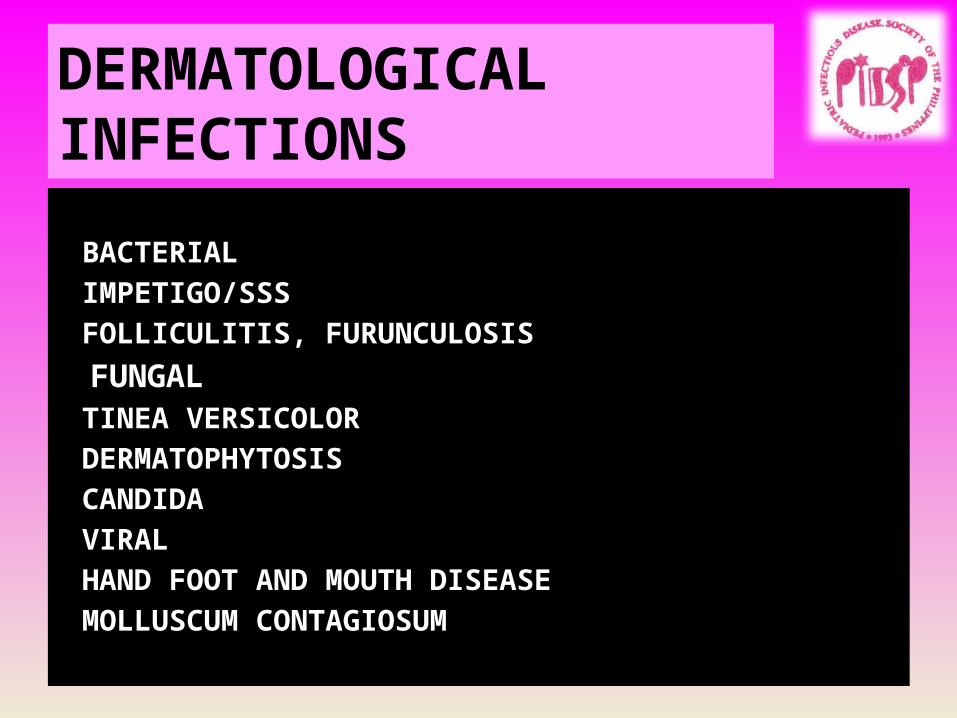
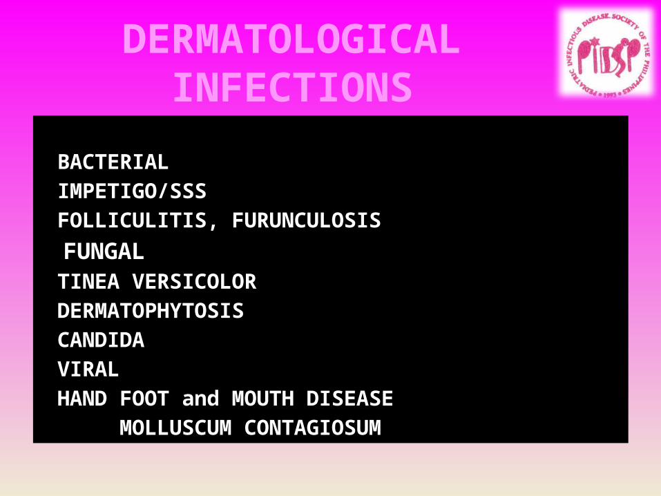
DERMATOLOGICAL INFECTIONS
BACTERIALIMPETIGO/SSSFOLLICULITIS, FURUNCULOSIS
FUNGALTINEA VERSICOLORDERMATOPHYTOSISCANDIDA
VIRALHAND FOOT AND MOUTH DISEASEMOLLUSCUM CONTAGIOSUM
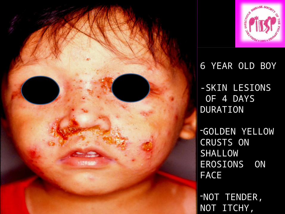
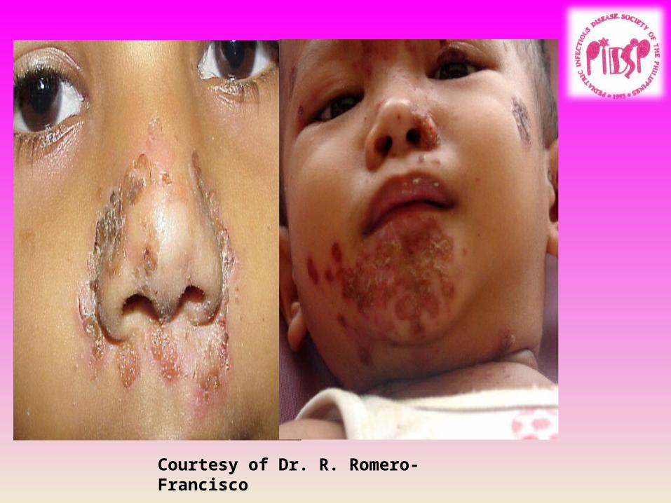
6 YEAR OLD BOY
-SKIN LESIONS OF 4 DAYS DURATION
-GOLDEN YELLOW CRUSTS ON SHALLOW EROSIONS ON FACE
-NOT TENDER, NOT ITCHY, SLIGHT FEVER
DIAGNOSIS?????
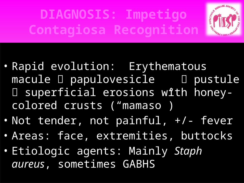
DIAGNOSIS: Impetigo Contagiosa Recognition
• Rapid evolution: Erythematous macule papulovesicle pustule superficial erosions with honey-colored crusts (“mamaso”)
• Not tender, not painful, +/- fever• Areas: face, extremities, buttocks• Etiologic agents: Mainly Staph aureus, sometimes
GABHS
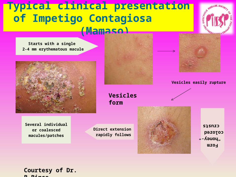
Typical clinical presentation of Impetigo Contagiosa
(Mamaso)Starts with a single
2-4 mm erythematous macule
Form “honey-colored” crusts
Vesicles easily rupture
Direct extension rapidly follows
Several individual or coalesced
macules/patches
Courtesy of Dr. B.Bince
Vesicles form
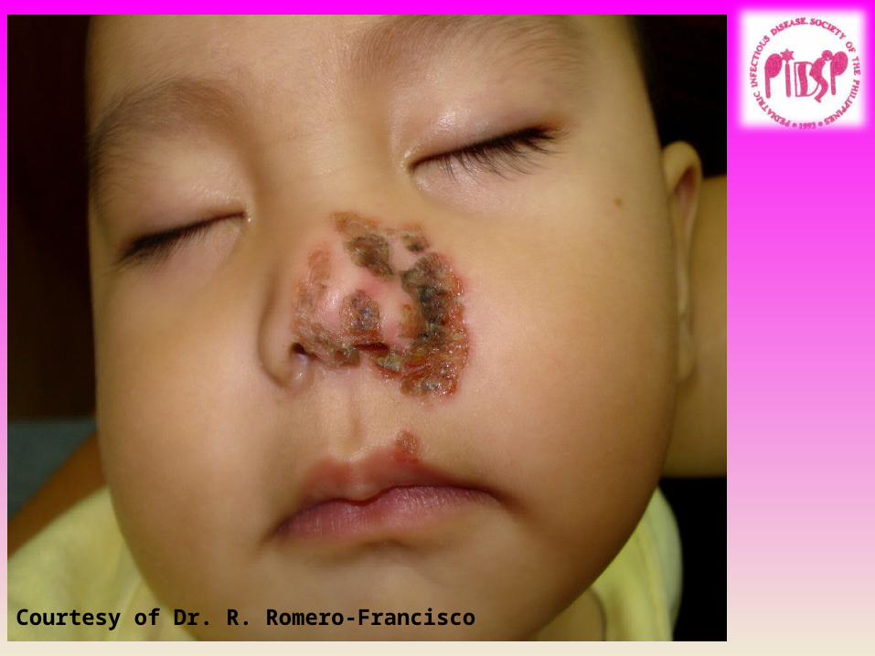
Courtesy of Dr. R. Romero-Francisco
Courtesy of Dr. R. Romero-Francisco
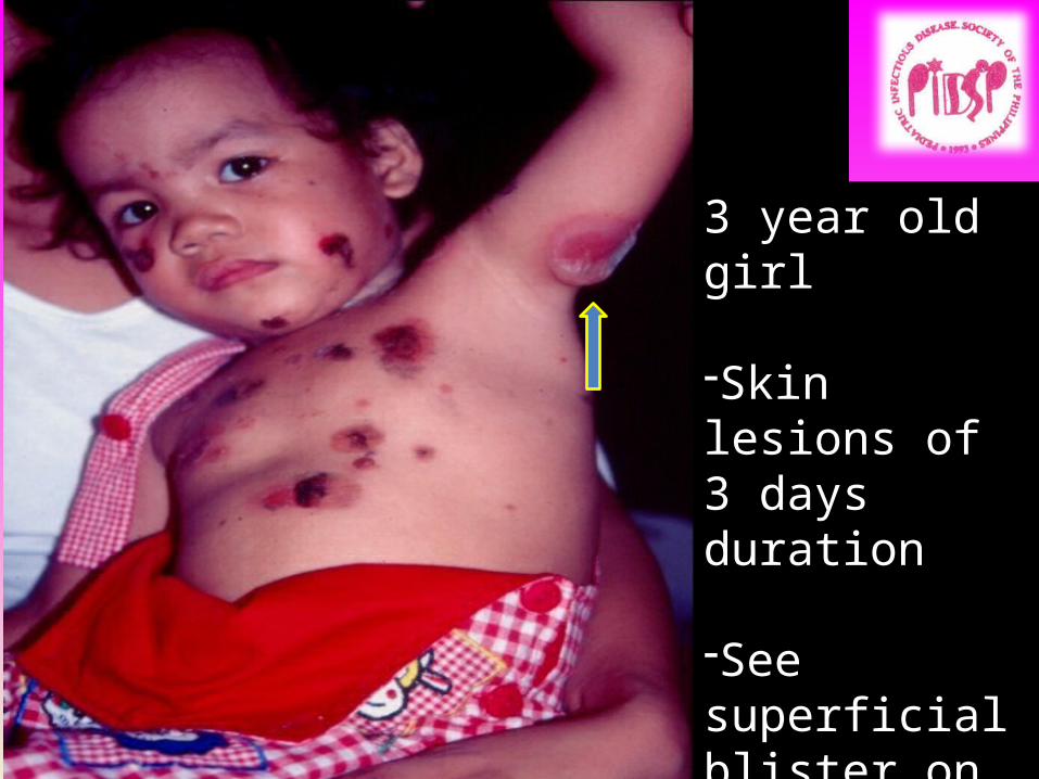
3 year old girl
-Skin lesions of 3 days duration
-See superficial blister on left underarm
Diagnosis????
IMPETIGO CONTAGIOSA
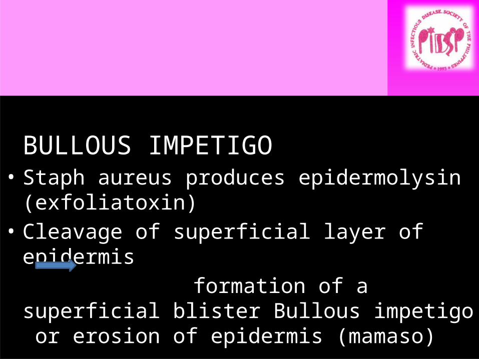
BULLOUS IMPETIGO• Staph aureus produces epidermolysin
(exfoliatoxin)• Cleavage of superficial layer of epidermis formation of a superficial blister Bullous
impetigo or erosion of epidermis (mamaso)
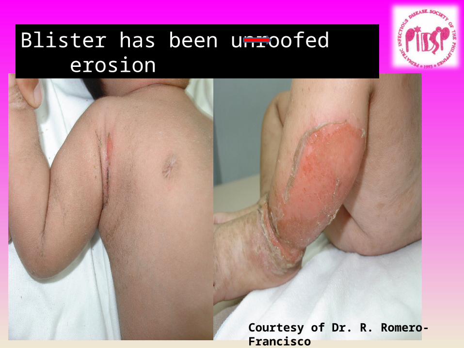
Blister has been unroofed erosion
Courtesy of Dr. R. Romero-Francisco
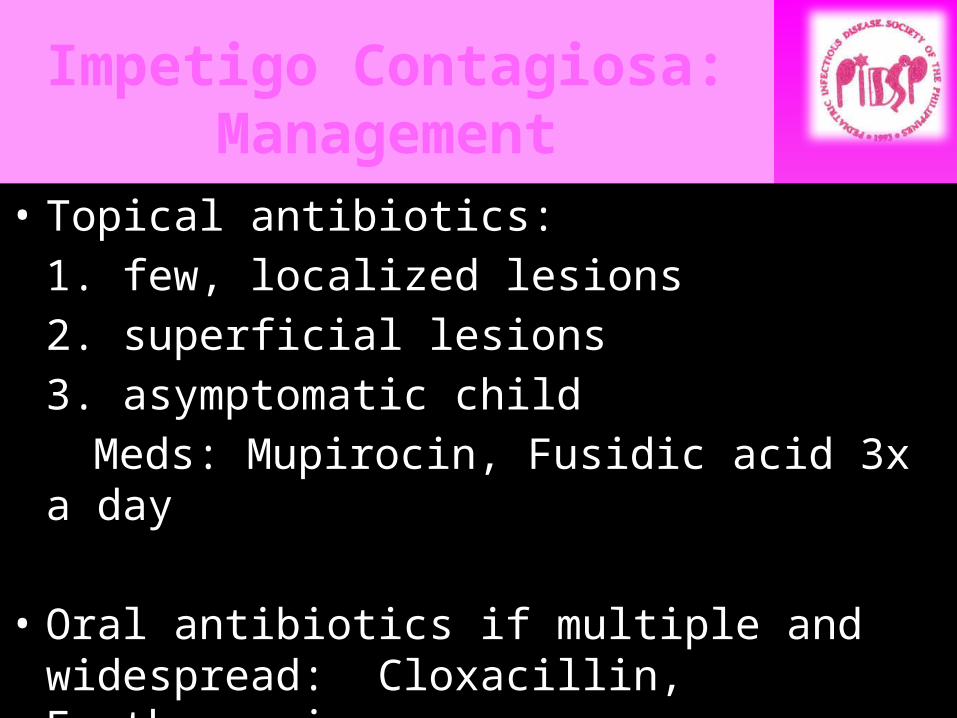
Impetigo Contagiosa: Management
• Topical antibiotics:1. few, localized lesions2. superficial lesions3. asymptomatic child
Meds: Mupirocin, Fusidic acid 3x a day
• Oral antibiotics if multiple and widespread: Cloxacillin, Erythromycin
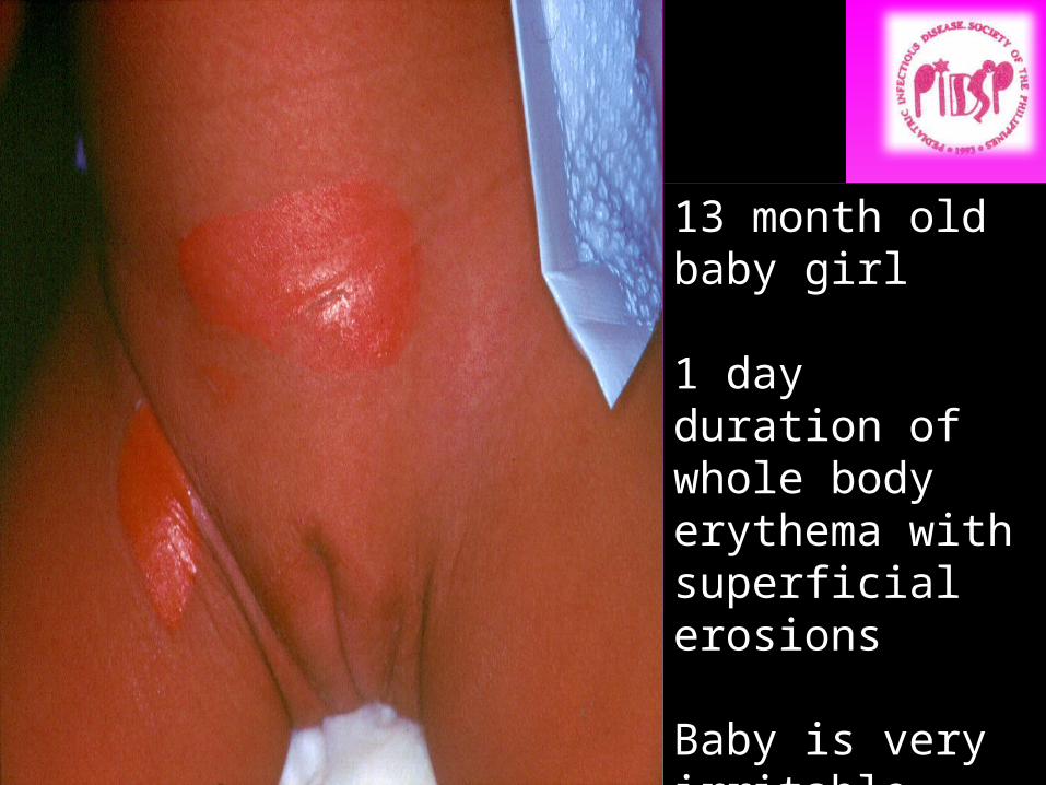
13 month old baby girl
1 day duration of whole body erythema with superficial erosions
Baby is very irritable
Diagnosis??????

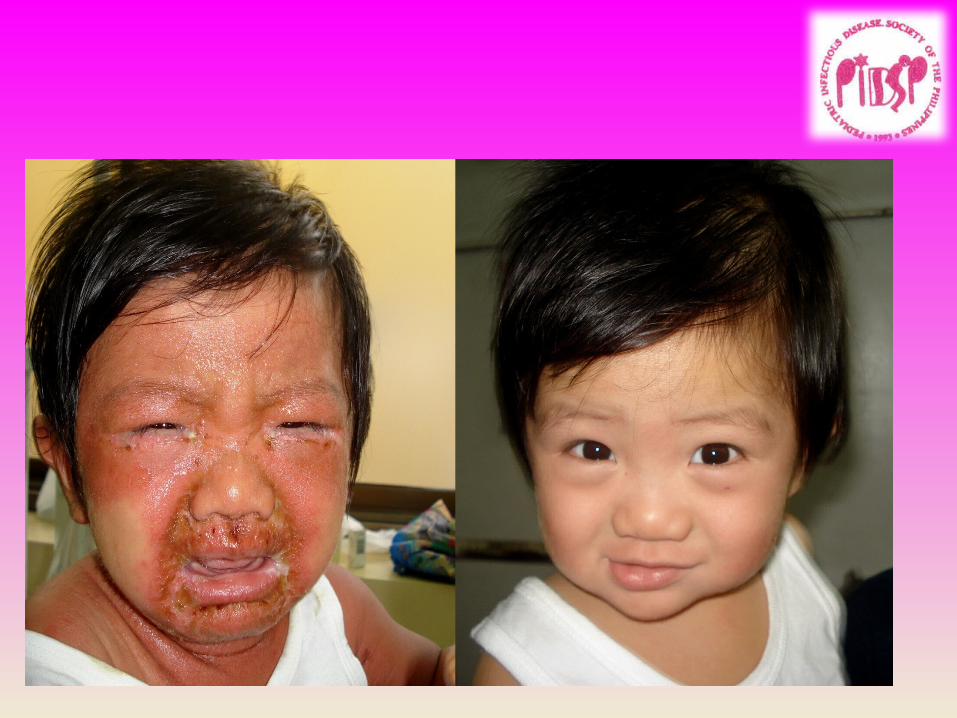
Staphylococcal Scalded Skin Syndrome or SSSS
• A child less than 5 y/o with diffuse tender erythema scarlatiniform eruption accentuated in flexures and periorificial areas “wrinkled” appearance and superficial desquamation
• Severe cases with diffuse sterile flaccid blisters and erosions

• Characteristic facies: peri-orificial erythema and scaling distinctive radial crusting and fissuring
• May have pharyngitis, conjunctivitis and superficial erosions of the lips with sparing of oral mucosa
SSSS: Recognition
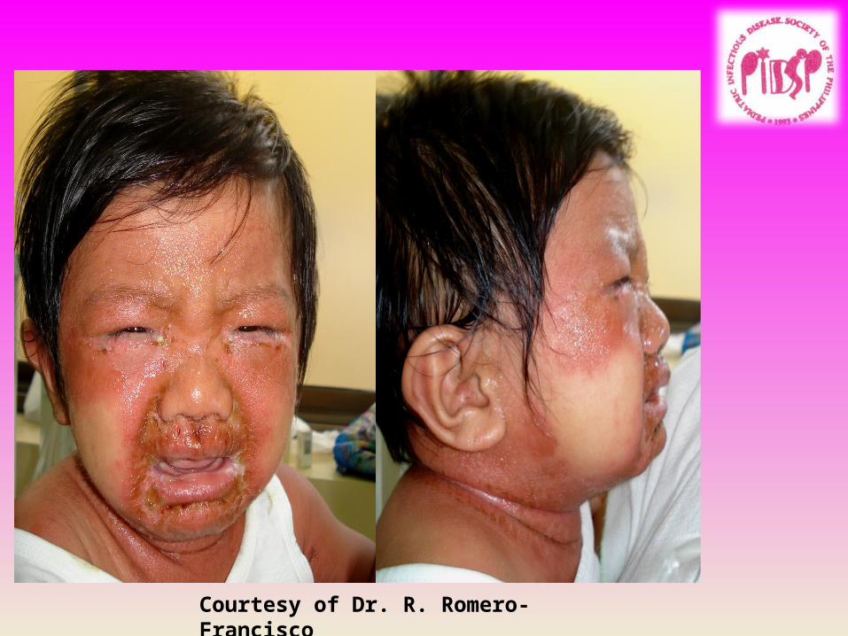
Courtesy of Dr. R. Romero-Francisco
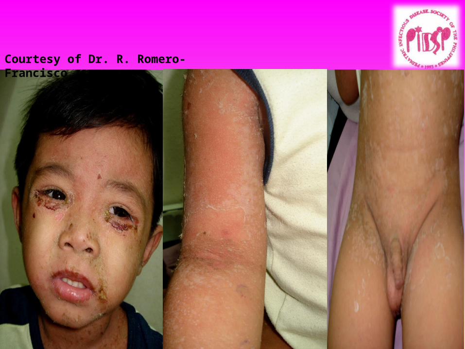
Courtesy of Dr. R. Romero-Francisco
Staphylococcal Scalded Skin Syndrome (SSSS)
• A toxin mediated infection• Due to exfoliative toxins A, B released by
Staphylococcus aureus phage Type II
SSSS: Management: Remember that this is a Systemic Staph infection
• Anti-Staph antibiotics for 7-10 days• Aggressive fluid and electrolyte management• Denuded phase: NSS compresses• Desquamation phase: emollients• Heals without scarring in 10-14 days
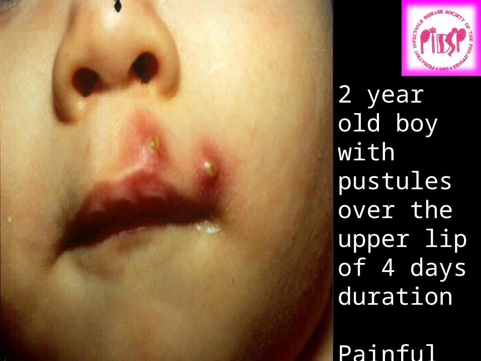
2 year old boy with pustules over the upper lip of 4 days duration
Painful
Diagnosis??

C ARB UNCLES
Folliculitis
Fu-run-cles
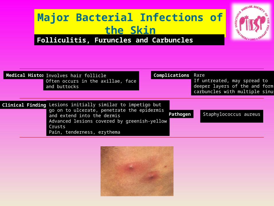
Major Bacterial Infections of the Skin
Folliculitis, Furuncles and Carbuncles
Medical History Involves hair follicleOften occurs in the axillae, faceand buttocks
Complications RareIf untreated, may spread to deeper layers of the and form carbuncles with multiple sinuses
Clinical Findings Lesions initially similar to impetigo butgo on to ulcerate, penetrate the epidermisand extend into the dermisAdvanced lesions covered by greenish-yellowCrustsPain, tenderness, erythema
Pathogen Staphylococcus aureus
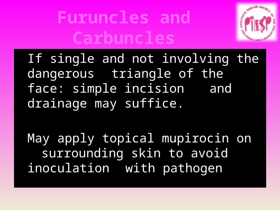
Furuncles and Carbuncles
If single and not involving the dangerous triangle of the face: simple incision and drainage may suffice.
May apply topical mupirocin on surrounding skin to avoid inoculation with pathogen
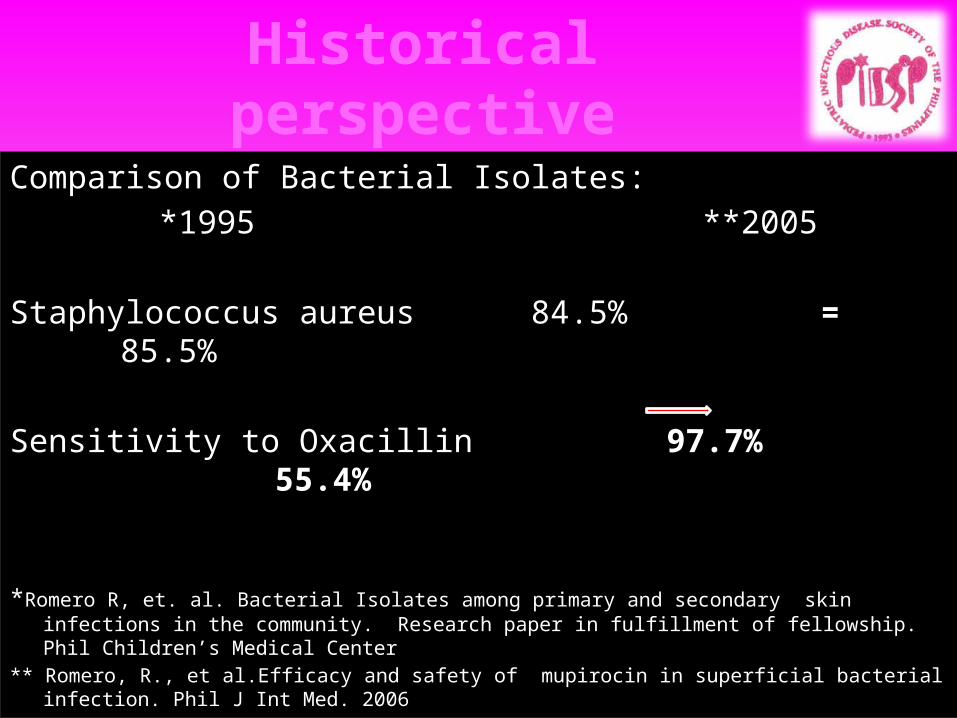
Historical perspective
Comparison of Bacterial Isolates: *1995 **2005
Staphylococcus aureus 84.5% = 85.5%
Sensitivity to Oxacillin 97.7% 55.4%
*Romero R, et. al. Bacterial Isolates among primary and secondary skin infections in the community. Research paper in fulfillment of fellowship. Phil Children’s Medical Center
** Romero, R., et al.Efficacy and safety of mupirocin in superficial bacterial infection. Phil J Int Med. 2006

Why the sudden change?
• Possibilities: (Philippine scenario)–Incomplete intake of prescribed antibiotics–Self medication – availability of antibiotics
from local drugstores w/o prescription–Application of “penicillin” powder on
infected wounds
Community Acquired Methicillin Resistant Staph Aureus (CA-MRSA)
What is MRSA (CDC Definition)?
MRSA is, by definition, any strain of Staphylococcus aureus bacteria that has developed resistance to beta-lactam antibiotics which include the penicillins (methicillin, dicloxacillin, nafcillin, oxacillin, etc.) and the cephalosporins.
Community acquired MRSA is a hybrid strain from a previously hospitalized patient who developed MRSA and the strain normally found in the community.

Community Acquired Methicillin Resistant Staph Aureus (CA-MRSA)
•The resistance of MRSA to beta-lactam antibiotics is due to the presence of the mecA gene sequence.
•The mecA gene produces transpeptidase PBP2a (penicillin-binding peptide) that decreases the bacterial affinity of the beta-lactam antibiotics.
•Most CA-MRSA hybrid strains may acquire a virulence factor not seen with HA-MRSA

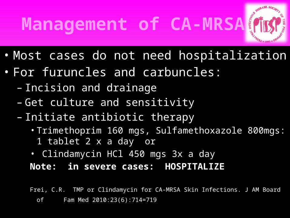
Management of CA-MRSA
• Most cases do not need hospitalization• For furuncles and carbuncles:– Incision and drainage– Get culture and sensitivity– Initiate antibiotic therapy• Trimethoprim 160 mgs, Sulfamethoxazole 800mgs: 1 tablet 2 x
a day or• Clindamycin HCl 450 mgs 3x a day
Frei, C.R. TMP or Clindamycin for CA-MRSA Skin Infections. J AM Board of Fam Med
2010:23(6):714=719
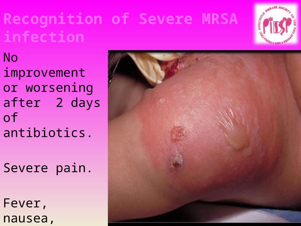
No improvement or worsening after 2 days of antibiotics.
Severe pain.
Fever, nausea, vomiting, other constitutional signs and symptoms
Recognition of Severe MRSA infection
Management of CA-MRSA
• Most cases do not need hospitalization• For furuncles and carbuncles:– Incision and drainage– Get culture and sensitivity– Initiate antibiotic therapy• Trimethoprim 160 mgs, Sulfamethoxazole 800mgs: 1 tablet 2 x
a day or• Clindamycin HCl 450 mgs 3x a dayNote: in severe cases: HOSPITALIZE
Frei, C.R. TMP or Clindamycin for CA-MRSA Skin Infections. J AM Board of Fam Med
2010:23(6):714=719
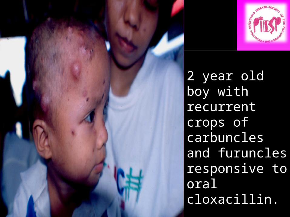
2 year old boy with recurrent crops of carbuncles and furuncles responsive to oral cloxacillin.
Problem: Why recurrent?

Recurrent Furunculosis
• Frequent attacks of furuncles/carbuncles: (1 or more episodes per month despite oral antibiotics)Look for source of staphylococcus!
May have to do culture of anterior nares of patient or caregiver(s)If +: Apply mupirocin 4x a day for 5 days to
anterior nares Or Rifampicin plus Cloxacillin for 7 days
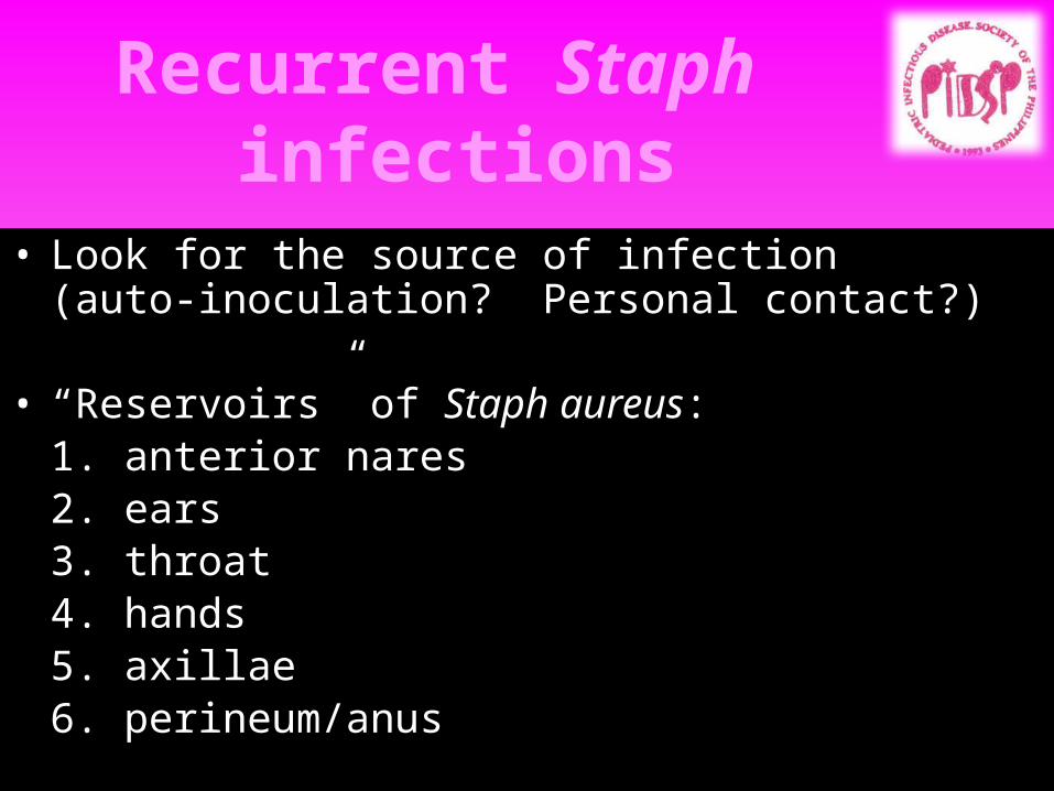
Recurrent Staph infections
• Look for the source of infection (auto-inoculation? Personal contact?)
• “Reservoirs” of Staph aureus:1. anterior nares2. ears3. throat4. hands5. axillae6. perineum/anus
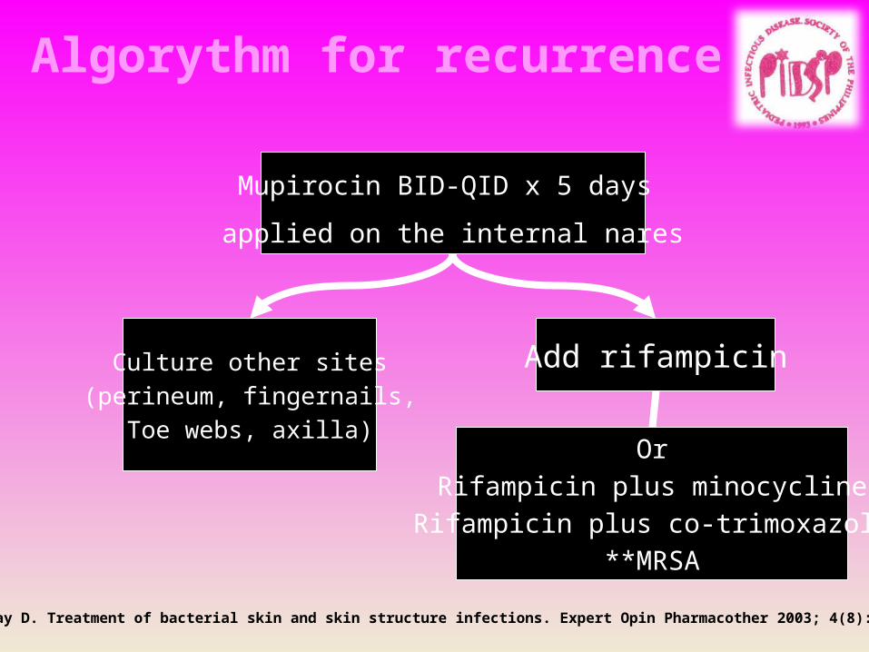
Algorythm for recurrence
Mupirocin BID-QID x 5 days
applied on the internal nares
Culture other sites(perineum, fingernails,
Toe webs, axilla)
Add rifampicin
OrRifampicin plus minocycline
Rifampicin plus co-trimoxazole**MRSA
Guay D. Treatment of bacterial skin and skin structure infections. Expert Opin Pharmacother 2003; 4(8): 1259-75.

Fungal InfectionsCandidaPityriasis versicolorTinea capitis
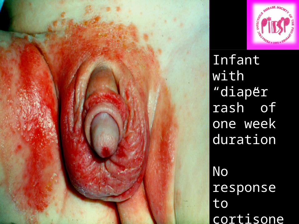
Diaper Candidiasis
Infant with “diaper rash” of one week duration
No response to cortisone cream
Diagnosis?
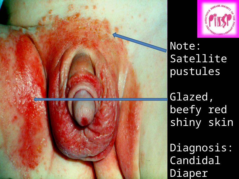
Diaper Candidiasis
Note:Satellite pustules
Glazed, beefy red shiny skin
Diagnosis:Candidal Diaper Dermatitis
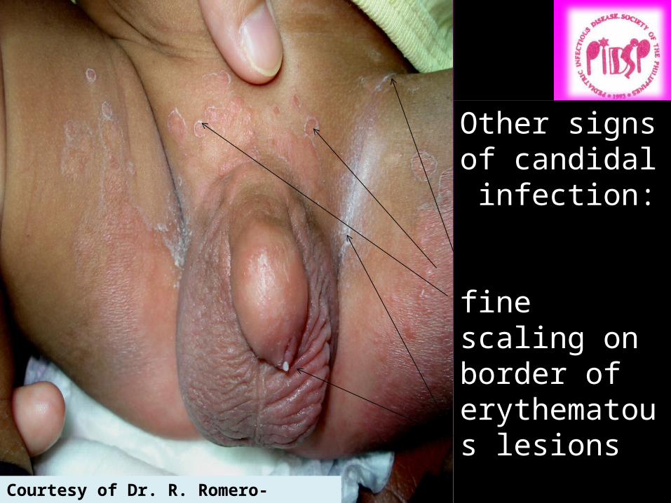
Other signs of candidal infection:
fine scaling on border of erythematous lesions
White cheesy material Courtesy of Dr. R. Romero-Francisco
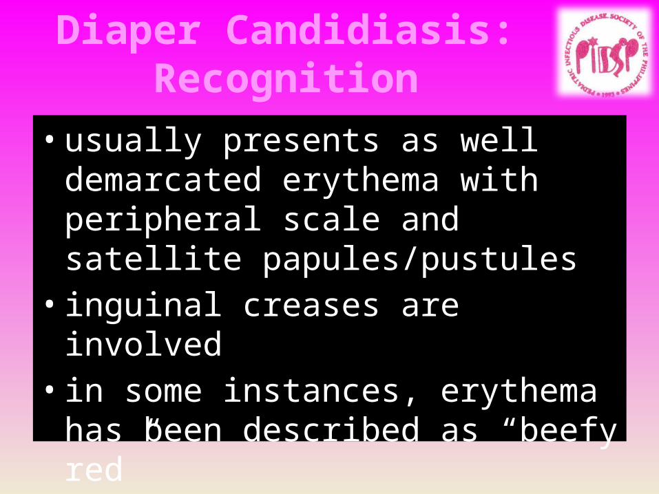
Diaper Candidiasis: Recognition
• usually presents as well demarcated erythema with peripheral scale and satellite papules/pustules
• inguinal creases are involved• in some instances, erythema has been
described as “beefy red”
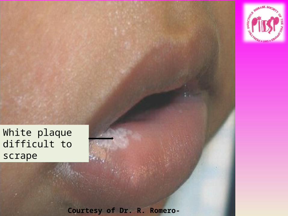
White plaquedifficult to scrape
Courtesy of Dr. R. Romero-Francisco
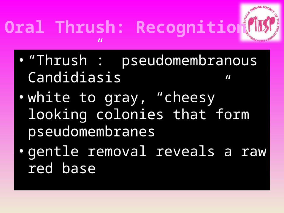
Oral Thrush: Recognition• “Thrush”: pseudomembranous
Candidiasis• white to gray, “cheesy” looking colonies
that form pseudomembranes• gentle removal reveals a raw red base
Candidiasis: Management
• Topical anti-candidal agent (nystatin or an azole preparation) +/- topical steroid
• NOTE: after the eruption has cleared, continue the anti-candidal agent for three more days
• Oral mycostatin or fluconazole if recurrent and extensive
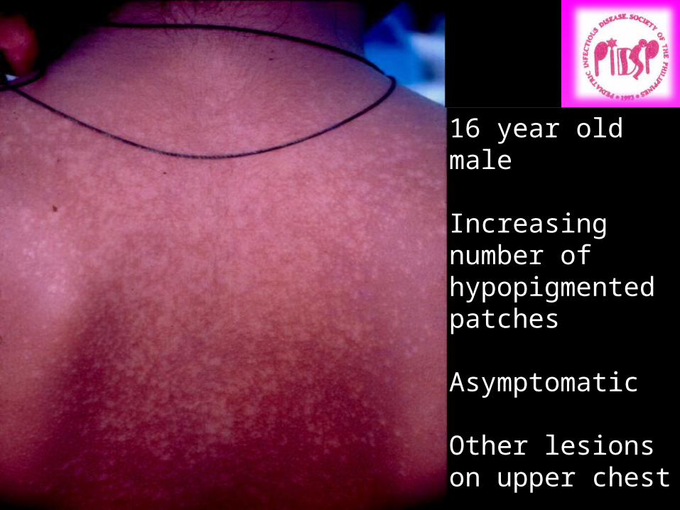
16 year old male
Increasing number of hypopigmentedpatches
Asymptomatic
Other lesions on upper chest
Diagnosis????
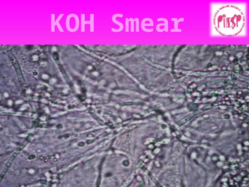
KOH Smear
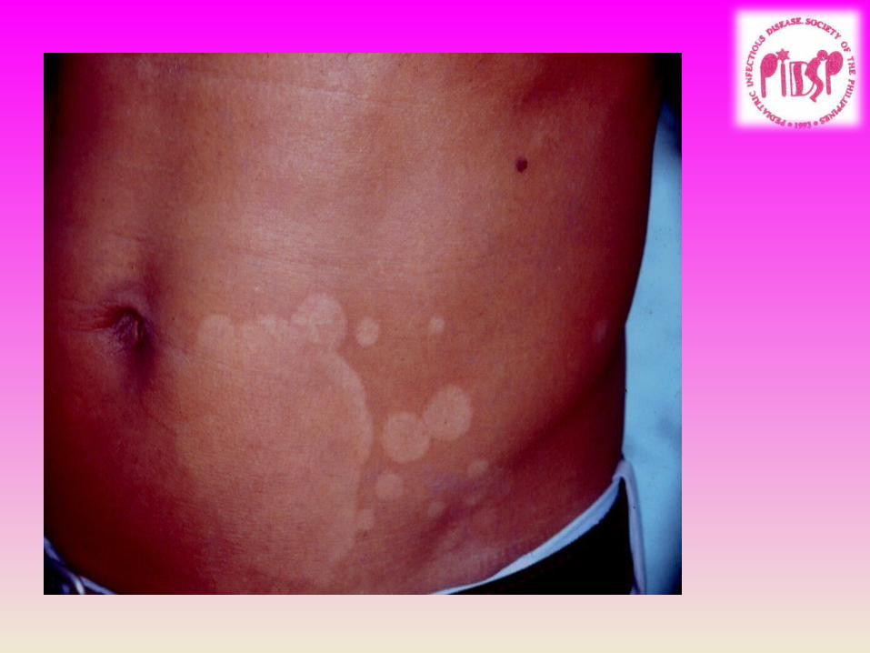
Pityriasis Versicolor: Recognition
• Small round to oval macules or minimally elevated plaques with “wrinkling” and superficial scale (“fingernail sign”)
• Lesions may be erythematous to brownish to hypopigmented (“an-an”)
• Most common on the chest, back and proximal arms
• Face involved in younger children• May be mildly pruritic• Etiologic agent: Pityrosporum ovale or
Malassezia furfur
Pityriasis Versicolor: Recognition
Pityriasis versicolor: Management
• Selenium sulfide or Zinc pyrithione 10-15 mins/day for 1-2 weeks
• Ketoconazole shampoo 5 mins/day for 3 days
• Ketoconazole cream• Oral ketoconazole discouraged• Advise on residual pigmentation
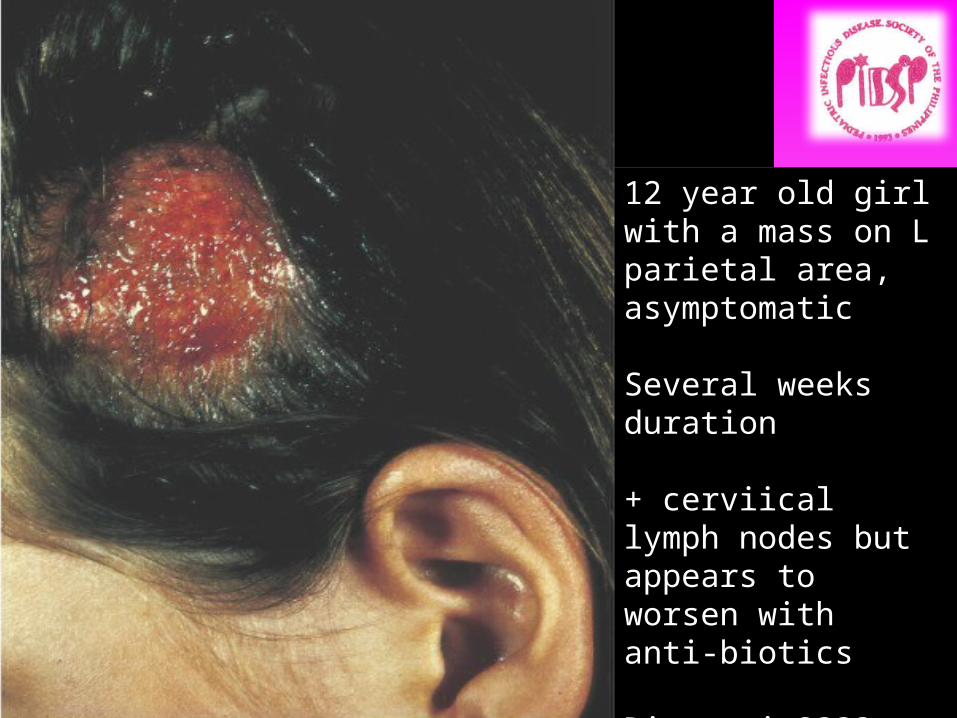
12 year old girl with a mass on L parietal area, asymptomatic
Several weeks duration
+ cerviical lymph nodes but appears to worsen with anti-biotics
Diagnosis????
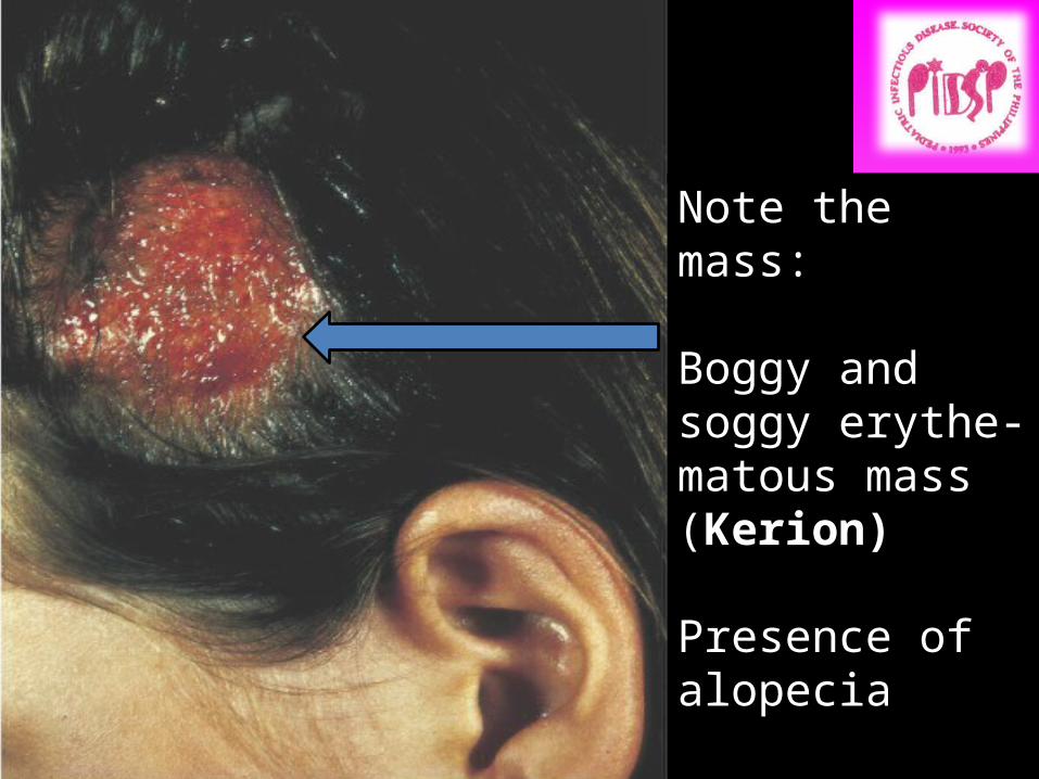
Note the mass:
Boggy and soggy erythe-matous mass (Kerion)
Presence of alopecia
Diagnosis:Tinea Capitis
Courtesy of Dr. R. Romero-Francisco
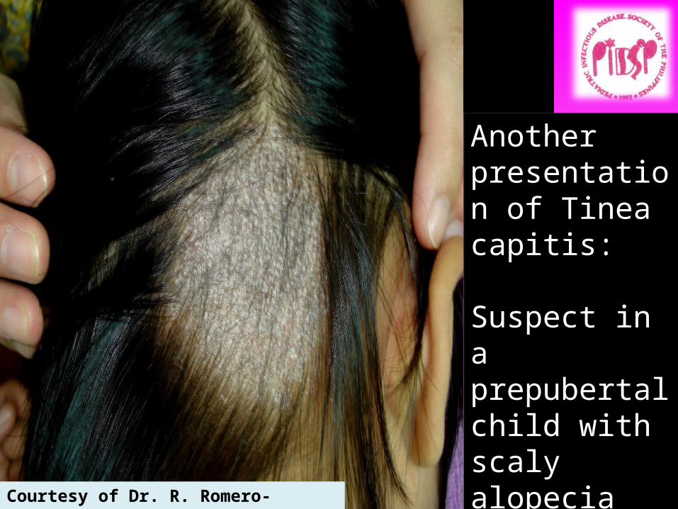
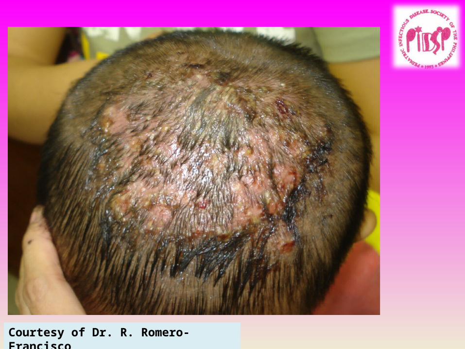
Another presentation of Tinea capitis:
Suspect in a prepubertal child with scaly alopecia
Courtesy of Dr. R. Romero-Francisco
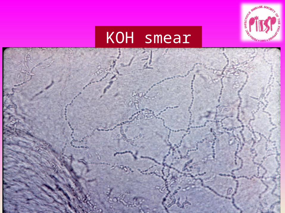
KOH smear
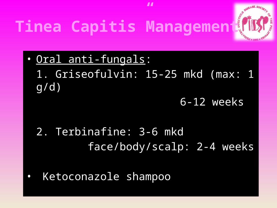
Tinea Capitis”Management
• Oral anti-fungals:1. Griseofulvin: 15-25 mkd (max: 1 g/d)
6-12 weeks
2. Terbinafine: 3-6 mkd face/body/scalp: 2-4 weeks
• Ketoconazole shampoo
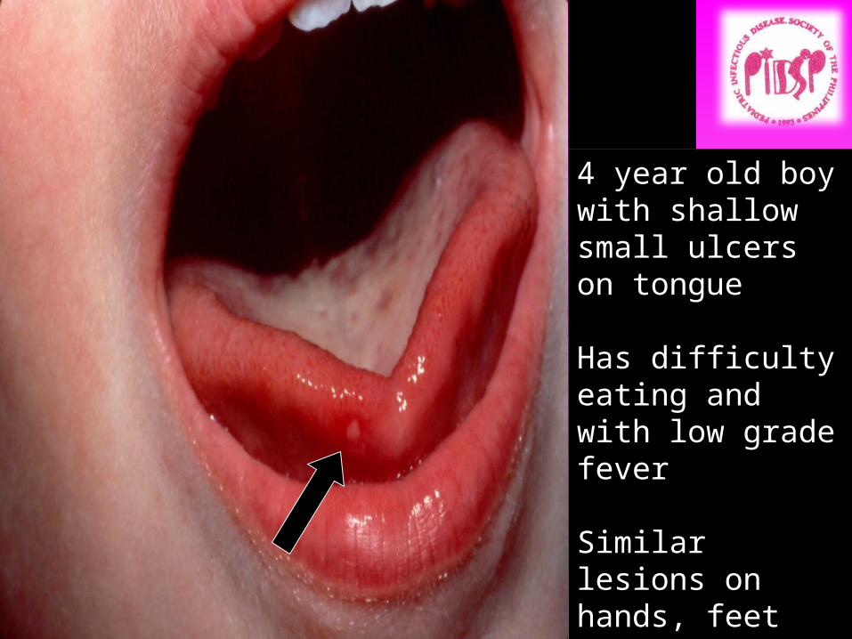
4 year old boy with shallow small ulcers on tongue
Has difficulty eating and with low grade fever
Similar lesions on hands, feet and buttocks
Diagnosis?????

Courtesy of Dr. R. Romero-Francisco
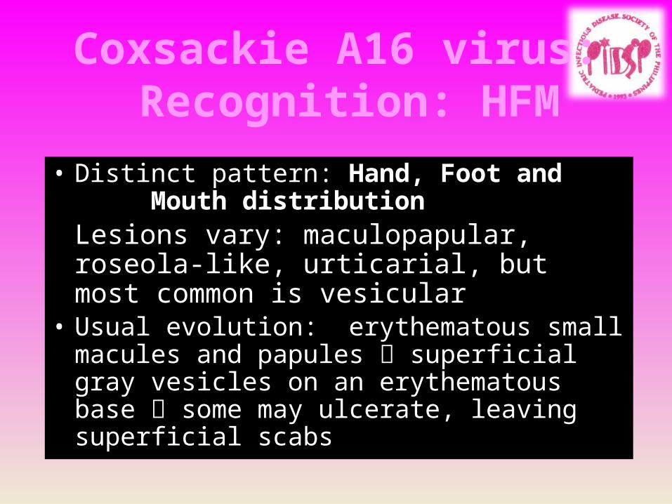
Coxsackie A16 virus: Recognition: HFM
• Distinct pattern: Hand, Foot and Mouth distributionLesions vary: maculopapular, roseola-like, urticarial, but most common is vesicular
• Usual evolution: erythematous small macules and papules superficial gray vesicles on an erythematous base some may ulcerate, leaving superficial scabs
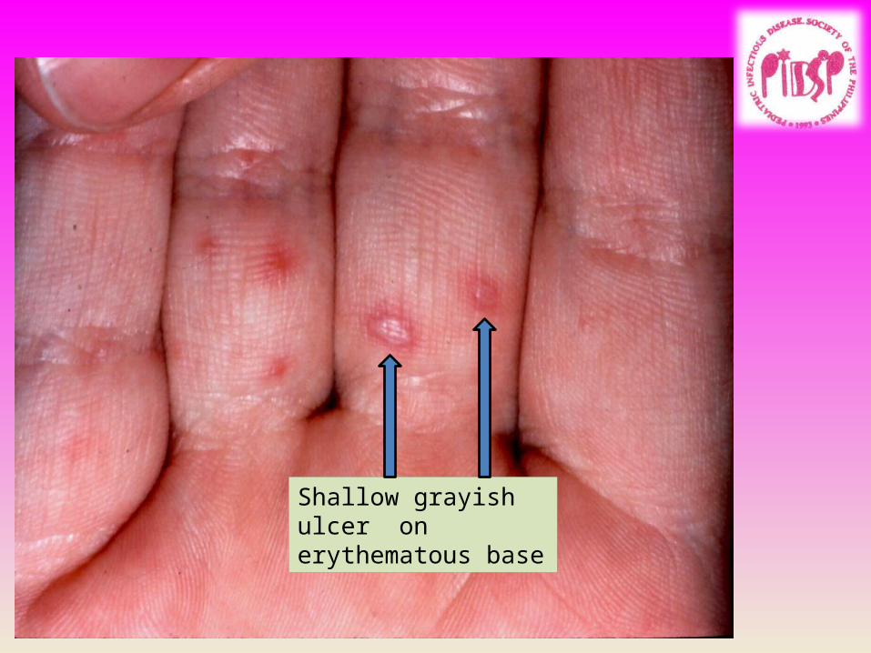
Shallow grayish ulcer on erythematous base
Coxsackie A16 virus: Recognition
• Hand, Foot and Mouth disease> areas involved: mouth, hands and feet, buttocks; may also be seen on face and extremities> rash usually lasts for 2-7 days> (+/-) fever, sore mouth, anorexia, malaise, abdominal pain
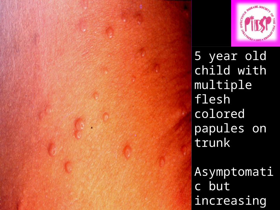
5 year old child with multiple flesh colored papules on trunk
Asymptomatic but increasing in number
Diagnosis????
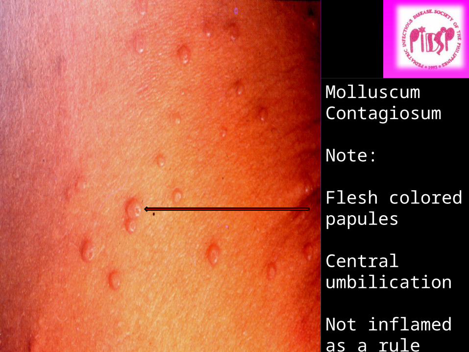
MolluscumContagiosum
Note:
Flesh colored papules
Central umbilication
Not inflamed as a rule


Flesh colored papules Central umbilication
Molluscum contagious: Recognition
• Flesh colored to pinkish to pearly white discrete papules with central umbilication
• Most common areas: axillae, lateral trunk, lower abdomen, thighs, face
• May have a dermatitis in 10% of cases• Etiologic agent: Molluscipox virus

Molluscum contagiosum: Management
• “Benign neglect”: spontaneous resolution in 6-9 months
• May have a more persistent, progressive course**Tx options:
1. Curettage2. topical Cantharidin3. Tretinoin cream4. Imiquimod cream
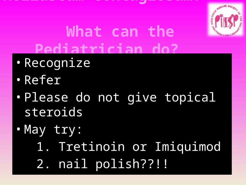
Molluscum contagiosum: What can the Pediatrician do?
• Recognize• Refer• Please do not give topical steroids• May try:
1. Tretinoin or Imiquimod2. nail polish??!!
DERMATOLOGICAL INFECTIONS
BACTERIALIMPETIGO/SSSFOLLICULITIS, FURUNCULOSIS
FUNGALTINEA VERSICOLORDERMATOPHYTOSISCANDIDA
VIRALHAND FOOT and MOUTH DISEASE
MOLLUSCUM CONTAGIOSUM

THANK YOU FOR YOUR KIND ATTENTION