Embed Size (px)
Citation preview

H. Jeremy Bockholt and Mark Scully-1-National Alliance for Medical Image Computing
DetectingWhiteMatterLesionsinLupus
H. Jeremy BockholtMark Scully
Slicer3 Training Compendium
Version 2.26/25/2009

H. Jeremy Bockholt and Mark Scully-2-National Alliance for Medical Image Computing
LearningobjectiveThis tutorial demonstratesan automated, multi-levelmethod to segment whitematter brain lesions inlupus.
Following this tutorial,you’ll be able to load scansinto Slicer3, and segmentand measure the volume ofwhite matter lesions on theprovided data-set.

H. Jeremy Bockholt and Mark Scully-3-National Alliance for Medical Image Computing
Prerequisites
This tutorial assumes that you have already completedthe tutorial Data Loading and Visualization. Tutorialsfor Slicer3 are available at the following location:
• Slicer3 tutorialshttp://www.na-mic.org/Wiki/index.php/Slicer3.2:Training

H. Jeremy Bockholt and Mark Scully-4-National Alliance for Medical Image Computing
Material
This course requires the following installation:
•The current version of Slicer 3.5.x Software which can be installed from:– http://www.slicer.org/pages/Downloads
•The White Matter Lesion module extension to Slicer 3– (see follow on instructions)
•The Lupus Lesion Tutorial Data, which can be downloaded from:– http://www.nitrc.org/frs/?group_id=180
•n.b., a reliable internet connection will be required for downloading the data
Disclaimer
It is the responsibility of the user of 3DSlicer to comply with both the terms ofthe license and with the applicable laws, regulations and rules.

H. Jeremy Bockholt and Mark Scully-5-National Alliance for Medical Image Computing
MethodsThe method makes use of local morphometricfeatures based on multiple MR sequences,including T1-weighted, T2-weighted, and FluidAttenuated Recovery from ten subjects. Afterpreprocessing, including co-registration, brainextraction, bias correction, and intensitystandardization, 49 features were calculatedfor each brain voxel based on localmorphometry. At each level of segmentation asupervised classifier takes advantage of adifferent subset of the features toconservatively segment lesion voxels, passingon more difficult voxels to the next classifier.This multi-level approach allows for a fastlesion classification method with tunable trade-off between sensitivity and specificity, withaccuracy comparable to a human rater.

H. Jeremy Bockholt and Mark Scully-6-National Alliance for Medical Image Computing
GettingtheModule• To add the external
module, Select theExtensionsManagement Wizardfrom the View menuwithin Slicer. Clicknext to search theexternal site for theappropriate moduleto install.

H. Jeremy Bockholt and Mark Scully-7-National Alliance for Medical Image Computing
InstallingtheModuleSelect
LesionSegmentationApplicationsfrom the list
Click Download &Install.
Click Finish whendownloadcompletes andrestart Slicer to usethe external module

H. Jeremy Bockholt and Mark Scully-8-National Alliance for Medical Image Computing
Joint Intensity Standardization Volume.nhdrJoint Intensity Standardization Volume.raw.gzJoint Intensity Standardization Volume1.nhdrJoint Intensity Standardization Volume1.raw.gzJoint Intensity Standardization Volume2.nhdrJoint Intensity Standardization Volume2.raw.gzLesionSegmentTutorial.mrmlPredict Lesions Volume.nhdrPredict Lesions Volume.rawPredict Lesions Volume1.nhdrPredict Lesions Volume1.rawlesionSegmentation.modellupus002_FLAIR_reg+bias.nii.gzlupus002_T1_reg+bias.nii.gzlupus002_T2_reg+bias.nii.gzlupus002_brain_mask.nii.gzlupus003_FLAIR_reg+bias.nii.gzlupus003_T1_reg+bias.nii.gzlupus003_T2_reg+bias.nii.gzlupus003_brain_mask.nii.gzsvm.model
TutorialDataThis course is built upon two scans ofpatients with lupus that have T1, T2,and FLAIR images. These imageshave been co-registered and brainextracted.The following summary shows thecontents of thedata/LesionSegmentationTutorialdirectory once download anduncompressed

H. Jeremy Bockholt and Mark Scully-9-National Alliance for Medical Image Computing
Slicer3GUI
The Graphical UserInterface (GUI) ofSlicer3 integrates fivecomponents:
•the Menu Toolbar
•the Module GUI Panel
•the 3D Viewer
•the Slice Viewer
•the Slice and 3D ViewController
Slice Viewer
3DViewerModule GUIPanel
Slice and3D View
Controller
Menu Toolbar

H. Jeremy Bockholt and Mark Scully-10-National Alliance for Medical Image Computing
Step1:Setup
Load the scene by selecting File ImportScene feature from the menu. Navigate thefilesystem to locate the MRML scene thatyou have downloaded. By loading the sceneyou will load the reference data sets that areneeded for this tutorial.

H. Jeremy Bockholt and Mark Scully-11-National Alliance for Medical Image Computing
Step1:Results
After the scene loads, you willhave the data sets that are neededfor this tutorial and may continueon to step 2. Confirm that you havethe data sets listed in the scenedisplay on the left.

H. Jeremy Bockholt and Mark Scully-12-National Alliance for Medical Image Computing
Step2:Setup
The next step is to select the IntensityStandardization module: Select and expandthe Module menu, First select Segmentation,next select Lesion Segmentation, finally selectJoin Intensity Standardization.

H. Jeremy Bockholt and Mark Scully-13-National Alliance for Medical Image Computing
Step2:Setup
In order to run the joint intensity standardizestep, confirm the input parameters to themodule that are listed on the left, you willalso need to scroll down and confirm theoutput stats parameters shown on the nextslide.

H. Jeremy Bockholt and Mark Scully-14-National Alliance for Medical Image Computing
Step2:Running
Finishing configuring the intensitystandardize procedure to run by confirmingthe output stats parameters as shown on theleft.
Next, Click Apply to run this step, fortypical computers, expect 2-5 minutes ofprocessing time.

H. Jeremy Bockholt and Mark Scully-15-National Alliance for Medical Image Computing
Step3:Setup
The next step is to select the Predict Lesionsmodule: Select and expand the Modulemenu, First select Segmentation, next selectLesion Segmentation, finally select PredictLesions

H. Jeremy Bockholt and Mark Scully-16-National Alliance for Medical Image Computing
Step3:Running
Finishing configuring the Predict Lesionsprocedure to run by confirming the input andoutput parameters as shown on the left.
Next, Click Apply to run this step, fortypical computers, expect 90-120 minutes ofprocessing time.

H. Jeremy Bockholt and Mark Scully-17-National Alliance for Medical Image Computing
Step3:Results
Once complete the results will load as alesion mask volume, as well as, a heat map,or lesion probability volume

H. Jeremy Bockholt and Mark Scully-18-National Alliance for Medical Image Computing
ExampleMeasurement
As a use case of the resuls, you may use theLabel Statistics Module to summarize thelesion load. As shown Select Statistics andthen select LabelStatistics.

H. Jeremy Bockholt and Mark Scully-19-National Alliance for Medical Image Computing
ExampleMeasurement
After selecting Flair as input GrayscaleVolume and Predict Lesions Volume as theInput Labelmap, a summary of the volume isprovided in the Label Statistics

H. Jeremy Bockholt and Mark Scully-20-National Alliance for Medical Image Computing
SetupExampleModel
You may also create a model of theclassified lesions by using the Model Makermodule. To do this, select Surface Modelsand then select the Grayscale Model Maker.

H. Jeremy Bockholt and Mark Scully-21-National Alliance for Medical Image Computing
ResultsExampleModel
To the left shows how the Predict LesionsVolume can be rendered using the modelmaker and above shows the rendered result.
H. Jeremy Bockholt and Mark Scully-22-National Alliance for Medical Image Computing
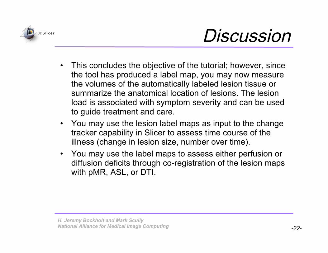
Discussion• This concludes the objective of the tutorial; however, since
the tool has produced a label map, you may now measurethe volumes of the automatically labeled lesion tissue orsummarize the anatomical location of lesions. The lesionload is associated with symptom severity and can be usedto guide treatment and care.
• You may use the lesion label maps as input to the changetracker capability in Slicer to assess time course of theillness (change in lesion size, number over time).
• You may use the label maps to assess either perfusion ordiffusion deficits through co-registration of the lesion mapswith pMR, ASL, or DTI.

H. Jeremy Bockholt and Mark Scully-23-National Alliance for Medical Image Computing
Conclusion• This capability provides an intuitive graphical
user interface to interact with the data• The tool has been built in an open-source
environment and is readily available to thescientific community

H. Jeremy Bockholt and Mark Scully-24-National Alliance for Medical Image Computing
ForMoreInformation• Register as a user of this 3dSlicer Module using
the NITRC resource to keep updated on anychanges or additions to either the capability ortutorial– http://www.nitrc.org/projects/lupuslesion/
• You may also send e-mail message with anyquestions or concerns to Jeremy Bockholt([email protected])

H. Jeremy Bockholt and Mark Scully-25-National Alliance for Medical Image Computing
Acknowledgments
National Alliance for Medical Image ComputingNIH U54EB005149
And other support:
DOE DE-FG02-99ER6274NIH 5R01HL077422-02NIH P41 RR13218NIH U24-RR021992