Embed Size (px)
Citation preview
INVERTEBRATE MICROBIOLOGY
Detection and Identification of Species-Specific BacteriaAssociated with Synanthropic Mites
Jan Hubert & Jan Kopecký & M. Alejandra Perotti & Marta Nesvorná & Henk R. Braig &
Markéta Ságová-Marečková & Lilia Macovei & Ludek Zurek
Received: 5 August 2011 /Accepted: 12 October 2011 /Published online: 5 November 2011# Springer Science+Business Media, LLC 2011
Abstract Internal bacterial communities of synanthropicmites Acarus siro, Dermatophagoides farinae, Lepidogly-phus destructor, and Tyrophagus putrescentiae (Acari:Astigmata) were analyzed by culturing and culture-independent approaches from specimens obtained fromlaboratory colonies. Homogenates of surface-sterilized miteswere used for cultivation on non-selective agar and DNAextraction. Isolated bacteria were identified by sequencing ofthe 16S rRNA gene. PCR amplified 16S rRNA genes wereanalyzed by terminal restriction fragment length polymor-phism analysis (T-RFLP) and cloning sequencing. Fluores-cence in situ hybridization using universal bacterial probeswas used for direct bacterial localization. T-RFLP analysis of16S rRNA gene revealed distinct species-specific bacterialcommunities. The results were further confirmed by cloningand sequencing (284 clones). L. destructor and D. farinae
showed more diverse communities then A. siro and T.putrescentiae. In the cultivated part of the community, themean CFUs from four mite species ranged from 5.2×10² to1.4×103 per mite. D. farinae had significantly higher CFUsthan the other species. Bacteria were located in the digestiveand reproductive tract, parenchymatical tissue, and inbacteriocytes. Among the clones, Bartonella-like bacteriaoccurring in A. siro and T. putresecentiae represented adistinct group related to Bartonellaceae and to Bartonella-like symbionts of ants. The clones of high similarity toXenorhabdus cabanillasii were found in L. destructor and D.farinae, and one clone related to Photorhabdus temperata inA. siro. Members of Sphingobacteriales cloned from D.farinae and A. siro clustered with the sequences of“Candidatus Cardinium hertigii” and as a separate novelcluster.
Electronic supplementary material The online version of this article(doi:10.1007/s00248-011-9969-6) contains supplementary material,which is available to authorized users.
J. Hubert (*) : J. Kopecký :M. Nesvorná :M. Ságová-MarečkováCrop Research Institute,Drnovská 507,Praha 6-Ruzyně 161 06, Czech Republice-mail: [email protected]
J. HubertMedical Center Prague,Mezi Vodami 205,Praha 4 143 00, Czech Republic
M. A. PerottiSchool of Biological Sciences, Environmental Biology,University of Reading,Whiteknights,Reading, Berkshire RG6 6AS, UK
H. R. BraigSchool of Biological Sciences, Bangor University,Bangor LL57 2UW Wales, UK
L. MacoveiThe Forsyth Institute,Boston, MA 02142, USA
L. ZurekDepartment of Entomology, Kansas State University,Manhattan, KS 66506, USA
L. ZurekDepartment of Diagnostic Medicine and Pathobiology,Kansas State University,Manhattan, KS 66506, USA
Microb Ecol (2012) 63:919–928DOI 10.1007/s00248-011-9969-6
Introduction
Mites are among the most diverse and successful of allinvertebrates. It was estimated that 48,200 species of mitesexist [23]. They have exploited an incredible array ofhabitats, and because of their microscopic size they golargely unnoticed. Many mites live freely in the soil orwater, but there is also a large number of species living asparasites on plants or animals. In urban habitats, synan-thropic mites (Acari: Astigmata) are the most abundant andfrequent pests.
Synanthropic mites represent a serious threat to humanhealth due to the production of human allergens [8] anddissemination of pathogenic fungi [18, 29, 31, 52]. Theyare also important pests of stored food products, where thedirect losses are caused by mite feeding on food or feedincluding grain, oilseeds, dried fruit, dried meat, andcheeses [33].
Only a few studies focused on bacteria and eukaryotesassociated with mites although bacteria seem to be closelyassociated with their life strategies and feeding [28, 46, 47].A diverse bacterial community was detected in the sheepscab mite Psoroptes ovis [26], while relatively poor(diversity and abundance) bacterial communities werefound in Dermatophagoides pteronyssinus and Dermato-phagoides farinae [43, 51, 62, 63, 66]. Tyrophagusputrescentiae interaction with bacteria was reported incontaminated Iberian ham causing damage of ham [19].Different bacterial communities were determined in the gutand on surface of T. putrescentiae originating from differenthabitats based on the cultivation methods [53]. Smrž andČatská [54] suggested that bacteria associated with T.putrescentiae including Serratia, Pseudomonas, Brevudi-monas, and Stenotrophomons spp. were symbiotic andparticipated in chitin digestion and utilization. In A. siro,the transmission microscopy (TEM) observation of the gutshowed that the postcolonic diverticula (sometimes called“Malphigian tubules [32]”) harbored symbiotic bacteria[55]. In addition, TEM observations of the gut in thisspecies revealed that bacteria in the gut are active andproduce exoenzymes (“hydrolyzed halos” around bacteria)[30].
In this study, we focused on four species of medicalimportance Acarus siro, D. farinae, Lepidoglyphus destruc-tor, and T. putrescentiae. D. farinae and T. putrescentiaeinhabit house dust, while A. siro, L. destructor, and T.putrescentiae infested stored products, especially in areaswith humid climate [2, 56]. We determined their internalbacterial communities by cultivation approach, terminalrestriction fragment length polymorphism analysis (T-RLFP), and cloning/sequencing of the 16S rRNA gene aswell as fluorescence in situ hybridization (FISH) to showdifferences among the species of mites.
Material and Methods
Sample Preparation
Four species of synantropic mites A. siro L., T. putrescentiae(Schrank) (Acari: Acarididae), L. destructor (Schrank) (Acari:Glycyphagidae), and D. farinae Hughes (Acari: Pyroglyphi-dae) originated from the laboratory cultures maintained atCrop Research Institute, Prague, Czech Republic (details inErban and Hubert [12]) were used in this study.
The stored-product mite diet (SPMd) used for A. siro andT. putrescentiae was a mixture of oat flakes, wheat germs,and Pangamin—dried yeast extract (Rapeto, Bezdruzice,Czech Republic) (ratio 10:10:1 wt). The house-dust mitediet (HDMd) used for L. destructor and D. farinae wascomposed of dog food (Ontario-pet, Placek, Podebrady,Czech Republic), wheat germ, Aqua-tropic—dried fishfood (Lon-Bio, Praha, Czech Republic), Pangamin, andgelatin (SERVA Electrophoresis GmbH, Heidelberg,Germany) (ratio 10:10:3:1 wt). Both diets were powderedand sieved and heated in thermostat (70°C) for 0.5 h tosuppress the microbial growth [12]. About 1.5 g of the dietwas placed into Iwaki tissue cell chambers (P-Lab, Praha,Czech Republic) and chambers were maintained in Secadordesiccators (P-Lab) at 85% relative humidity and 25±3°C indarkness. From each culture, 200 mg of each mite specieswas transferred to Eppendorf tubes, surface sterilized (Zureket al. 2000), and homogenized in 100 μl of phosphatebuffered saline (PBS) (MP Biomedicals, Aurora, OH, USA)with the Potter–Elvehjem homogenizer (Kavalier Sázava,Czech Republic) and used for bacterial culturing and DNAextraction.
T-RLFP
Total DNA was extracted from mite homogenates using theDNAEASY Tissue kit (Qiagen, Valencia, CA, USA) andcleaned with GENECLEAN® TURBO kit (MP Biomedicals,Solon, OH). The bacterial 16S rRNA gene fragments werePCR amplified with 6-carboxyfluorescein (FAM) labeledforward primer 27f (5′-AGAGTTTGATCMTGGCKCAG[49]) and reverse primer pH (5′-AAGGAGGTGATCCAGCCGCA [5]). PCR protocol consisted of initial dena-turation at 94°C at 2 min, followed by 35 cycles of 45 s at94°C, 45 s at 57°C, 1 min 30 s at 72°C, and a final extensionstep at 72°C for 5 min. The amplicon was purified andcleaved by AluI. After inactivation of the restriction enzymeand purification with Sigma SpinTM Post-Reaction Clean-UpColumns (Sigma-Aldrich, St. Louis, MO, USA), fragmentanalysis was performed at Genomac International, s.r.o.(Prague, Czech Republic) with a 96-capillary sequencer(Applied Biosystems, Foster City, CA, USA). T-RFLPanalyses were performed in one run for all samples.
920 J. Hubert et al.
Bacterial Isolation and Identification
The mite homogenates were serially diluted to 10−6, platedon TSB (tryptone soy broth) agar (HiMedia, Mumbai,India), and cultivated for 48 h at 37°C. Ten homogenatesper species were used. The CFUs were counted andindividual colony morphotypes were identified using PCRamplification and sequencing of the 16S rRNA gene.Specifically, the genomic DNA was prepared by boilingthe cells with subsequent centrifugation, and the superna-tant was used as a template. The primers and PCRconditions are described below. The CFUs were recalcu-lated per one mite and data were processed by analysis ofvariance (ANOVA) with Tukey’s post hoc comparisonusing STATISTIX (Analytical Software, Tallahassee, FL,
USA). As control, both rearing diets (10 mg) werehomogenized, diluted, and spread on the plates by thesame protocol as the mite homogenates.
16S rRNA Gene Cloning and Sequencing
PCR amplification of 16S rRNA gene was performedwith universal bacterial primers—UF: 5′-AGA GTTTGA TYM TGGC 3′ (position 8–23) and UR: 5′-GYTACC TTG TTA CGA CTT (position 1496–1514) [3].The amplification was done for DNA extracted from bothmite homogenates and bacterial isolates. Amplificationwas done on a Peltier Thermal Cycler (MJ Research,Waltham, MA, USA). A total volume of 25 μl PCRreaction mixture contained a final concentration of200 μM dNTPs, 3 mM MgCl2, forward and reverseprimers (100 nM each), 0.5 U Taq polymerase (allPromega, Madison, WI, USA), and 300 ng templateDNA. The amplification conditions were as follows:2 min at 94°C followed by 30 cycles of 90 s at 94°C,90 s at 50°C, and 60 s at 72°C, followed by finalextension for 10 min at 72°C and 4°C hold [3]. ResultingPCR products were purified with GFX PCR DNA andGel Band Purification Kit (Amersham Bioscience, UK).For identification of isolated bacteria, sequences weredetermined on an ABI 3700 DNA Analyzer at the K-StateDNA Sequencing Facility using the same bacterialuniversal primers and analyzed as described above. Forculture-independent approach, the PCR amplicons origi-nating from the mite homogenates were cloned usingpGEM®-T Easy Vector (Promega). Selected clones (ca.70 per one species) were sequenced in the UC-RiversideCore Instrumentation facility from the T6 and SP6primers on the ABI 3730xl sequencer. Nearly full-lengthsequences were assembled with CodonCode Aligner,version 1.5.2 (CodonCode Corporation, Dedham, MA,USA) and assigned to bacterial taxonomy using Ribo-somal Database Project naïve Bayesian rRNA classifier[64].
Phylogenetic Analysis
An initial alignment of partial 16S rRNA genesequences was performed using Muscle 3.6 [11].Phylogeny was inferred by Bayesian analysis usingMarkov chain Monte Carlo sampler in Phase 2.0 software[34]. Neighbor-joining method was based on the Jukes–Cantor distance matrix (1,000 bootstrap resamplings) inPhylip 3.67 package [16], and maximum-likelihoodmethod with Jukes–Cantor substitution model (1,000bootstrap resamplings) in Phyml software [22]. Theresulting phylograms were finalized using MEGA version4 [61].
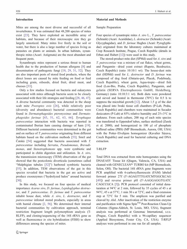
Table 1 The comparison of colony forming units (CFU) of cultivatedpart of bacterial community among tested species of mites
Mite species N Bacterial CFU
A. siro 12 637±525 a
D. farinae 10 1395±999 b
L. destructor 10 537±188 a
T. putrescentiae 10 521±360 a
CFU was recalculated per one mite and the means±standarddeviations are presented. The letters indicate Tukey’s differences
N numbers of replicates
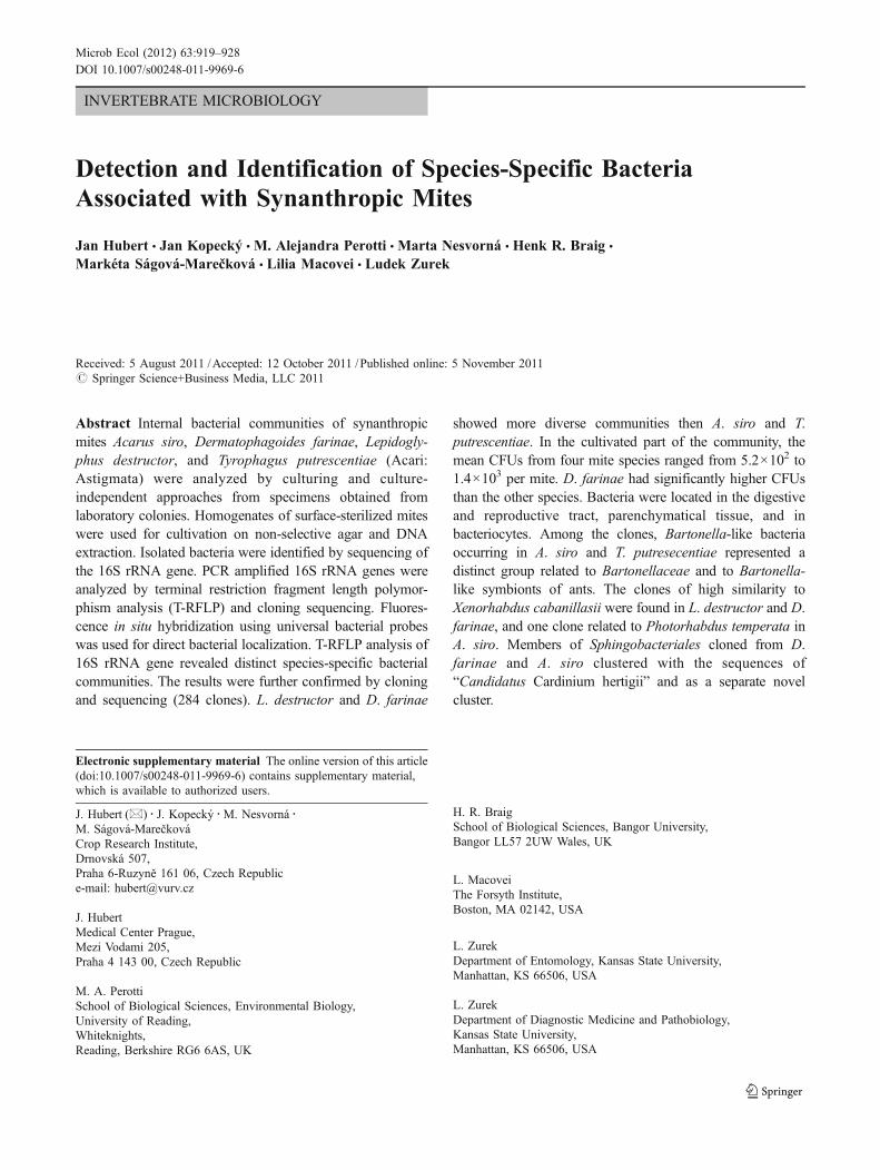
Figure 1 Bacterial communities associated with Lepidoglyphusdestructor (Ld), Tyrophagus putrescentiae (Tp), Dermatophagoidesfarinae (Df), and Acarus siro (As). T-RFLP profiles of bacterialdiversity expressed by Sammon’s method of multidimensional scaling.Three replicates were sampled and processed independently
Species-Specific Bacteria Associated with Synanthropic Mites 921
FISH
Fluorescence in situ hybridization was performed usinguniversal bacterial probes [45]. Mite specimens werefixed in 4% formaldehyde and preparation of whole-mitespecimens for the hybridizations followed the FISHprotocol described in Perrotti et al. [45]. For the hybrid-izations, samples were incubated at 45°C in darkness for amaximum of 20 h, then washed for 1 h with the samehybridization buffer followed by washing in PBTA(phosphate buffer with Triton X-100 plus sodium azide)at room temperature. Samples were mounted in PBS/glycerol and examined under the microscope. Thebacterial probes EUB-338 [1] and EUB-338 II and III[9] used in this experiment were equimolar mixed in thehybridization buffer (following remarks of ProBase). No-probe and competition suppression controls using excessunlabeled probes were performed. The hybridized speci-mens were visualized using Confocal Zeiss LSM510microscope with Coherent Multiphoton laser. Lengthmeasurements were performed by the Zeiss LSM ImageExaminer.
Results
T-RFLP
Based on T-RFLP analysis of 16S rRNA gene, distinctbacterial communities were identified among the testedmite species. Specifically, the bacterial communities wereseparated by the type of diet along the first axis inSammon’s multidimensional scaling (Fig. 1).
Bacterial Isolation
In the isolated bacteria from homogenized and surface-sterilized mites, the mean bacterial CFU numbers rangedfrom 5.2×10² to 1.4×10³ per mite. Significant differencesin CFU numbers were observed among mite species (F(3,38)=4.96, P=0.005); Tukey’s comparison showed that D. farinaecarried significantly higher CFU number than the other mitespecies (Table 1).
The 16S rRNA genes were sequenced from 54 coloniesrepresenting the culturable part of bacterial communitiesassociated with L. destructor (10 sequences), D. farinae (9),A. siro (18), and T. putrescentiae (17), and from 30 coloniesisolated from uninoculated rearing diets. The sequenceswere deposited in GenBank under accession nos.JN392772–JN392855 (Supplementary Table S1). Therewere no remarkable differences among the types of thediet (Table S1).
Cloning
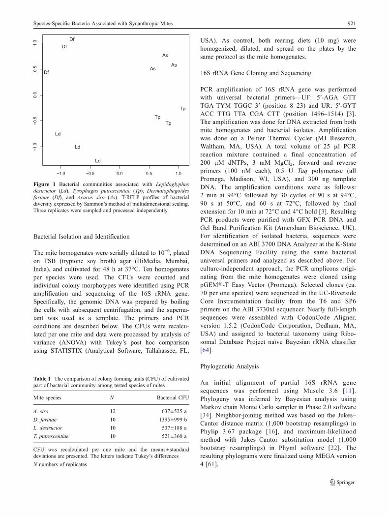
The 16S rRNA genes were cloned and 280 sequencesrepresenting the bacterial communities associated with L.destructor (67 clones), D. farinae (74), A. siro (68), and T.putrescentiae (71) were deposited in GenBank underaccession nos. JN236220–JN236499 (SupplementaryTable S1). The phylogenetic assignment of cloned bacteriashowed that the communities differed among the species(Fig. 2).
Rhizobiales were represented by 60 cloned sequences ofunidentified bacteria related to Bartonellaceae (Fig. 3). Thesequences showed separated species-specific clusters orig-inating from A. siro and T. putrescentiae, respectively. In
Figure 2 Phylogenetic assign-ment of bacterial 16S rRNAgene sequences cloned fromDNA extracted from mite wholebody homogenates (a) andamplified from the isolatesrepresenting isolated bacterialcommunity members (b)
922 J. Hubert et al.
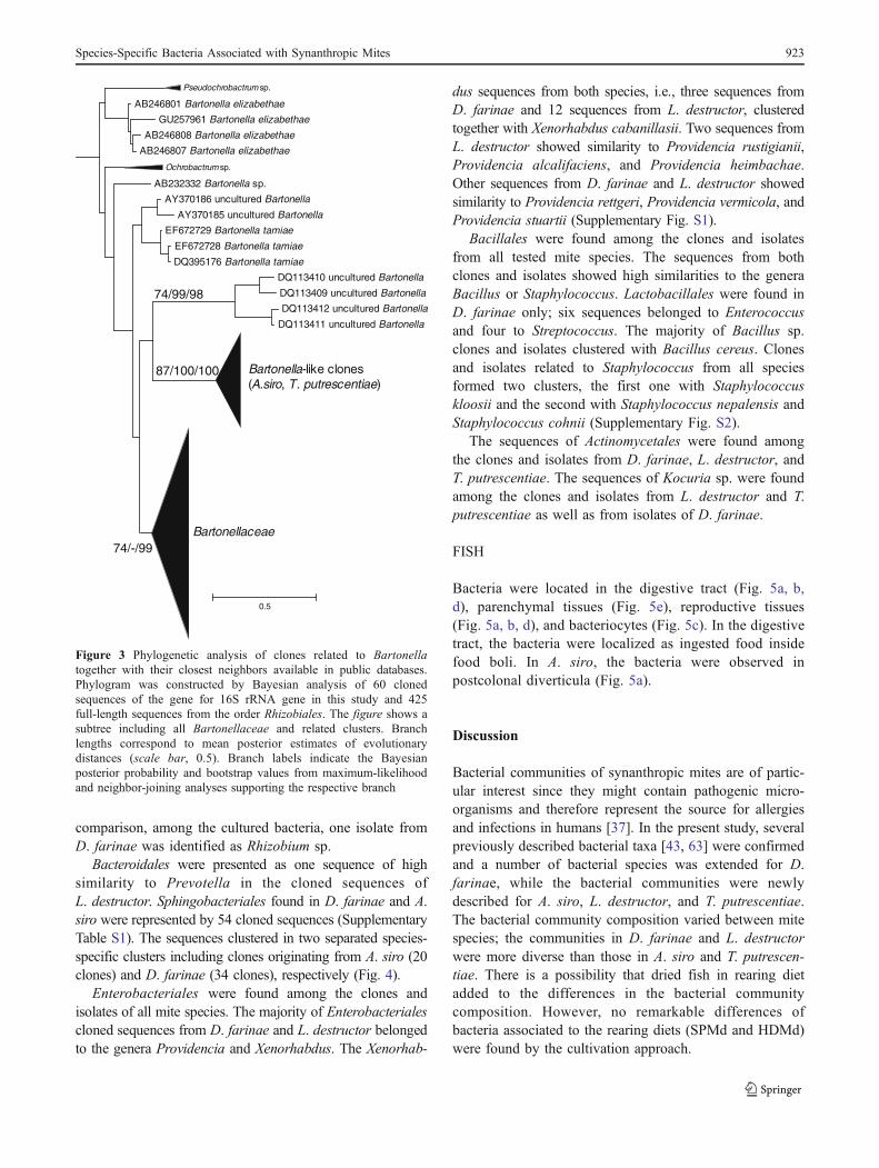
comparison, among the cultured bacteria, one isolate fromD. farinae was identified as Rhizobium sp.
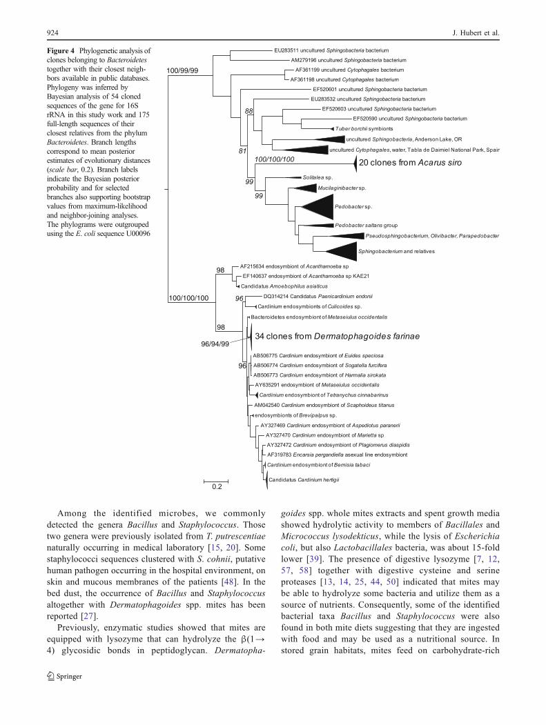
Bacteroidales were presented as one sequence of highsimilarity to Prevotella in the cloned sequences ofL. destructor. Sphingobacteriales found in D. farinae and A.siro were represented by 54 cloned sequences (SupplementaryTable S1). The sequences clustered in two separated species-specific clusters including clones originating from A. siro (20clones) and D. farinae (34 clones), respectively (Fig. 4).
Enterobacteriales were found among the clones andisolates of all mite species. The majority of Enterobacterialescloned sequences from D. farinae and L. destructor belongedto the genera Providencia and Xenorhabdus. The Xenorhab-
dus sequences from both species, i.e., three sequences fromD. farinae and 12 sequences from L. destructor, clusteredtogether with Xenorhabdus cabanillasii. Two sequences fromL. destructor showed similarity to Providencia rustigianii,Providencia alcalifaciens, and Providencia heimbachae.Other sequences from D. farinae and L. destructor showedsimilarity to Providencia rettgeri, Providencia vermicola, andProvidencia stuartii (Supplementary Fig. S1).
Bacillales were found among the clones and isolatesfrom all tested mite species. The sequences from bothclones and isolates showed high similarities to the generaBacillus or Staphylococcus. Lactobacillales were found inD. farinae only; six sequences belonged to Enterococcusand four to Streptococcus. The majority of Bacillus sp.clones and isolates clustered with Bacillus cereus. Clonesand isolates related to Staphylococcus from all speciesformed two clusters, the first one with Staphylococcuskloosii and the second with Staphylococcus nepalensis andStaphylococcus cohnii (Supplementary Fig. S2).
The sequences of Actinomycetales were found amongthe clones and isolates from D. farinae, L. destructor, andT. putrescentiae. The sequences of Kocuria sp. were foundamong the clones and isolates from L. destructor and T.putrescentiae as well as from isolates of D. farinae.
FISH
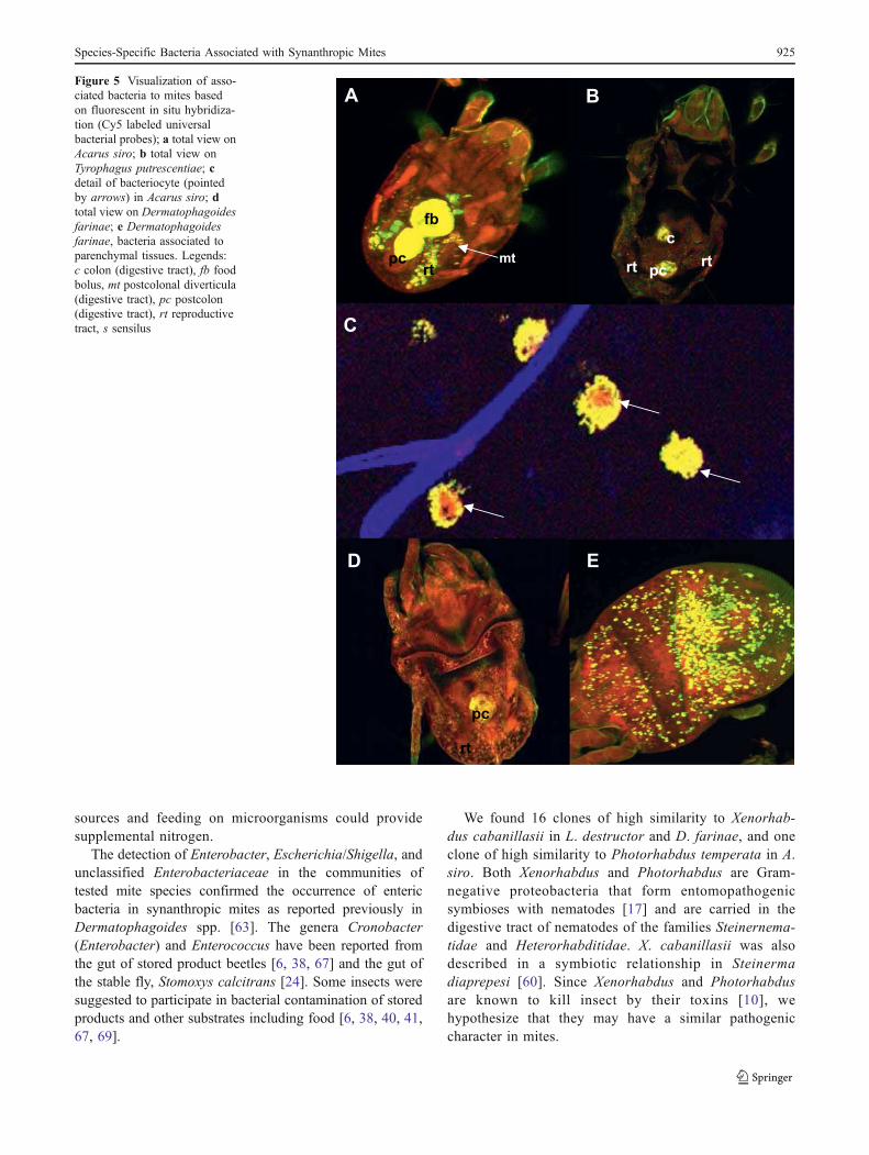
Bacteria were located in the digestive tract (Fig. 5a, b,d), parenchymal tissues (Fig. 5e), reproductive tissues(Fig. 5a, b, d), and bacteriocytes (Fig. 5c). In the digestivetract, the bacteria were localized as ingested food insidefood boli. In A. siro, the bacteria were observed inpostcolonal diverticula (Fig. 5a).
Discussion
Bacterial communities of synanthropic mites are of partic-ular interest since they might contain pathogenic micro-organisms and therefore represent the source for allergiesand infections in humans [37]. In the present study, severalpreviously described bacterial taxa [43, 63] were confirmedand a number of bacterial species was extended for D.farinae, while the bacterial communities were newlydescribed for A. siro, L. destructor, and T. putrescentiae.The bacterial community composition varied between mitespecies; the communities in D. farinae and L. destructorwere more diverse than those in A. siro and T. putrescen-tiae. There is a possibility that dried fish in rearing dietadded to the differences in the bacterial communitycomposition. However, no remarkable differences ofbacteria associated to the rearing diets (SPMd and HDMd)were found by the cultivation approach.
Pseudochrobactrum sp.
AB246801 Bartonella elizabethae
GU257961 Bartonella elizabethae
AB246808 Bartonella elizabethae
AB246807 Bartonella elizabethae
Ochrobactrum sp.
AB232332 Bartonella sp.
AY370186 uncultured Bartonella
AY370185 uncultured Bartonella
EF672729 Bartonella tamiae
EF672728 Bartonella tamiae
DQ395176 Bartonella tamiae
DQ113410 uncultured Bartonella
DQ113409 uncultured Bartonella
DQ113412 uncultured Bartonella
DQ113411 uncultured Bartonella
Bartonella-like clones (A.siro, T. putrescentiae)
Bartonellaceae
0.5
87/100/100
74/99/98
74/-/99
Figure 3 Phylogenetic analysis of clones related to Bartonellatogether with their closest neighbors available in public databases.Phylogram was constructed by Bayesian analysis of 60 clonedsequences of the gene for 16S rRNA gene in this study and 425full-length sequences from the order Rhizobiales. The figure shows asubtree including all Bartonellaceae and related clusters. Branchlengths correspond to mean posterior estimates of evolutionarydistances (scale bar, 0.5). Branch labels indicate the Bayesianposterior probability and bootstrap values from maximum-likelihoodand neighbor-joining analyses supporting the respective branch
Species-Specific Bacteria Associated with Synanthropic Mites 923
Among the identified microbes, we commonlydetected the genera Bacillus and Staphylococcus. Thosetwo genera were previously isolated from T. putrescentiaenaturally occurring in medical laboratory [15, 20]. Somestaphylococci sequences clustered with S. cohnii, putativehuman pathogen occurring in the hospital environment, onskin and mucous membranes of the patients [48]. In thebed dust, the occurrence of Bacillus and Staphylococcusaltogether with Dermatophagoides spp. mites has beenreported [27].
Previously, enzymatic studies showed that mites areequipped with lysozyme that can hydrolyze the β(1→4) glycosidic bonds in peptidoglycan. Dermatopha-
goides spp. whole mites extracts and spent growth mediashowed hydrolytic activity to members of Bacillales andMicrococcus lysodekticus, while the lysis of Escherichiacoli, but also Lactobacillales bacteria, was about 15-foldlower [39]. The presence of digestive lysozyme [7, 12,57, 58] together with digestive cysteine and serineproteases [13, 14, 25, 44, 50] indicated that mites maybe able to hydrolyze some bacteria and utilize them as asource of nutrients. Consequently, some of the identifiedbacterial taxa Bacillus and Staphylococcus were alsofound in both mite diets suggesting that they are ingestedwith food and may be used as a nutritional source. Instored grain habitats, mites feed on carbohydrate-rich
100/99/99
100/100/100
98
98
96
96/94/99
Figure 4 Phylogenetic analysis ofclones belonging to Bacteroidetestogether with their closest neigh-bors available in public databases.Phylogeny was inferred byBayesian analysis of 54 clonedsequences of the gene for 16SrRNA in this study work and 175full-length sequences of theirclosest relatives from the phylumBacteroidetes. Branch lengthscorrespond to mean posteriorestimates of evolutionary distances(scale bar, 0.2). Branch labelsindicate the Bayesian posteriorprobability and for selectedbranches also supporting bootstrapvalues from maximum-likelihoodand neighbor-joining analyses.The phylograms were outgroupedusing the E. coli sequence U00096
924 J. Hubert et al.
sources and feeding on microorganisms could providesupplemental nitrogen.
The detection of Enterobacter, Escherichia/Shigella, andunclassified Enterobacteriaceae in the communities oftested mite species confirmed the occurrence of entericbacteria in synanthropic mites as reported previously inDermatophagoides spp. [63]. The genera Cronobacter(Enterobacter) and Enterococcus have been reported fromthe gut of stored product beetles [6, 38, 67] and the gut ofthe stable fly, Stomoxys calcitrans [24]. Some insects weresuggested to participate in bacterial contamination of storedproducts and other substrates including food [6, 38, 40, 41,67, 69].
We found 16 clones of high similarity to Xenorhab-dus cabanillasii in L. destructor and D. farinae, and oneclone of high similarity to Photorhabdus temperata in A.siro. Both Xenorhabdus and Photorhabdus are Gram-negative proteobacteria that form entomopathogenicsymbioses with nematodes [17] and are carried in thedigestive tract of nematodes of the families Steinernema-tidae and Heterorhabditidae. X. cabanillasii was alsodescribed in a symbiotic relationship in Steinermadiaprepesi [60]. Since Xenorhabdus and Photorhabdusare known to kill insect by their toxins [10], wehypothesize that they may have a similar pathogeniccharacter in mites.
Figure 5 Visualization of asso-ciated bacteria to mites basedon fluorescent in situ hybridiza-tion (Cy5 labeled universalbacterial probes); a total view onAcarus siro; b total view onTyrophagus putrescentiae; cdetail of bacteriocyte (pointedby arrows) in Acarus siro; dtotal view on Dermatophagoidesfarinae; e Dermatophagoidesfarinae, bacteria associated toparenchymal tissues. Legends:c colon (digestive tract), fb foodbolus, mt postcolonal diverticula(digestive tract), pc postcolon(digestive tract), rt reproductivetract, s sensilus
Species-Specific Bacteria Associated with Synanthropic Mites 925
Bartonella species are Gram-negative bacteria that arehuman pathogens, and their potential vectors are arthropods[4]. Bartonella spp. are common symbiotic bacteria in ticks[35] and recently have been described from D. farinae andD. pteronyssinus [63] but were not found in Psoroptes ovis[26]. Valerio et al. [63] suggested Bartonella sp. as adominant symbiont in house dust mites. The Bartonella-like bacteria were detected in A. siro and T. putrescentiae inthis study. Our clones were related to Bartonellaceae, butformed a separate cluster close to Bartonella-like symbiontsof ants of the genus Tetraponera. In ants, the Bartonella-like symbionts were present in the intestine [59] while in A.siro, an abundant bacterial community resided in postcolo-nal diverticula (this study and Sobotnik et al. [55]). Thehigh incidence (63%) of Bartonella-like bacteria among theobtained sequences suggested that postcolonal diverticulawas the main colonization site for Bartonella-like bacteriain A. siro. However, Bartonella-like bacteria were alsofound in T. putrescentiae, which is probably lackingpostcolonal diverticula [53]. The localization of Bartonella-like bacteria in mite body is still open.
Some bacterial species in the phylum Bacteroidetesare known as manipulators of arthropod reproduction thatincludes cytoplasmic incompatibility [31]. Members of theclade “Candidatus Cardinium hertigii” were found invarious arthropods and nematodes [21], and they are alsocommon in spider mites [42]. Cytophaga-like organismswere observed in the related oribatid mite Oppiella nova[65]. The previous study showed the absence of Cardi-nium in Rhizoglyphus robini and T. putrescentiae [68],while the evidence of symbiotic proteobacteria closelyrelated to endosymbionts from ticks and spider mites wasreported in Dermatophagoides mites [63]. In this study,the cloned sequences of Sphingobacteriales formed twoseparate clusters. In D. farinae, the clones clustered to thesequences of “Ca. Cardinium hertigii”. In A. siro, thecloned sequences of Sphingobacteriales formed a separatecluster distinct from “Ca. Cardinium hertigii”. Theposition of the Sphingobacteriales sequences from A. siroindicated that they may constitute a new taxon ofendosymbiotic bacteria.
Finally, bacteria were present not only in the digestivetract of mites but also in the reproductive tract, parenchy-mal tissue, and in bacteriocytes in the examined mitespecies as shown in the results of FISH with the universaleubacterial primers. Similar distribution of bacteria wasfound in Brevipalpus spp. mites in which Cardinium cellswere detected in every body part including legs and palpsusing electron microscopy [36]. The presence of bacteria inbacteriocytes is probably novel for mites.
Mite species used in this study belong to pests of human-made habitats. It was documented that they act as vectorstransporting fungi of medical importance [18, 24, 31]. In
this study, we found that some Enterobacteriaceae andBacillales related to pathogenic species (e.g., S. cohnii)occur in house-dust and stored-product mites. Consequently,mites can distribute them through human habitats by the sameway as they spread fungi [52].
Acknowledgments This work was supported by the Czech ScienceFoundation (grant no. GA525/09/1872); the Ministry of Education,Youth and Sports of the Czech Republic (grant no. ME09013); and bythe OECD (project AGR/PROG-JA00038773). M.A.P. is supportedby the Royal Society, UK.
References
1. Amann RI, Binder BJ, Olson RJ, Chisholm SW, Devereux R, StahlDA (1990) Combination of 16S rRNA-targeted oligonucleotideprobes with flow cytometry for analyzing mixed microbial popula-tions. Appl Environ Microbiol 56:1919–1925
2. Arlian LG, Morgan MS (2003) Biology, ecology, and prevalenceof dust mites. Immunol Allergy Clin North Am 23:443–468
3. Barbieri E, Paster BJ, Hughes D, Zurek L, Moser DP, Teske A,Sogin ML (2001) Phylogenetic characterization of epibioticbacteria in the accessory nidamental gland and egg capsules ofthe squid Loligo pealei (Cephalopoda:Loliginidae). EnvironMicrobiol 3:151–167
4. Billeter SA, Levy MG, Chomel BB, Breitschwerdt EB (2008)Vector transmission of Bartonella species with emphasis on thepotential for tick transmission. Med Vet Entomol 22:1–15
5. Bruce KD, Hiorns WD, Hobman JL, Osborn AM, Strike P,Ritchie DA (1992) Amplification of DNA from native populationsof soil bacteria by using the polymerase chain reaction. ApplEnviron Microbiol 58:3413–3416
6. Channaiah LH, Subramanyam B, Zurek L (2010) Survival ofEnterococcus faecalis OG1RF:pCF10 in poultry and cattle feed:vector competence of the red flour beetle, Tribolium castaneum(Herbst). J Food Prot 73:568–573
7. Childs M, Bowman CE (1981) Lysozyme activity in 6 species ofeconomically important astigmatid mites. Comp Biochem Physiol70B:615–617
8. Colloff MJ (2009) Dust mites. CSIRO, Collingwood, p 5839. Daims H, Brühl A, Amann R, Schleifer KH, Wagner M (1999)
The domain-specific probe EUB338 is insufficient for thedetection of all bacteria: development and evaluation of a morecomprehensive probe set. Syst Appl Microbiol 22:434–444
10. Ffrench-Constant RH, Dowling A, Waterfield NR (2007) Insec-ticidal toxins from Photorhabdus bacteria and their potential usein agriculture. Toxicon 49:436–451
11. Edgar RC (2004) MUSCLE: multiple sequence alignment withhigh accuracy and high throughput. Nucleic Acids Res 32:1792–1797
12. Erban T, Hubert J (2008) Digestive function of lysozyme insynanthropic acaridid mites enables utilization of bacteria as afood source. Exp Appl Acarol 44:199–212
13. Erban T, Hubert J (2010) Determination of pH in regions of themidguts of acaridid mites. J Insect Sci 10(42):1–12
14. Erban T, Hubert J (2010) Comparative analyses of proteases inseven species of synanthropic acaridid mites. Arch Insect BiochPhysiol 75:187–206
15. da Silva Ezequiel O, Gazeta GS, de Oliveira GN, de-Barros RAM,Serra-Freire NM (2001) Mites as vectors of fungi and bacteria:observation during natural infestation in the Pathology Laboratory.Entomologia y Vectores 8:481–489 [Portuguese, English abstract]
926 J. Hubert et al.
16. Felsenstein J (1989) PHYLIP—phylogeny inference package(version 3.2). Cladistics 5:164–166
17. Forst S, Dowds B, Boemare N, Stackebrandt E (1997) Xenorhab-dus and Photorhabdus spp.: bugs that kill bugs. Annu RevMicrobiol 51:47–72
18. Franzolin MR, Gambale W, Cuero RG, Correa B (1999)Interaction between toxigenic Apergillus flavus link and mites(Tyrophagus putrescentiae Schrank) on maize grains: effects onfungal growth and aflatoxin production. J Stored Prod Res35:215–224
19. García N (2004) Efforts to control mites on Iberian ham byphysical methods. Exp Appl Acarology 32:41–50
20. Gazeta GS, Norberg AN, Aboud-Dutra AE, Serra-Freire NM(2000) Tyrophagus putrescentiae (Schrank, 1781) as a vector ofpathogenic bacteria: laboratory observation. Entomologia yVectores 7:49–55 [Portuguese, English abstract]
21. Gotoh T, Noda H, Ito S (2007) Cardinium symbionts causecytoplasmic incompatibility in spider mites. Heredity 98:13–20
22. Guindon S, Gascuel O (2003) A simple, fast, and accuratealgorithm to estimate large phylogenies by maximum likelihood.Syst Biol 52:696–704
23. Halliday RB, O’Connor BM, Baker AS (2000) Globaldiversity of mites. In: Raven PH, Williams T (eds) Natureand human society: the quest for a sustainable world.Proceedings of the 1997 forum on biodiversity. NationalAcademies, Washington, pp 192–212
24. Hamilton JV, LehaneMJ, Braig HR (2003) Isolation of Enterobactersakazakii from midgut of Stomoxys calcitrans. Emerging Infect Dis9:1355–1356
25. Harris J, Mason DE, Li J, Burdick KW, Backes BJ, Chen T,Shipway A, Van Heeke G, Gough L, Ghaemmaghami A, ShakibF, Debaene F, Winssinger N (2004) Activity profile of dust miteallergen extract using substrate libraries and functional proteomicmicroarrays. Chem Biol 11:1361–1372
26. Hogg JC, Lehane MJ (1999) Identification of bacterial speciesassociated with the sheep scab mite (Psoroptes ovis) by usingamplified genes coding for 16S rRNA. Appl Environ Microbiol65:4227–4229
27. Horak B, Dutkiewicz J, Solarz K (1996) Microflora andacarofauna of bed dust from homes in Upper Silesia, Poland.Ann Allergy Asthma Immunol 76:41–50
28. Hoy MA, Jeyaprakash A (2005) Microbial diversity in thepredatory mite Metaseiulus occidentalis (Acari: Phytoseiidae)and its prey, Tetranychus urticae (Acari: Tetranychidae). BiolControl 32:427–441
29. Hubert J, Stejskal V, Munzbergová Z, Kubátová A, Vaňová M,Žďárková E (2004) Mites and fungi in heavily infested stores inthe Czech Republic. J Econ Entomol 97:2144–2153
30. Hubert J, Križková-Kudliková I, Stejskal V (2004) Review ofdigestive enzymes of stored product and house dust mites.Proceedings of the V symposium of the European association ofacarologists “acarine biodiversity in the natural and humansphere”, Berlin 2004. Phytophaga 14:695–710
31. Hubert J, Jarošík V, Mourek J, Kubatová A, Žďárková E (2004)Astigmatid mite growth and fungi preference (Acari: Acaridida):comparisons in laboratory experiments. Pedobiologia 48:205–214
32. Hughes TE (1950) The physiology of the alimentary canal ofTyrophagus farinae. Quart J Microsc Sci 91:54–60
33. Hughes AM (1976) The mites of stored food and houses.Technical bulletin of the UK ministry of agriculture, fisheriesand food, Her Majesty’s Stationery Office, London 400 pp.
34. Jow H, Hudelot C, Rattray M, Higgs P (2002) Bayesianphylogenetics using an RNA substitution model applied to earlymammalian evolution. Mol Biol Evol 19:1591–1601
35. Kim CM, Kim JY, Yi YH, Lee MJ, Cho MR, Shah DH, Klein TA,Kim HC, Song JW, Chong ST, O’Guinn ML, Lee JS, Lee IY, Park
JH, Chae JS (2005) Detection of Bartonella species from ticks,mites and small mammals in Korea. J Vet Sci 6:327–334
36. Kitajima EW, Groot TVM, Novelli VM, Freitas-Astúa J, AlbertiG, de-Moraes GJ (2007) In situ observation of the Cardiniumsymbionts of Brevipalspus (Acari: Tenuipalpidae) by electronmicroscopy. Exp Appl Acarol 42:263–271
37. Langer K, Breuer K, Kapp A, Werfel T (2007) Staphylococcusaureus-derived enterotoxins enhance house dust mite-inducedpatch test reactions in atopic dermatitis. Exp Dermatol 16:124–129
38. Larson Z, Subramanyam B, Zurek L, Herrman T (2008) Diversityand antibiotic resistance of enterococci associated with the storedproduct insects collected from feed mills. J Stored Prod Res44:198–203
39. Mathaba LT, Pope CH, Lenzo J, Hartofillis M, Peake H, MoritzRL, Simpson RJ, Bubert A, Thompson PJ, Stewart GA (2002)Isolation and characterization of a 13.8-kDa bacteriolytic enzymefrom house dust mite extracts: homology with prokaryoticproteins suggests that the enzyme could be bacterially derived.FEMS Immunol Med Microbiol 33:77–88
40. Mramba F, Broce AB, Zurek L (2006) Isolation of Enterobactersakazakii from stable flies, Stomoxys calcitrans L. (Diptera:Muscidae). J Food Prot 69:671–673
41. Mramba F, Broce AB, Zurek L (2007) Vector competence ofstable flies, Stomoxys calcitrans L. (Diptera: Muscidae), forEnterobacter sakazakii. J Vector Ecol 32:134–139
42. Nakamura Y, Kawai S, Yukuhiro F, Ito S, Gotoh T, Kisimoto R,Yanase T, Matsumoto Y, Kageyama D, Noda H (2009) Prevalenceof Cardinium bacteria in planthoppers and spider mites andtaxonomic revision of “Candidatus Cardinium hertigii” based ondetection of a new Cardinium group from biting midges. ApplEnviron Microbiol 75:6757–6763
43. Oh H, Ishii A, Tongu Y, Itano K (1986) Microorganismsassociated with the house dust mite, Dermatophagoides. Jpn JSanit Zool 37:229–235 [in Japanese, English abstract]
44. Ortego F, Sánchez-Ramos I, Ruiz M, Castañera P (2000) Character-ization of proteases from a stored product mite, Tyrophagusputrescentiae. Arch Insect Biochem Physiol 43:116–124
45. Perotti MA, Allen JM, Reed DL, Braig HR (2007) Host–symbiontinteractions of the primary endosymbiont of human head andbody lice. FASEB J 21:1058–1066
46. Perotti MA, Braig HR (2004) Endosymbionts of Acari. Phyto-phaga 14:457–476
47. Perotti MA, Braig HR (2011) Eukaryotic ectosymbionts of Acari.J Appl Entomol 135:514–523
48. Rózalska M, Szewczyk EM (2008) Staphylococcus cohniihemolysins—isolation, purification and properties. Folia Micro-biol (Praha) 53:521–526
49. Sagová-Marečková M, Čermák L, Novotná J, Plhačková K,Forstová J, Kopecký J (2008) Innovative methods for soil DNApurification tested in soils with widely differing characteristics.Appl Environ Microbiol 74:2902–2907
50. Sánchez-Ramos I, Hernández CA, Castañera P, Ortego F (2004)Proteolytic activities in body and faecal extracts of the storagemite, Acarus farris. Med Vet Entomol 18(4):378–386
51. Saleh SM, Keladan NL, Shaker N (1991) Control of Europeanhouse dust mite Dermatophagoides pteronyssinus (Trouessart)with Bacillus spp. Acarologia (Acarologia) 32:257–260
52. Sinha RN (1979) Ecology of microflora in stored grain. Ann TechAgri 28:191–209
53. Smrž J (2003) Microanatomical and biological aspects of bacterialassociations in Tyrophagus putrescentiae (Acari: Acaridida). ExpAppl Acarol 31:105–113
54. Smrž J, Čatská V (2010) Mycophagous mites and their internalassociated bacteria cooperate to digest chitin in soil. Symbiosis52:33–40
Species-Specific Bacteria Associated with Synanthropic Mites 927
55. Šobotník J, Alberti G, Weyda F, Hubert J (2008) Ultrastructure ofthe digestive tract in Acarus siro (Acari: Acaridida). J Morphol269:54–71
56. Stejskal V, Hubert J (2008) Risk of occupational allergy to storedgrain arthropods and false pest-risk perception in Czech grainstores. Ann Agric Environ Med 15:29–35
57. Stewart GA, Lake FR, Thompson PJ (1991) Faecally derivedhydrolytic enzymes from Dermatophagoides pteronyssinus: phys-icochemical characterization of potential allergens. Int ArchAllergy Appl Immunol 95:248–256
58. Stewart GA, Bird CH, Krska KD, Colloff MJ, Thompson PJ(1992) A comparative study of allergenic and potentiallyallergenic enzymes from Dermatophagoides pteronyssinus, D.farinae and Euroglyphus maynei. Exp Appl Acarol 16:165–180
59. Stoll S, Gadau J, Gross J, Feldhaar H (2007) Bacterial microbiotaassociated with ants of the genus Tetraponera. Biol J Linnean Soc90:399–412
60. Tailliez P, Pagès S, Ginibre N, Boemare N (2006) New insightinto diversity in the genus Xenorhabdus, including the descriptionof ten novel species. Int J Syst Evol Microbiol 56:2805–2818
61. Tamura K, Dudley J, Nei M, Kumar S (2007) MEGA4: MolecularEvolutionary Genetics Analysis (MEGA) software version 4.0.Mol Biol Evol 24:1596–1599
62. Trivedi B, Valerio C, Slater JE (2003) Endotoxin content ofstandardized allergen vaccines. J Allergy Clin Immunol 111:777–783
63. Valerio CR, Murray P, Arlian LG, Slater JE (2005) Bacterial 16Sribosomal DNA in house dust mite cultures. J Allergy ClinImmunol 116:1296–1300
64. Wang Q, Garrity GM, Tiedje JM, Cole JR (2007) Naïve Bayesianclassifier for rapid assignment of rRNA sequences into the newbacterial taxonomy. Appl Environ Microbiol 73:5261–5267
65. Weeks AR, Velten R, Stouthamer R (2003) Incidence of a newsex-ratio-distorting endosymbiotic bacterium among arthropods.Proc Biol Sci 270:1857–1865
66. Yella L, Morgan MS, Arlian LG (2011) Population growth andallergen accumulation of Dermatophagoides pteronyssinus cul-tured at 20 and 25°C. Exp Appl Acarol 53:103–119
67. Yezerski A, Cussatt G, Glick D, Evancho M (2005) The effects ofthe presence of stored product pests on the microfauna of a flourcommunity. J Appl Microbiol 98:507–515
68. Zchori-Fein E, Perlman SJ (2004) Distribution of the bacterialsymbiont Cardinium in arthropods. Mol Ecol 13:2009–2016
69. Zurek L, Schal C, Watson DW (2000) Diversity and contribution ofthe intestinal bacterial community to the development of Muscadomestica (Diptera: Muscidae) larvae. J Med Entomol 37:924–928
928 J. Hubert et al.