Embed Size (px)
Citation preview
Detection of Mucosal Human Papillomavirus Types 6/11 in Cutaneous Lesions from Transplant Recipients
Chantal Soler, * Yvette Chardonnet, * Patrice Allibert, t Sylvie Euvrard, * Daniel Schmitt, * and Bernard Mandrandt *INSERM U346 Affiliee CNRS, Pavilion R, Hopital E. Herriot, Lyon, and j-UMRI03 CNRS/ BioMerieux, Ecole Normale Superieure, Lyon, France
Transplant recipients develop multiple cutaneous lesions. We have identified human papillomavirus (HPV) DNA in these lesions using three different techniques, namely polymerase chain reaction (peR), in situ hybridization, and Southern blotting. By peR, HPV DNA was detected in 43 of 62 samples: warts, actinic keratoses, Bowen's disease, and squamous cell carcinomas. Surprisingly, HPV 6/11, usually associated with mucosa, were frequently found in benign, premalignant, and malignant cutaneous lesions (30/43 cases). Some of these biopsies were simultaneously tested by in situ hybridization and/or Southern blotting. By in situ hybridization, HPV 6/11 were identified in two warts and
Immunosuppressed patients such as transplant recipients develop numerous cutaneous lesions, profuse verrucosis, and carcinomas on sun-exposed areas [1,2). In these benign and malignant lesions HPV DNA has been detected [3-6). Several HPV types have been detected in such biopsies: HPV 1,
2, 3, 4, and 10 were frequent in skin warts [3,7), as in the general population; benign HPV 2 and 4 were also found in squamous cell carcinomas [4). oncogenic HPV 16 was seen in benign skin warts [7) and in periungual squamous cell carcinomas [8,9). HPV 6/11, which usually infect benign genital lesions such as condylomas or benign laryngeal papillomas of the general population [10,11), have also been detected in perianal squamous cell carcinomas from transplant recipients [12). This investigation reports the detection of mucosal HPV types 6/11 in different cutaneous lesions from transplant recipients using peR and, in some specimens, in situ hybridization and Southern blotting.
MATERIALS AND METHODS
Biopsy Specimens Sixty-two cutaneous biopsies were excised from 22 renal and five cardiac transplant recipients with multiple lesions on sun-exposed areas (face, arm, hand, leg, top of back, and chest). Ten skin biopsies were taken from normal individuals with similar lesions, and six genital warts and five juvenile laryngeal papillomas were also tested.
Each biopsy was cut into two parts: one was snap frozen and stored at - 20" C and the other was fixed in Bouin's or Baker's solution and embedded in paraffin. Four-micrometer frozen sections were layered on aminopropyltriethoxysilane (Aldrich Chemic, Strasbourg, France)-treated slides and fixed in acetone for itl sitJl hybridization. One slide from each paraffin-embedded lesion was routinely stained with hematoxylin-eosin for histologic examination.
Manuscript received June 24, 1992; accepted for publication May 3,1993. Reprint requests to: Dr. Y. Chardon net, INSERM U346 Affiliee CNRS,
Pavillon R, Hopital E. Herriot, 69437 Lyon Cedex 03, France. Abbreviation: dUTP, deoxy uridine triphosphate.
one squamous cell carcinoma among 29 tissue specimens tested. Of the three samples examined by Southern blotting, HPV 6/11 were detected in one squamous cell carcinoma. In patients from a control population cutaneous biopsies did not exhibit HPV types 6/11 except in Bowen's disease; HPV types 1 or 2 were mainly found in benign warts. These findings suggest that in transplant recipients, HPV can lose their specificity towards mucosa or cutaneous epithelium. The significance of the presence of HPV 6/11 in skin lesions remains unknown. Key words: HPV 6/11/skin lesions/renal transplant recipients/cardiac transplant recipients. ] Invest DermatoI101:286-291, 1993
Polymerase Chain Reaction Studies Total cellular DNA was extracted from biopsies by phenol-chloroform-isoamyl alcohol and precipitated with ethanol. PCR was performed on cellular DNAs using a Perkin-Elmer Cetus DNA thermal cycler according to the procedure and precautions previously described [13], with primers chosen in E6 - E7 region ofHPV types 5, 6/11 , 16, and 18 (Table I). The amplification protocol was annealing for 1 min at 55"C, extension for 1 min at 72"C, and denaturation for 1 min at 95"C, except for HPV 5, for which the annealing temperature was 58"C. Amplified products were detected on ethidium bromide-stained gels and Southern blots were hybridized with radiolabeled specific oligonucleotides (Table I) .
To prevent artefactual results, cellular and plasmid DNAs were prepared in different rooms. Furthermore, some cellular DNAs used in these experiments were extracted in different years. Buffer and primers were submitted to UV for 3 hand aliquoted. Positive and negative controls were included in the reactions.
In Sit" Hybridization HPV la, 2a, 5, 6a, II, 16, and 18 (provided by G. Orth, Paris and H . zur Hausen, Heidelberg) cloned in pBR322 plasmid were prepared and purified on cesium chloride gradients. Plasmid DNAs were biotinylated by nick translation using biotinylated ll-dUTP (Sigma, Saint Louis, MO). The sections were processed under stringent conditions at Tm-17"C and DNA-DNA hybrids were detected with a three-step procedure using streptavidin-alkaline phosphatase complex [14).
Southern Blot Ten micrograms of total cellular DNA from eight biopsyspecimens (three from transplant recipients and five from controls) were digested with Pst I (Boehringer, Mannheim, Germany) in buffer from the supplier and electrophoresed on a 1 % agarose gel. The fragments were then transferred onto a Gene Screen Plus membrane (NEN , Boston, MA). Plasmid HPV types la, 2a, 5, 6a, 11a, 16, and 18 probes were labeled by nick translation using 32P_dCTP (specific activity: 800 Cijmmol; Amersham). Membranes were prehybridized in 10 ml of 50% deionized formamide, 1 % sodium dodecyl sulfate (SDS), 1 M NaCI, 10% dextran sulfate, for 6 h at 42"C. One milliliter of solution containing about 5 X 10⁶ cpm of labeled probe (specific activity: 8 X 10⁷ cpm/J1g DNA) and 100 J1g of salmon sperm DNA were added after denaturation by heating in boiling water for 10 min and chilling on ice. Hybridization was carried out overnight at 42"C. The membranes were carefully washed successively in 2 X sodium saline citrate (1 X SSC: 0.15 M NaCI, 0.015 M sodium citrate), 2 X SSC plus 1% SDS,
0022-202X/93/S06.00 Copyright © 1993 by The Society for Investigative Dermatology, Inc.
286
VOL. tOt, NO.3 SEPTEMBER 1993 HPV 6/ 11 IN CUTANEOUS LESIONS 287
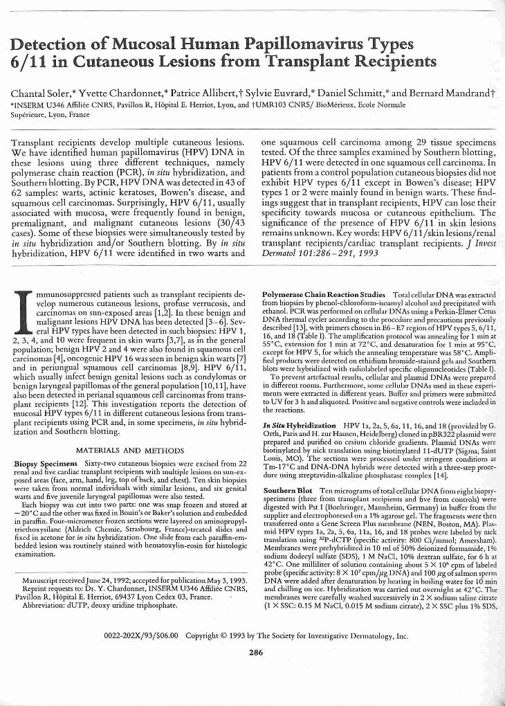
Table I. Primers and Oligoprobes for HPV 5, 6,11,16, and 18 Chosen in E6-E7 Region
HPV Localization Sequences Size of Fragments Tm
Primers HPV5 609 - 630 5' TTCATAAGGTGAGGAACGCCTG 3'
797 - 897 3' CGAAAGGTTGTCGATGACTGGC 5' 288 bp HPV6/11 514 - 533 5' TACACTGCTGGACAACATGC 3'
797 - 815 3' CACACAGGGTAGACGCGTG 5' 301 bp HPV 16 548 - 571 5' CCCAGCTGTAATCATGCATGGAGA 3'
778 - 801 3'ACCTTCTGGACAATTACCCGTGTG5' 253 bp PVH 18 540 - 560 5' CGACAGGAACGACTCCAACGA 3'
718-741 3' TCAATTAGTAGTTGTAAATGGTCG 5' 201 bp Probes
HPV 5 797-818 5'CAACGAAAGGATCTCTTACAAA3' 50SC 69 °C 62.3°C 59.2°C
HPV 6/1 t 584-602 5'GACCTGTTGCTGTGGATGT3' HPV16 700 - 717 5' CCGGACAGAGCCCATTAC 3' HPV18 601-620 5' TAAGGCAACATTGCAAGACA 3'
and 0.1 X SSC solutions. They were further exposed to X-OMA T (Kodak) films for 1-14 d using cassettes with intensifying screens. Membranes were successively hybridized with the different HPV probes after dehybridization according to the manufacturer's instructions and control for the loss of radioactivi ty .
RESULTS
All biopsy specimens we:e examine~ after stai~ing. with ~ema.tox.ylin-eosin and were classified according to their histologic cntena. Data comparing histologic diagnosis and results of experimental studies are shown in Table II .
Transplant Recipient Lesions PCR amplification of HPV types 5, 6/11, 16, and 18 was performed on skin lesions. Of the 62 samples, 43 (69%) were HPV DNA positive; 30 of 43 (69%) exhibited HPV 6/11 DNA, four of 11 (36%) benign vulgaris warts, seven of seven premalignant actinic keratoses, four of four premalignant Bowen's disease, and 15 of 21 (74%) squamous cell carcinomas (Table II).
Among 30 biopsies containing HPV 6/11 DNA by PCR (Table III), 12 samples (three warts, four actinic keratoses, three Bowen's disease, two squamous cell carcinomas) contained these HPV types; the other probes gave negative reactions. HPV 5 and HPV 6/11 were found in seven cases (one actinic keratosis, six squamous cell carcinomas). The other 11 samples contained HPV 6/11 and HPV 16, 18, and/or 5 (one wart, two actinic keratoses, one Bowen's disease, seven squamous cell carcinomas).
After HPV 6/11 amplification and hybridization of Southern blots with radioactive probes, bands of various intensities were seen, indicating that various amounts ofHPV 6/11 DNA were present in these samples. Figure 1 shows an example of results obtained after amplification of squamous cell carcinoma DNAs. In samples con-
~ainin~ more than one. type of DNA, HPV 6/11 were predominant In bemgn and premalignant lesions, but in squamous cell carcinomas they were present in smaller amounts than in oncogenic types.
J:IPV 6(11 DNA :vas identified (Table III) not only in seven patients wI~h sl11gl~ biopsies but also in seven other patients having s~veral excls.ed leslOns. In this latter case, samples were taken at ~dferent. penods, therefore excluding the possibility of contaminatIOn dunng DNA extraction. Furthermore, amplification without DNA or amplification of either MRC5 cell DNA or dermal DNA extracted from normal skin gave negative results in every case. Because HPV: 6/1.1 were usually found in benign mucosal lesions, PCR amplifications and analyses of amplification products were repeated at least two or three times . Similar results were detected consistently.
Twenty-nine of the 62 biopsies (Table IV) were a.lso screened for the presence of HPV DNA by in situ hybridization with HPV type 1,2,5,6/11,16, and 18 probes; 12 of29 contained HPV DNA. The positive signal was always nuclear and was located in intermediate and upp~r epithelial cell layers. Furthermore, it was usually in foci of a few 111fected cells. HPV 6/11 were identified in three samples: two warts and one squamous cell carcinoma (Fig 2); these results were confirmed with PCR. In warts, HPV 6/11 were associated with HPV 1 and/or 2 a.nd 1~; in the squamous cell carcinoma, on.ly HPV types 6/1 .1 v.:ere Ide~tl.fied: Three.to five sections per sample were tested by III SItu hybndlzatlon, which gave similar reactions.
Tissue sections were negative when the DNA probe was omitted or replace.d by the. p~R322 probe. HPV 16 and 18 probes were assayed With CaSb, SlHa, and HeLa cells containing, respectively, 600 copies ofHPV 16, one to two copies ofHPV 16, and 10-50 copies of HPV 18. CaSki and SiHa cells were positive only with HPV 16 p~obe, HeLa cells o~ly with HPV 18 probe.
Companng the results obtallled by PCR and in situ hybridization
Table II. Frequency of HPV 6/11 Detected by PCR in Skin Lesions from Transplant Recipients and Normal Population'
Lesions
Warts Keratoacanthomas Actinic keratoses Bowen's disease Squamous cell carcinomas Total Controls
Genital condylomas Latyngeal papillomas
Total
18 2
12 6
24 62
• PCR amplification of HPV types 5, 6/11, 16, and 18. • HPV DNA +, number of lesions containing HPV DNA.
Transplant Recipients
HPVb DNA+
11 0 7 4
21 43
, HPV 6/11 +, number of lesions containing HPV types 6/11 DNA.
HPV' 6/11 +
4 (36%) 0 7 (100%) 4 (36%)
15(71%) 30 (69%)
Non-Transplant Recipients
HPVb HPV' Total DNA+ 6/11 +
3 0 0
3 2 2 4 2 0
10 4 2
6 6 6 5 5 5
288 SOLER ET AL THE JOURNAL OF INVESTIGATIVE DERMATOLOGY
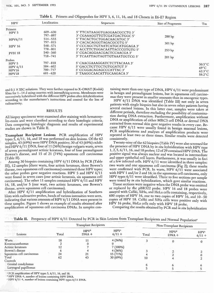
Table III. Detection of HPV 6/11 by peR" in 30 Skin Lesions from Transplant Recipients
Patients Lesions
Renal transplant recipients A Warts B Warts;
actinic keratosis
Squamous cell carcinomas
C Warts D Warts E Actinic keratosis;
squamous cell carcinoma F Actinic keratosis;
Bowen's disease G Actinic keratosis;
Bowen's disease H Actinic keratosis I Squamous cell carcinoma
J Squamous cell carcinoma K Squamous cell carcinoma
Cardiac transplant recipients L Actinic keratosis M Bowen's disease
N Squamous cell carcinomas
'peR amplification of HPV types 5, 6/11, 16, and 18 . • +, Positive HPV type DNA.
Number
1 2 3 4 5 6 7 8 9
10 11 12 13 14 15 16 17 18 19 20 21 22 23
24 25 26 27 28 29 30
' +/-, Weak positivity; the othet HPV types tested were negative.
with HPV type 5, 6/11,16, and 18 probes, an HPV DNA was more frequently detected by PCR than by in situ hybridization, 25 of 29 (86%) and eight of 29 (27%), respectively. HPV 6/11 were also more common with PCR than with in situ hybridization (Table IV), 20 of25 (80%) and 3 of8 (37%), respectively. By peR, HPV 6/11 were identified in HPV-positive samples (three of six warts, six of six actinic keratoses, four of five Bowen's disease, and seven of nine squamous cell carcinomas), whereas by in situ hybridization they
HPV Types
Localization 5 6/11 16 18
Arm +h Finger + Neck + Finger + + Back + + + Hand + Back + Back + + Back + + Back + + Neck + + Cheek + + Forehead + Nose + Back + + Temple + + Nose + Leg + Ear + Leg + Lip + + + Neck + + +/-' +/-Hand + + Hand + + + Arm + + Face + Neck + + Sternum + + Temple + + Ear + + +
were detectable only in two of three warts and one of four squamous cell carcinomas, the other samples being negative.
To avoid false-positive reactions of peR, the results were examined according to the patients, the location of the lesions, and the time of excision. HPV 6/11 were found in cutaneous lesions from 14 of 22 (64%) patients with a renal transplant and three of five (60%) patients with a cardiac transplant. These biopsies were taken from various body sites. Furthermore, when several specimens of
_1037bp 1 2 3 4 5 6 7 8 9 M
_475
301bp_ 315 301bp_
141
B
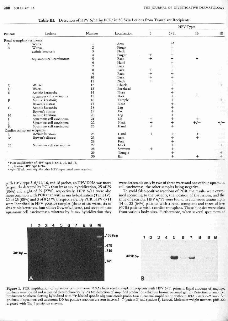
Figure 1. PCR amplification of squamous cell carcinoma DNAs from renal transplant recipients with HPV 6/11 primers. Equal amounts of amplified products were loaded and separated electrophoretically. A) No detection of amplified product on ethidium bromide-stained gel. B) Detection of amplified product on Southern blotting hybridized with 32P-labeled specilic oligonucleotide probe. Lallf 1, control amplification without DNA. Lalle5 2-9, amplified products of squamous cell carcinoma DNAs; positive reactions are seen in falles 5 - 7 (patient B) and (patient I) . Latle M, Molecular weight markers, pBR 322 digested with Taq I restriction enzyme.
VOL. 101, NO.3 SEPTEMBER 1993 HPV 6/ 11 IN CUTANEOUS LESIONS 289
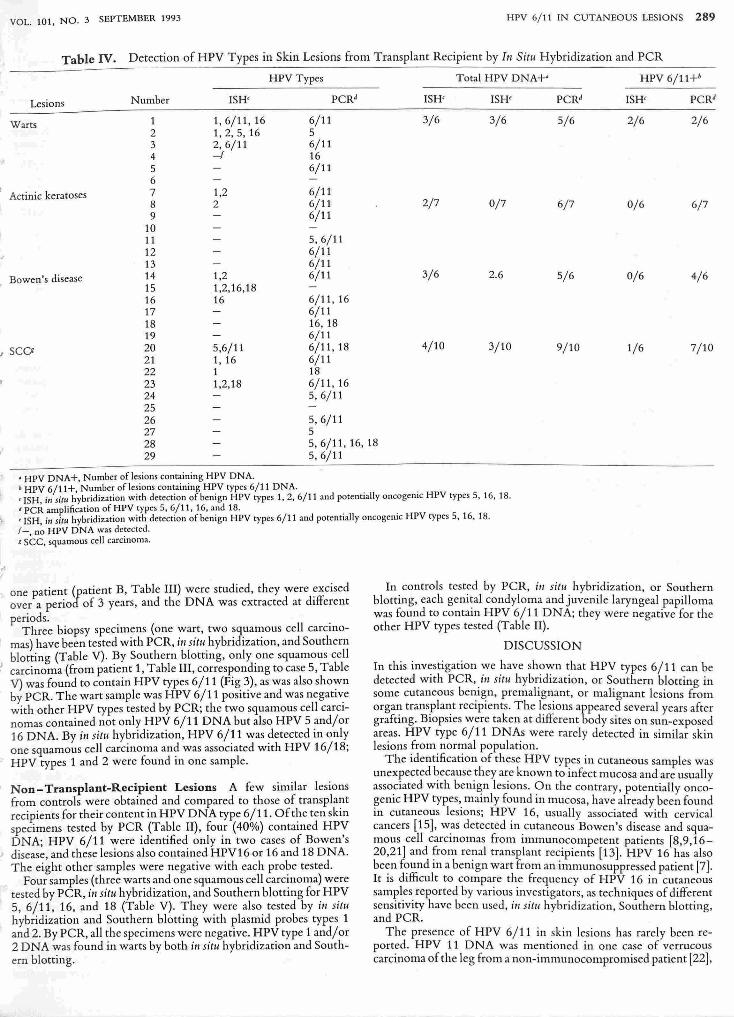
Table IV. Detection of HPV Types in Skin Lesions from Transplant Recipient by III Sitll Hybridization and PCR
HPV Types Total HPV DNA+' HPV 6/11+b
Lesions Number ISH' PCRJ ISH' ISH' PCRJ ISH' PCRJ
1 1,6/11,16 6/11 3/6 3/6 5/6 2/6 2/6 2 1,2,5,16 5
Warts
3 2,6/11 6/11 4 -I 16 5 6/11 6 7 1,2 6/11 8 2 6/11 2/7 0/7 6/7 0/6 6/7 9 6/11
, Actinic keratoses
10 11 5,6/11 12 6/11 13 6/11 14 1,2 6/11 3/6 2.6 5/6 0/6 4/6 Bowen's disease 15 1,2,16,18 16 16 6/11, 16 17 6/11 18 16,18 19 6/11 20 5,6/11 6/11,18 4/10 3/10 9/10 1/6 7/10 21 1, 16 6/11
) SeQ
22 1 18 23 1,2,18 6/11,16 24 5,6/11 25 26 5,6/11 27 5 28 5,6/11,16,18 29 5,6/11
• HPV DNA+, Number of lesions containing HPV DNA. ; HPV 6/11+, Number of lesions containing HPV types 6/11 DNA. . , ISH, i" sitll hybridization with detection of benign HPV types 1, 2, 6/11 ond potentially oncogeOlc HPV types 5, 16, 18. i PCR amplification of HPV types 5, 6/11, 16, and 18. . 'ISH, in sill< hybridization with detection of benign HPV types 6/11 and potentially oncogelllc HPV types 5,16,18. 1- no HPV DNA was detected.
I ...
I sec, squamous cell carcinoma.
one patient (patient B, Table III) were studied, they were ~xcised over a period of 3 years, and the DNA was extracted at different periods.. .
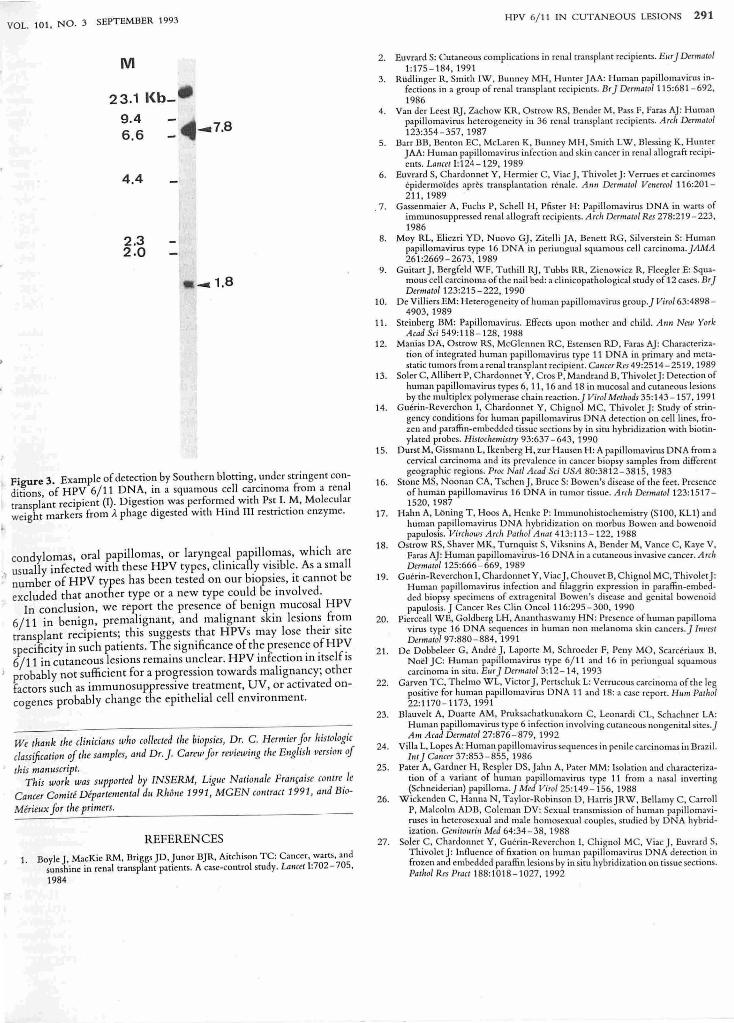
Three biopsy speCimens (one wart, two squamous cell carC1l10-mas) have been tested with PCR, ill situ hybridization, and Southern blotting (Table V). By Southern blotting, only one squamous cell
) carcinoma (from patient 1, Table III, corresponding to case 5, Table V) was found to contain HPV types 6/11 (Fig 3), as was also shown by PCR. The wart sample was HPV 6/11 positive and was negative with other HPV types tested by PCR; the two squamous cell carcinomas contained not only HPV 6/11 DNA but also HPV 5 and/or 16 DNA. By ill situ hybridization, HPV 6/11 was detected in only one squamous cell carcinoma and was associated with HPV 16/18; HPV types 1 and 2 were found in one sample.
Non - Transplant-Recipient Lesions A few similar lesions from controls were obtained and compared to those of transplant recipients for their content in HPV DNA type 6/11. Of the ten skin specimens tested by PCR (Table II), four (40%) contained HPV DNA; HPV 6/11 were identified only in two cases of Bowen's
, disease, and these lesions also contained HPV16 or 16 and 18 DNA. The eight other samples were negative with each probe tested.
Four samples (three warts and one squamous cell carcinoma) were tested by PCR, in situ hybridization, and Southern blotting for HPV 5, 6/11, 16, and 18 (Table V). They were also tested by in situ hybridization and Southern blotting with plasmid probes types 1 and 2. By PCR, all the specimens were negative. HPV type 1 and/or 2 DNA was found in warts by both in situ hybridization and Southern blotting.
In controls tested by PCR, i,·, situ hybridization, or Southern blotting, each geni~al condyloma and juvenile laryngeal papilloma was found to conta1l1 HPV 6/11 DNA; they were negative for the other HPV types tested (Table II).
DISCUSSION
In this investigation we have shown that HPV types 6/11 can be detected with PCR,.in sitlt hyb~idization, or Southern blotting in some cutaneous bel1lgn, premalIgnant, or malignant lesions from organ trans'pla~t recipients. The lesions appeared several years after graftmg. BIOpSies were taken at different body sites on sun-exposed areas. HPV type 6/11 DNAs were rarely detected in similar skin lesions from normal population.
The identification of these HPV types in cutaneous samples was unexpected ~ecause they ~e known to infect mucosa and are usually associated With bel1lgn leSIOns. On the contrary, potentially oncogenic HPV types,. mainly found in mucosa, have already been found 111 cutaneous leSions; HPV 16, usually associated with cervical cancers [15], w.as detected in cutaneous Bowen's disease and squamous cell carcmomas from Immunocompetent patients [8,9,16-20,21] and ~rom re~lal transplant recipients [13]. HPV 16 has also be~n f~und 111 a bel1lgn wart from an immunosuppressed patient [7]. It IS difficult to compare the frequency of HPV 16 in cutaneous sam~les. reported by various investigators, as techniques of different sensltlvlty have been used, in situ hybridization, Southern blotting, and PCR.
The presence of HPV 6/11 in skin lesions has rarely been reported. HPV 11 DNA was mentioned in one case of verrucous carcinoma of the leg from a non-immunocompromised patient [22],
290 SOLER ET AL
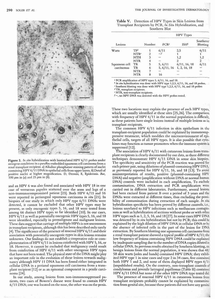
Figure 2. In situ hybridization with biotinylated HPV 6/11 probes under stringent conditions in a paraffin-embedded squamous cell carcinoma from a renal transplant recipient. a) Alkaline phosphatase staining pattern of nuclei containing HPV 6/11 DNA in epithelial cells from upper layers. b) Detail of positive nuclei at higher magnification. D, Dermis; E, Epidermis. Bar, 100/.lm in (a) and 25 /lm in (b).
and an HPV 6 was also found and associated with HPV 18 in one case of verrucous papules scattered over the arms and legs of a non-immunocompromised patient [23]. Both HPV 6/11 and 16 were reported in periungual squamous carcinoma in situ [21] . In biopsies of our study in which only HPV type 6/11 DNAs were detected, it cannot be excluded that other HPV types may be present, as only oncogenic types 5, 16, and 18 were tested from among 66 distinct HPV types so far identified (10] . In our cases, HPV 6/11 as well as potentiaIly oncogenic HPV types 5,16, and 18 were identified, especially in premalignant and malignant lesions. These data suggest that carriage of multiple HPVs is not uncommon in transplant recipients, although this has been described only rarely [4]. The significance of the presence of mucosa.! HPV 6/11 and their association with potentially oncogenic types in skin lesions remains unknown. Our findings do not establish a cooperative role or complementation ofHPV 6/11 in lesions coinfected with HPV 5,16, or 18. However, it cannot be excluded that malignancy could result from coinfection of HPV 6/11 with other oncogenic HPY types that have not been tested. The physical state of the DNA might play an important role in the evolution of these lesions towards malignancy although HPV 11 DNA has been found either integrated in cellular DNA in a perianal squamous cell carcinoma from a transplant recipient [12] or as an episomal component in a penile carcinoma [24J .
In our study, among lesions from non-immunosuppressed patients, two cases of Bowen's disease were found to contain HPV 6/11 DNA; one was located on the nose, the other was on the penis.
THE JOURNAL OF INVESTIGATIVE DERMATOLOGY
Table V. Detection of HPV Types in Skin Lesions from Transplant Recipients by PCR, 111 Sit", Hybridization, and
Southern Blot
HPV Types
Southem' Lesions Number peR" ISH; Blotting
Warts TFd 1 6/11 2,5 6/11 NTR' 2 - f 1 1 NTH 3 1,2 1,2 NTR 4 2 2
Squamous cell TR 5 5,6/11 6/11,16,18 6/11 carcmomas TR 6 5,6/11, 16 1,2,16,18
NTR 7 NTR 8 16
• peR amplification of HPV types 5, 6/ 11, 16, and 18. • I" lit" hybridization was done with HPV type 1,2,5, 6/11,16, and 18 probes. ' Southern blotting was done with HPV rype 1,2,5, 6/11,16, and 18 probes j TR, transplant recipients. , NTR, non-transplant recipients. J -, no Hl'V DNA was detected with the HPV probes tested.
These two locations may explain the presence of such HPV types, which are usually identified at these sites [25,26]. The comparison with frequency of HPV 6/11 in the normal population is difficult, as these patients have single lesions instead of multiple lesions as in transplant recipients.
The common HPV 6/11 infection in skin epithelium in the transplant-recipient population could be explained by immunosuppressive treatment, which modifies the microenvironment of epithelial cells, targets of all HPV types. It is also possible that cytokines may function as tumor promoters when the immune system is suppressed [12].
The association of HPV 6/11 with cutaneous lesions from transplant recipients is clearly documented by our data, as three different techniques demonstrate HPV 6/11 DNA in some skin biopsies. The specificity and sensitivity of the PCR reaction was proved for each primer pair, using dilutions of plasmid-containing HPV DNA, as previously reported for HPV 6/11,16, and 18 [13]. To avoid misinterpretation of results, positive (plasmid-containing HPV DNA) and negative (amplification without DNA or normal human DNA) controls were included in each amplification. To exclude contamination, DNA extraction and PCR amplification were carried out in different laboratories. Furthermore. several lesions had been excised from patient B over a period 0(3 years and the DNAs were extracted at different times, thus excluding the possibility of contamination during extraction of each sample. III situ hybridization specificity has been proved by different controls, i.e., lesions unrelated to HPV infections such as molluscum contagiosum as well as hybridization of sections without probe or with other HPV types such as 1,2,5,16, and 18 [27]. In some cases HPV DNA was detected by iI, situ hybridization but not by PCR; this could be explained by the high dilution ofHPV DNA in cellular DNA or by the absence of infected cells in the part of the lesion for DNA extraction. By Southern blotting one squamous cell carcinoma from a renal transplant patient showed a typical profile ofHPV 6/11; the low frequency oflesions containing HPV DNA could be explained by inadequate sampling due to the number of DNA copies diluted in cellular DNA. In previous results obtained by Southern blotting, on benign lesions from the normal population, it was shown that 48/ 58 skin warts (sales and hands) contained HPV DNA; they exhibited HPV type 1 in nine cases and type 2 in 34 cases, five containe~ both HPV 1 and 2, and none of them displayed HPV types 6/11 (unpublished results). On the contrary, non-transplant controls of condylomas and juvenile laryngeal papillomas (Table II) contained HPV 6/11 DNA but none of the other HPV DNA type tested did.
The presence of mucosal HPV 6/11 in cutaneous lesions from transplant recipients probably cannot be explained by contamina· tion from genital site, because these patients did not have any geni
VOL. 101, NO. 3 SEPTEMBER 1993
M
23.1 I<b-9,4 6,6
4.4
2,3 2.0
_7.8
_1 .8
Figure 3. Example of detection by Southern blotting, under stringent conditions of HPV 6/11 DNA, in a squamous cell carcinoma from a renal t anspl;nt recipient (I). Digestion was performed with Pst 1. M, Molecular ~eight markers from A phage digested with Hind III restriction enzyme.
condylomas, oral papillomas, or larynge~l . papillomas, which are usually infected with these HPV types, cltl11cally vIsible. As a small
;. number of HPV types has been tested on our biopsies, it cannot be excluded that another type or a new type could be involved.
In conclusion, we report the presence of benign mucosal HPV 6/11 in benign, premal.ignant, and malignant skin lesions. fr~m transplant recipients;. tim sugge~ts that HPVs may lose their site specificity in such patients. The slgl11ficance of t~e pres.enc~ o.fHPV 6/11 in cutaneous lesions remains unclear. HPV 111fection 111 Itself IS
I probably not sufficient for a progressIOn towards maltgnancy; other factors such as immunosuppressive treatment, UV, or activated oncogenes probably change the epithelial cell environment.
We tirallk the clitlicialls who collected the biopsies, Dr. C. Hermier for histologic classiJicatio" of tire samples, alld Dr. J. Carew for revicwillg tire ElIglisll /Jersioll of
this manuscript. This work was supported by INSERM, Ligue Natiolla le Frallfaise col/tre Ie
Cancer Camilli Depa rtemet/tal dll RI,cme 1991, MGEN COl/tract 1991, alld Bio
Merieux for the primers.
REFERENCES
1. Boyle J, MacKie RM, Briggs JD, Junor BJR, Aitchison TC: Cancer, warts, and sunshine in renal transplant pauents. A case-control study. LAllret 1:702- 705,
1984
HPV 6/11 IN CUTANEOUS LESIONS 291
2. Euvrard S: C utaneous complications in renal transplant recipients. Ellr] Dermatol 1:175-184, 1991
3. Rildlinger 11., Smith IW. Bunney MH , Hunte.r JAA: Human papillomavirus infections in a group of renal transplanr recipients. Br] Dermalo/115:681- 692. 1986
4. Van der Leest RJ. Zachow K11. , Ostrow RS, Bender M. Pass F. Faras AJ: Human papillomavirus he.terogeneity in 36 renal transplant recipients. Arch Dermalol 123:354 - 357, 1987
5. Barr BB. Benton EC. McLaren K, Bunney MH, Smith LW. Blessing K, Hunter JAA: Human papillomavirus infection and skin cancer in renal allograft recipients. Lallcel 1:124 - 129, 1989
6. Euvrard S, C hardonnet Y, Hermier C, Viac J, Thivolet J: Ve.rrues et carcinomes epidermoIdes aprcs transplanration renale. AmI Dermalol Venereal 116:201 -211,1989
7. Gassenmaier A, Fuchs P, Schell H, Pfister H: Papillomavirus DNA in warts of immunosuppressed renal allograft recipients. Arch Dermalol Res 278:219-223, 1986
8. May RL, Eliezri YO, Nuovo GJ, Zitelli JA, Benett RG, Si lverstein S: Human papillomavirus type 16 DNA in periungual squamous cell ca.rcinoma.]AMA 261:2669-2673,1989
9. GuitartJ , Bergfeld WF. Tuthill RJ , Tubbs 11.11., Zienowicz 11., Fleegler E: Squamous cell carcinoma of the nail bed: a c1il1icopathological srudy of 12 cases. Br] Derlllalo/123:215-222,1990
10. De Villiers EM: Heterogeneity of human papillomavirus group.] viroI63:4898-4903,1989
11. Steinberg BM: PapillomavinlS. Effects upon mother and ch ild. AIIII New York Acad Sci 549: 11 8 - 128, 1988
12. Mal1ias DA, Ostrow RS, McGlennen RC, Estensen RD. Faras AJ: Characterization of integrated human papillolllavirus type 11 DNA in primaty and metastatic rumors from a renal transplant recipient. Gimcer R es 49:2514 - 2519, 1989
13. Soler C , Allibert P, ChardolUtet Y, Cros P, Mandrand B, ThivoletJ: Detection of human papillomavirus types 6, 11, 16 and 18 in mucosal and cutaneous lesions by the multiplex polymerase chain reaction.] Virol Melhods 35:143-157.1991
14. Guerin-Reverchon I, C hard0llllet Y, C higno l MC, Thivolet J: Srudy of stringency conditions for human papillomavirus DNA detection on cell lines, frozen and paraffin-embedded tissue sections by in siru hybridization with biotinylated probes. Hisiochelllisiry 93:637 -643, 1990
15. Durst M, Gissmann L, Ikenberg H, zur Hausen H: A papillomavirus DNA from a cervical carci noma and its preva.lcncc in cancer biopsy samples fronl different geographic regions. Proc Nat! Acad Sci USA 80:3812-3815, 1983
16. Stone MS, Noonan CA, TschenJ, Bruce S: Bowen's disease of the feet . Presence of human papillomavirus 16 DNA in tumor tissue. Arch Der",aloI123:1517 -1520,1987
17. Hahn A, LOning T, Haas A, Henke P: Immunohistochemistry (SI00, KLl) and human papillomavirus DNA hybridization on morbus Bowen and bowenoid papulosis. VircholVs Arch PalllOl AlloI413:113 - 122, 1988
18. Ostrow RS, Shaver MK, Turuquist S, Viksnins A, Bender M, Vance C, Kaye V, Faras AJ: Human papillomavinls-16 DNA in a cutaneous invasive cancer. Arc/, Der",0101125:666-669,1989
19. Guerin-Reverchon I. Chardonnct Y, Viac J, C hou vet B, Chignol MC. Thivolet J: Human papillomavirus infection and filaggrin expression in paraffin-embedded biopsy specimens of extragenita l Bowen 's disease and genital bowenoid papulosis. J Cance r Res C lin Oncol 116:295 - 300, 1990
20. Pierceall WE, Goldberg LH, Ananthaswamy HN: Presence of In lin an papilloma virus type 16 DNA sequences in human non melanoma skin cancers.] Illvest Derlllalol 97 :880 - 884, 1991
21. De Dobbclcer G. Andre J, L1porte M, Schroeder F, Peny MO, Scarccriaux B, Noel JC: Human papillomavirus rype 6/11 and 16 in periungual squamous carcinoma in situ . Ellr] Dennato/3:12 - 14, 1993
22. Garven TC, Thelma WL, Victor J , Pcrtschuk L: Verrucous carcinoma of the leg positive for human papillomavirus DNA II and 18: a case report. HIIIII Palhol 22:1170-1173,1991
23. Blauvelt A, Duarte AM, Pruksachatkunakorn C . Leonardi CL, Schachner LA: Human papillomavirus type 6 infection involving cutaneous nongcnital sites.] Alii Acad Derlllolo/27:876-879, 1992
24. Villa L, Lopes A: Human papi ll omavirus sequences in penile carcinomas in Brazil. 1111] Callcer 37:853-855,1986
25. Pater A, Gardner H, Respler OS, Jahn A, Pater MM: Isolation and characterization of a variant of human papillomavirus type 11 from a nasal inverting (Schneiderian) papilloma.] Met! Viro/25 :149-156, 1988
26. Wickenden C, Hanna N, Taylor-Robinson D, HarrisJRW, Bellamy C, Carroll P, Malcolm ADB, Coleman DV: Sexual transmission of human papillomaviruses 1I1 heterosexual and male homosexua l couples, studied by DNA hybridization. Cellilollrill Med 64:34-38,1988
27. Soler C, Chardonnet Y, Gucri n-Reverchon I, Chignol MC, Viac J, Euvrard S, Thivolet J: Influence of fixation on human papi llomavirus DNA detection in frozen and embedded paraffin lesions by in situ hybridization on tissue sections. PatllOl Res Proct 188:1018 -1027,1992