Embed Size (px)
Citation preview
International Journal of Scientific & Engineering Research, Volume 8, Issue 1, January-2017 1226 ISSN 2229-5518
IJSER © 2017 http://www.ijser.org
Detection of Pneumocystis jirovecii Copy Number in BronchoAlveolar Lavage and Nasal swab Fluids of
Immunocompromised Patients Ghada B. Alomashi Ali T. Al-Damarchi
Marwa S. Al-Juboori College of medicine / university of Al-Qadisiyah
0TAbstract: The current study was tried to identification the of Pneumocystis jirovecii copy number in immunocompromised patients suffering from pulmonary disease, known as (PCP) Pneumocystis pneumonia by using real-time PCR (qPCR) and DNA sequencing for Phylogenetic tree analysis study of local Pneumocystis jirovecii confirmative detection based mitochondrial large subunit ribosomal RNA gene method by used PCR. The present study was conducted on 93 samples 53 samples were bronchoaleveolarlavage BAL were collected from patients whom suffer from (Pneumonia, chronic obstructive Pulmonary disease, Lung cancer, Tuberculosis, and HIV, Transplantation, Asthma, Hepatitis, Diabetes mellitus of both types with chest infection). And 40 sample nasal swabs were taken from infants suffer from pneumonia as diagnostic by pediatrician , BAL samples collected from Baghdad Medical City hospital and from Educational Al-Diwaniyah hospital while nasal swabs samples collected from Maternity and childhood teaching hospital in Al-Diwaniyah city at the period from the beginning of November 2015 to the end of May 2016.
0T The result of real-time PCR for detect target genes (qpcr mtLSU rRNA gene, PCR-ssrRNA gene) was showed positive samples were 72 and negative samples were 21, the rate of detection of pneumocystis jirovecii by qPCR method were 77.4%. DNA sequencing technique was used for genetic confirmation and phylogenetic tree analysis of Pneumocystis jirovecii as well as direct submission of some local Pneumocystis jirovecii isolates in NCBI-gene bank data base.
0TKey words : Pneumocystis jirovecii , BronchoAlveolar , Immunocompromised Patients
—————————— ——————————
1. Introduction
Pneumocystis jirovecii is one of the most recurrent and severe
opportunistic infections in immunocompromised patients it is the
causative agent and responsible for a severe pulmonary disease,
known as (PCP) Pneumocystis pneumonia (Calderón-Sandubete
IJSER
International Journal of Scientific & Engineering Research, Volume 8, Issue 1, January-2017 1227 ISSN 2229-5518
IJSER © 2017 http://www.ijser.org
et al ., 2002; Cushion ,2004). It's also regarded as one of the most
frequent and serious complications, affecting immunocompromised
patients such as those with HIV infection (Aliouat-Denis et al., 2009).
And it is an important cause of morbidity and mortality among them
(Elvin et al., 1994; Morris and Norris , 2012). In chronic
immunosuppressive medications and are thus at a risk for PCP is
rapidly growing such as malignancies, solid organ transplant recipients
and patients treated by immunosuppressive agents are susceptible to
this infection (Sulieman et al., 2014; Sassi et al.,2012). And some
other conditions such as diabetes and severe malnutrition increase the
risk of PCP (Sanno et al., 2007; Morrow et al., 2014 and Morris et
al., 2004). Cough is generally non-productive but may be productive
cough in up to a third of patients, exertional dyspnea, Fever,
tachypnea, chest pain, there may be signs of AIDS such as thrush,
oral hairy leukoplakia, kaposi's sarcoma , scattered crackles and
wheeze may be present, or (rarely) signs of focal consolidation,
extrapulmonary disease may manifest as hepatosplenomegaly,
IJSER
International Journal of Scientific & Engineering Research, Volume 8, Issue 1, January-2017 1228 ISSN 2229-5518
IJSER © 2017 http://www.ijser.org
lymphadenopathy or ocular disease (Cisse et al., 2012). In the
diagnosis of Pneumocystis jirovecii culturing is not dependable yet , so
confirmation of the diagnosis requires identification of organisms in
bronchoalveolarlavage and staining of the cyst wall or trophozoite
(Bowling et al., 1973). Monoclonal antibodies for detecting P. jiroveci
are available and have a sensitivity greater than 90% for detecting P.
jiroveci in induced sputum from HIV-infected patients, immunological
diagnostic method of Pneumocystis jiroveci pneumonia (PJP) can be
difficult in patients without HIV infection (Catherinot et al .,2010).
Real-time PCR SYBR green-based real-time PCR
technique(Espy,2006).Was conducted for rapid detection of
Pneumocystis jirovecii according to method described by (Huggett et
al.,2015). The current study was aimed to highlight the role of
Pneumocystis jirovecii in patients who suffering from pneumonia with
some risk factors, to achieve this goal, the following objectives were
conducted :
1- Bronchoalveolarlavage (BAL) fluid were collected from
patients who suffer from pneumonia with some risk factors
(immunocompromised patients) .
IJSER
International Journal of Scientific & Engineering Research, Volume 8, Issue 1, January-2017 1229 ISSN 2229-5518
IJSER © 2017 http://www.ijser.org
2- Nasal swabs were taken from infants suffer from pneumonia as
diagnostic by pediatrician.
3- Extraction of DNA and amplified with specific primers of target
genes (qpcr mtLSU rRNA gene, PCR-ssrRNA gene).
4- Determination of target genes copy number of Bronchoalveolar
lavage (BAL) and nasal swabs using quantitative -PCR method.
5- DNA sequencing technique was used for genetic confirmation and
phylogenetic tree analysis of Pneumocystis jirovecii as well as
direct submission of some local Pneumocystis jirovecii isolates in
NCBI-gene bank data base.
2. Sample Collection and Diagnosis Method
Patients: BAL were collected from 53 patients whom suffer from
(Pneumonia, chronic obstructive Pulmonary disease, Lung cancer,
Tuberculosis, and HIV, Transplantation, Asthma, Hepatitis, Diabetes
mellitus of both types with chest infection). And 40 nasal swabs were
taken from infants suffer from pneumonia as diagnostic by pediatrician.
BAL is a saline wash of the airways bronchi and air sacs alveolar was
down the fluid were aspirate and collected in sterile container were
taken from patients were stored at -4°c in refrigerator nasal swab
after diagnosis of pneumonia by pediatrician taken nasal swabs and
place in the culture medium provided . Molecular technique for
IJSER
International Journal of Scientific & Engineering Research, Volume 8, Issue 1, January-2017 1230 ISSN 2229-5518
IJSER © 2017 http://www.ijser.org
detection Pneumocystis jirovecii using qPCR, in both BAL and nasal
swab samples . DNA extraction for qPCR DNA extraction from BAL
and nasal swab samples was using a commercial Genomic DNA
extraction kit (Reagent Genomic DNA extraction kit, Geneaid. USA)
.The extracted genomic DNA was checked by using Nanodrop
spectrophotometer (THERMO. USA), that check and measurement the
purity of DNA through reading the absorbance in at (260 /280 nm)
.The Real-Time PCR primers based mitochondrial large subunit
ribosomal RNA gene were used for specific detection of Pneumocystis
jirovecii as well as PCR primers based small subunit ribosomal RNA
gene were used for phylogenetic tree analysis study, these primers
were designed in this study using NCBI-Gene bank data base and
primer3 plus and these primers provided by (Bioneer company,
Korea).as table 1.
IJSER
International Journal of Scientific & Engineering Research, Volume 8, Issue 1, January-2017 1231 ISSN 2229-5518
IJSER © 2017 http://www.ijser.org
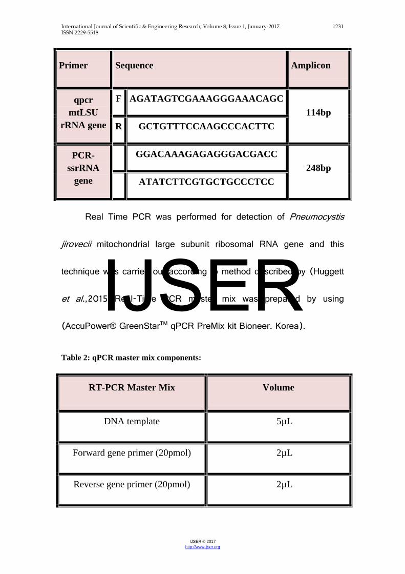
Primer Sequence Amplicon
qpcr mtLSU
rRNA gene
F AGATAGTCGAAAGGGAAACAGC 114bp
R GCTGTTTCCAAGCCCACTTC
PCR-ssrRNA
gene
GGACAAAGAGAGGGACGACC 248bp
ATATCTTCGTGCTGCCCTCC
Real Time PCR was performed for detection of Pneumocystis
jirovecii mitochondrial large subunit ribosomal RNA gene and this
technique was carried out according to method described by (Huggett
et al.,2015).Real-Time PCR master mix was prepared by using
(AccuPower® GreenStarTM qPCR PreMix kit Bioneer. Korea).
Table 2: qPCR master mix components:
RT-PCR Master Mix Volume
DNA template 5µL
Forward gene primer (20pmol) 2µL
Reverse gene primer (20pmol) 2µL
IJSER
International Journal of Scientific & Engineering Research, Volume 8, Issue 1, January-2017 1232 ISSN 2229-5518
IJSER © 2017 http://www.ijser.org
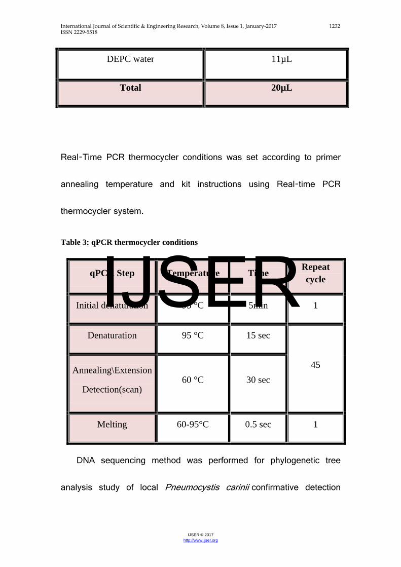
DEPC water 11µL
Total 20µL
Real-Time PCR thermocycler conditions was set according to primer
annealing temperature and kit instructions using Real-time PCR
thermocycler system.
Table 3: qPCR thermocycler conditions
Repeat cycle Time Temperature qPCR Step
1 5min 95 °C Initial denaturation
45
15 sec 95 °C Denaturation
30 sec 60 °C Annealing\Extension
Detection(scan)
1 0.5 sec 60-95°C Melting
DNA sequencing method was performed for phylogenetic tree
analysis study of local Pneumocystis carinii confirmative detection
IJSER
International Journal of Scientific & Engineering Research, Volume 8, Issue 1, January-2017 1233 ISSN 2229-5518
IJSER © 2017 http://www.ijser.org
based mitochondrial large subunit ribosomal RNA gene. The
sequencing of the PCR product of ribosomal RNA gene, where the
248bp PCR product was purified from agarose gel by using (EZ EZ-
10 Spin Column DNA Gel Extraction Kit, Biobasic. Canada).
3. Statistical Analysis
Data were analyzed using statical package for social science (spss) version
(22), T-test chi-square test, and Mann Whitney test were used For compression
between the cases groups. Numeric data were presented as mean and standard
deviation were as categorical data were presented as number and percentage
the level of significance was considered at p-value as ≤ 0.05.
4.Results
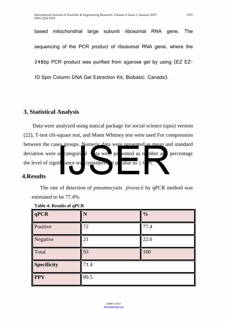
The rate of detection of pneumocystis jirovecii by qPCR method was
estimated to be 77.4%. Table 4: Results of qPCR
qPCR N %
Positive 72 77.4
Negative 21 22.6
Total 93 100
Specificity 71.4
PPV 89.5
IJSER
International Journal of Scientific & Engineering Research, Volume 8, Issue 1, January-2017 1234 ISSN 2229-5518
IJSER © 2017 http://www.ijser.org
NPV 66.7
* PPV: positive predictive values
* NPV: negative predictive values
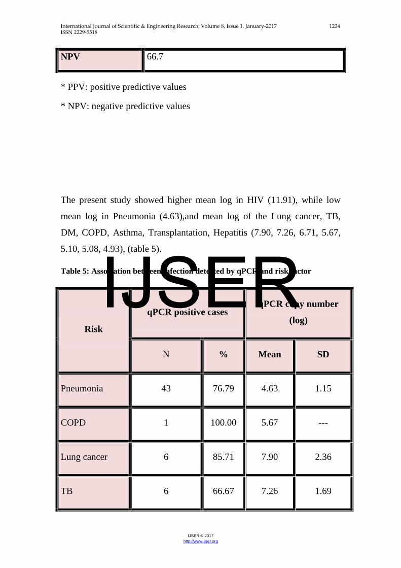
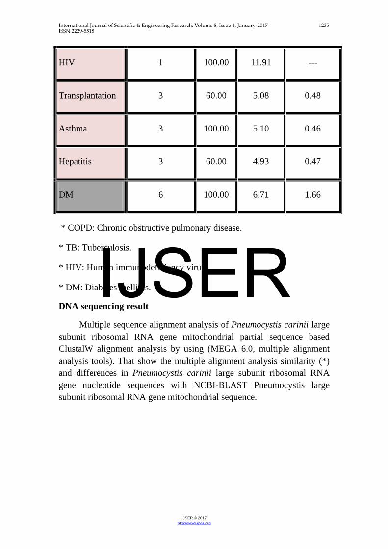
The present study showed higher mean log in HIV (11.91), while low
mean log in Pneumonia (4.63),and mean log of the Lung cancer, TB,
DM, COPD, Asthma, Transplantation, Hepatitis (7.90, 7.26, 6.71, 5.67,
5.10, 5.08, 4.93), (table 5).
Table 5: Association between infection detected by qPCR and risk factor
Risk
qPCR positive cases qPCR copy number
(log)
N % Mean SD
Pneumonia 43 76.79 4.63 1.15
COPD 1 100.00 5.67 ---
Lung cancer 6 85.71 7.90 2.36
TB 6 66.67 7.26 1.69
IJSER
International Journal of Scientific & Engineering Research, Volume 8, Issue 1, January-2017 1235 ISSN 2229-5518
IJSER © 2017 http://www.ijser.org
HIV 1 100.00 11.91 ---
Transplantation 3 60.00 5.08 0.48
Asthma 3 100.00 5.10 0.46
Hepatitis 3 60.00 4.93 0.47
DM 6 100.00 6.71 1.66
* COPD: Chronic obstructive pulmonary disease.
* TB: Tuberculosis.
* HIV: Human immunodeficiency virus.
* DM: Diabetes mellitus.
DNA sequencing result
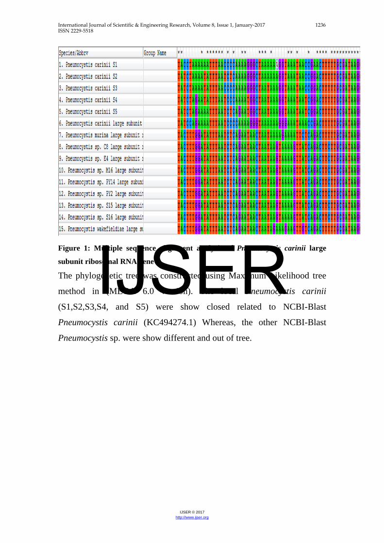
Multiple sequence alignment analysis of Pneumocystis carinii large subunit ribosomal RNA gene mitochondrial partial sequence based ClustalW alignment analysis by using (MEGA 6.0, multiple alignment analysis tools). That show the multiple alignment analysis similarity (*) and differences in Pneumocystis carinii large subunit ribosomal RNA gene nucleotide sequences with NCBI-BLAST Pneumocystis large subunit ribosomal RNA gene mitochondrial sequence.
IJSER
International Journal of Scientific & Engineering Research, Volume 8, Issue 1, January-2017 1236 ISSN 2229-5518
IJSER © 2017 http://www.ijser.org
Figure 1: Multiple sequence alignment analysis of Pneumocystis carinii large
subunit ribosomal RNA gene .
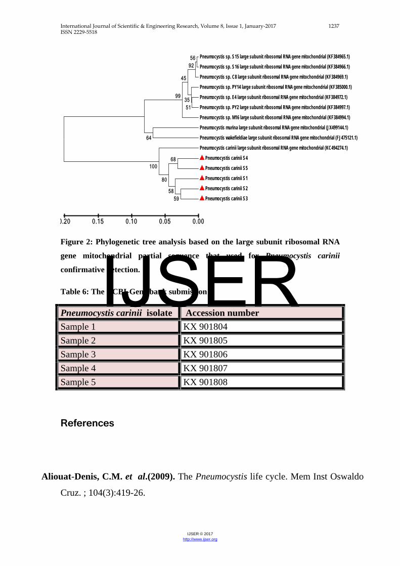
The phylogenetic tree was constructed using Maximum Likelihood tree
method in (MEGA 6.0 version). The local Pneumocystis carinii
(S1,S2,S3,S4, and S5) were show closed related to NCBI-Blast
Pneumocystis carinii (KC494274.1) Whereas, the other NCBI-Blast
Pneumocystis sp. were show different and out of tree.
IJSER
International Journal of Scientific & Engineering Research, Volume 8, Issue 1, January-2017 1237 ISSN 2229-5518
IJSER © 2017 http://www.ijser.org
Figure 2: Phylogenetic tree analysis based on the large subunit ribosomal RNA
gene mitochondrial partial sequence that used for Pneumocystis carinii
confirmative detection.
Table 6: The NCBI-Gene bank submission
Pneumocystis carinii isolate Accession number Sample 1 KX 901804 Sample 2 KX 901805 Sample 3 KX 901806 Sample 4 KX 901807 Sample 5 KX 901808
References
Aliouat-Denis, C.M. et al.(2009). The Pneumocystis life cycle. Mem Inst Oswaldo
Cruz. ; 104(3):419-26.
Pneumocystis sp. S15 large subunit ribosomal RNA gene mitochondrial (KF384965.1)
Pneumocystis sp. S16 large subunit ribosomal RNA gene mitochondrial (KF384966.1)
Pneumocystis sp. C8 large subunit ribosomal RNA gene mitochondrial (KF384969.1)
Pneumocystis sp. PY14 large subunit ribosomal RNA gene mitochondrial (KF385000.1)
Pneumocystis sp. E4 large subunit ribosomal RNA gene mitochondrial (KF384972.1)
Pneumocystis sp. PY2 large subunit ribosomal RNA gene mitochondrial (KF384997.1)
Pneumocystis sp. M16 large subunit ribosomal RNA gene mitochondrial (KF384994.1)
Pneumocystis murina large subunit ribosomal RNA gene mitochondrial (JX499144.1)
Pneumocystis wakefieldiae large subunit ribosomal RNA gene mitochondrial (FJ475121.1)
Pneumocystis carinii large subunit ribosomal RNA gene mitochondrial (KC494274.1)
Pneumocystis carinii S4
Pneumocystis carinii S5
Pneumocystis carinii S1
Pneumocystis carinii S2
Pneumocystis carinii S3
5692
5135
45
99
59
68
58
80
100
64
0.000.050.100.150.20
IJSER
International Journal of Scientific & Engineering Research, Volume 8, Issue 1, January-2017 1238 ISSN 2229-5518
IJSER © 2017 http://www.ijser.org
Bowling, M. C.; Smith, I. M. and Wescott ,S. L. (1973). A rapid staining
procedure for Pneumocystis (arinii. Am. J. Med. Technol. 39:267-268.
Calderón-Sandubete, E.J. et al.(2002). Historical perspective on Pneumocystis
carinii infection. Protist. ;153:303–10.
Catherinot, E.; Lanternier, F. and Bougnoux, M.E.( 2010). Pneumocystis
jirovecii pneumonia. Infect Dis Clin North Am24:107–138.
Cisse, O.H. ; Pagni, M. and Hauser, P.M.( 2012).De novo assembly of the
Pneumocystis jirovecii genome from a single bronchoalveolar lavage fluid
specimen from a patient. MBio. Dec 26;4(1):e00428-12.
Cushion, M.T.( 2004). Pneumocystis: unraveling the cloak of obscurity. Trends
Microbiol 12: 243-229.
Elvin , K. ; Lidman , C. and Tynell, E. et al. (1994). Natural history of
asymptomatic and symptomatic apatients. Scand J Infect Dis., 26(6): 643-51.
Espy, M. J. (2006). "Real-Time PCR in Clinical Microbiology: Applications for Routine Laboratory Testing". Clinical Microbiology Reviews. 19 (3).Retrieved 11 July 2016.
Huggett, J. F.; O'Grady, J. and Bustin, S. A. (2015) . qPCR, dPCR, NGS–
ajourney. Biomol.Detect.Quantif. 3,A1–A5. 10.1016/j.bdq.2015.01.001.
Morris ,A. and Norris, K.A.( 2012).Colonization by Pneumocystis jirovecii and its role in disease. Clin Microbiol .Rev. ; 25(2):297-317.
Morris, A.; Lundgren, J.D. and Masur, H. et al. (2004).Current epidemiology of
Pneumocystis pneumonia. Emerg Infect Dis. Oct;10(10):1713-20.
Morrow, B.M.; Samuel, C.M. and Zampoli, M. et al. (2014). Pneumocystis
pneumonia in South African children diagnosed by molecular methods. BMC
Res Notes. Jan 10;7(1):26.
IJSER
International Journal of Scientific & Engineering Research, Volume 8, Issue 1, January-2017 1239 ISSN 2229-5518
IJSER © 2017 http://www.ijser.org
Sanno, K.,et al . (2007). Pneumocystis pneumonia in a patient with type 2 diabetes
mellitus. Intern Med. 46(14):1131-3.
Sassi , M .,et al. (2012). Outbreaks of pneumocystis pneumonia in 2 renal transplant
centers linked to a single strain of pneumocystis: implications for transmission
and virulence. Clin Infect Dis. 54(10):1437-44.
Sulieman , S.E.; Metjian, T.A.; Zaoutis, T.E.; and Fisher, B.T.(2014).
Pneumocystis pneumonia: epidemiology and options for prophylaxis in Non-
HIV immunocompromised pediatric patients. Curr Fungal Infect Rep. 8(1):45-
55.
IJSER


![pneumocystis jirovecii (carinii) * IOOml 5ml 2ml * 0.5m] 0 ... · pneumocystis jirovecii (carinii) * IOOml 5ml 2ml * 0.5m] 0.29/0ifidEâ 0.50/03< +51J—Y * 0.50/034 2 3 4 5 6 7 8](https://img.pdfslide.net/doc/110x75/5f8507d94c3d7978ec0519a4/pneumocystis-jirovecii-carinii-iooml-5ml-2ml-05m-0-pneumocystis-jirovecii.jpg)