Embed Size (px)
Citation preview

JOURNAL OF VIROLOGY, Dec. 2009, p. 12702–12713 Vol. 83, No. 240022-538X/09/$12.00 doi:10.1128/JVI.01184-09Copyright © 2009, American Society for Microbiology. All Rights Reserved.
Determinants of the Hepatitis C Virus Nonstructural Protein 2Protease Domain Required for Production of Infectious Virus�
Thomas G. Dentzer,1,2 Ivo C. Lorenz,1† Matthew J. Evans,1‡ and Charles M. Rice1*Center for the Study of Hepatitis C, Laboratory of Virology and Infectious Disease, The Rockefeller University, 1230 York Avenue,
New York, New York 10065,1 and Laboratoire de Retrovirologie, Centre de Recherche Public-Sante, 84 rue Val Fleuri,Luxembourg L-1526, Luxembourg2
Received 9 June 2009/Accepted 28 September 2009
The hepatitis C virus (HCV) nonstructural protein 2 (NS2) is a dimeric multifunctional hydrophobic proteinwith an essential but poorly understood role in infectious virus production. We investigated the determinantsof NS2 function in the HCV life cycle. On the basis of the crystal structure of the postcleavage form of the NS2protease domain, we mutated conserved features and analyzed the effects of these changes on polyproteinprocessing, replication, and infectious virus production. We found that mutations around the protease activesite inhibit viral RNA replication, likely by preventing NS2-3 cleavage. In contrast, alterations at the dimerinterface or in the C-terminal region did not affect replication, NS2 stability, or NS2 protease activity butdecreased infectious virus production. A comprehensive deletion and mutagenesis analysis of the C-terminalend of NS2 revealed the importance of its C-terminal leucine residue in infectious particle production. Thecrystal structure of the NS2 protease domain shows that this C-terminal leucine is locked in the active site, andmutation or deletion of this residue could therefore alter the conformation of NS2 and disrupt potentialprotein-protein interactions important for infectious particle production. These studies begin to dissect theresidues of NS2 involved in its multiple essential roles in the HCV life cycle and suggest NS2 as a viable targetfor HCV-specific inhibitors.
An estimated 130 million people are infected with hepatitisC virus (HCV), the etiologic agent of non-A, non-B viral hep-atitis. Transmission of the virus occurs primarily through bloodor blood products. Acute infections are frequently asymptom-atic, and 70 to 80% of the infected individuals are unable toeliminate the virus. Of the patients with HCV-induced chronichepatitis, 15 to 30% progress to cirrhosis within years to de-cades after infection, and 3 to 4% of patients develop hepato-cellular carcinoma (17). HCV infection is a leading cause ofcirrhosis, end-stage liver disease, and liver transplantation inEurope and the United States (7), and reinfection after livertransplantation occurs almost universally. There is no vaccineavailable, and current HCV therapy of pegylated alpha inter-feron in combination with ribavirin leads to a sustained re-sponse in only about 50% of genotype 1-infected patients.
The positive-stranded RNA genome of HCV is about 9.6 kbin length and encodes a single open reading frame flanked by5� and 3� nontranslated regions (5� and 3� NTRs). The trans-lation product of the viral genome is a large polyprotein con-taining the structural proteins (core, envelope proteins E1 andE2) in the N-terminal region and the nonstructural proteins(p7, nonstructural protein 2 [NS2], NS3, NS4A, NS4B, NS5A,and NS5B) in the C-terminal region. The individual proteins
are processed from the polyprotein by various proteases. Thehost cellular signal peptidase cleaves between core/E1, E1/E2,E2/p7, and p7/NS2, and signal peptide peptidase releases corefrom the E1 signal peptide. Two viral proteases, the NS2-3protease and the NS3-4A protease, cleave the remainder of theviral polyprotein in the nonstructural region (22, 27). Thestructural proteins package the genome into infectious parti-cles and mediate virus entry into a naïve host cell; the non-structural proteins NS3 through NS5B form the RNA replica-tion complex. p7 and NS2 are not thought to be incorporatedinto the virion but are essential for the assembly of infectiousparticles (14, 36); however, their mechanisms of action are notunderstood.
NS2 (molecular mass of 23 kDa) is a hydrophobic proteincontaining several transmembrane segments in the N-terminalregion (5, 9, 32, 39). The C-terminal half of NS2 and theN-terminal third of NS3 form the NS2-3 protease (10, 11, 26,37). NS2 is not required for the replication of subgenomicreplicons, which span NS3 to NS5B (20). However, cleavage atthe NS2/3 junction is necessary for replication in chimpanzees(16), the full-length replicon (38), and in the infectious tissueculture system (HCVcc) (14). Although cleavage can occur invitro in the absence of microsomal membranes, synthesis of thepolyprotein precursor in the presence of membranes greatlyincreases processing at the NS2/3 site (32). In vitro studiesindicate that purified NS2-3 protease is active in the absence ofcellular cofactors (11, 37). In addition to its role as a protease,NS2 has been shown to be required for assembly of infectiousintracellular virus (14). The N-terminal helix of NS2 was firstimplicated in infectivity by the observation that an intergeno-typic breakpoint following this transmembrane segment re-sulted in higher titers of infectious virus (28). Structural and
* Corresponding author. Mailing address: Laboratory of Virologyand Infectious Disease, Center for the Study of Hepatitis C, TheRockefeller University, 1230 York Ave., New York, NY 10065. Phone:(212) 327-7046. Fax: (212) 327-7048. E-mail: [email protected].
† Present address: International AIDS Vaccine Initiative, AIDSVaccine Design & Development Laboratory, Brooklyn, NY 11220.
‡ Present address: Department of Microbiology, Mount Sinai School ofMedicine, New York, NY 10029.
� Published ahead of print on 7 October 2009.
12702
by guest on Novem
ber 24, 2009 jvi.asm
.orgD
ownloaded from
functional characterization of the NS2 transmembrane regionhas shown that this domain is essential for infectious virusproduction (13). In particular, a central glycine residue in thefirst NS2 helix plays a critical role in HCV infectious virusassembly (13). The NS2 protease domain, but not its catalyticactivity, is also essential for infectious virus assembly, whereasthe unprocessed NS2-3 precursor is not required (13, 14).
The crystal structure of the postcleavage NS2 protease do-main (NS2pro, residues 94 to 217), revealed a dimeric cysteineprotease containing two composite active sites (Fig. 2C; [21]).Two antiparallel �-helices make up the N-terminal subdomain,followed by an extended crossover region, which positions the�-sheet-rich C-terminal subdomain near the N-terminal regionof the partner monomer. Two of the conserved residues of thecatalytic triad (His 143, Glu 163) are located in the loop regionafter the second N-terminal helix of one monomer, while thethird catalytic residue, Cys 184, is located in the C-terminalsubdomain of the other monomer. Creation of this unusualpair of composite active sites through NS2 dimerization hasbeen shown to be essential for autoproteolytic cleavage (21).The structure of NS2pro further demonstrated that the C-terminal residue of NS2 remains bound in the active siteafter cleavage, suggesting a possible mechanism for restric-tion of this enzyme to a single proteolytic event (21). Herewe have used the crystal structure of NS2pro, along withsequence alignments, to target conserved residues in each ofthe NS2pro structural regions. Our mutational analysis re-vealed that the residues in the dimer crossover region andthe C-terminal subdomain are important for infectious virusproduction. In contrast, the majority of amino acids in theactive site pocket were not required for infectivity. Interest-ingly, we observed that the extreme C-terminal leucine ofNS2 is absolutely essential for generation of infectious virus,as mutations, deletions, and extensions into NS3 are verypoorly tolerated. This analysis begins to dissect the deter-minants of the multiple functions of this important proteasein the HCV life cycle.
MATERIALS AND METHODS
Plasmid constructs. Mono- and bicistronic (see Fig. 1A) genomes were gen-erated by standard molecular biology techniques and verified by restrictionenzyme digestion and sequencing of PCR-amplified segments. Descriptions ofthe cloning strategies are provided below, and plasmid and primer sequences areavailable upon request.
(i) J6/H77NS2/JFH and mutant derivatives. J6/H77NS2/JFH constructs con-tain genotype 2a (J6) from core to p7, genotype 1a (strain H77) NS2 andgenotype 2a (JFH) NS3 to NS5B. To create this construct, the J6/JFH plasmid(18) was used as a template to PCR amplify the J6/JFH E2 3� end through thep7 sequence with forward oligonucleotide RU-O-5739 (5�-CCGCCTTGTCGACTGGTC) and reverse oligonucleotide RU-O-5855 (5�-CTCCGTGTC-CAACGCGTAAGCCTGTTGGGGC). The H77 NS2 through half of JFH-1NS3 region was PCR amplified from H77/JFH (18) with the forward oligonu-cleotide RU-O-5854 (5�-CAGGCTTACGCGTTGGACACGGAGGTGGCC)and reverse oligonucleotide RU-O-5721 (5�-GCTACCGAGGGGTTAAGCACT). Since these PCR products overlap at the p7/NS2 junction, they were usedas template in a second round of PCR with the outside oligonucleotides RU-O-5855 and RU-O-5721 to generate a PCR product encoding the J6 E2 C-terminalregion though p7, the H77 NS2, and JFH-1 NS3 N-terminal region. This productwas digested with restriction endonucleases BsaBI and AvrII and ligated into theBsaBI/AvrII-digested J6/JFH plasmid to create the final J6/H77NS2/JFH plas-mid. An adaptive change at G1145A (chimeric genome numbering) encodingsubstitution A269T (chimeric polyprotein numbering) was found to increaseinfectious virus titers of J6/H77NS2/JFH, and was included in all J6/H77NS2/
JFH-based genomes constructed. Mutant derivatives of J6/H77NS2/JFH werecreated by site-directed mutagenesis using the AfeI/BbvCI restriction sites.
(ii)J6/H77NS2/JFH(NS2-IRES-NS3)andmutantderivatives.J6/H77NS2/JFH-(NS2-IRES-NS3) encodes a stop codon after NS2, an encephalomyocarditis virus(EMCV) internal ribosome entry site (IRES), a start codon, and the remainderof the JFH-1 polyprotein starting with NS3. Mutant derivatives of J6/H77NS2/JFH(NS2-IRES-NS3) were created by site-directed mutagenesis using the PmeI/MluI restriction sites.
(iii) J6/H77NS2/JFH(NS2-IRES-nsGluc2AUbi) and mutant derivatives. J6/H77NS2/JFH(NS2-IRES-nsGluc2AUbi) is similar to J6/H77NS2/JFH(NS2-IRES-NS3) but encodes a Gaussia luciferase gene followed by the foot andmouth disease virus 2A peptide and a ubiquitin monomer (nsGluc2AUbi cas-sette) between the EMCV IRES and NS3 (14). The N-terminal signal sequenceof Gaussia luciferase has been deleted so that the reporter is not secreted.Mutant derivatives of J6/H77NS2/JFH(NS2-IRES-nsGluc2AUbi) were createdby site-directed mutagenesis using the PmeI/MluI restriction sites. NS2 exten-sions into NS3 were subcloned using BbvCI/SpeI/KpnI restriction sites.
Cell culture. Huh-7.5 cells were cultured in Dulbecco’s modified Eagle me-dium (Invitrogen) supplemented with 0.1 mM nonessential amino acids and 10%fetal bovine serum (complete medium). Cells were grown at 37°C in 5% CO2.
RNA transcription. In vitro transcripts were generated as previously described(18). Briefly, plasmid DNA was linearized by XbaI and purified by using aMinelute column (Qiagen, Valencia, CA). RNA was transcribed from 1 �g ofpurified template by using the T7 Megascript kit (Ambion, Austin, TX) or the T7RNA polymerase kit (Promega, Madison, WI). Reaction mixtures were incu-bated at 37°C for 3 h, followed by a 15-min digestion with 3 U of DNase I(Ambion). RNA was purified by using an RNeasy kit (Qiagen) with an additionalon-column DNase treatment. RNA was quantified by absorbance at 260 nm anddiluted to 0.5 �g/�l. Prior to storage at �80°C, RNA integrity was determined byagarose gel electrophoresis and visualization by ethidium bromide staining.
RNA electroporation. Huh-7.5 cells were electroporated with RNA as previ-ously described (18). Briefly, Huh-7.5 cells were treated with trypsin, washedtwice with ice-cold RNase-free AccuGene phosphate-buffered saline (PBS) (Bio-Whittaker, Rockland, ME), and resuspended at 1.75 � 107 cells/ml in PBS. Then,2 �g of each RNA was combined with 0.4 ml of cell suspension and immediatelypulsed using a BTX ElectroSquare Porator ECM 830 (820 V, 99 �s, five pulses).Electroporated cells were incubated at room temperature for 10 min prior toresuspension in 15 ml or 30 ml complete medium for nonreporter and reporterconstructs, respectively. Resuspended cells were plated into 24-well, 6-well, andP100 tissue culture dishes.
Assays for RNA replication. At 4 or 8, 24, 48, and 72 h postelectroporation,cells in 24-well plates were washed with Dulbecco PBS and lysed by the additionof Renilla lysis buffer (Promega, Madison, WI) or RLT buffer (Qiagen) contain-ing 1% �-mercaptoethanol for assay of replication by luciferase activity or quan-titative reverse transcription-PCR (qRT-PCR), respectively. For luciferase as-says, the lysates were thawed prior to the addition of Renilla substrate (Promega)according to the manufacturer’s instructions. The luciferase activity was mea-sured by using a Berthold Centro LB 960 96-well luminometer. For qRT-PCRanalysis, prior to storage at �80°C, the lysates were homogenized by centrifu-gation through a QiaShredder column (Qiagen) for 2 min at 14,000 � g. TotalRNA was isolated by RNeasy kit (Qiagen) and quantified by determining theabsorbance at 260 nm. A total of 50 ng of total cellular RNA was used perreaction mixture. qRT-PCRs were performed on a LightCycler 480 (Roche,Basel, Switzerland) using the LightCycler amplification kit (Roche) withprimers directed against the viral 3� NTR. We assembled 20-�l reactionmixtures according to the manufacturer’s instructions as previously described(14).
Assays for infectious virus production. At 4 or 8, 24, 48, and 72 h postelec-troporation, the cell culture medium was harvested and replaced with freshcomplete medium. Harvested cell culture supernatants were clarified by using a0.45-�m-pore-size filter and stored in aliquots at �80°C. For detection of infec-tious virus production by qRT-PCR or luciferase assay, naïve cells were infectedwith clarified cell culture supernatants and incubated for 72 h prior to analysis.Determination of infectious virus production by limiting dilution assay was per-formed as described previously (14, 18). Briefly, clarified cell culture superna-tants were serially diluted and used to infect approximately 3 � 103 cells platedin 96-well dishes. At 3 days postinfection, cells were washed with Dulbecco PBS,fixed with ice-cold methanol, and stained for the presence of NS5A expression asdescribed previously. The 50% tissue culture infectious dose (TCID50) wascalculated using the Reed and Muench method (18).
Anti-NS2 MAb (6H6). The immunogen was a recombinant NS2 proteasedomain of strain H77 (residues 94 to 217 with an amino-terminal hexahistidinetag) expressed in Escherichia coli and purified by fast protein liquid chromatog-
VOL. 83, 2009 MUTATIONAL ANALYSIS OF THE NS2 PROTEASE 12703
by guest on Novem
ber 24, 2009 jvi.asm
.orgD
ownloaded from
raphy under denaturing conditions (6 M guanidinium hydrochloride) or in thepresence of detergent (21). BALB/c mice were immunized three times intraperi-toneally with 50 �g NS2 purified under denaturing conditions, followed by a finalboost with 50 �g NS2 purified in the presence of detergent. The preimmune andtest bleeds were assayed for the presence of NS2-specific polyclonal antibodies bya standard enzyme-linked immunosorbent assay, as well as by Western blotting.Screening of hybridoma supernatants produced 6H6, an isotype immunoglobulinG1 monoclonal antibody (MAb).
SDS-PAGE and immunoblotting. Cells were lysed at 72 h postelectroporationwith RLT buffer (Qiagen) containing 1% �-mercaptoethanol and homogenizedby centrifugation through a QiaShredder column (Qiagen) for 2 min at 14,000 �g. The lysates were separated by sodium dodecyl sulfate-polyacrylamide gelelectrophoresis (SDS-PAGE). After transfer to nitrocellulose membrane, theblots were blocked for 1 h with 5% milk–PBS-T (PBS plus 0.1% Tween). ForNS2 detection, MAb 6H6 (1.0 mg/ml), was diluted 1:1,000 in PBS-T. For NS5Adetection, MAb 9E10 (18) was diluted 1:1,000 in PBS-T. For �-actin detection,mouse anti-�-actin MAb (Sigma, St. Louis, MO) was diluted 1:20,000 in PBS-T.After 1-h incubation at room temperature and extensive washing with PBS-T,horseradish peroxidase-conjugated rabbit anti-mouse immunoglobulin second-ary antibody (Pierce, Rockford, IL) was added at 1:10,000 dilution in 5% milk–PBS-T for 45 min at room temperature. After additional washing, blots weredeveloped with SuperSignal West Pico chemiluminescent substrate (Pierce).
RESULTS
Creation and characterization of triple chimeric HCV ge-nomes. The crystal structure of NS2pro was solved using theH77 genotype 1a protein (21). In order to facilitate a structure-based mutational analysis of NS2, we created J6/JFH genomesencoding the H77 NS2 sequence. Three different full-lengthHCV genome constructs were used (Fig. 1A). J6/H77NS2/JFHis a monocistronic genome encoding J6 core through p7, H77NS2, and JFH-1 NS3 through NS5B; the 5� and 3� NTRs of allthe genomes are derived from JFH-1. J6/H77NS2/JFH repli-cated with kinetics similar to those of J6/JFH but releasedapproximately 50-fold-less infectious virus (data not shown).By passaging transfected cells, we selected a single nucleotidechange, G1145A (chimeric genome nucleotide numbering) en-coding an A269T mutation in the J6 E1 protein. This substi-tution enhanced infectious particle production (data notshown) and was included in all genomes constructed. To studythe functions of NS2 in infectious virus production indepen-dent of its autoproteolytic cleavage requirements, we createdbicistronic genomes. J6/H77NS2/JFH(NS2-IRES-NS3) is iden-tical to J6/H77NS2/JFH but with the addition of a stop codonafter H77 NS2 and an encephalomyocarditis virus internalribosome entry site upstream of NS3. This genome allowsexpression of the viral replicase independently of NS2-3 cleav-age, and thus uncouples processing from replication and vi-rus production. J6/H77NS2/JFH(NS2-IRES-nsGluc2AUbi) isidentical to J6/H77NS2/JFH(NS2-IRES-NS3), but it encodesthe reporter gene Gaussia luciferase immediately downstreamof the EMCV IRES. Cleavage of the reporter protein from theviral polyprotein is mediated by the foot-and-mouth diseasevirus 2A peptide and cleavage after the C terminus of theubiquitin monomer by host ubiquitin carboxy-terminal hydro-lase (31). This generates nonsecreted Gaussia luciferase (ns-Gluc) and the proper N terminus of NS3. The triple chimericgenomes were tested for replication and infectious virus pro-duction at various times postelectroporation of Huh-7.5 cellswith in vitro-transcribed RNA; 72-h time points are shown(Fig. 1). All chimeric genomes were viable, although RNAreplication levels and infectious particle production weresomewhat reduced compared to the parental J6/JFH. As ex-
FIG. 1. HCV genomes used in this study. (A) Schematic represen-tation of HCV genomes. (I) J6/JFH with J6 C-NS2 shown in gray andJFH NS3-NS5B in white. (II) J6/H77NS2/JFH with J6 (gray), H77 NS2(dark gray), and JFH (white) with adaptive mutation in E1 (black dot).(III) Bicistronic construct similar to that shown for construct II butwith an EMCV IRES between NS2 and NS3. (IV) Bicistronic reporterconstruct similar to construct III with EMCV IRES, plus nonsecretedGaussia luciferase, foot and mouth disease virus 2A and ubiquitincleavage sites (nsGluc2AUbi) between NS2 and NS3 (checkered box).5� UTR and 3�UTR, 5� untranslated region and 3� untranslated region,respectively. (B) RNA replication of J6/JFH and J6/H77/JFH genomesmeasured by quantitative RT-PCR at 72 h postelectroporation. HCVRNA copies normalized to 50 ng of total RNA. (C) Infectious virusproduction of bicistronic constructs at 72 h postelectroporation, asmeasured by limiting dilution assay (TCID50). WT, wild type of eachgenome indicated; GNN, corresponding polymerase-defective control.The means plus standard errors of the means (error bars) of threeindependent experiments with two different RNA preparations areshown.
12704 DENTZER ET AL. J. VIROL.
by guest on Novem
ber 24, 2009 jvi.asm
.orgD
ownloaded from
pected, genomes containing a mutation of the NS5B RNA-dependent RNA polymerase motif GDD to GNN did notreplicate (Fig. 1B and C).
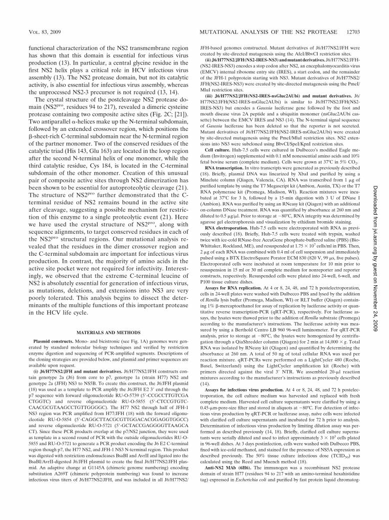
The monoclonal antibody 6H6 recognizes a C-terminalepitope of NS2. In Western blot, MAb 6H6 strongly recognizedNS2 from strains H77 (genotype 1a) and JFH-1 (Jc1 genome,genotype 2a) (16, 28) and showed a weak signal for strain J6(J6/JFH genome, genotype 2a) (18); it did not react with Con1(genotype 1b) (2) (Fig. 2A). Strain H77 NS2 is also recognizedin an enzyme-linked immunosorbent assay, immunoprecipita-tion, and immunofluorescence (data not shown). Epitope map-ping with an NS2 peptide library revealed that 6H6 bindingoccurs close to the NS2 C terminus (H77 NS2 amino acids 197to 208, GQEILLGPADG). Sequence alignments in this regionshowed variability between NS2 genotypes that correlated withthe Western blot results (Fig. 2B). The epitope of MAb 6H6 isshown on the NS2 crystal structure in Fig. 2C.
NS2 catalytic-cleft residues are required for NS2-3 cleavage.Previous studies have shown that the catalytic activity of theNS2-3 protease is not required for infectious virus production(14). To investigate whether residues surrounding the activesite pocket were required for the generation of infectious prog-
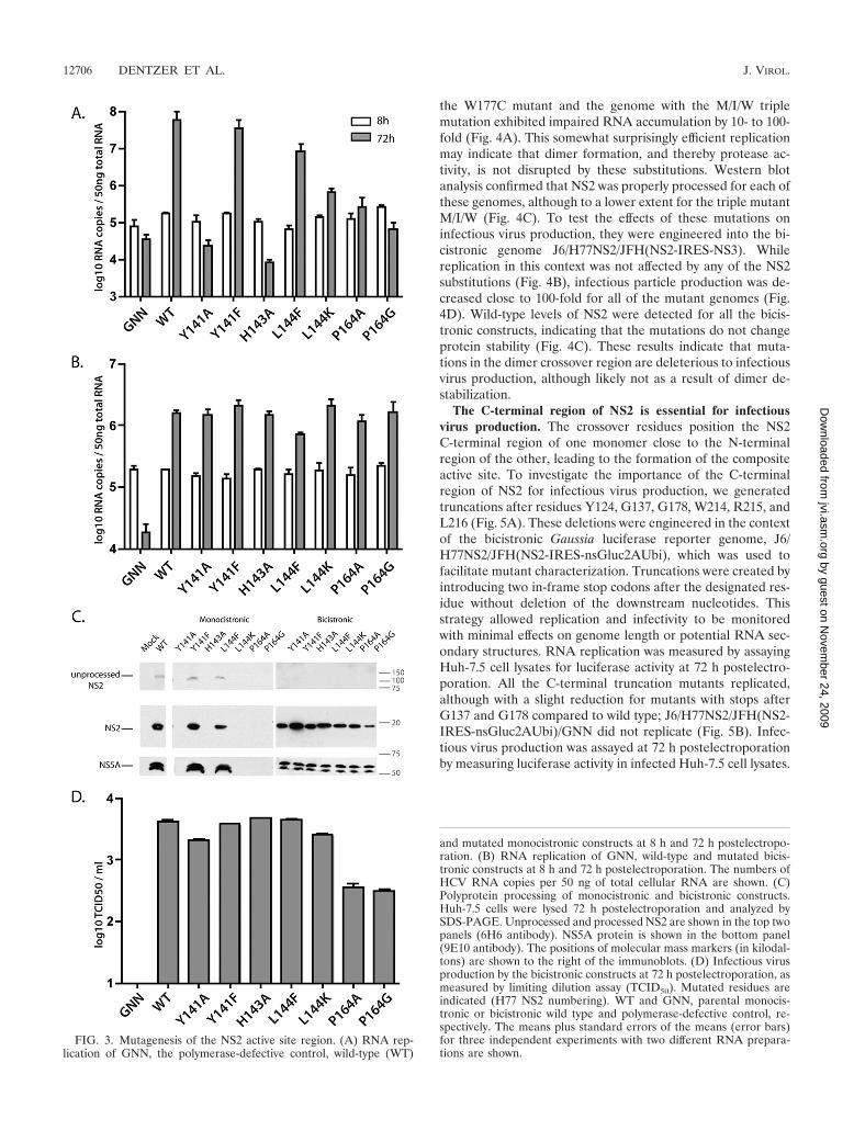
eny, we mutated individual residues in this region. Y141 andL144 are highly conserved amino acids surrounding the pro-tease active site, H143 is part of the catalytic triad, and P164 isan unusual cis-proline residue important for active site geom-etry. We mutated these residues to alanine and/or to less con-servative substitutions in the context of the monocistronic J6/H77NS2/JFH and bicistronic J6/H77NS2/JFH(NS2-IRES-NS3) genome, and assayed replication by quantitative RT-PCR for intracellular HCV RNA at 72 h postelectroporation.We confirmed that the active site mutation H143A abolishedreplication in the full-length monocistronic background (10),and we found that Y141A also severely impaired RNA accu-mulation; a Y141F substitution, which preserved the aromaticring, did not have a dramatic effect (Fig. 3A). Replication wasdecreased by mutation of L144 to bulky residues (L144F,L144K) and by substitutions of P164 (P164A, P164G). Consis-tent with the requirement for NS2-3 cleavage, robustly repli-cating genomes showed processed NS2 by Western blotting(Fig. 3C).
To test the effects of NS2 active cleft mutations on infectiousvirus production, we engineered these substitutions into thebicistronic J6/H77NS2/JFH(NS2-IRES-NS3) genome. In thiscontext, all mutants replicated as efficiently as the wild-typevirus did; J6/H77NS2/JFH(NS2-IRES-NS3)/GNN did not rep-licate (Fig. 3B). Infectious virus production was measured byinoculating naïve Huh-7.5 cells with filtered culture superna-tants harvested at 72 h postelectroporation and calculating theTCID50/ml. Mutations Y141A, Y141F, H143A, L144F, andL144K were not impaired or only slightly impaired in terms ofinfectious titers compared to wild-type J6/H77NS2/JFH(NS2-IRES-NS3), whereas substitution of P164 mutated to Ala orGly decreased infectious virus production by about 10-fold(Fig. 3D). Western blotting of cell lysates harvested at 72 hpostelectroporation revealed that all of the mutants expressedreadily detectable levels of NS2 (Fig. 3C).
These results indicate that mutations at the NS2 active cleftcan inhibit replication of a monocistronic genome, likely byaffecting NS2-3 processing, but that the catalytic activity is notrequired for infectious virus production of a bicistronic ge-nome. The moderate deleterious effect of cis-proline 164 mu-tations on infectivity may indicate a more global impact of thisunusual residue on NS2 architecture.
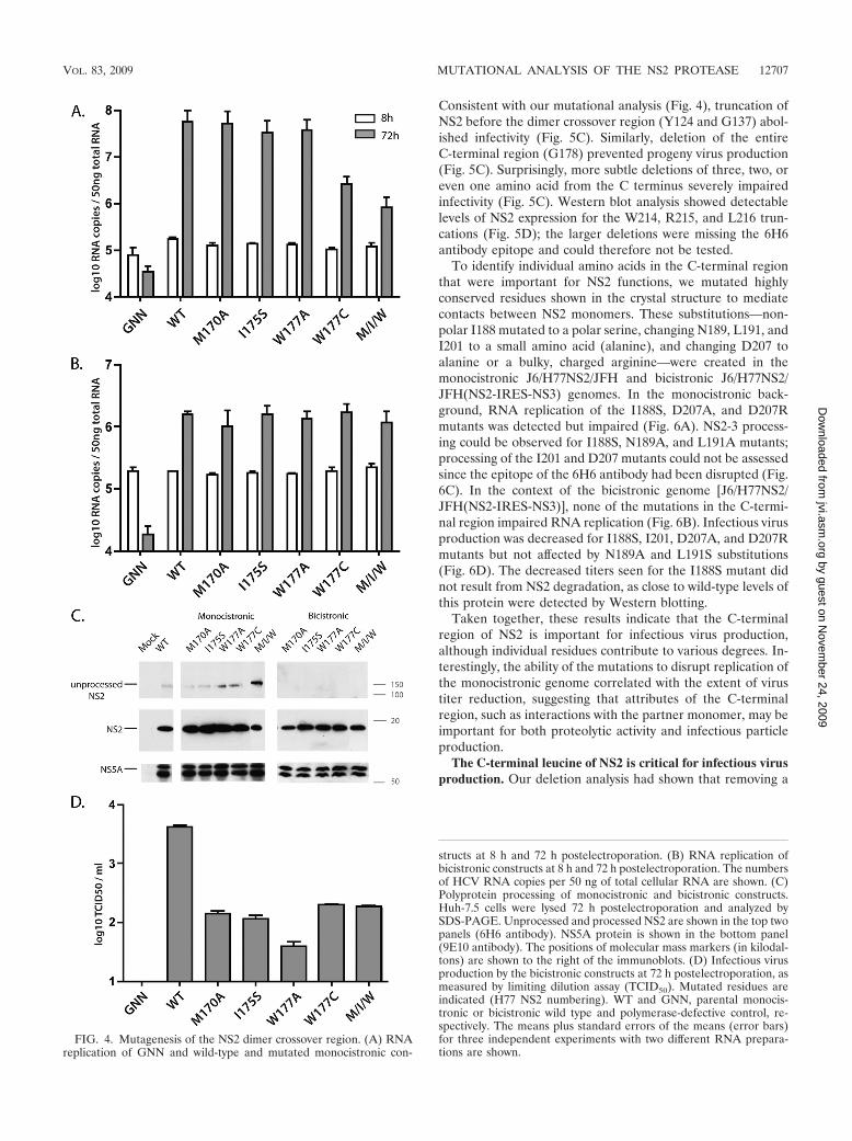
Residues in the NS2 dimer crossover region are importantfor infectious virus production. NS2 dimerization creates twocomposite active sites and has been shown to be essential forproteolytic cleavage at the NS2/NS3 junction (21). Althoughthe critical residues for dimer formation and stabilization arenot known, amino acids in the crossover region between thetwo monomers may be envisioned to participate. To test theeffects of mutations in the dimer crossover region on replica-tion and infectious virus production, we created several sub-stitutions of highly conserved amino acids in the context ofmonocistronic and bicistronic genomes: a triple mutation withM170A, I175A, and W177A (M/I/W) and individual mutationsM170A, I175S, W177A, and W177C. The single isoleucine-to-serine substitution was chosen in order to change a nonpolarresidue to a polar residue, with predicted disruption to thedimer interface. In the monocistronic J6/H77NS2/JFH back-ground, replication levels close to those of the wild type wereobserved for the M170A, I175S, and W177A mutants, whereas
FIG. 2. Characterization of anti-NS2 antibody (6H6). (A) Westernblot comparing the reactivity of the 6H6 anti-NS2 antibody and 9E10anti-NS5A antibody against genotype 1a, 1b, and 2a proteins. Huh-7.5cells were infected with a recombinant vaccinia helper virus expressingthe T7 polymerase, followed by transfection with plasmids coding fora full-length HCV genomes under the control of a T7 promoter. Thelysates were harvested 16 h posttransfection. The positions of molec-ular mass markers (in kilodaltons) are indicated to the right of the blot.(B) Amino acid sequence alignment of the 6H6 epitope region. Vari-ation between protein sequences is indicated in red. (C) Crystal struc-ture of the dimeric NS2 protease domain (21) with monomers shownin red and blue with the 6H6 antibody epitope shown in yellow.
VOL. 83, 2009 MUTATIONAL ANALYSIS OF THE NS2 PROTEASE 12705
by guest on Novem
ber 24, 2009 jvi.asm
.orgD
ownloaded from
the W177C mutant and the genome with the M/I/W triplemutation exhibited impaired RNA accumulation by 10- to 100-fold (Fig. 4A). This somewhat surprisingly efficient replicationmay indicate that dimer formation, and thereby protease ac-tivity, is not disrupted by these substitutions. Western blotanalysis confirmed that NS2 was properly processed for each ofthese genomes, although to a lower extent for the triple mutantM/I/W (Fig. 4C). To test the effects of these mutations oninfectious virus production, they were engineered into the bi-cistronic genome J6/H77NS2/JFH(NS2-IRES-NS3). Whilereplication in this context was not affected by any of the NS2substitutions (Fig. 4B), infectious particle production was de-creased close to 100-fold for all of the mutant genomes (Fig.4D). Wild-type levels of NS2 were detected for all the bicis-tronic constructs, indicating that the mutations do not changeprotein stability (Fig. 4C). These results indicate that muta-tions in the dimer crossover region are deleterious to infectiousvirus production, although likely not as a result of dimer de-stabilization.
The C-terminal region of NS2 is essential for infectiousvirus production. The crossover residues position the NS2C-terminal region of one monomer close to the N-terminalregion of the other, leading to the formation of the compositeactive site. To investigate the importance of the C-terminalregion of NS2 for infectious virus production, we generatedtruncations after residues Y124, G137, G178, W214, R215, andL216 (Fig. 5A). These deletions were engineered in the contextof the bicistronic Gaussia luciferase reporter genome, J6/H77NS2/JFH(NS2-IRES-nsGluc2AUbi), which was used tofacilitate mutant characterization. Truncations were created byintroducing two in-frame stop codons after the designated res-idue without deletion of the downstream nucleotides. Thisstrategy allowed replication and infectivity to be monitoredwith minimal effects on genome length or potential RNA sec-ondary structures. RNA replication was measured by assayingHuh-7.5 cell lysates for luciferase activity at 72 h postelectro-poration. All the C-terminal truncation mutants replicated,although with a slight reduction for mutants with stops afterG137 and G178 compared to wild type; J6/H77NS2/JFH(NS2-IRES-nsGluc2AUbi)/GNN did not replicate (Fig. 5B). Infec-tious virus production was assayed at 72 h postelectroporationby measuring luciferase activity in infected Huh-7.5 cell lysates.
FIG. 3. Mutagenesis of the NS2 active site region. (A) RNA rep-lication of GNN, the polymerase-defective control, wild-type (WT)
and mutated monocistronic constructs at 8 h and 72 h postelectropo-ration. (B) RNA replication of GNN, wild-type and mutated bicis-tronic constructs at 8 h and 72 h postelectroporation. The numbers ofHCV RNA copies per 50 ng of total cellular RNA are shown. (C)Polyprotein processing of monocistronic and bicistronic constructs.Huh-7.5 cells were lysed 72 h postelectroporation and analyzed bySDS-PAGE. Unprocessed and processed NS2 are shown in the top twopanels (6H6 antibody). NS5A protein is shown in the bottom panel(9E10 antibody). The positions of molecular mass markers (in kilodal-tons) are shown to the right of the immunoblots. (D) Infectious virusproduction by the bicistronic constructs at 72 h postelectroporation, asmeasured by limiting dilution assay (TCID50). Mutated residues areindicated (H77 NS2 numbering). WT and GNN, parental monocis-tronic or bicistronic wild type and polymerase-defective control, re-spectively. The means plus standard errors of the means (error bars)for three independent experiments with two different RNA prepara-tions are shown.
12706 DENTZER ET AL. J. VIROL.
by guest on Novem
ber 24, 2009 jvi.asm
.orgD
ownloaded from
Consistent with our mutational analysis (Fig. 4), truncation ofNS2 before the dimer crossover region (Y124 and G137) abol-ished infectivity (Fig. 5C). Similarly, deletion of the entireC-terminal region (G178) prevented progeny virus production(Fig. 5C). Surprisingly, more subtle deletions of three, two, oreven one amino acid from the C terminus severely impairedinfectivity (Fig. 5C). Western blot analysis showed detectablelevels of NS2 expression for the W214, R215, and L216 trun-cations (Fig. 5D); the larger deletions were missing the 6H6antibody epitope and could therefore not be tested.
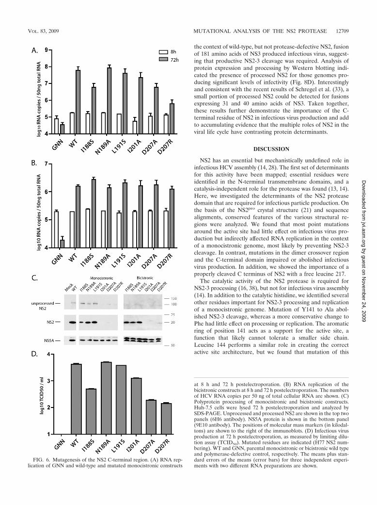
To identify individual amino acids in the C-terminal regionthat were important for NS2 functions, we mutated highlyconserved residues shown in the crystal structure to mediatecontacts between NS2 monomers. These substitutions—non-polar I188 mutated to a polar serine, changing N189, L191, andI201 to a small amino acid (alanine), and changing D207 toalanine or a bulky, charged arginine—were created in themonocistronic J6/H77NS2/JFH and bicistronic J6/H77NS2/JFH(NS2-IRES-NS3) genomes. In the monocistronic back-ground, RNA replication of the I188S, D207A, and D207Rmutants was detected but impaired (Fig. 6A). NS2-3 process-ing could be observed for I188S, N189A, and L191A mutants;processing of the I201 and D207 mutants could not be assessedsince the epitope of the 6H6 antibody had been disrupted (Fig.6C). In the context of the bicistronic genome [J6/H77NS2/JFH(NS2-IRES-NS3)], none of the mutations in the C-termi-nal region impaired RNA replication (Fig. 6B). Infectious virusproduction was decreased for I188S, I201, D207A, and D207Rmutants but not affected by N189A and L191S substitutions(Fig. 6D). The decreased titers seen for the I188S mutant didnot result from NS2 degradation, as close to wild-type levels ofthis protein were detected by Western blotting.
Taken together, these results indicate that the C-terminalregion of NS2 is important for infectious virus production,although individual residues contribute to various degrees. In-terestingly, the ability of the mutations to disrupt replication ofthe monocistronic genome correlated with the extent of virustiter reduction, suggesting that attributes of the C-terminalregion, such as interactions with the partner monomer, may beimportant for both proteolytic activity and infectious particleproduction.
The C-terminal leucine of NS2 is critical for infectious virusproduction. Our deletion analysis had shown that removing a
FIG. 4. Mutagenesis of the NS2 dimer crossover region. (A) RNAreplication of GNN and wild-type and mutated monocistronic con-
structs at 8 h and 72 h postelectroporation. (B) RNA replication ofbicistronic constructs at 8 h and 72 h postelectroporation. The numbersof HCV RNA copies per 50 ng of total cellular RNA are shown. (C)Polyprotein processing of monocistronic and bicistronic constructs.Huh-7.5 cells were lysed 72 h postelectroporation and analyzed bySDS-PAGE. Unprocessed and processed NS2 are shown in the top twopanels (6H6 antibody). NS5A protein is shown in the bottom panel(9E10 antibody). The positions of molecular mass markers (in kilodal-tons) are shown to the right of the immunoblots. (D) Infectious virusproduction by the bicistronic constructs at 72 h postelectroporation, asmeasured by limiting dilution assay (TCID50). Mutated residues areindicated (H77 NS2 numbering). WT and GNN, parental monocis-tronic or bicistronic wild type and polymerase-defective control, re-spectively. The means plus standard errors of the means (error bars)for three independent experiments with two different RNA prepara-tions are shown.
VOL. 83, 2009 MUTATIONAL ANALYSIS OF THE NS2 PROTEASE 12707
by guest on Novem
ber 24, 2009 jvi.asm
.orgD
ownloaded from

single NS2 residue, the C-terminal leucine 217, almost com-pletely abolished infectious virus production. The crystal struc-ture of the postcleavage form of NS2pro shows that leucine 217remains in the active site through hydrogen bond interactionswith the catalytic triad, a conformation that is proposed to limitthe enzyme to a single autoproteolytic cleavage (21). To inves-tigate the importance of this C-terminal residue for infectivity,L217 was mutated to a variety of amino acids—isoleucine,valine, alanine, tryptophan, asparagine, or lysine—in the con-text of the bicistronic reporter virus, J6/H77NS2/JFH(NS2-IRES-nsGluc2AUbi). The L217 mutants replicated, althoughat levels somewhat reduced compared to that of the wild type(Fig. 7A). Infectious virus production was markedly impairedfor all mutants tested apart from L217I, which showed a re-duction of infectious titers of about 10-fold (Fig. 7B). Thesedefects were not a result of NS2 instability, as the mutantproteins were readily detected by Western blotting (Fig. 7C).These data suggest that infectious virus production specificallyrequires a leucine at the C terminus of NS2, although anisoleucine can function to some degree.
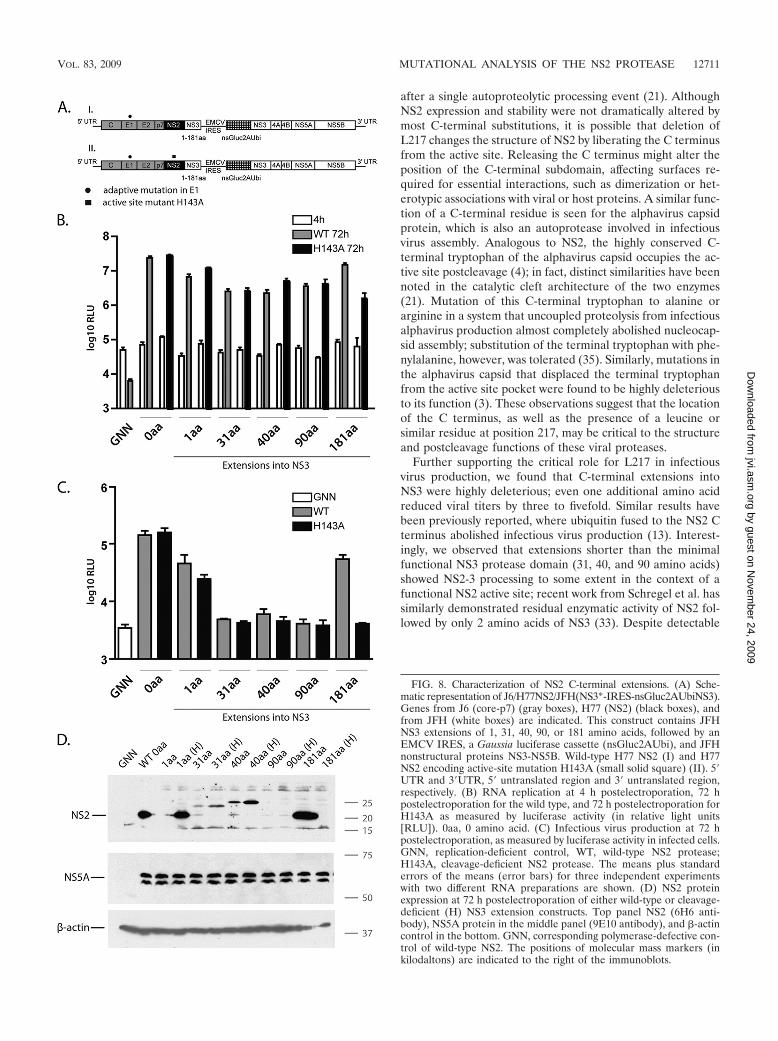
Additional residues fused to the C terminus of NS2 abolishinfectious virus production. NS2 requires residues 1 to 181 ofNS3 for optimal proteolytic activity (30). Our finding of acritical role for the NS2 C-terminal leucine in infectivity sug-gested divergent requirements for infectious virus productionand proteolysis. To investigate the effect of adding residuesfrom NS3 on viral titers, we created J6/H77NS2/JFH bicis-tronic reporter genomes in which NS2 was followed by 1, 31,40, 90, or 181 amino acids of NS3, a stop codon, the EMCVIRES, the Gaussia luciferase-2AUbi cassette, and full-lengthNS3 (Fig. 8A). These extensions were created in the context ofwild-type H77 NS2, as well as in the context of the H143Aactive site mutation to prevent NS2-3 cleavage. At 72 h post-electroporation, the NS3 fusion constructs replicated, althoughextensions of 1, 31, 40, and 90 amino acids impaired this pro-cess up to 10-fold, in both the wild-type and H143A back-grounds. Mutant virus with the 181-amino-acid extensionshowed replication levels comparable to that of the parentalgenome in the wild-type background but more drastically im-paired RNA replication in the H143A background; the reasonsfor this discrepancy are not known (Fig. 8B). Infectious virusproduction was severely impaired by uncleavable NS3 fusionsof 31 to 181 amino acids (Fig. 8C). Even the addition of asingle NS3 residue decreased infectivity by three- to fivefold. In
FIG. 5. Characterization of NS2 C-terminal truncations. (A) NS2pro
dimer with monomers shown in red and blue (21). The positions of theC-terminal truncations are indicated in yellow. (B) RNA replication at4 h and 72 h postelectroporation of GNN, wild type, and C-terminaltruncations in the bicistronic Gaussia luciferase reporter virus back-
ground, as measured by luciferase activity (in relative light units[RLU]). (C) Infectious virus production of NS2 C-terminal truncationsat 72 h after infection of naïve Huh-7.5 cells with supernatants har-vested 72 h postelectroporation. Truncation points are indicated(H77 NS2 numbering). WT, wild type [J6/H77NS2/JFH(NS2-IRES-nsGluc2AUbi)]; GNN, J6/H77NS2/JFH(NS2-IRES-nsGluc2AUbi)/GNN.The means plus standard errors of the means (error bars) for threeindependent experiments with two different RNA preparations areshown. (D) Polyprotein processing of NS2 truncations. Huh-7.5 cells lysed72 h postelectroporation and analyzed by SDS-PAGE. NS2 expression isshown in the top panel (6H6 antibody), NS5A protein in the middle panel(9E10 antibody), and �-actin control in the bottom. The antibody epitopeis not present in the NS2 truncations Y124, G137, and G178. The posi-tions of molecular mass markers (in kilodaltons) are shown to the right ofthe immunoblots.
12708 DENTZER ET AL. J. VIROL.
by guest on Novem
ber 24, 2009 jvi.asm
.orgD
ownloaded from
the context of wild-type, but not protease-defective NS2, fusionof 181 amino acids of NS3 produced infectious virus, suggest-ing that productive NS2-3 cleavage was required. Analysis ofprotein expression and processing by Western blotting indi-cated the presence of processed NS2 for those genomes pro-ducing significant levels of infectivity (Fig. 8D). Interestinglyand consistent with the recent results of Schregel et al. (33), asmall portion of processed NS2 could be detected for fusionsexpressing 31 and 40 amino acids of NS3. Taken together,these results further demonstrate the importance of the C-terminal residue of NS2 in infectious virus production and addto accumulating evidence that the multiple roles of NS2 in theviral life cycle have contrasting protein determinants.
DISCUSSION
NS2 has an essential but mechanistically undefined role ininfectious HCV assembly (14, 28). The first set of determinantsfor this activity have been mapped; essential residues wereidentified in the N-terminal transmembrane domains, and acatalysis-independent role for the protease was found (13, 14).Here, we investigated the determinants of the NS2 proteasedomain that are required for infectious particle production. Onthe basis of the NS2pro crystal structure (21) and sequencealignments, conserved features of the various structural re-gions were analyzed. We found that most point mutationsaround the active site had little effect on infectious virus pro-duction but indirectly affected RNA replication in the contextof a monocistronic genome, most likely by preventing NS2-3cleavage. In contrast, mutations in the dimer crossover regionand the C-terminal domain impaired or abolished infectiousvirus production. In addition, we showed the importance of aproperly cleaved C terminus of NS2 with a free leucine 217.
The catalytic activity of the NS2 protease is required forNS2-3 processing (16, 38), but not for infectious virus assembly(14). In addition to the catalytic histidine, we identified severalother residues important for NS2-3 processing and replicationof a monocistronic genome. Mutation of Y141 to Ala abol-ished NS2-3 cleavage, whereas a more conservative change toPhe had little effect on processing or replication. The aromaticring of position 141 acts as a support for the active site, afunction that likely cannot tolerate a smaller side chain.Leucine 144 performs a similar role in creating the correctactive site architecture, but we found that mutation of this
FIG. 6. Mutagenesis of the NS2 C-terminal region. (A) RNA rep-lication of GNN and wild-type and mutated monocistronic constructs
at 8 h and 72 h postelectroporation. (B) RNA replication of thebicistronic constructs at 8 h and 72 h postelectroporation. The numbersof HCV RNA copies per 50 ng of total cellular RNA are shown. (C)Polyprotein processing of monocistronic and bicistronic constructs.Huh-7.5 cells were lysed 72 h postelectroporation and analyzed bySDS-PAGE. Unprocessed and processed NS2 are shown in the top twopanels (6H6 antibody). NS5A protein is shown in the bottom panel(9E10 antibody). The positions of molecular mass markers (in kilodal-tons) are shown to the right of the immunoblots. (D) Infectious virusproduction at 72 h postelectroporation, as measured by limiting dilu-tion assay (TCID50). Mutated residues are indicated (H77 NS2 num-bering). WT and GNN, parental monocistronic or bicistronic wild typeand polymerase-defective control, respectively. The means plus stan-dard errors of the means (error bars) for three independent experi-ments with two different RNA preparations are shown.
VOL. 83, 2009 MUTATIONAL ANALYSIS OF THE NS2 PROTEASE 12709
by guest on Novem
ber 24, 2009 jvi.asm
.orgD
ownloaded from

residue to Phe allowed detectable levels of NS2-3 processing,whereas Lys apparently abolished the function of the activesite. Proline 164 has a cis conformation that is thought to bendthe peptide backbone to establish the correct geometry of theGlu 163 side chain for catalysis; mutation of P164 to Ala or Glyprevented replication of a monocistronic genome. The major-ity of active site changes had little effect on infectious virusproduction in a bicistronic genome. Substitutions of P164,however, decreased infectious titers. It is possible that muta-tion of this cis-proline dramatically affects NS2 structure; in-deed, the P164G substitution appeared to slightly destabilizethe protein. It is also possible that mutations of P164 affectNS2 dimer formation, as this proline is important for position-ing the linker between the N- and C-terminal subdomains.Alternatively, P164 may directly participate in infectious virusassembly independent of a role in catalysis. Studies of theNS2-3 protein of the distantly related pestivirus, classical swinefever virus, have similarly shown that the NS2 protease activityis dispensable for infectious virus production (23). However, ahistidine-to-arginine mutation within the active site abolishedinfectivity without affecting NS2-3 expression (23).
The crystal structure of NS2pro shows a crossover region thatpositions the subdomains for creation of the composite activesites (21). We hypothesized that mutations in this region woulddisrupt dimer stability and NS2-3 processing. RNA replicationof a monocistronic genome, however, was impaired only bysubstitution of W177 to Cys or by a triple mutation of M170A/I175A/W177A. This suggests that single point mutations donot have a drastic effect on dimer integrity; indeed, the signif-icant buried surface area between the monomers indicates thatthe NS2 dimer is highly stable (21). Although NS2-3 processingwas not greatly impacted by mutations in the crossover se-quence, infectious virus production was impaired by all substi-tutions we tested in this region. A number of the crossoverresidues are exposed on the surface of the NS2pro dimer (Fig.9). Mutations in this region may disrupt associations with viralor host proteins involved in infectious virus production. In-deed, NS2 has been suggested to participate in a number ofgenetic or physical interactions, including with structural pro-teins core and E2 as well as with nonstructural proteins p7,NS3, NS4A, and NS5A (15, 19, 25, 29, 34, 40). The A269Tadaptive mutation identified here suggests a genetic interac-tion between NS2 and E1. Cellular kinase CKII also appears toassociate with and phosphorylate NS2 (8), and NS2 may inter-act with additional host factors to influence apoptosis (6) andcellular gene expression (5).
In addition to mediating contacts between monomers, theC-terminal subdomain of NS2 contributes the catalytic cysteineto the composite active site. Deletion analysis revealed thateven a single amino acid truncation at the C terminus severelyimpaired infectious virus production. Furthermore, the major-ity of substitutions at the terminal L217 were highly deleteriousto infectivity. Interestingly, previous reports have shown thatmost modifications of L217 have little effect on NS2-3 process-ing (12, 30). Our finding of an essential role for L217 in infec-tious virus production helps explain its high level of conserva-tion across all genotypes. The structure of the postcleavageform of NS2pro shows that L217 occupies the active sitethrough contacts with H143 and C184 (13, 21). This confor-mation has been suggested to render the protease inactive
FIG. 7. Mutagenesis of the NS2 C-terminal leucine 217. (A) RNAreplication at 4 h and 72 h postelectroporation of GNN, wild type, andC-terminal mutants in the context of the bicistronic Gaussia luciferasereporter virus, as measured by luciferase activity (in relative light units[RLU]). (B) Infectious virus production of genomes bearing Leu 217mutations at 72 h after infection of naïve Huh-7.5 cells with superna-tants harvested 72 h postelectroporation. WT, wild type [J6/H77NS2/JFH(NS2-IRES-nsGluc2AUbi)]; GNN, J6/H77NS2/JFH(NS2-IRES-nsGluc2AUbi)/GNN. The means plus standard errors of the means(error bars) for three independent experiments with two differentRNA preparations are shown. (C) Polyprotein processing of 72 hpostelectroporation. Huh-7.5 cells were lysed and analyzed by SDS-PAGE. NS2 expression is shown in the top panel (6H6 antibody),NS5A protein in the middle panel (9E10 antibody), and �-actin controlin the bottom. The positions of molecular mass markers (in kilodal-tons) are shown to the right of the immunoblots.
12710 DENTZER ET AL. J. VIROL.
by guest on Novem
ber 24, 2009 jvi.asm
.orgD
ownloaded from
after a single autoproteolytic processing event (21). AlthoughNS2 expression and stability were not dramatically altered bymost C-terminal substitutions, it is possible that deletion ofL217 changes the structure of NS2 by liberating the C terminusfrom the active site. Releasing the C terminus might alter theposition of the C-terminal subdomain, affecting surfaces re-quired for essential interactions, such as dimerization or het-erotypic associations with viral or host proteins. A similar func-tion of a C-terminal residue is seen for the alphavirus capsidprotein, which is also an autoprotease involved in infectiousvirus assembly. Analogous to NS2, the highly conserved C-terminal tryptophan of the alphavirus capsid occupies the ac-tive site postcleavage (4); in fact, distinct similarities have beennoted in the catalytic cleft architecture of the two enzymes(21). Mutation of this C-terminal tryptophan to alanine orarginine in a system that uncoupled proteolysis from infectiousalphavirus production almost completely abolished nucleocap-sid assembly; substitution of the terminal tryptophan with phe-nylalanine, however, was tolerated (35). Similarly, mutations inthe alphavirus capsid that displaced the terminal tryptophanfrom the active site pocket were found to be highly deleteriousto its function (3). These observations suggest that the locationof the C terminus, as well as the presence of a leucine orsimilar residue at position 217, may be critical to the structureand postcleavage functions of these viral proteases.
Further supporting the critical role for L217 in infectiousvirus production, we found that C-terminal extensions intoNS3 were highly deleterious; even one additional amino acidreduced viral titers by three to fivefold. Similar results havebeen previously reported, where ubiquitin fused to the NS2 Cterminus abolished infectious virus production (13). Interest-ingly, we observed that extensions shorter than the minimalfunctional NS3 protease domain (31, 40, and 90 amino acids)showed NS2-3 processing to some extent in the context of afunctional NS2 active site; recent work from Schregel et al. hassimilarly demonstrated residual enzymatic activity of NS2 fol-lowed by only 2 amino acids of NS3 (33). Despite detectable
FIG. 8. Characterization of NS2 C-terminal extensions. (A) Sche-matic representation of J6/H77NS2/JFH(NS3*-IRES-nsGluc2AUbiNS3).Genes from J6 (core-p7) (gray boxes), H77 (NS2) (black boxes), andfrom JFH (white boxes) are indicated. This construct contains JFHNS3 extensions of 1, 31, 40, 90, or 181 amino acids, followed by anEMCV IRES, a Gaussia luciferase cassette (nsGluc2AUbi), and JFHnonstructural proteins NS3-NS5B. Wild-type H77 NS2 (I) and H77NS2 encoding active-site mutation H143A (small solid square) (II). 5�UTR and 3�UTR, 5� untranslated region and 3� untranslated region,respectively. (B) RNA replication at 4 h postelectroporation, 72 hpostelectroporation for the wild type, and 72 h postelectroporation forH143A as measured by luciferase activity (in relative light units[RLU]). 0aa, 0 amino acid. (C) Infectious virus production at 72 hpostelectroporation, as measured by luciferase activity in infected cells.GNN, replication-deficient control, WT, wild-type NS2 protease;H143A, cleavage-deficient NS2 protease. The means plus standarderrors of the means (error bars) for three independent experimentswith two different RNA preparations are shown. (D) NS2 proteinexpression at 72 h postelectroporation of either wild-type or cleavage-deficient (H) NS3 extension constructs. Top panel NS2 (6H6 anti-body), NS5A protein in the middle panel (9E10 antibody), and �-actincontrol in the bottom. GNN, corresponding polymerase-defective con-trol of wild-type NS2. The positions of molecular mass markers (inkilodaltons) are indicated to the right of the immunoblots.
VOL. 83, 2009 MUTATIONAL ANALYSIS OF THE NS2 PROTEASE 12711
by guest on Novem
ber 24, 2009 jvi.asm
.orgD
ownloaded from

NS2-3 processing, however, genomes encoding NS3 extensionsstill did not support infectious virus production. This couldindicate that insufficient quantities of mature NS2 are pro-duced by suboptimal cleavage or that short fragments of NS3impair infectivity, possibly by acting as dominant-negative in-hibitors of interactions between NS2 and full-length NS3 (15).The finding that fused residues from NS3 are deleterious to therole of HCV NS2 infectivity contrasts with the related pestivi-ruses, in which the uncleaved NS2-3 precursor is essential forinfectious virus production (1, 23). The possibility that NS2and NS3 form functional associations during virion morpho-genesis, however, suggests conserved strategies between HCVand other members of the family Flaviviridae (24).
Using monocistronic and bicistonic genomes, we were ableto analyze the effects of NS2 mutations on protease activity andpostcleavage functions. Our results add to accumulating evi-dence that the determinants of these two essential roles aredivergent. Previous work has demonstrated that the transmem-brane domains of NS2 play critical roles in infectivity (13) butare not absolutely required for protease activity (10, 26, 32, 37).
Conversely, the active cleft is essential for the protease func-tion but predominantly dispensable for infectivity (13, 14; thisstudy). A number of substitutions in the dimer crossover regionand C-terminal subdomain affected infectious titers, but notprotease activity, and L217 was found to be dispensable forprocessing but critical for infectious virus production. Simi-larly, the finding that NS2 functions in assembly do not tolerateC-terminal extensions contrasts with the requirement for theNS3 protease domain for optimal NS2-3 cleavage. These dif-ferences highlight the two distinct functions of NS2 and suggestthat further analysis of these roles may reveal important reg-ulatory switches.
In conclusion, we dissected the determinants of the NS2protease domain required for infectious virus production. Wefound critical roles for residues in the dimer crossover regionand at the extreme C terminus of the protein, and we showedthat C-terminal extensions into NS3 are deleterious to infec-tivity. These insights increase our understanding of the multi-functional NS2 protein and may facilitate exploiting this targetfor antiviral drug development.
ACKNOWLEDGMENTS
We thank Maryline Panis and Anesta Webson for laboratory sup-port and technical assistance. We are grateful to Catherine L. Murray,Christopher T. Jones, Cynthia de la Fuente, and Kimberly D. Ritolafor reagents, constructs, helpful discussions, and critical reading of themanuscript.
This study was supported by The Greenberg Medical ResearchInstitute, NIH Public Health Service grant (AI075099), and the StarrFoundation. T.G.D. is supported by the Fonds National de la Recher-che Luxembourg.
REFERENCES
1. Agapov, E. V., C. L. Murray, I. Frolov, L. Qu, T. M. Myers, and C. M. Rice.2004. Uncleaved NS2-3 is required for production of infectious bovine viraldiarrhea virus. J. Virol. 78:2414–2425.
2. Blight, K. J., J. A. McKeating, and C. M. Rice. 2002. Highly permissive celllines for subgenomic and genomic hepatitis C virus RNA replication. J. Vi-rol. 76:13001–13014.
3. Choi, H. K., S. Lee, Y. P. Zhang, B. R. McKinney, G. Wengler, M. G.Rossmann, and R. J. Kuhn. 1996. Structural analysis of Sindbis virus capsidmutants involving assembly and catalysis. J. Mol. Biol. 262:151–167.
4. Choi, H. K., L. Tong, W. Minor, P. Dumas, U. Boege, M. G. Rossmann, andG. Wengler. 1991. Structure of Sindbis virus core protein reveals a chymot-rypsin-like serine proteinase and the organization of the virion. Nature354:37–43.
5. Dumoulin, F. L., A. von dem Bussche, J. Li, L. Khamzina, J. R. Wands, T.Sauerbruch, and U. Spengler. 2003. Hepatitis C virus NS2 protein inhibitsgene expression from different cellular and viral promoters in hepatic andnonhepatic cell lines. Virology 305:260–266.
6. Erdtmann, L., N. Franck, H. Lerat, J. Le Seyec, D. Gilot, I. Cannie, P.Gripon, U. Hibner, and C. Guguen-Guillouzo. 2003. The hepatitis C virusNS2 protein is an inhibitor of CIDE-B-induced apoptosis. J. Biol. Chem.278:18256–18264.
7. Everhart, J. E., Y. Wei, H. Eng, M. R. Charlton, D. H. Persing, R. H.Wiesner, J. J. Germer, J. R. Lake, R. K. Zetterman, and J. H. Hoofnagle.1999. Recurrent and new hepatitis C virus infection after liver transplanta-tion. Hepatology 29:1220–1226.
8. Franck, N., J. Le Seyec, C. Guguen-Guillouzo, and L. Erdtmann. 2005.Hepatitis C virus NS2 protein is phosphorylated by the protein kinase CK2and targeted for degradation to the proteasome. J. Virol. 79:2700–2708.
9. Grakoui, A., D. W. McCourt, C. Wychowski, S. M. Feinstone, and C. M. Rice.1993. A second hepatitis C virus-encoded proteinase. Proc. Natl. Acad. Sci.USA 90:10583–10587.
10. Grakoui, A., D. W. McCourt, C. Wychowski, S. M. Feinstone, and C. M. Rice.1993. Characterization of the hepatitis C virus-encoded serine proteinase:determination of proteinase-dependent polyprotein cleavage sites. J. Virol.67:2832–2843.
11. Hijikata, M., H. Mizushima, T. Akagi, S. Mori, N. Kakiuchi, N. Kato, T.Tanaka, K. Kimura, and K. Shimotohno. 1993. Two distinct proteinaseactivities required for the processing of a putative nonstructural precursorprotein of hepatitis C virus. J. Virol. 67:4665–4675.
FIG. 9. Summary of critical NS2pro residues. (A) Exposed surfacerendering of the NS2pro dimer. Mutations showing abolished infectiousvirus production are shown in red (M170, I175, W177, D207, andL217), mutations showing impaired infectious virus production areshown in orange (P164 and I188), and mutations with no significanteffect on infectious virus production are shown in green (Y141, H143,L144, N189, and L191). (B) NS2pro dimer rotated 90° around thehorizontal axis as shown in panel A.
12712 DENTZER ET AL. J. VIROL.
by guest on Novem
ber 24, 2009 jvi.asm
.orgD
ownloaded from

12. Hirowatari, Y., M. Hijikata, Y. Tanji, H. Nyunoya, H. Mizushima, K.Kimura, T. Tanaka, N. Kato, and K. Shimotohno. 1993. Two proteinaseactivities in HCV polypeptide expressed in insect cells using baculovirusvector. Arch. Virol. 133:349–356.
13. Jirasko, V., R. Montserret, N. Appel, A. Janvier, L. Eustachi, C. Brohm, E.Steinmann, T. Pietschmann, F. Penin, and R. Bartenschlager. 2008. Struc-tural and functional characterization of nonstructural protein 2 for its role inhepatitis C virus assembly. J. Biol. Chem. 283:28546–28562.
14. Jones, C. T., C. L. Murray, D. K. Eastman, J. Tassello, and C. M. Rice. 2007.Hepatitis C virus p7 and NS2 proteins are essential for production of infec-tious virus. J. Virol. 81:8374–8383.
15. Kiiver, K., A. Merits, M. Ustav, and E. Zusinaite. 2006. Complex formationbetween hepatitis C virus NS2 and NS3 proteins. Virus Res. 117:264–272.
16. Kolykhalov, A. A., E. V. Agapov, K. J. Blight, K. Mihalik, S. M. Feinstone,and C. M. Rice. 1997. Transmission of hepatitis C by intrahepatic inoculationwith transcribed RNA. Science 277:570–574.
17. Liang, T. J., B. Rehermann, L. B. Seeff, and J. H. Hoofnagle. 2000. Patho-genesis, natural history, treatment, and prevention of hepatitis C. Ann.Intern. Med. 132:296–305.
18. Lindenbach, B. D., M. J. Evans, A. J. Syder, B. Wolk, T. L. Tellinghuisen,C. C. Liu, T. Maruyama, R. O. Hynes, D. R. Burton, J. A. McKeating, andC. M. Rice. 2005. Complete replication of hepatitis C virus in cell culture.Science 309:623–626.
19. Liu, Q., R. A. Bhat, A. M. Prince, and P. Zhang. 1999. The hepatitis C virusNS2 protein generated by NS2-3 autocleavage is required for NS5A phos-phorylation. Biochem. Biophys. Res. Commun. 254:572–577.
20. Lohmann, V., F. Korner, J. Koch, U. Herian, L. Theilmann, and R. Barten-schlager. 1999. Replication of subgenomic hepatitis C virus RNAs in ahepatoma cell line. Science 285:110–113.
21. Lorenz, I. C., J. Marcotrigiano, T. G. Dentzer, and C. M. Rice. 2006. Struc-ture of the catalytic domain of the hepatitis C virus NS2-3 protease. Nature442:831–835.
22. Moradpour, D., F. Penin, and C. M. Rice. 2007. Replication of hepatitis Cvirus. Nat. Rev. Microbiol. 5:453–463.
23. Moulin, H. R., T. Seuberlich, O. Bauhofer, L. C. Bennett, J. D. Tratschin,M. A. Hofmann, and N. Ruggli. 2007. Nonstructural proteins NS2-3 andNS4A of classical swine fever virus: essential features for infectious particleformation. Virology 365:376–389.
24. Murray, C. L., C. T. Jones, and C. M. Rice. 2008. Architects of assembly:roles of Flaviviridae non-structural proteins in virion morphogenesis. Nat.Rev. Microbiol. 6:699–708.
25. Murray, C. L., C. T. Jones, J. Tassello, and C. M. Rice. 2007. Alaninescanning of the hepatitis C virus core protein reveals numerous residuesessential for production of infectious virus. J. Virol. 81:10220–10231.
26. Pallaoro, M., A. Lahm, G. Biasiol, M. Brunetti, C. Nardella, L. Orsatti, F.Bonelli, S. Orru, F. Narjes, and C. Steinkuhler. 2001. Characterization of the
hepatitis C virus NS2/3 processing reaction by using a purified precursorprotein. J. Virol. 75:9939–9946.
27. Penin, F., J. Dubuisson, F. A. Rey, D. Moradpour, and J. M. Pawlotsky. 2004.Structural biology of hepatitis C virus. Hepatology 39:5–19.
28. Pietschmann, T., A. Kaul, G. Koutsoudakis, A. Shavinskaya, S. Kallis, E.Steinmann, K. Abid, F. Negro, M. Dreux, F. L. Cosset, and R. Barten-schlager. 2006. Construction and characterization of infectious intrageno-typic and intergenotypic hepatitis C virus chimeras. Proc. Natl. Acad. Sci.USA 103:7408–7413.
29. Pietschmann, T., V. Lohmann, A. Kaul, N. Krieger, G. Rinck, G. Rutter, D.Strand, and R. Bartenschlager. 2002. Persistent and transient replication offull-length hepatitis C virus genomes in cell culture. J. Virol. 76:4008–4021.
30. Reed, K. E., A. Grakoui, and C. M. Rice. 1995. Hepatitis C virus-encodedNS2-3 protease: cleavage-site mutagenesis and requirements for bimolecularcleavage. J. Virol. 69:4127–4136.
31. Ryan, M. D., A. M. King, and G. P. Thomas. 1991. Cleavage of foot-and-mouth disease virus polyprotein is mediated by residues located within a 19amino acid sequence. J. Gen. Virol. 72:2727–2732.
32. Santolini, E., L. Pacini, C. Fipaldini, G. Migliaccio, and N. Monica. 1995.The NS2 protein of hepatitis C virus is a transmembrane polypeptide. J. Vi-rol. 69:7461–7471.
33. Schregel, V., S. Jacobi, F. Penin, and N. Tautz. 2009. Hepatitis C virus NS2is a protease stimulated by cofactor domains in NS3. Proc. Natl. Acad. Sci.USA 106:5342–5347.
34. Selby, M. J., E. Glazer, F. Masiarz, and M. Houghton. 1994. Complexprocessing and protein:protein interactions in the E2:NS2 region of HCV.Virology 204:114–122.
35. Skoging, U., and P. Liljestrom. 1998. Role of the C-terminal tryptophanresidue for the structure-function of the alphavirus capsid protein. J. Mol.Biol. 279:865–872.
36. Steinmann, E., F. Penin, S. Kallis, A. H. Patel, R. Bartenschlager, and T.Pietschmann. 2007. Hepatitis C virus p7 protein is crucial for assembly andrelease of infectious virions. PLoS Pathog. 3:e103.
37. Thibeault, D., R. Maurice, L. Pilote, D. Lamarre, and A. Pause. 2001. In vitrocharacterization of a purified NS2/3 protease variant of hepatitis C virus.J. Biol. Chem. 276:46678–46684.
38. Welbourn, S., R. Green, I. Gamache, S. Dandache, V. Lohmann, R. Barten-schlager, K. Meerovitch, and A. Pause. 2005. Hepatitis C virus NS2/3 pro-cessing is required for NS3 stability and viral RNA replication. J. Biol. Chem.280:29604–29611.
39. Yamaga, A. K., and J. H. Ou. 2002. Membrane topology of the hepatitis Cvirus NS2 protein. J. Biol. Chem. 277:33228–33234.
40. Yi, M., Y. Ma, J. Yates, and S. M. Lemon. 2007. Compensatory mutations inE1, p7, NS2, and NS3 enhance yields of cell culture-infectious intergenotypicchimeric hepatitis C virus. J. Virol. 81:629–638.
VOL. 83, 2009 MUTATIONAL ANALYSIS OF THE NS2 PROTEASE 12713
by guest on Novem
ber 24, 2009 jvi.asm
.orgD
ownloaded from