Embed Size (px)
Citation preview

JOURNAL OF CLINICAL MICROBIOLOGY, June 2002, p. 1947–1957 Vol. 40, No. 60095-1137/02/$04.00�0 DOI: 10.1128/JCM.40.6.1947–1957.2002Copyright © 2002, American Society for Microbiology. All Rights Reserved.
Development of a Sensitive and Specific Enzyme-LinkedImmunosorbent Assay for Detecting and Quantifying
CMY-2 and SHV �-LactamasesAndrea M. Hujer,1 Malcolm G. P. Page,2 Marion S. Helfand,3 Bethany Yeiser,1
and Robert A. Bonomo1*Research Service, Louis Stokes Veterans Affairs Medical Center, Cleveland, Ohio 441061; Basilea Pharmaceutica Ltd.,
Basel, Switzerland2; and Infectious Diseases Division, University Hospitals, Cleveland, Ohio 441063
Received 6 September 2001/Returned for modification 31 October 2001/Accepted 1 March 2002
Polyclonal rabbit antibodies against SHV-1 and CMY-2 �-lactamases were produced and characterized, andenzyme-linked immunosorbent assays (ELISAs) were developed. Immunoblots revealed that the anti-SHV-1antibody recognized SHV-1 but did not recognize TEM-1, K-1, OXA-1, or any AmpC �-lactamase tested. Theanti-CMY-2 antibody detected Escherichia coli CMY-2, Enterobacter cloacae P99, Klebsiella pneumoniae ACT-1,and the AmpC �-lactamases of Enterobacter aerogenes, Morganella morganii, and Citrobacter freundii. Nocross-reactivity of the anti-CMY-2 antibody was seen against laboratory strains of E. coli possessing TEM-1,SHV-1, K-1, or OXA-1 �-lactamases. Operating conditions for performing ELISAs were optimized. Bothanti-CMY-2 and anti-SHV-1 antibodies detected picogram quantities of purified protein in ELISAs. Thereactivity of the anti-CMY-2 antibody was tested against a number of AmpC �-lactamases by assaying knownquantities of purified enzymes in ELISAs (AmpC �-lactamases of M. morganii, C. freundii, E. coli, and E.cloacae). As the homology to CMY-2 �-lactamase decreased, the minimum level needed for detection increased(e.g., 94% homology recognized at 1 ng/ml and 71% homology recognized at 10 ng/ml). The ELISAs were usedto assay unknown clinical isolates for AmpC and SHV �-lactamases, and the results were confirmed with PCRamplification of blaAmpC and blaSHV genes. Overall, we found that our ELISAs were at least 95% sensitive andspecific for detecting SHV and AmpC �-lactamases. The ELISA format can facilitate the identification ofAmpC and SHV �-lactamases and can be used to quantify relative amounts of �-lactamase enzymes in clinicaland laboratory isolates.
Common molecular techniques available for identifying�-lactamases in clinical samples include DNA hybridization(Southern blotting), amplification by PCR with specific prim-ers, and analytical isoelectric focusing (aIEF) (29). For mostapplications, DNA hybridization with digoxigenin-labeledprobes that allow recognition of sequences with less homology(conditions of low stringency) is adequate but can be labor-intensive and time-consuming. aIEF is a very sensitive tech-nique that can be performed rapidly (26). Unfortunately, aIEFcan be imprecise in determining the identities of �-lactamaseswith pIs of greater than 7.6. PCR-based methods of detectionrequire the construction of specific primers that may not beable to amplify closely related �-lactamase genes. Even whenmultiple PCR primer sets and susceptibility tests are used toidentify �-lactamases, samples can remain uncharacterized(32).
Immunological methods have long been applied to the anal-ysis and classification of �-lactamases (4, 5, 7–9, 11, 13, 17, 18,22–24, 31, 33, 34). Antibody-based methods offer an advantagein that they are easily performed and highly sensitive. Poly-clonal antibodies recognize multiple epitopes and can detectclosely related variants of �-lactamases. Hence, polyclonal an-
tibodies were used early on to classify �-lactamases and un-derstand catalysis (33).
Most recently, antibodies against �-lactamases have beendeveloped to assess enzyme expression in a number of studiesinvestigating the effects of point mutations. Studies with anti-ROB-1 �-lactamase, anti-TEM-1 �-lactamase, and anti-PSE-4�-lactamase antibodies have addressed in a qualitative mannerthe effects of point mutations on steady-state �-lactamase lev-els (10, 14, 25, 27, 30). To our knowledge, only two anti-�-lactamase antibodies have been used in both Western blot andenzyme-linked immunosorbent assay (ELISA) formats to mea-sure TEM-1 and PC1 �-lactamases (15, 22).
Here, we describe the production, purification, and charac-terization of polyclonal anti-SHV-1 and anti-CMY-2 �-lacta-mase antibodies. We chose to develop these antibodies be-cause SHV-1 is the second most common class A �-lactamasefound in Escherichia coli and Klebsiella pneumoniae, andCMY-2 �-lactamase is the most common plasmid-determinedAmpC �-lactamase (3, 21). Our goals were to use these anti-bodies to detect the presence of SHV and AmpC �-lactamasesin clinical and laboratory strains and to develop a highly sen-sitive ELISA for each. The ELISA format can facilitate thesimultaneous screening of multiple clinical isolates for thepresence of SHV and AmpC �-lactamases. Furthermore,ELISAs can be used to measure (quantitate) relative �-lacta-mase production. In the research setting, the ELISA formatcan be a very useful tool for studying the induction and relativeamounts of SHV and AmpC �-lactamases.
* Corresponding author. Mailing address: Infectious Disease, LouisStokes Veterans Affairs Medical Center, 10701 East Blvd., Cleveland,OH 44106. Phone: (216) 791-3800, ext. 4788. Fax: (216) 231-3482.E-mail: [email protected].
1947
on August 22, 2019 by guest
http://jcm.asm
.org/D
ownloaded from

MATERIALS AND METHODS
Bacterial strains and plasmids. Escherichia coli 20 and Klebsiella pneumoniae15571 are clinical isolates recovered from patients at University Hospitals andthe Louis Stokes Veterans Affairs Medical Center (LSVAMC), respectively, inCleveland, Ohio (28). These bacteria were the parent strains used for the sub-sequent cloning and isolation of CMY-2 and SHV-1 �-lactamases. E. coli DH5�
and E. coli DH10B were obtained from Gibco BRL Life Technologies (GrandIsland, N.Y.). E. coli J53-2 was previously described (28). The E. coli straincontaining the OXA-1 �-lactamase was a kind gift from George A. Jacoby(Lahey Clinic, Burlington, Mass.). Proteus vulgaris harboring K-1, Enterobactercloacae with P99, and K. pneumoniae containing ACT-1 �-lactamases were kindgifts from Patricia Bradford (Wyeth-Ayerst Laboratories, Pearl River, N.Y.).The Enterobacter aerogenes strain with an AmpC �-lactamase was a kind gift fromReuben Ramphal (University of Florida, Gainesville).
A total of 101 clinical isolates were studied in validating our ELISAs. FredTenover (Centers for Disease Control and Prevention, Atlanta, Ga.) and JanPatterson (University of Texas, Southwest, San Antonio) kindly provided theclinical isolates with uncharacterized �-lactamases, in set 1 and set 2, respec-tively. The identities of isolates in set 1 were unknown. Set 2 consisted of 14 K.pneumoniae isolates. Set 3 comprised 46 K. pneumoniae isolates kindly providedby David Paterson (University of Pittsburgh, Pittsburgh, Pa.). In addition, DonnaO’Black (University of Cincinnati, Cincinnati, Ohio) provided 11 E. coli, 1Klebsiella oxytoca, and 3 K. pneumoniae isolates. Two E. cloacae, one K. oxytoca,one E. coli, four K. pneumoniae, two Hafnia alvei, one Morganella morganii, andfour Citrobacter freundii isolates were collected and kindly provided by Curtis J.Donskey (LSVAMC).
Plasmid pUC18, encoding the TEM-1 �-lactamase, was a kind gift from LouisB. Rice (LSVAMC). The SHV-1 �-lactamase was cloned in pBC SK(�) (Strat-agene, La Jolla, Calif.) as previously described (28). E. coli J53-2-derived strains194 and 194-61 possess plasmid p194 or a subclone of p194 in pBC SK(�); bothencode the CMY-2 �-lactamase. All bacteria were grown in Luria-Bertani (LB)broth with either ampicillin or chloramphenicol selection.
�-Lactamase protein expression and purification. The SHV-1 and CMY-2�-lactamases expressed in E. coli were liberated by periplasmic fractionation andpurified according to previously described methods (19, 20; M. S. Helfand, A. M.Hujer, and R. A. Bonomo, submitted for publication). In brief, a 5-ml overnightculture of E. coli DH10B or DH5� harboring the SHV-1 or CMY-2 �-lactamasegene cloned into a high-copy-number phagemid vector, pBC SK(�), was used toinoculate 1.5 liters of LB broth containing 100 �g of ampicillin or 20 �g ofchloramphenicol (Sigma Chemical Co., St. Louis, Mo.)/ml. Cells were grownovernight, pelleted, and stored at �20°C until �-lactamase purification. Cellswere resuspended in 200 ml of 50 mM Tris HCl (pH 7.4) with freshly preparedlysozyme (Sigma) added to a final concentration of 10 �g/ml and incubated for15 min at room temperature. EDTA was added to a 1 mM concentration withconstant mixing. The crude lysate was filtered through a 0.22-�m-pore-sizeNalgene bottle-top filter (Fisher, Pittsburgh, Pa.) and concentrated by using aDiaflo 10-kDa ultrafiltration membrane (Amicon Inc., Beverly, Mass.). The�-lactamase was purified from the crude lysate by preparative isoelectric focusingin an Ultrodex/Ampholine (pH gradient, 3.5 to 10) gel bed prepared accordingto the manufacturer’s specifications (Amersham Pharmacia Biotech, Piscataway,N.J.). The Ultradex gel was run overnight (4°C) at a constant power of 8 W ona Multiphor II isoelectric focusing apparatus (Amersham Pharmacia Biotech).�-Lactamase activity in the gel was identified by using the chromogenic cepha-losporin nitrocefin (Becton Dickinson, Cockeysville, Md.). This visual identifi-cation was accomplished by applying a solution of 100 �M nitrocefin to the filterpaper. A yellow-to-pink color change was observed in the �-lactamase-contain-ing area of the gel.
Areas of the gel containing �-lactamase activity were cut out, and �-lactamasewas eluted with 20 mM diethanolamine (pH 8.3). Ampholines were removedfrom the eluate by dialysis against 20 mM diethanolamine (pH 8.3). The samplewas then concentrated and resolved with 5% stacking–12% separating sodiumdodecyl sulfate (SDS)-polyacrylamide gel electrophoresis (PAGE). Purity wasassessed by Coomassie brilliant blue R250 staining. The protein concentrationwas determined by a Bio-Rad (Hercules, Calif.) protein assay with bovine serumalbumin (BSA) as a standard.
Purified AmpC �-lactamases isolated from C. freundii, E. cloacae, M. morganii,Pseudomonas aeruginosa, and Staphylococcus aureus PC1 were obtained fromRoche Laboratories, Basel, Switzerland. Homology of these enzymes to CMY-2�-lactamase was defined by DNA analysis comparisons (Table 1) carried out byusing DNASIS for Windows (Hitachi Software Genetic Systems, South SanFrancisco, Calif.).
Custom antibody production. Custom antibodies were produced by GenosysBiotech, Inc., The Woodlands, Tex., with 1.5 mg of purified SHV-1 and 1.5 mgof purified CMY-2 �-lactamases. Each rabbit was immunized with 200 �g ofCMY-2 or SHV-1 �-lactamase in complete Freund’s adjuvant. This step wasfollowed by immunization with 100 �g of SHV-1 or CMY-2 �-lactamase inincompete Freund’s adjuvant every 2 weeks thereafter for 10 weeks. Bleedingwas performed on days 49, 63, and 77 of the immunization-antibody produc-tion schedule. Approximately 25 ml of serum was obtained per bleed. A totalof four rabbits were used for anti-SHV-1 and anti-CMY-2 antibody produc-tion.
Anti-CMY-2 and anti-SHV-1 antibody purification. Polyclonal immunoglob-ulin G antibodies were isolated from the rabbit serum by using Protein G columnpurification. Five milliliters of serum was added to 10 ml of phosphate-bufferedsaline (PBS) (pH 7.4), and the mixture was run over a 5-ml Hi-Trap protein Gcolumn (Sigma) with positive pressure at a flow rate of 0.5 ml/min. The boundanti-SHV-1 or anti-CMY-2 antibody was eluted from the column with 0.1 Mglycine (pH 2.7) in 1-ml aliquots and neutralized with 100 �l of 1 M Tris HCl (pH8.8). The antibody concentration was determined by measuring the optical den-sity (OD) at 260 nm (OD260) with a Fisher spectrophotometer. Approximately 15mg of purified immunoglobulin G was obtained per 5 ml of serum for bothanti-SHV-1 and anti-CMY-2 antibodies on one column pass. The antibody elu-ate was dialyzed against PBS (pH 7.4) and divided into 0.5-ml aliquots for finalstorage at �20°C.
Immunoblotting. Clinical and laboratory isolates possessing �-lactamases weregrown in LB broth to an OD600 of 0.5. Fifty microliters of each culture was mixedwith SDS-PAGE sample buffer, resulting in final concentrations of 62.5 mM Trisbase (pH 6.8), 2% glycerol, 2% SDS, 100 mM dithiothreitol, and 0.02% bromo-phenol blue. These samples were then boiled. A 7.5-�l aliquot was subjected toelectrophoresis and transferred to polyvinylidene difluoride membranes (Boehr-inger Mannheim, Indianapolis, Ind.). After overnight incubation in blockingbuffer (5% BSA [Amresco, Solon, Ohio], 20 mM Tris HCl-buffered 150 mMsaline [pH 7.5]), �-lactamase present on the blots was detected by incubationwith either 1 �g of anti-SHV-1 or anti-CMY-2 antibody/ml or a 1:100 dilution ofanti-TEM antibody, kindly provided by T. Palzkill (Baylor College of Medicine,Houston, Tex.). Antibody incubation of the immunoblots was carried out for 3 hat room temperature. The membranes were washed four times for 10 min eachtime in Tris-buffered saline (pH 7.4) and subsequently incubated with a 1:2,000dilution of horseradish peroxidase-conjugated protein G (Bio-Rad). All anti-body-protein G incubations were done in blocking buffer. After four morewashes, the membranes were processed for film exposure by using an ECL kit inaccordance with the manufacturer’s protocol (Amersham Pharmacia Biotech).
Antibody recognition determinations by immunoblotting. Purified SHV-1 andCMY-2 �-lactamases were diluted across a range of concentrations. The follow-ing amounts were used for SDS-PAGE loading: 100, 50, 10, 1, 0.1, and 0.01 ng.The protein samples were electrophoresed and transferred to polyvinylidenedifluoride membranes as stated above. Two separate blots were probed with 1 �gof anti-SHV-1 or anti-CMY-2 antibody/ml. The level of antibody recognition foreach was determined by immunoblotting with chemiluminescence exposure tofilm for 1 min.
ELISAs for SHV and AmpC �-lactamases. Ninety-six-well Immulon-4 enzymeimmunoassay plates (Fisher) were coated overnight with 4 �g of anti-SHV-1 oranti-CMY-2 polyclonal antibody/ml diluted in carbonate buffer (pH 9.5). Plateswere washed six times with PBS containing 0.05% Tween 20 (Bio-Rad) and
TABLE 1. Percent homology to CMY-2 and anti-CMY-2antibody recognitiona
�-Lactamase % DNA homology Recognition by antibody
CMY-2 100 �C. freundii AmpC 94 �P99 74 �ACT-1 74 �E. cloacae AmpC 71 �M. morganii AmpC 54 �PC 1 45 �K-1 29 �SHV-1 27 �TEM-1 27 �OXA-1 26 �
a Homology percentages were based on DNA sequence comparisons per-formed by using DNASIS. �, recognition; �, no recognition.
1948 HUJER ET AL. J. CLIN. MICROBIOL.
on August 22, 2019 by guest
http://jcm.asm
.org/D
ownloaded from

blocked with 5% BSA in PBS for 2 h at room temperature. Purified SHV-1 andAmpC �-lactamases were serially diluted to fixed concentrations, or 100-�laliquots of overnight bacterial cultures were boiled for 10 min and seriallydiluted. These dilutions were applied to the enzyme immunoassay plates, incu-bated for 2 h, washed, and incubated for an additional hour with 2 �g ofbiotinylated anti-SHV-1 or anti-CMY-2 polyclonal antibody/ml.
Concentrations of coating and biotinylated detecting antibodies were variedinitially to empirically determine the best concentrations for assessing the pres-ence of SHV or AmpC �-lactamase. Between all steps following sample incu-bation, washes were done six times with 0.05% Tween 20–PBS. Plates were thenincubated with a 1:3,000 dilution of streptavidin-horseradish peroxidase (ZymedLaboratories, South San Francisco, Calif.) for 30 min, followed by developmentwith o-phenylenediamine and H2O2 diluted in citric acid buffer (pH 5.5) (Sigma).Development was terminated by the addition of H2SO4 to a concentration of 0.5M. OD492 values were obtained with a Cerus ELISA plate reader and comparedto those for serially diluted, purified SHV-1 or CMY-2 �-lactamase used as aninternal point of reference. Averaging the OD values of the standards, plottingOD against concentration in nanograms per milliliter, and generating a slope
determined the amount of CMY-2 or SHV �-lactamase present in the samples.The slope of the line was then used to calculate the amount of �-lactamasepresent.
PCR analysis for the presence of �-lactamases. SHV �-lactamase primers (5�ATGCGTTATATTCGCCTGTG 3� and 5� TGCTTTGTTATTCGGGCCAA 3�[Genosys Biotech]) were used to amplify the blaSHV gene (GenBank accessionnumber AF124984). AmpC �-lactamase primers (5� ATCAAAACTGGCAGCCG 3� and 5� GAGCCCGTTTTATGGACCCA 3� [Genosys Biotech]) weredesigned from homologous regions of the P99, CMY-2, and ACT-1 bla genes(GenBank accession numbers X07274, X91840, and U58495, respectively); theywere used to amplify AmpC �-lactamase genes.
A 10-�l aliquot of an overnight culture was diluted 1:10 with water and boiled for10 min. PCR amplification was then performed with 10 �l of this dilution as theDNA template. PCR conditions included 35 cycles of amplification at a denaturationtemperature of 94oC for 1 min, an annealing temperature of 60°C for 1 min, and anextension temperature of 72oC for 1 min. This step was followed by a final extensionat 72oC for 10 min. PCR products were run on 1% agarose gels, stained withethidium bromide, and photographed with UV illumination. �X174 replicative-form
FIG. 1. Immunoblotting. (a) Immunoblot of various amounts of purified CMY-2 �-lactamase probed with 1 �g of anti-CMY-2 antibody/ml. (b)Immunoblot of various amounts of purified SHV-1 �-lactamase probed with 1 �g of anti-SHV-1 antibody/ml. (c) Immunoblot of various�-lactamase-producing strains probed with 1 �g of anti-CMY-2 antibody/ml. Strains, listed from left to right, included E coli DH10B carryingplasmid pBC SK(�) with the SHV-1 �-lactamase, strains producing K-1 and ACT-1 �-lactamases, strain DH5�/pUC18 producing the TEM-1�-lactamase, a cefepime-resistant E. aerogenes strain producing a �-lactamase (EA), and a strain expressing the P99 Amp C �-lactamase; inaddition, E. coli J53-2-derived strains 194-61 and 194 and E. coli strain 20 (EC20) are clinical and laboratory strains producing CMY-2 �-lactamase.(d) Identical immunoblots of strains E. coli DH10B/pUC18 producing TEM-1 �-lactamase, E. coli DH10B/pBC SK(�) producing SHV-1�-lactamase, and E. coli J53-2-derived 194-61 producing CMY-2 �-lactamase probed with anti-TEM antibody (1:100 dilution) or 1 �g of anti-SHVantibody/ml.
VOL. 40, 2002 ELISAs FOR CMY-2 AND SHV �-LACTAMASES 1949
on August 22, 2019 by guest
http://jcm.asm
.org/D
ownloaded from

DNA HaeIII fragments (Gibco BRL Life Technologies) were used to assess PCRproduct sizes.
Statistical analysis. Statistical analysis was performed on data gathered fromthe 101 clinical isolates. Sensitivity, specificity, and negative and positive predic-tive values were calculated for the SHV and AmpC ELISAs; a kappa statistic wasassigned for each as well (2, 16).
RESULTS
Antibody recognition and lower limit of detection. Immuno-blots revealed that both anti-SHV-1 and anti-CMY-2 antibod-ies had a lower limit of antigen detection at 1 ng of purified
FIG. 2. Effects of varying coating and detecting antibody concentrations in the SHV ELISA. Purified SHV-1 �-lactamase was diluted acrossa range of concentrations and run in triplicate for each detecting antibody concentration (2, 4, and 6 �g of biotinylated anti-SHV antibody/ml).A separate standard curve was generated for each detecting antibody concentration and was used to calculate SHV amounts in nanograms permilliliter. Values plotted are the means for triplicate samples. The sample range never exceeded 7% of the plotted value and was not included inthe graph. The effect of varying the coating antibody concentration was examined in a similar manner.
1950 HUJER ET AL. J. CLIN. MICROBIOL.
on August 22, 2019 by guest
http://jcm.asm
.org/D
ownloaded from

SHV-1 or CMY-2, respectively (Fig. 1a and b). Antibody rec-ognition of �-lactamases in bacterial lysates was examined forboth antibodies in a similar manner (Fig. 1c and d). Anti-CMY-2 antibody was capable of detecting CMY-2 (present inthe bacterial lysates of E. coli strains designated 194-61, 194,and 20), P99, E. aerogenes AmpC, and ACT-1 �-lactamases(Fig. 1c). SHV-1, TEM-1, K-1, and OXA-1 (not shown) �-lac-tamases were not recognized by anti-CMY-2 antibody. Anti-SHV-1 antibody recognition was also determined by immuno-blot analysis. Duplicate blots of strains producing TEM-1,SHV-1, and CMY-2 were probed with either anti-TEM oranti-SHV antibody. Anti-TEM antibody recognized bothTEM-1 and SHV-1 �-lactamases. However, anti-SHV-1 anti-body was capable of recognizing only SHV (Fig. 1d). No cross-reactivity was observed for TEM-1 or any AmpC �-lactamase.
Development of the AmpC and SHV �-lactamase ELISAs.In order to optimize signal strength and to keep nonspecificbackground to a minimum, we determined the optimal con-centrations of detecting and coating antibodies for theELISAs. The results of testing detecting antibody concentra-
tions of 2, 4, and 6 �g/ml are summarized in Fig. 2b. PurifiedSHV-1 diluted across a concentration range of picograms tonanograms per milliliter was run in triplicate and detected with2, 4, or 6 �g of biotinylated anti-SHV-1 antibody/ml. Thecoating antibody concentration was kept constant at 4 �g/ml.Separate standard curves were generated for each concentra-tion and used to calculate SHV-1 standard quantities. Al-though absolute OD values varied slightly, calculated purifiedSHV-1 concentrations differed by less than 1% between dif-ferent detecting antibody concentrations. Similar results wereobtained when biotinylated anti-CMY-2 detecting antibodyconcentrations were varied (data not shown). With these re-sults in mind, we chose to use 2 �g/ml as the biotinylateddetecting antibody concentration for both the SHV and theAmpC ELISAs. This concentration provided the ability to de-tect picogram-per-milliliter levels of �-lactamase while keepingnonspecific binding to a minimum.
Shown in Fig. 2a are the results of varying the anti-SHV-1coating antibody concentrations in the ELISA format. Thedetecting antibody concentration was kept constant at 2 �g/ml.
FIG. 3. ELISA standard curves. Purified SHV-1 and CMY-2 �-lactamases at known concentrations were run in duplicate and used to generatea standard curve for every ELISA that was performed.
VOL. 40, 2002 ELISAs FOR CMY-2 AND SHV �-LACTAMASES 1951
on August 22, 2019 by guest
http://jcm.asm
.org/D
ownloaded from

A similar approach was undertaken for the anti-CMY-2 anti-body. A coating antibody concentration of 4 �g/ml for bothantibodies allowed the greatest recognition and the least non-specific binding. Operating conditions for the ELISAs weredetermined to be 4 �g of coating antibody/ml and 2 �g ofbiotinylated detecting antibody/ml.
Generation of ELISA standard curves. Purified SHV-1 orCMY-2 �-lactamases, at known concentrations, were used togenerate a standard curve for every ELISA performed (Fig. 3).The slope of the line generated was used to calculate theamount of �-lactamase present in the samples.
Defining AmpC ELISA recognition. Since cross-reactivitywas observed between anti-CMY-2 antibody and AmpC �-lac-tamases other than CMY-2, we wanted to further define which�-lactamases anti-CMY-2 antibody recognized and at whatlevel they could be detected. We measured the concentrationsof purified AmpC �-lactamases with the Bio-Rad protein assayin order to load identical quantities to determine the lowerlimits of recognition for other AmpC �-lactamases. Figure 4demonstrates that the AmpC �-lactamases from C. freundii, E.cloacae, and M. morganii and E. coli CMY-2 were recognizedin the AmpC ELISA. DNA homology comparisons between
the sequences of these �-lactamases and the CMY-2 �-lacta-mase sequence are shown in Table 1. By loading known con-centrations of the various purified AmpC enzymes, we showedthat as the homology to CMY-2 decreased, the minimum con-centration of that enzyme that could be reliably detected in-creased. The CMY-2 �-lactamase (100% homology) could bedetected at less than 100 pg/ml, and the AmpC �-lactamase ofC. freundii (94% homology) could be detected at 1 ng/ml. TheAmpC �-lactamase of E. cloacae (71% homology) could bedetected at 10 ng/ml. The AmpC �-lactamase of M. morganii(54% homology) could be detected only at a level of 500 ng/ml.The AmpC �-lactamase of P. aeruginosa could not be detectedat 100 ng/ml (data not shown). DNA homology comparisonswere also done for SHV-1 (Table 2). From immunoblot anal-ysis, we know that the anti-SHV-1 antibody recognized onlySHV.
FIG. 4. Minimum concentrations of various AmpC �-lactamases needed in order to be detected by the AmpC ELISA. By assaying knownconcentrations of the various purified enzymes, we demonstrated that as DNA sequence homology to CMY-2 decreased, the minimum concen-tration of the �-lactamase that could be reliably detected increased. Purified AmpC �-lactamases were CMY-2 from E. coli (CMY-2), C. freundii(C. f.), E. cloacae (E. cloa), M. morganii (M. mor), and S. aureus PC1 (PC1).
TABLE 2. Percent homology to SHV-1 and anti-SHV-1antibody recognitiona
�-Lactamase % DNA homology Recognition by antibody
SHV-1 100 �TEM-1 63 �K-1 41 �P99 29 �ACT-1 27 �CMY-2 27 �OXA-1 27 �
a Homology percentages were based on DNA sequence comparisons per-formed by using DNASIS. �, recognition; �, no recognition.
TABLE 3. Results for set 1 (unknowns)
Sample
Result for:
SHV AmpC
ELISAa PCRb ELISAa PCRb
1 � � � �2 � � � �3 � � � �4 � � � �5 � � � �6 � � � �7 � � � �8 � � � �9 � � � �10 � � � �11 � � � �
a All negative ELISA values had an OD of �0.05. �, positive; �, negative.b There were 35 cycles of PCR amplification. �, positive; �, negative.
1952 HUJER ET AL. J. CLIN. MICROBIOL.
on August 22, 2019 by guest
http://jcm.asm
.org/D
ownloaded from

Testing clinical isolates with the SHV and AmpC ELISAs.To validate our ELISAs, we tested three sets of isolates. Set 1isolates (unknown clinical isolates; Table 3) were screened withthe SHV and AmpC ELISAs. Figure 5a graphically depictsAmpC ELISA results along with the results for two positivecontrols (P99 �-lactamase and cefepime-resistant E. aerogenesAmpC �-lactamase). All unknown samples were diluted 1:5with 0.1% BSA in PBS. The OD492 cutoff value taken as apositive sample was 0.05 OD unit for both ELISAs. Unknownsamples 3, 5, 9, and 10 tested positive for the presence of anAmpC �-lactamase in the ELISA. This finding was confirmedby PCR analysis (Fig. 5b and Table 3), with the exception thatsample 4 was weakly positive for an AmpC �-lactamase in thePCR but not in the ELISA. An SHV ELISA was also per-
formed on the unknown isolates in set 1; all samples werenegative in the ELISA for the SHV enzyme. This finding wasconfirmed by PCR amplification specific for the blaSHV gene(Fig. 5b).
Set 2 isolates were assayed in a manner similar to that for set1 isolates. Thirteen of 14 isolates were positive in the PCR forthe SHV gene. The SHV ELISA correctly detected this �-lac-tamase in 12 of the 13 isolates (Table 4). All isolates in set 2tested negative for the presence of an AmpC �-lactamase inboth PCR and ELISA analyses (Table 4).
With set 3, we extended our analysis to include 75 addi-tional clinical isolates. PCR amplification for the SHV generevealed 44 positive isolates; 43 of these possessed a positivesignal in the SHV ELISA. SHV PCR demonstrated 32 neg-
FIG. 5. ELISA validation. (a) Eleven clinical isolates (set 1) were blindly screened with the AmpC ELISA. Shown are AmpC ELISA resultsand results for two positive controls (P99 �-lactamase and cefepime-resistant E. aerogenes [EA]). All unknown samples were diluted 1:5 with 0.1%BSA in PBS. (b) PCR analysis of 11 clinical isolates (set 1) with SHV primers and AmpC primers. PCR amplicons were run on a 1% ethidiumbromide-stained agarose gel. Lanes: M, DNA sizing standard (�X174 replicative-form DNA HaeIII fragments); A, K. pneumoniae containingACT-1 �-lactamase amplicons; P, P99 �-lactamase amplicons; C, 194-61 CMY-2 �-lactamase amplicons; B, blank; S, SHV �-lactamase amplicons.
VOL. 40, 2002 ELISAs FOR CMY-2 AND SHV �-LACTAMASES 1953
on August 22, 2019 by guest
http://jcm.asm
.org/D
ownloaded from

ative isolates; all were negative in the SHV ELISA, with theexception of isolate 42 (Table 5). The ampC PCR producedsimilar results, with 18 positive and 58 negative isolates. Ofthe 18 isolates positive for the ampC gene, 16 produced apositive signal in the ELISA; of the 58 PCR-negative iso-lates, 56 were negative in the ELISA (Table 5).
Statistical results. The performance characteristics of theAmpC and SHV ELISAs are summarized in Table 6.
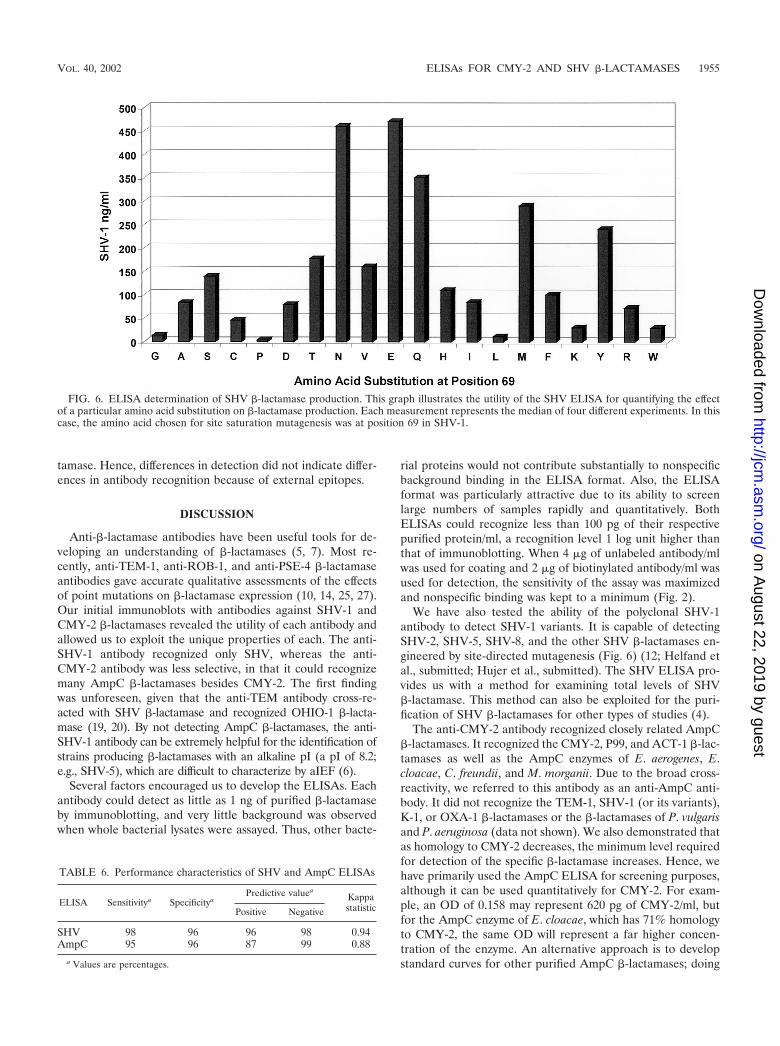
Measuring SHV �-lactamase expression. In order to showthat the SHV ELISA could be used to quantify differences inthe total amounts of SHV, we assayed a variety of SHV �-lac-tamase mutants in E. coli DH10B (12; Helfand et al., submit-ted; A. M. Hujer, K. M. Hujer, V. E. Anderson, and R. A.Bonomo, submitted for publication). Our results indicated thatboth immunoblotting (data not shown) and the ELISA format(Fig. 6) can be used to assess relative levels of steady-stateSHV �-lactamase amounts. The mutants tested possessed anamino acid substitution in the hydrophobic core of the �-lac-
TABLE 4. Results for set 2 (K. pneumoniae)
Sample
Result for:
SHV AmpC
ELISA (OD)a PCRb ELISAa PCRb
12 � (0.057) � � �13 � (0.085) � � �14 � (0.056) � � �15 � (0.048) � � �16 � (0.055) � � �17 � (0.340) � � �18 � (0.179) � � �19 � (0.101) � � �20 � (0.077) � � �21 � (0.170) � � �22 � (0.013) � � �23 � (0.075) � � �24 � (0.165) � � �25 � (0.057) � � �
a Values in parentheses are ODs. Symbols: �, positive; �, negative.b There were 35 cycles of PCR amplification. Symbols: �, positive; �, nega-
tive.
TABLE 5. Results for set 3 (various organisms)
Sample no. andorganism
Result for:Sample no. and
organism
Result for:
SHV AmpC SHV AmpC
ELISAa PCRb ELISAa PCRb ELISAa PCRb ELISA (OD)a PCRb
26 E. coli � (0.024) � � (0.147) �27 E. coli � (0.035) � � (1.335) �28 E. coli � (0.027) � � (0.452) �29 E. coli � (0.023) � � (0.857) �30 E. coli � (0.026) � � (0.322) �31 E. coli � (0.039) � � (0.609) �32 E. coli � (0.013) � � (0.853) �33 E. coli � (0.023) � � (1.206) �34 E. coli � (0.017) � � (2.595) �35 E. coli � (0.018) � � (0.050) �36 E. coli � (0.021) � � (0.067) �37 K. oxytoca � (1.300) � � (0.017) �38 K. oxytoca � (0.091) � � (0.049) �39 K. pneumoniae � (0.070) � � (0.175) �40 K. pneumoniae � (0.081) � � (0.217) �41 K. pneumoniae � (0.010) � � (0.038) �42 K. pneumoniae � (0.099) � � (0.027) �43 K. pneumoniae � (0.000) � � (0.028) �44 K. pneumoniae � (0.012) � � (0.032) �45 K. pneumoniae � (1.283) � � (0.021) �46 K. pneumoniae � (0.552) � � (0.030) �47 K. pneumoniae � (1.044) � � (0.030) �48 K. pneumoniae � (1.165) � � (0.039) �49 K. pneumoniae � (0.566) � � (0.024) �50 K. pneumoniae � (0.671) � � (0.021) �51 K. pneumoniae � (0.634) � � (0.024) �52 K. pneumoniae � (0.505) � � (0.017) �53 K. pneumoniae � (1.129) � � (0.024) �54 K. pneumoniae � (0.298) � � (0.038) �55 K. pneumoniae � (2.059) � � (0.020) �56 K. pneumoniae � (0.567) � � (0.017) �57 K. pneumoniae � (0.510) � � (0.036) �58 K. pneumoniae � (0.157) � � (0.016) �59 K. pneumoniae � (0.683) � � (0.028) �60 K. pneumoniae � (0.738) � � (0.024) �61 K. pneumoniae � (0.970) � � (0.024) �62 K. pneumoniae � (1.463) � � (0.034) �63 K. pneumoniae � (0.389) � � (0.019) �
a Values in parentheses are ODs. Symbols: �, positive; �, negative.b There were 35 cycles of PCR amplification. Symbols: �, positive; �, negative.
64 K. pneumoniae � (1.454) � � (0.017) �65 K. pneumoniae � (1.711) � � (0.014) �66 K. pneumoniae � (0.477) � � (0.021) �67 K. pneumoniae � (0.331) � � (0.021) �68 K. pneumoniae � (0.062) � � (0.031) �69 K. pneumoniae � (0.148) � � (0.034) �70 K. pneumoniae � (0.398) � � (0.006) �71 K. pneumoniae � (2.401) � � (0.012) �72 K. pneumoniae � (2.181) � � (0.024) �73 K. pneumoniae � (0.475) � � (0.021) �74 K. pneumoniae � (0.775) � � (0.017) �75 K. pneumoniae � (0.855) � � (0.016) �76 K. pneumoniae � (0.545) � � (0.032) �77 K. pneumoniae � (0.067) � � (0.027) �78 K. pneumoniae � (0.039) � � (0.019) �79 K. pneumoniae � (0.392) � � (0.017) �80 K. pneumoniae � (0.068) � � (0.027) �81 K. pneumoniae � (0.000) � � (0.009) �82 K. pneumoniae � (0.004) � � (0.028) �83 K. pneumoniae � (0.000) � � (0.016) �84 K. pneumoniae � (0.077) � � (0.021) �85 K. pneumoniae � (0.955) � � (0.012) �86 K. pneumoniae � (0.138) � � (0.016) �87 E. cloacae � (0.004) � � (0.114) �88 C. freundii � (0.003) � � (2.756) �89 K. pneumoniae � (0.001) � � (2.969) �90 C. freundii � (0.004) � � (1.650) �91 E. cloacae � (0.000) � � (0.203) �92 C. freundii � (0.000) � � (0.035) �93 K. pneumoniae � (0.002) � � (0.041) �94 H. alvei � (0.001) � � (0.021) �95 K. oxytoca � (0.014) � � (0.028) �96 E. coli � (0.007) � � (0.032) �97 C. freundii � (0.000) � � (0.020) �98 K. pneumoniae � (0.008) � � (0.025) �99 K. pneumoniae � (0.441) � � (0.013) �100 M. morganii � (0.002) � � (0.013) �101 H. alvei � (0.006) � � (0.048) �
1954 HUJER ET AL. J. CLIN. MICROBIOL.
on August 22, 2019 by guest
http://jcm.asm
.org/D
ownloaded from
tamase. Hence, differences in detection did not indicate differ-ences in antibody recognition because of external epitopes.
DISCUSSION
Anti-�-lactamase antibodies have been useful tools for de-veloping an understanding of �-lactamases (5, 7). Most re-cently, anti-TEM-1, anti-ROB-1, and anti-PSE-4 �-lactamaseantibodies gave accurate qualitative assessments of the effectsof point mutations on �-lactamase expression (10, 14, 25, 27).Our initial immunoblots with antibodies against SHV-1 andCMY-2 �-lactamases revealed the utility of each antibody andallowed us to exploit the unique properties of each. The anti-SHV-1 antibody recognized only SHV, whereas the anti-CMY-2 antibody was less selective, in that it could recognizemany AmpC �-lactamases besides CMY-2. The first findingwas unforeseen, given that the anti-TEM antibody cross-re-acted with SHV �-lactamase and recognized OHIO-1 �-lacta-mase (19, 20). By not detecting AmpC �-lactamases, the anti-SHV-1 antibody can be extremely helpful for the identification ofstrains producing �-lactamases with an alkaline pI (a pI of 8.2;e.g., SHV-5), which are difficult to characterize by aIEF (6).
Several factors encouraged us to develop the ELISAs. Eachantibody could detect as little as 1 ng of purified �-lactamaseby immunoblotting, and very little background was observedwhen whole bacterial lysates were assayed. Thus, other bacte-
rial proteins would not contribute substantially to nonspecificbackground binding in the ELISA format. Also, the ELISAformat was particularly attractive due to its ability to screenlarge numbers of samples rapidly and quantitatively. BothELISAs could recognize less than 100 pg of their respectivepurified protein/ml, a recognition level 1 log unit higher thanthat of immunoblotting. When 4 �g of unlabeled antibody/mlwas used for coating and 2 �g of biotinylated antibody/ml wasused for detection, the sensitivity of the assay was maximizedand nonspecific binding was kept to a minimum (Fig. 2).
We have also tested the ability of the polyclonal SHV-1antibody to detect SHV-1 variants. It is capable of detectingSHV-2, SHV-5, SHV-8, and the other SHV �-lactamases en-gineered by site-directed mutagenesis (Fig. 6) (12; Helfand etal., submitted; Hujer et al., submitted). The SHV ELISA pro-vides us with a method for examining total levels of SHV�-lactamase. This method can also be exploited for the puri-fication of SHV �-lactamases for other types of studies (4).
The anti-CMY-2 antibody recognized closely related AmpC�-lactamases. It recognized the CMY-2, P99, and ACT-1 �-lac-tamases as well as the AmpC enzymes of E. aerogenes, E.cloacae, C. freundii, and M. morganii. Due to the broad cross-reactivity, we referred to this antibody as an anti-AmpC anti-body. It did not recognize the TEM-1, SHV-1 (or its variants),K-1, or OXA-1 �-lactamases or the �-lactamases of P. vulgarisand P. aeruginosa (data not shown). We also demonstrated thatas homology to CMY-2 decreases, the minimum level requiredfor detection of the specific �-lactamase increases. Hence, wehave primarily used the AmpC ELISA for screening purposes,although it can be used quantitatively for CMY-2. For exam-ple, an OD of 0.158 may represent 620 pg of CMY-2/ml, butfor the AmpC enzyme of E. cloacae, which has 71% homologyto CMY-2, the same OD will represent a far higher concen-tration of the enzyme. An alternative approach is to developstandard curves for other purified AmpC �-lactamases; doing
FIG. 6. ELISA determination of SHV �-lactamase production. This graph illustrates the utility of the SHV ELISA for quantifying the effectof a particular amino acid substitution on �-lactamase production. Each measurement represents the median of four different experiments. In thiscase, the amino acid chosen for site saturation mutagenesis was at position 69 in SHV-1.
TABLE 6. Performance characteristics of SHV and AmpC ELISAs
ELISA Sensitivitya SpecificityaPredictive valuea
KappastatisticPositive Negative
SHV 98 96 96 98 0.94AmpC 95 96 87 99 0.88
a Values are percentages.
VOL. 40, 2002 ELISAs FOR CMY-2 AND SHV �-LACTAMASES 1955
on August 22, 2019 by guest
http://jcm.asm
.org/D
ownloaded from

so might allow us to examine �-lactamase induction specific tobacterial strains with known AmpC �-lactamases.
A direct application of the ELISAs is in the qualitativescreening of clinical isolates. Our analysis of 101 isolates is anexample of such an application. To verify the ELISA results,PCR amplification was performed on all samples with primersdesigned from homologous regions of several blaAmpC genesand SHV primers designed to amplify the blaSHV gene.
Overall, there was excellent agreement between the PCRand ELISA results. Given these data, we calculated the sensi-tivity of the SHV ELISA to be 98% and the specificity to be96%. The kappa statistic, which determines how reliable datainterpretation is by measuring agreement and providing anidea of how much the data are removed from random distri-bution, was calculated to be 0.94 for the SHV ELISA. Thesenumbers argue for a very accurate assaying method. Also,many samples can be processed simultaneously, far more thancan be processed by PCR and gel loading in the same timeframe.
The results for the AmpC ELISA were similar. We calcu-lated the sensitivity of the AmpC ELISA to be 95% and thespecificity to be 96%. The kappa statistic was calculated to be0.88.
The positive and negative predictive values for each ELISAwere very high, indicating that these ELISAs are a good way ofassaying for the presence of the respective �-lactamases. It isalso interesting that two isolates of C. freundii were PCR pos-itive but AmpC ELISA negative. This species is known to havean inducible AmpC �-lactamase. By growing these isolates inthe presence of cefoxitin, we may be able to induce the AmpC�-lactamase and detect its presence with an ELISA.
A novel way to use the SHV ELISA is in the quantificationof SHV �-lactamase. We examined the total levels of SHV�-lactamase as a result of point mutations in a number ofexperiments (10, 12; Helfand et al., submitted; Hujer et al.,submitted). Currently, this application has been used for theanalysis of site saturation mutagenesis of Ambler positionsGly238, Asn104, Ser130, and Met69 (1, 12; Helfand et al.,submitted; Hujer et al., submitted).
In conclusion, polyclonal antibodies were raised to detectand quantify SHV and AmpC �-lactamases. Low-level detec-tion (less than 100 pg) and selective recognition of SHV by theSHV ELISA allowed us to quantify differences in the amountsof the SHV class A �-lactamase. The AmpC ELISA possesseda similar detection threshold. The polyclonal AmpC antibodywas less selective in that it could recognize other AmpC �-lac-tamases. The AmpC ELISA will continue to be used primarilyas a qualitative screening tool for clinical isolates. It has notescaped our attention that this technology can be modified todetect pre- and postinduction AmpC �-lactamases in clinicalisolates. In the research setting, this ELISA can also be used toquantitatively measure differences in �-lactamase expressionafter induction of AmpC enzymes. Used appropriately, immu-nology-based technology (ELISA and immunoblotting) canpermit the detection of �-lactamases in clinical strains and canpermit careful study of the effects of point mutations on �-lac-tamases and exploration of issues of induction and regulatorymechanisms affecting �-lactamase production. The sensitivityand specificity of both ELISAs are comparable to those ofmany commercially available diagnostic tests and indicate that
the ELISAs represent a very rapid and accurate way of screen-ing for the presence of AmpC and/or SHV �-lactamases inclinical isolates.
ACKNOWLEDGMENTS
This work was supported by grants from the Veterans Affairs Med-ical Center Merit Review Program and Merck Research Laboratories.
We thank P. N. Rather, R. M. Rerko, and C. R. Bethel for carefulreview of the manuscript and valuable advice.
REFERENCES
1. Ambler, R. P., A. F. Coulson, J. M. Frere, J. M. Ghuysen, B. Joris, M.Forsman, R. C. Levesque, G. Tiraby, and S. G. Waley. 1991. A standardnumbering scheme for the class A �-lactamases. Biochem. J. 276:269–270.
2. Barker, L. R., and J. C. Roberts. 1994. The diagnostic process, p. 8–10. InL. R. Barker, J. R. Burton, and P. D. Zieve (ed.), Principles of ambulatorymedicine, 4th ed. The Williams & Wilkins Co., Baltimore, Md.
3. Bauernfeind, A., Y. Chong, and K. Lee. 1998. Plasmid-encoded AmpC �-lac-tamases: how far have we gone 10 years after the discovery? Yonsei Med. J.39:520–525.
4. Bibi, E. 1989. Purification of TEM-1 �-lactamase by immunoaffinity chro-matography. Biochem. J. 263:309–311.
5. Bibi, E., and R. Laskov. 1990. Selection and application of antibodies mod-ifying the function of �-lactamase. Biochim. Biophys. Acta 1035:237–241.
6. Bush, K., G. A. Jacoby, and A. A. Medeiros. 1995. A functional classificationscheme for �-lactamases and its correlation with molecular structure. Anti-microb. Agents Chemother. 39:1211–1233.
7. Chambers, S. J., G. M. Wyatt, and M. R. Morgan. 2001. Investigation of theinteraction between �-lactams and a metallo-�-lactamase from Bacilluscereus using a monoclonal antibody. Anal. Biochem. 288:149–155.
8. Crook, J., J. A. Tharpe, S. E. Johnson, D. B. Williams, A. R. Stinson, R. R.Facklam, E. W. Ades, G. M. Carlone, and J. S. Sampson. 1998. Immunore-activity of five monoclonal antibodies against the 37-kilodalton common cellwall protein (PsaA) of Streptococcus pneumoniae. Clin. Diagn. Lab. Immu-nol. 5:205–210.
9. Fuji-Kuriyama, Y., M. Yamamoto, and S. Sugawara. 1977. Purification andproperties of �-lactamase from Proteus morganii. J. Bacteriol. 131:726–734.
10. Giakkoupi, P., A. M. Hujer, V. Miriagou, E. Tzelepi, R. A. Bonomo, and L. S.Tzouvelekis. 2001. Substitution of Thr for Ala-237 in TEM-17, TEM-12 andTEM-26: alterations in �-lactam resistance conferred on Escherichia coli.FEMS Microbiol. Lett. 201:37–40.
11. Hirai, K., K. Sato, N. Matsubara, R. Katsumata, M. Inoue, and S. Misu-hashi. 1980. Immunological properties of �-lactamase that hydrolyzes cefu-roxime and cefotaxime. Antimicrob. Agents Chemother. 20:262–264.
12. Hujer, A. M., K. M. Hujer, and R. A. Bonomo. 2001. Mutagenesis of aminoacid residues in the SHV-1 �-lactamase: the premier role of Gly238Ser inpenicillin and cephalosporin resistance. Biochim. Biophys. Acta 1547:37–50.
13. Ishii, Y., M. Ichikawa, K. Yamaguchi, K. Takano, and M. Inoue. 1991.Localization of cephalosporinase in Enterobacter cloacae by immunocyto-chemical examination. J. Antibiot. 44:1088–1095.
14. Juteau, J. M., E. Billings, J. R. Knox, and R. C. Levesque. 1992. Site-saturation mutagenesis and three-dimensional modelling of ROB-1 define asubstrate binding role of Ser130 in class A �-lactamases. Protein Eng. 7:693–701.
15. Kamata, S. I., A. Oshkawa, O. Ito, N. Kakiichi, K. Komine, T. Matsunaga,M. Hayashi, M. Sugiyama, H. Otsuka, S. Ura, and K. Uchida. 1992. Pre-liminary experiment for detection of penicillinase by enyme-linked immu-nosorbent assay and Western blotting technique. J. Vet. Med. Sci. 54:395–397.
16. Landis, J. R., and G. G. Koch. 1977. An application of hierarchical kappa-type statistics in the assessment of agreement among multiple observers.Biometrics 33:363–374.
17. Le Goffic, F., J. Andrillon-Speigel, and R. Letarte. 1974. Immunologicalstudy of anti-�-lactamase antibodies by acidimetric methods. Antimicrob.Agents Chemother. 6:676–679.
18. Letarte, R., M. Devaud-Felix, J. C. Pechere, and D. Allard-Leprohon. 1977.Enzymatic and immunological characterization of a new cephalosporinasefrom Enterobacter aerogenes. Antimicrob. Agents Chemother. 12:201–205.
19. Lin, S., M. Thomas, D. M. Shlaes, S. D. Rudin, J. R. Knox, V. Anderson, andR. A. Bonomo. 1998. Kinetic analysis of an inhibitor-resistant variant of theOHIO-1 �-lactamase, an SHV-family class A enzyme. Biochem. J. 333:395–400.
20. Lin, S., M. Thomas, S. Mark, V. Anderson, and R. A. Bonomo. 1999.OHIO-1 �-lactamase mutants: the Arg244Ser mutant and resistance to�-lactams and �-lactamase inhibitors. Biochim. Biophys. Acta 1432:125–136.
21. Medeiros, A. A. 1997. Evolution and dissemination of �-lactamases acceler-ated by generations of �-lactam antibiotics. Clin. Infect. Dis. 24(Suppl.1):S19-S45.
1956 HUJER ET AL. J. CLIN. MICROBIOL.
on August 22, 2019 by guest
http://jcm.asm
.org/D
ownloaded from

22. Morin, C. J., P. C. Patel, R. C. Levesque, and R. Letarte. 1987. Monoclonalantibodies to TEM-1 plasmid-mediated �-lactamase. Antimicob. AgentsChemother. 31:1761–1767.
23. Murakami, K., and T. Yoshida. 1985. Monoclonal antibodies against species-specific cephalosporinase of Pseudomonas aeruginosa. Eur. J. Biochem. 146:693–697.
24. Murata, T., S. Minami, K. Yasuda, S. Iyobe, M. Inoue, and S. Mitsuhashi.1981. Purification and properties of cephalosporinase from Pseudomonasaeruginosa. J. Antibiot. 34:1164–1170.
25. Palzkill, T., Q. Q. Le, K. V. Venkatachalam, M. LaRocco, and H. Ocera.1994. Evolution of antibiotic resistance: several different amino acid substi-tutions in an active site loop alter the substrate profile of �-lactamase. Mol.Microbiol. 12:217–229.
26. Paterson, D. L., L. B. Rice, and R. A. Bonomo. 2001. A rapid method ofextraction and analysis of extended spectrum �-lactamases from clinicalstrains of Klebsiella pneumoniae. Clin. Microbiol. Infect. 7:709–711.
27. Petrosino, J. F., and T. Palzkill. 1996. Systematic mutagenesis of the activesite omega loop of TEM-1 �-lactamase. J. Bacteriol. 178:1821–1828.
28. Rice, L. B., L. L. Carias, A. M. Hujer, M. Bonafede, R. Hutton, C. Hoyen, andR. A. Bonomo. 2000. High-level expression of chromosomally encodedSHV-1 �-lactamase and an outer membrane protein change confer resis-tance to ceftazidime and piperacillin-tazobactam in a clinical isolate of Kleb-siella pneumoniae. Antimicrob. Agents Chemother. 44:362–367.
29. Rice, L. B., and R. A. Bonomo. 1996. Genetic and biochemical mechanismsof bacterial resistance to antimicrobial agents, p. 453–501. In V. Lorian (ed.),Antibiotics in laboratory medicine, 4th ed. The Williams & Wilkins Co.,Baltimore, Md.
30. Savoie, A., F. Sanschagrin, T. Palzkill, N. Voyer, and R. C. Levesque.2000. Structure-function analysis of alpha-helix H4 using PSE-4 as amodel enzyme representative of class A �-lactamases. Protein Eng. 13:267–274.
31. Sawai, T., M. Kanno, and K. Tsukamoto. 1982. Characterization of eight�-lactamases of gram-negative bacteria. J. Bacteriol. 152:567–571.
32. Steward, C., J. K. Rasheed, S. K. Hubert, J. W. Biddle, P. M. Raney, G. J.Anderson, P. P. Williams, K. L. Brittain, A. Oliver, J. E. McGowan, Jr., andF. C. Tenover. 2001. Characterization of clinical isolates of Klebsiella pneu-moniae from 19 laboratories using the National Committee for ClinicalLaboratory Standards extended-spectrum �-lactamase detection methods.J. Clin. Microbiol. 39:2864–2872.
33. Sykes, R. B., et al. 1979. Detection, assay, and immunology of �-lactamases,p.17–49. In J. M. T. Hamilton-Miller and J. T. Smith (ed.), �-Lactamases.Academic Press, Inc. (London), Ltd., London, England.
34. Tajima, M., Y. Takenouchi, S. Sugawara, M. Inoue, and S. Mitsuhashi. 1980.Purification and properties of chromosomally mediated �-lacatamase fromCitrobacter freundii GN7391. J. Gen. Microbiol. 121:449–456.
VOL. 40, 2002 ELISAs FOR CMY-2 AND SHV �-LACTAMASES 1957
on August 22, 2019 by guest
http://jcm.asm
.org/D
ownloaded from