Embed Size (px)
Citation preview
79J. Mamm. Ova Res. Vol. 24, 79�91, 2007
�Mini Review�
Development of an Uteroplacental Microarray and Analysis of the Expression Profile of Placental Genes during Bovine GestationKazuyoshi Hashizume1*, Keiichiro Kizaki1, Koichi Ushizawa2, Misa Hosoe2, Kei Imai3 and Toru Takahashi3
1Laboratory of Veterinary Physiology, Department of Veterinary Medicine, Faculty of Agriculture, Iwate University, 3-18-8 Ueda, Morioka, Iwate 020-8550, Japan2Reproductive Biology Research Unit, Division of Animal Sciences, National Institute of Agrobiological Sciences, 2 Ikenodai, Tsukuba, Ibaraki 305-8602, Japan3Department of Technology, National Livestock Breeding Center, 1 Odakurahara, Odakura, Nishigo, Fukushima 961-8511, Japan
Abstract: Microarray technology provides new insightsinto the field of reproductive biology as well as otherfields of medicine and biology. We fabricated a bovineuteroplacental cDNA microarray and investigated thekey fac tors invo lved in the es tab l ishment andmaintenance of gestation. Microarray-based globalgene expression analyses on bovine placenta andtrophoblast cells suggest that the expression profiles ofspecific genes depend on the cells and tissues in whichthey are expressed as well as the time of the gestationperiod. This custom-made microarray revealed thattrophoblast-specific genes such as placental lactogen,pregnancy-associated glycoproteins, prolactin-relatedproteins, and those of the sulfotransferase family weremainly expressed in the trophoblast giant cells and thattheir expression increased as gestation progressed.The expression of these genes was extremely temporaland spatial. Further, the expression of the transcriptionfactor AP-2 increased in the trophoblast giant cells asgestation progressed. Thus, the AP-2 gene family mayplay a major role in regulating the functions of bovinetrophoblast giant cells. Microarray technology providesin fo rma t i on no t on l y on t housands o f genessimultaneously but also on their regulatory mechanismsin cells. Bioinformatic tools could greatly aid biologicaland biomedical research; therefore, active efforts mustbe undertaken in this field.
Key words: Custom-made cDNA microarray, Bovine,Gestation, Bioinformatics
Introduction
Viviparity is an evolutionary feature of mammals. Anessential feature of mammalian pregnancy is thepresence of a feto-maternal interface, i.e., the placenta.The placenta connects the mother and the fetus andplays a crucial role in fetal growth and the maintenanceof pregnancy. However, despite a large body ofresearch that has been conducted to elucidate themechanisms underlying implantation, placentation,fetogenesis, and delivery, the detailed mechanismsrema in unc lea r . The c omp lex ce l l - t o - ce l lcommunication occurring during gestation, whichextends for approximately 280 days in catt le, ismodulated by hormones, cytokines, and growth factors.Placentomes in catt le are composed of the fetalcotyledonary and maternal caruncular tissues [1].Establishment of the placenta involves successiveprocesses, namely, implantation and placentation.During these processes, fetal trophoblast cells invadethe maternal endometrium. This is a complex process,and successful implantation requires appropriatecommunication between the embryo and the maternalendometrium. The onset of gestation is orchestrated byvarious molecules that are synthesized and secreted bythe trophoblast cells [2�4]. In cattle, the maternal andfetal tissues are distinguished as the cotyledon, which is
Received: May 28, 2007Accepted: June 14, 2007*To whom correspondence should be addressed.e-mail: [email protected]
80 J. Mamm. Ova Res. Vol. 24, 2007
the fetal component, and the caruncle, which is thematernal component [1 ] . The main reason forreproduct ive wastage in fa rm animals is ear lyembryonic loss [2, 4]. Placental dysfunction leads topremature termination of pregnancy. These conditionseventually decrease animal productivity. To eliminateor reduce the effects of these factors, more detailedinformation is required regarding the mechanisms andmolecular cascade involved in the establishment andmaintenance of pregnancy.
Innovating technologies provide new insights intovarious fields of scientific; however, they also lead tomisconceptions and unreasonably high expectationswith regard to resolving complex issues. Since the lastdecade, molecular technologies have improved ourunderstanding of genomic information. Molecularbiology techniques such as northern and Southernblotting have been used on a large scale; however, themajor limitations of these techniques are that theypermit simultaneous analysis of only a limited number ofgenes and that they do not provide insights intodi f ferential gene expression [5]. An interest ingmolecular tool is microarray analysis, by whichinformation on several thousands of genes can beobtained simultaneously. It provides not only genomicbu t a lso func t iona l , p ro teomic , and metabo l icinformation [6, 7]. It is a useful tool for research in thefield of reproduction since numerous intricate issuespertaining to gestation, the functioning of the feto-maternal interface, immunological regulation in the fetalsemi-a l lograf t , product ion of p lacenta-spec i f icmolecules, expression and functions of retroviruses inplacenta, etc., are yet to be elucidated. During the lastdecade, cDNA and/or oligonucleotide array techniqueshave been developed to examine gene expression inva r i ous spec ies o f o rgan i sms and i n va r i ousphysiopathological conditions [8�12]. Moreover,microarray techniques have been used to study themultigenic regulation of implantation in humans andmice [13�17].
We have previously developed a bovine-specificmiroarray system to analyze placental functions,name ly , imp lan ta t i on , p l acen ta t i on , and t hemaintenance of gestation, in cattle [12, 18]. In thisreview, we mainly introduce the fabrication of cDNAmicroarray and practical application of this microarray tobovine uteroplacental tissues. Many reviews regardingthe applications of microarray technology, both practicaland theoretical, have been reported; these haveprovided detail information on the subject [19�23].
Construction of the Uteroplacental cDNA Microarray
Microarray techniques are based on the fundamentalprinciple of hybridization of 2 complementary DNAstrands, similar to northern blotting and Southernblotting. It enables simultaneous comparison of theexpression levels of thousands of genes. Microarraysare generally produced by spotting cDNA or oligo-DNAonto glass or silicon chips at a high density [9, 13, 21,24]. We first fabricated cDNA microarrays in 1999 byusing bovine uter ine and placental t issues andsubsequently generated a bovine liver microarraycomprising approximately 8,000 spotted clones [12, 18,25]. Recently, Takahashi et al. of the National Instituteo f Agrob io log ica l Sc iences (Tsukuba , Japan)constructed an 11-k oligoarray comprising uterine andplacental genes from the abovementioned microarrayand other bovine gene ESTs obtained from publicgenome databases such as DDBJ [12, 25, Takahashi etal., unpublished data]. In this review, we provide anoverview of cDNA microarray construction.
Tissue collection for constructing cDNA librariesWe collected endometrial and placental tissues from
Japanese black cows in order to establish cDNAlibraries. Tissue collection is one of most crucial factorsinvolved in constructing microarrays. Further, thecondition of the animals used for tissue collection is alsoa significant factor. Therefore, we collected samplesfrom different parts of the endometrium throughout theestrous cycle and during gestation; further, we alsosepara te l y co l l ec ted the co ty ledona ry andintercotyledonary fetal membranes, as describedpreviously [26, 27].
Library constructionTotal RNA was isolated from the tissues by using
Isogen (Nippon Gene, Toyama, Japan), according tothe manufacturer�s instructions. After preparingpoly(A)+ RNA from the total RNA, a phage cDNA librarywas constructed using the ZAP Express Vector kit(Stratagene, San Diego, CA). The cDNA fragmentsused to construct the library were approximately 500�2,500 bp in length. A phagemid cDNA library wasexcised in vivo from the phage library by using theExAssist helper phage (Stratagene).
Clone selection and construction of a normalized cDNAlibrary
We constructed a phagemid cDNA library containing
81Hashizume, et al.
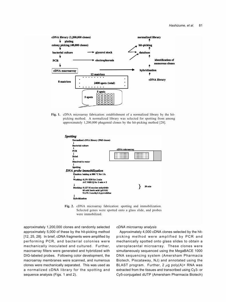
approximately 1,200,000 clones and randomly selectedapproximately 5,000 of these by the hit-picking method[12, 25, 28]. In brief, cDNA fragments were amplified byper fo rming PCR, and bac te r ia l co lon ies weremechanical ly inoculated and cultured. Further,macroarray filters were generated and hybridized withDIG-labeled probes. Following color development, themacroarray membranes were scanned, and numerousclones were mechanically separated. This was used asa normal ized cDNA l ibrary for the spot t ing andsequence analysis (Figs. 1 and 2).
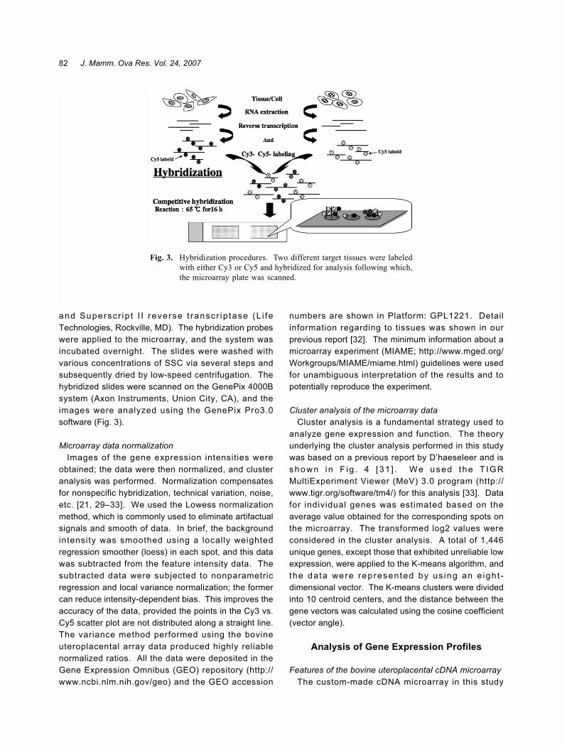
cDNA microarray analysisApproximately 4,000 cDNA clones selected by the hit-
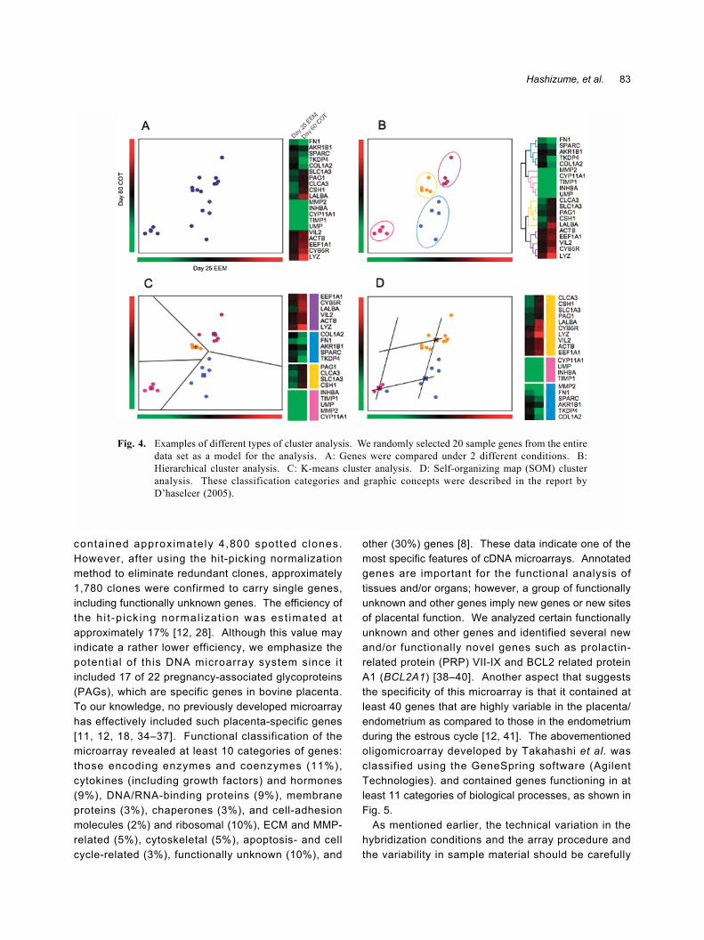
p i c k ing me thod we re amp l i f i ed by PCR andmechanically spotted onto glass slides to obtain auteroplacental microarray. These c lones weresimultaneously sequenced using the MegaBACE 1000DNA sequencing system (Amersham PharmaciaBiotech, Piscataway, NJ) and annotated using theBLAST program. Further, 2 µg poly(A)+ RNA wasextracted from the tissues and transcribed using Cy3- orCy5-conjugated dUTP (Amersham Pharmacia Biotech)
Fig. 1. cDNA microarray fabrication: establishment of a normalized library by the hit-picking method. A normalized library was selected for spotting from amongapproximately 1,200,000 phagemid clones by the hit-picking method [28].
Fig. 2. cDNA microarray fabrication: spotting and immobilization.Selected genes were spotted onto a glass slide, and probeswere immobilized.
82 J. Mamm. Ova Res. Vol. 24, 2007
and Superscr ip t I I reverse t ranscr ip tase (L i feTechnologies, Rockville, MD). The hybridization probeswere applied to the microarray, and the system wasincubated overnight. The slides were washed withvarious concentrations of SSC via several steps andsubsequently dried by low-speed centrifugation. Thehybridized slides were scanned on the GenePix 4000Bsystem (Axon Instruments, Union City, CA), and theimages were analyzed using the GenePix Pro3.0software (Fig. 3).
Microarray data normalizationImages of the gene expression intensities were
obtained; the data were then normalized, and clusteranalysis was performed. Normalization compensatesfor nonspecific hybridization, technical variation, noise,etc. [21, 29�33]. We used the Lowess normalizationmethod, which is commonly used to eliminate artifactualsignals and smooth of data. In brief, the backgroundintensity was smoothed using a locally weightedregression smoother (loess) in each spot, and this datawas subtracted from the feature intensity data. Thesubtracted data were subjected to nonparametricregression and local variance normalization; the formercan reduce intensity-dependent bias. This improves theaccuracy of the data, provided the points in the Cy3 vs.Cy5 scatter plot are not distributed along a straight line.The variance method performed using the bovineuteroplacental array data produced highly reliablenormalized ratios. All the data were deposited in theGene Expression Omnibus (GEO) repository (http://www.ncbi.nlm.nih.gov/geo) and the GEO accession
numbers are shown in Platform: GPL1221. Detailinformation regarding to tissues was shown in ourprevious report [32]. The minimum information about amicroarray experiment (MIAME; http://www.mged.org/Workgroups/MIAME/miame.html) guidelines were usedfor unambiguous interpretation of the results and topotentially reproduce the experiment.
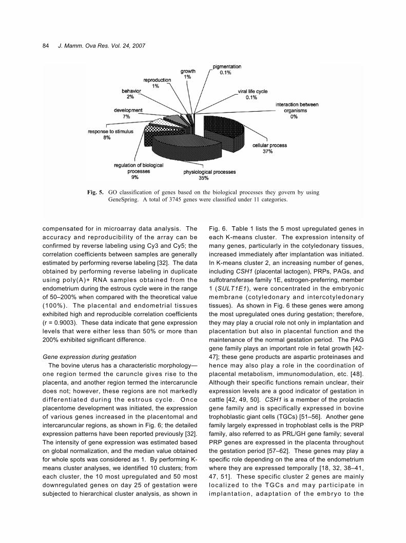
Cluster analysis of the microarray dataCluster analysis is a fundamental strategy used to
analyze gene expression and function. The theoryunderlying the cluster analysis performed in this studywas based on a previous report by D�haeseleer and isshown in F ig . 4 [ 31 ] . We us ed the T IGRMultiExperiment Viewer (MeV) 3.0 program (http://www.tigr.org/software/tm4/) for this analysis [33]. Datafor individual genes was estimated based on theaverage value obtained for the corresponding spots onthe microarray. The transformed log2 values wereconsidered in the cluster analysis. A total of 1,446unique genes, except those that exhibited unreliable lowexpression, were applied to the K-means algorithm, andthe data were represented by us ing an e ight -dimensional vector. The K-means clusters were dividedinto 10 centroid centers, and the distance between thegene vectors was calculated using the cosine coefficient(vector angle).
Analysis of Gene Expression Profiles
Features of the bovine uteroplacental cDNA microarrayThe custom-made cDNA microarray in this study
Fig. 3. Hybridization procedures. Two different target tissues were labeledwith either Cy3 or Cy5 and hybridized for analysis following which,the microarray plate was scanned.
83Hashizume, et al.
contained approximately 4,800 spot ted c lones.However, after using the hit-picking normalizationmethod to eliminate redundant clones, approximately1,780 clones were confirmed to carry single genes,including functionally unknown genes. The efficiency ofthe h i t -p ick ing normal izat ion was est imated atapproximately 17% [12, 28]. Although this value mayindicate a rather lower efficiency, we emphasize thepotential of this DNA microarray system since i tincluded 17 of 22 pregnancy-associated glycoproteins(PAGs), which are specific genes in bovine placenta.To our knowledge, no previously developed microarrayhas effectively included such placenta-specific genes[11, 12, 18, 34�37]. Functional classification of themicroarray revealed at least 10 categories of genes:those encoding enzymes and coenzymes (11%),cytokines (including growth factors) and hormones(9%), DNA/RNA-binding proteins (9%), membraneproteins (3%), chaperones (3%), and cell-adhesionmolecules (2%) and ribosomal (10%), ECM and MMP-related (5%), cytoskeletal (5%), apoptosis- and cellcycle-related (3%), functionally unknown (10%), and
other (30%) genes [8]. These data indicate one of themost specific features of cDNA microarrays. Annotatedgenes are important for the functional analysis oftissues and/or organs; however, a group of functionallyunknown and other genes imply new genes or new sitesof placental function. We analyzed certain functionallyunknown and other genes and identified several newand/or functionally novel genes such as prolactin-related protein (PRP) VII-IX and BCL2 related proteinA1 (BCL2A1) [38�40]. Another aspect that suggeststhe specificity of this microarray is that it contained atleast 40 genes that are highly variable in the placenta/endometrium as compared to those in the endometriumduring the estrous cycle [12, 41]. The abovementionedoligomicroarray developed by Takahashi et al. wasclassified using the GeneSpring software (AgilentTechnologies). and contained genes functioning in atleast 11 categories of biological processes, as shown inFig. 5.
As mentioned earlier, the technical variation in thehybridization conditions and the array procedure andthe variability in sample material should be carefully
Fig. 4. Examples of different types of cluster analysis. We randomly selected 20 sample genes from the entiredata set as a model for the analysis. A: Genes were compared under 2 different conditions. B:Hierarchical cluster analysis. C: K-means cluster analysis. D: Self-organizing map (SOM) clusteranalysis. These classification categories and graphic concepts were described in the report byD�haseleer (2005).
84 J. Mamm. Ova Res. Vol. 24, 2007
compensated for in microarray data analysis. Theaccuracy and reproducibil i ty of the array can beconfirmed by reverse labeling using Cy3 and Cy5; thecorrelation coefficients between samples are generallyestimated by performing reverse labeling [32]. The dataobtained by performing reverse labeling in duplicateusing poly(A)+ RNA samples obtained from theendometrium during the estrous cycle were in the rangeof 50�200% when compared with the theoretical value(100%). The placental and endometrial t issuesexhibited high and reproducible correlation coefficients(r = 0.9003). These data indicate that gene expressionlevels that were either less than 50% or more than200% exhibited significant difference.
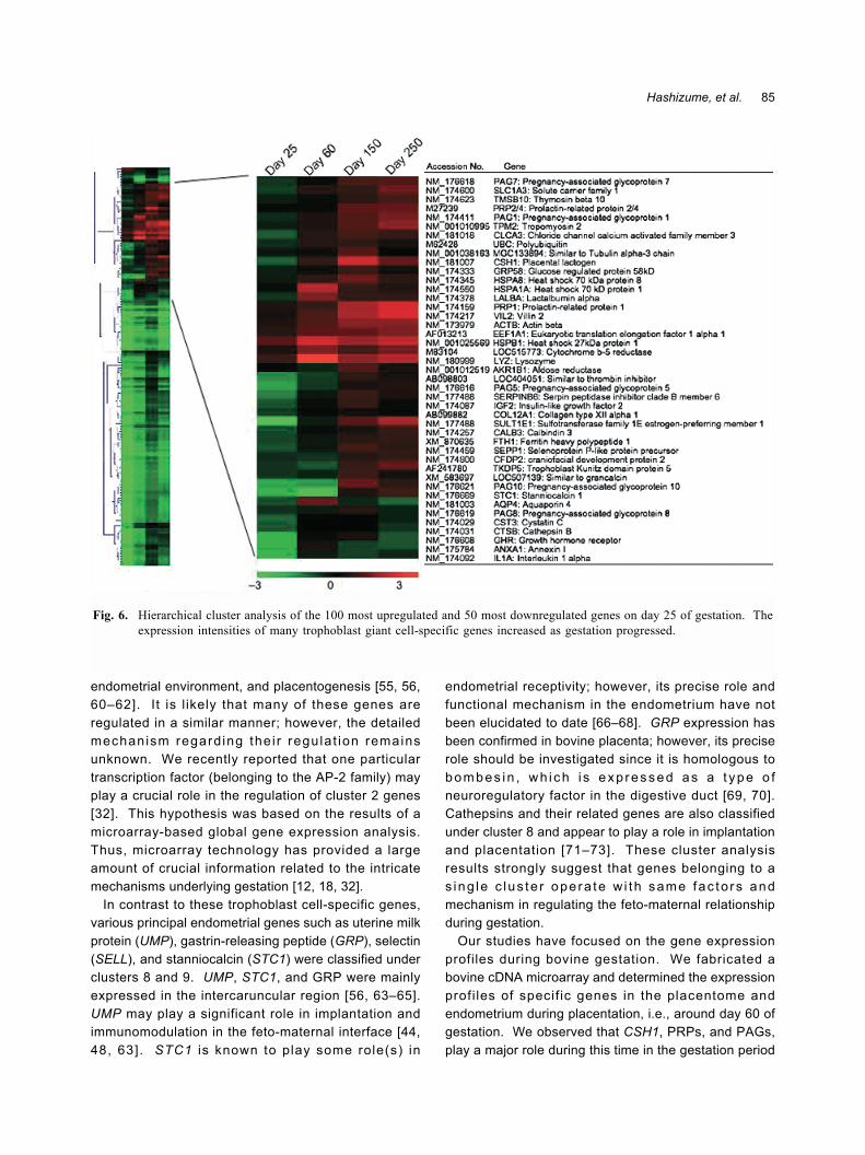
Gene expression during gestationThe bovine uterus has a characteristic morphology�
one region termed the caruncle gives rise to theplacenta, and another region termed the intercaruncledoes not; however, these regions are not markedlyd i f fe rent ia ted dur ing the es t rous cyc le . Onceplacentome development was initiated, the expressionof various genes increased in the placentomal andintercaruncular regions, as shown in Fig. 6; the detailedexpression patterns have been reported previously [32].The intensity of gene expression was estimated basedon global normalization, and the median value obtainedfor whole spots was considered as 1. By performing K-means cluster analyses, we identified 10 clusters; fromeach cluster, the 10 most upregulated and 50 mostdownregulated genes on day 25 of gestation weresubjected to hierarchical cluster analysis, as shown in
Fig. 6. Table 1 lists the 5 most upregulated genes ineach K-means cluster. The expression intensity ofmany genes, particularly in the cotyledonary tissues,increased immediately after implantation was initiated.In K-means cluster 2, an increasing number of genes,including CSH1 (placental lactogen), PRPs, PAGs, andsulfotransferase family 1E, estrogen-preferring, member1 (SULT1E1), were concentrated in the embryonicmembrane (cotyledonary and intercotyledonarytissues). As shown in Fig. 6 these genes were amongthe most upregulated ones during gestation; therefore,they may play a crucial role not only in implantation andplacentation but also in placental function and themaintenance of the normal gestation period. The PAGgene family plays an important role in fetal growth [42-47]; these gene products are aspartic proteinases andhence may also play a role in the coordination ofplacental metabolism, immunomodulation, etc. [48].Although their specific functions remain unclear, theirexpression levels are a good indicator of gestation incattle [42, 49, 50]. CSH1 is a member of the prolactingene family and is specifically expressed in bovinetrophoblastic giant cells (TGCs) [51�56]. Another genefamily largely expressed in trophoblast cells is the PRPfamily, also referred to as PRL/GH gene family; severalPRP genes are expressed in the placenta throughoutthe gestation period [57�62]. These genes may play aspecific role depending on the area of the endometriumwhere they are expressed temporally [18, 32, 38�41,47, 51]. These specific cluster 2 genes are mainlyloca l i zed to the TGCs and may pa r t i c ipa te inimplanta t ion , adapta t ion o f the embryo to the
Fig. 5. GO classification of genes based on the biological processes they govern by usingGeneSpring. A total of 3745 genes were classified under 11 categories.
85Hashizume, et al.
endometrial environment, and placentogenesis [55, 56,60�62]. It is l ikely that many of these genes areregulated in a similar manner; however, the detailedmechanism regarding the i r regulat ion remainsunknown. We recently reported that one particulartranscription factor (belonging to the AP-2 family) mayplay a crucial role in the regulation of cluster 2 genes[32]. This hypothesis was based on the results of amicroarray-based global gene expression analysis.Thus, microarray technology has provided a largeamount of crucial information related to the intricatemechanisms underlying gestation [12, 18, 32].
In contrast to these trophoblast cell-specific genes,various principal endometrial genes such as uterine milkprotein (UMP), gastrin-releasing peptide (GRP), selectin(SELL), and stanniocalcin (STC1) were classified underclusters 8 and 9. UMP, STC1, and GRP were mainlyexpressed in the intercaruncular region [56, 63�65].UMP may play a significant role in implantation andimmunomodulation in the feto-maternal interface [44,48, 63]. STC1 is known to play some role(s) in
endometrial receptivity; however, its precise role andfunctional mechanism in the endometrium have notbeen elucidated to date [66�68]. GRP expression hasbeen confirmed in bovine placenta; however, its preciserole should be investigated since it is homologous tobombes in , wh i ch i s exp ressed as a t ype o fneuroregulatory factor in the digestive duct [69, 70].Cathepsins and their related genes are also classifiedunder cluster 8 and appear to play a role in implantationand placentation [71�73]. These cluster analysisresults strongly suggest that genes belonging to as ing le c lus te r ope ra te w i th same fac to rs andmechanism in regulating the feto-maternal relationshipduring gestation.
Our studies have focused on the gene expressionprofiles during bovine gestation. We fabricated abovine cDNA microarray and determined the expressionprofi les of specif ic genes in the placentome andendometrium during placentation, i.e., around day 60 ofgestation. We observed that CSH1, PRPs, and PAGs,play a major role during this time in the gestation period
Fig. 6. Hierarchical cluster analysis of the 100 most upregulated and 50 most downregulated genes on day 25 of gestation. Theexpression intensities of many trophoblast giant cell-specific genes increased as gestation progressed.
86 J. Mamm. Ova Res. Vol. 24, 2007
[12]. Further, we examined the embryonic andextraembryonic gene expression during the peri-implantation period, and several specific gene clumpswere detected around the time of implantation [74].
Aberrations were detected in some trophoblast-specificgenes in somatic cell nuclear-transferred (SNT) clonedcattle cells; no major variations were observed in thegene expression in the cloned trophoblast cells [41].
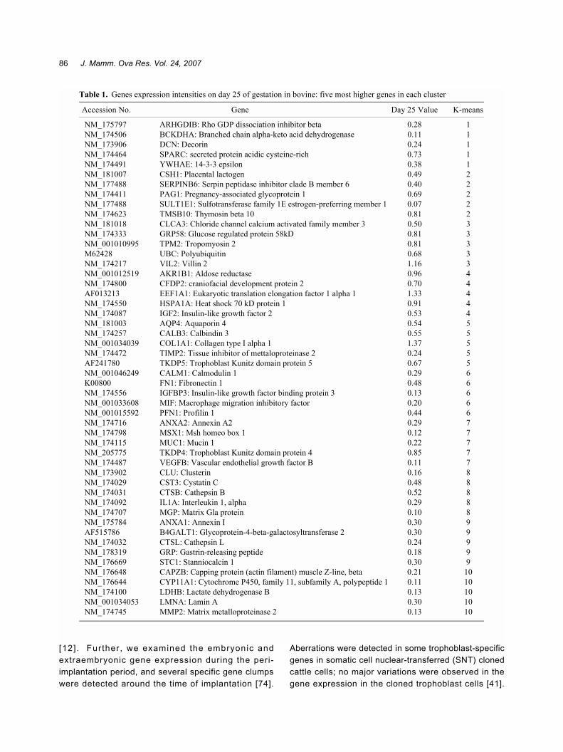
Table 1. Genes expression intensities on day 25 of gestation in bovine: five most higher genes in each cluster
Accession No. Gene Day 25 Value K-means
NM_175797 ARHGDIB: Rho GDP dissociation inhibitor beta 0.28 1NM_174506 BCKDHA: Branched chain alpha-keto acid dehydrogenase 0.11 1NM_173906 DCN: Decorin 0.24 1NM_174464 SPARC: secreted protein acidic cysteine-rich 0.73 1NM_174491 YWHAE: 14-3-3 epsilon 0.38 1NM_181007 CSH1: Placental lactogen 0.49 2NM_177488 SERPINB6: Serpin peptidase inhibitor clade B member 6 0.40 2NM_174411 PAG1: Pregnancy-associated glycoprotein 1 0.69 2NM_177488 SULT1E1: Sulfotransferase family 1E estrogen-preferring member 1 0.07 2NM_174623 TMSB10: Thymosin beta 10 0.81 2NM_181018 CLCA3: Chloride channel calcium activated family member 3 0.50 3NM_174333 GRP58: Glucose regulated protein 58kD 0.81 3NM_001010995 TPM2: Tropomyosin 2 0.81 3M62428 UBC: Polyubiquitin 0.68 3NM_174217 VIL2: Villin 2 1.16 3NM_001012519 AKR1B1: Aldose reductase 0.96 4NM_174800 CFDP2: craniofacial development protein 2 0.70 4AF013213 EEF1A1: Eukaryotic translation elongation factor 1 alpha 1 1.33 4NM_174550 HSPA1A: Heat shock 70 kD protein 1 0.91 4NM_174087 IGF2: Insulin-like growth factor 2 0.53 4NM_181003 AQP4: Aquaporin 4 0.54 5NM_174257 CALB3: Calbindin 3 0.55 5NM_001034039 COL1A1: Collagen type I alpha 1 1.37 5NM_174472 TIMP2: Tissue inhibitor of mettaloproteinase 2 0.24 5AF241780 TKDP5: Trophoblast Kunitz domain protein 5 0.67 5NM_001046249 CALM1: Calmodulin 1 0.29 6K00800 FN1: Fibronectin 1 0.48 6NM_174556 IGFBP3: Insulin-like growth factor binding protein 3 0.13 6NM_001033608 MIF: Macrophage migration inhibitory factor 0.20 6NM_001015592 PFN1: Profilin 1 0.44 6NM_174716 ANXA2: Annexin A2 0.29 7NM_174798 MSX1: Msh homeo box 1 0.12 7NM_174115 MUC1: Mucin 1 0.22 7NM_205775 TKDP4: Trophoblast Kunitz domain protein 4 0.85 7NM_174487 VEGFB: Vascular endothelial growth factor B 0.11 7NM_173902 CLU: Clusterin 0.16 8NM_174029 CST3: Cystatin C 0.48 8NM_174031 CTSB: Cathepsin B 0.52 8NM_174092 IL1A: Interleukin 1, alpha 0.29 8NM_174707 MGP: Matrix Gla protein 0.10 8NM_175784 ANXA1: Annexin I 0.30 9AF515786 B4GALT1: Glycoprotein-4-beta-galactosyltransferase 2 0.30 9NM_174032 CTSL: Cathepsin L 0.24 9NM_178319 GRP: Gastrin-releasing peptide 0.18 9NM_176669 STC1: Stanniocalcin 1 0.30 9NM_176648 CAPZB: Capping protein (actin filament) muscle Z-line, beta 0.21 10NM_176644 CYP11A1: Cytochrome P450, family 11, subfamily A, polypeptide 1 0.11 10NM_174100 LDHB: Lactate dehydrogenase B 0.13 10NM_001034053 LMNA: Lamin A 0.30 10NM_174745 MMP2: Matrix metalloproteinase 2 0.13 10
87Hashizume, et al.
Specific gene expression was examined using a bovinetrophoblast cell l ine (BT-1) as a model for in vitroanalysis. Mononucleate trophoblast cel ls wereobserved to differentiate into placental lactogen-expressing TGCs [55, 62, 75]. By using microarraytechniques and bioinformatics, recent studies haveinvestigated global gene expression profiles throughoutthe gestation period and have explored a commonregulatory factor for the expression of these genes.Many trophoblast- and/or endometrium-specific genesexhibited marked variations with temporal and spatialspecificity [32]. We identified several features of bovinegene expression during gestation. (1) Of the genes thatwere classif ied by K-means cluster analysis, ( i)trophoblast-specific genes such as CSH1, PAGs, PRP,and SULT1E1 were classified under cluster 2, and theexpression levels for most of these genes increased asgestation progressed and (ii) endometrium-specificgenes such as UMP, cathepsins, and SELL, wereclassified under clusters 8 and 9, and their expressionwas extremely temporal and spat ial . (2) TGCssimultaneously expressed various molecules, namely,placental lactogen, PRPs, PAGs, heparanase, theantiapoptosis gene BCL2A1, SULT1E1, etc. [38�40,43�47, 76]. (3) The transcription factor AP-2 may play amajor role in regulating bovine TGC functions since thisfamily of transcription factors was also expressed inTGCs and their expression increased as gestationprogressed. These gene express ion analysesemphasize the significance of the trophoblast celllineage since many genes such as CSH1 and AP-2 aredetected in TGCs in various rodent species as well as inhumans and ruminants [32, 77�80].
During the last decade, microarray technology hasdeveloped rapidly and has easy applications. It hasalready been applied in various fields such as biology,toxicology, and medicine, and the data have beenanalyzed by bioinfomatic methods [81�86]; however,the methods of analysis have not been standardized asyet [19, 20, 24, 87, 88]. Microarray technology canprovide information regarding not only specific genes intissues but also gene cascades and/or moleculari n te rac t i ons i n ce l l s an d t i ss ues . Cu r ren t l y ,bioinformatics tools are available to analyze thisinformation; however, there exists a communication gapbetween biologists and bioinformatics scientists.Bridging this gap is of paramount importance.
Acknowledgements
The authors express their sincere thanks to Drs.
Hiroko Ishiwata and Herath Chandra B of the NationalInstitute of Agrobiological Sciences; Drs. Akira Ito andTakashi Sato of the Tokyo University of Pharmacy andLife Sciences; and Dr. Gozo Tsujimoto and group(Department of Genomic Drug Discovery Science,Graduate School of Pharmaceutical Sciences, KyotoUniversity), part icularly Susumu Katsuma, AkiraHirasawa, Satoshi Shiojima, Hiroshi Ikawa, YasuhitoSuzuki, and Gozo Tsujimoto, for their assistance in thecDNA microarray fabrication and data analysis. Theauthors also thank Mrs. Misako Akiyama for her kindassistance. This study was supported in part by theOrganized Research Combination System, Hoga-kenkyu (16658105); Kiban B (17380172); Kiban C(17580284) of the Ministry of Education, Science andTechnology; and the Bio-oriented Technology ResearchAdvancement Institute, Japan. The authors alsoreceived a Research Project for Utilizing AdvancedTechnologies grant (05-1770) from the Ministry ofAgriculture, Forestry and Fisheries, Japan, and anAnimal Remodeling Project grant (05-201, 202) from theNational Institute of Agrobiological Sciences.
References
1) Wooding, F.B.P. and Flint, A.P. (1994) Placentation. In:Marshall�s Physiology of Reproduction Vol. 4. 4th ed.(Lamming, G.E., ed.), pp.233�460, Chapman & Hall,London.
2) Cross, J.C., Werb, Z. and Fisher, S.J. (1994) Implantationand the placenta: key pieces of the development puzzle.Science, 266, 1508�1518.
3) Jauniaux, E., Watson, A.L., Hempstock, J., Bao, Y.P.,Skepper, J.N. and Burton, G.J. (2000) Onset of maternalarterial blood flow and placental oxidative stress. Apossible factor in human early pregnancy failure. Am. J.Pathol., 15, 2111�2122.
4) Spencer, T.E., Johnson, G.A., Bazer, F.W., Burghardt, R.C.and Palmarini, M. (2007) Pregnancy recognition andconceptus implantation in domestic ruminants: roles ofprogesterone, interferons and endogenous retroviruses.Reprod. Fertil. Dev., 19, 65�78.
5) Hayes, P.C., Wolf, C.R. and Hayes, J.D. (1989) Blottingtechniques for the study of DNA, RNA, and proteins. BMJ,299, 965�968.
6) Schena, M., Shalon, D., Davis, R.W. and Brown, P.O.(1995) Quantitative monitoring of gene expression patternswith a complementary DNA microarray. Science, 270,467�470.
7) Shalon, D., Smith, S.J. and Brown, P.O. (1996) A DNAmicroarray system for analyzing complex DNA samplesusing two-color fluorescent probe hybridization. GenomeRes., 6, 639�645.
8) DeRisi, J., Penland, L., Brown, P.O., Bittner, M.L.,
88 J. Mamm. Ova Res. Vol. 24, 2007
Meltzer, P.S., Ray, M., Chen, Y., Su, Y.A. and Trent, J.M.(1996) Use of a cDNA microarray to analyse geneexpression patterns in human cancer. Nat. Genet., 14, 457�460.
9) Ahrendt, S.A., Halachmi, S., Chow, J.T., Wu, L.,Halachmi, N., Yang, S.C., Wehage, S., Jen, J. andSidransky, D. (1999) Rapid p53 sequence analysis inprimary lung cancer using an oligonucleotide probe array.Proc. Natl. Acad. Sci. USA., 96, 7382�7387.
10) Brazma, A. and Vilo, J. (2000) Gene expression dataanalysis. FEBS Lett., 480, 7�24.
11) Band, M.R., Olmstead, C., Everts, R.E., Liu, Z.L. andLewin, H.A. (2002) A 3800 gene microarray for cattlefunctional genomics: comparison of gene expression inspleen, placenta, and brain. Anim. Biotechnol., 13, 163�172.
12) Ishiwata, H., Katsuma, S., Kizaki, K., Patel, O.V., Nakano,H., Takahashi, T., Imai, K., Hirasawa, A., Shiojima, S.,Ikawa, H., Suzuki, Y., Tsujimoto, G., Izaike, Y., Todoroki,J. and Hashizume, K. (2003) Characterization of geneexpression profiles in early bovine pregnancy using acustom cDNA microarray. Mol. Reprod. Dev., 65, 9�18.
13) Tanaka, T.S., Jaradat, S.A., Lim, M.K., Kargul, G.J.,Wang, X., Grahovac, M.J., Pantano, S., Sano, Y., Piao, Y.,Nagaraja, R., Doi, H., Wood, W.H. 3rd., Becker, K.G. andKo, M.S. (2000) Genome-wide expression profiling ofmid-gestation placenta and embryo using a 15,000 mousedevelopmental cDNA microarray. Proc. Natl. Acad. Sci.USA., 97, 9127�9132.
14) Yoshioka, K., Matsuda, F., Takakura, K., Noda, Y.,Imakawa, K. and Sakai, S. (2000) Determination of genesinvolved in the process of implantation: application ofGeneChip to scan 6500 genes. Biochem. Biophys. Res.Commun., 272, 531�538.
15) Kao, L.C., Tulac, S., Lobo, S., Imani, B., Yang, J.P.,Germeyer, A., Osteen, K., Taylor, R.N., Lessey, B.A. andGiudice, L.C. (2002) Global gene profiling in humanendometrium during the window of implantat ion.Endocrinology, 143, 2119�2138.
16) Aronow, B.J., Richardson, B.D. and Handwerger, S. (2001)Microarray analysis of trophoblast differentiation: geneexpression reprogramming in key gene function categories.Physiol. Genomics, 6, 105�116.
17) Chen, H.W., Chen, J.J., Tzeng, C.R., Li, H.N., Cheng, Y.F.,Chang, C.W., Wang, R.S., Yang, P.C. and Lee, Y.T. (2002)Global analysis of differentially expressed genes in earlygestational deciduas and chorionic villi using a 9600human cDNA microarray. Mol. Hum. Reprod., 8, 475�484.
18) Hashizume, K. (2007) Analysis of uteroplacental-specificmolecules and their functions during implantation andplacentation in the bovine. J. Reprod. Dev., 53, 1�11.
19) Schulze, A. and Downward, J. (2001) Navigating geneexpression using microarrays�a technology review. Nat.Cell Biol., 3, E190�E195.
20) Mutch, D.M., Berger, A., Mansourian, R., Rytz, A. andRoberts, M.A. (2002) The limit fold change model: apractical approach for selecting differentially expressed
genes from microarray data. BMC. Bioinformatics., 3, 17.21) Tarca, A.L., Romero, R. and Draghici, S. (2006) Analysis
of microarray experiments of gene expression profiling.Am. J. Obstet. Gynecol., 195, 373�388.
22) Iwahashi, H., Kitagawa, E., Suzuki, Y., Ueda,Y., Ishizawa,Y.H., Nobumasa, H., Kuboki, Y., Hosoda, H. andIwahashi , Y. (2007) Evaluation of toxici ty of themycotoxin citrinin using yeast ORF DNA microarray andOligo DNA microarray. BMC. Genomics, 8, 95.
23) Perez-Diez, A., Morgun, A. and Shulzhenko, N. (2007)Microarrays for cancer diagnosis and classification. Adv.Exp. Med. Biol., 593, 74�85.
24) White, C.A. and Salamonsen, L.A. (2005) A guide to issuesin microarray analysis: application to endometrial biology.Reproduction, 130, 1�13.
25) Herath, C.B., Shiojima, S., Ishiwata, H., Katsuma, S.,Kadowaki, T., Ushizawa, K., Imai, K., Takahashi, T.,Hirasawa, A., Tsujimoto, G. and Hashizume, K. (2004)Pregnancy-associated changes in genome-wide geneexpression profiles in the l iver of cow throughoutpregnancy. Biochem. Biophys. Res. Commun., 313, 666�680.
26) Nikitenko, L., Morgan, G., Kolesnikov, S.I. and Wooding,F .B .P . ( 1 998 ) Imm unocy toche mica l and i n s i t uhybridization studies of the distribution of calbindin D 9kin the bovine placenta throughout pregnancy. J. Histchem.Cytochem., 46, 679�688.
27) Regnault, T.R., Orbus, R.J., De Vrijer, B., Davidsen, M.L.,Galan, H.L., Wilkening, R.B. and Anthony, R.V. (2002)Placental expression of VEGF, PIGF, and their receptors ina model of placental insufficiency-intrauterine growthrestriction (PI-IUGR). Placenta., 23, 132�144.
28) Katsuma, S., Shiojima, S., Hirasawa, A., Suzuki, Y.,Ikawa, H., Takagaki, K., Kaminishi, Y., Murai, M., Ohgi,T., Yano, J. and Tsujimoto, G. (2001) Functional genomicsearch of G-protein coupled receptors (GPCR) usingmicroarrays with normalized cDNA library. MethodsEnzymol., 345, 585�600.
29) Werner, T. (2001) Cluster analysis and promoter modellingas bioinformatics tools for the identification of target genesfrom expression array data. Pharmacogenomicsm, 2, 25�36.
30) Yang, Y.H., Dudoit, S., Luu, P., Lin, D.M., Peng, V., Ngai,J. and Speed, T.P. (2002) Normalization for cDNAmicroarray data: a robust composite method addressingsingle and multiple slide system variation. Nucleic. AcidsRes., 30, e15.
31) D�haeseleer, P. (2005) How does gene expressionclustering work? Nat. Biotechnol., 23, 1499�1501.
32) Ushizawa, K., Takahashi, T., Hosoe, M., Ishiwata, H.,Kaneyama, K., Kizaki, K. and Hashizume, K. (2007)Global gene expression analysis and regulation of theprincipal genes expressed in bovine placenta in relation tothe transcription factor AP-2 family. Reprod. Biol.Endocrinol, 5, 17.
33) Saeed, A.I., Sharov, V., White, J., Li, J., Liang, W.,Bhagabati, N., Braisted, J., Klapa, M., Currier, T.,
89Hashizume, et al.
Thiagarajan, M., Sturn, A., Snuffin, M., Rezantsev, A.,Popov, D., Ryltsov, A., Kostukovich, E., Borisovsky, I.,Liu, Z., Vinsavich, A., Trush, V. and Quackenbush, J.(2003) TM4: a free, open-source system for microarraydata management and analysis. Biotechniques, 34, 374�378.
34) Bauersachs, S., Ulbrich, S.E., Gross, K., Schmidt, S.E.,Meyer, H.H., Einspanier, R., Wenigerkind, H., Vermehren,M., Blum, H., Sinowatz, F. and Wolf, E. (2005) Geneexpression profiling of bovine endometrium during theoestrous cycle: detection of molecular pathways involvedin functional changes. J. Mol. Endocrinol., 34, 889�908.
35) Everts, R.E., Band, M.R., Liu, Z.L., Kumar, C.G., Liu, L.,Loor, J.J., Oliveira, R. and Lewin, H.A. (2005) A 7872cDNA microarray and its use in bovine functionalgenomics. Vet. Immunol. Immunopathol., 105, 235�245.
36) Smith, S.L., Everts, R.E., Tian, X.C., Du, F., Sung, L.Y.,Rodriguez-Zas, S.L., Jeong, B.S., Renard, J.P., Lewin,H.A. and Yang, X. (2005) Global gene expression profilesreveal significant nuclear reprogramming by the blastocyststage after cloning. Proc. Natl. Acad. Sci. USA., 102,17582�17587.
37) Bauersachs, S., Ulbrich, S.E., Gross, K., Schmidt, S.E.,Meyer, H.H., Wenigerkind, H., Vermehren, M., Sinowatz,F., Blum, H. and Wolf, E. (2006) Embryo-inducedtranscriptome changes in bovine endometrium revealspecies-specific and common molecular markers of uterinereceptivity. Reproduction, 132, 319�331.
38) Ushizawa, K., Takahashi, T., Hosoe, M., Kaneyama, K.and Hashizume, K. (2005) Cloning and expression of twonew prolactin-related proteins, prolactin-related protein-VII I and - IX , in bov ine p lacenta . Reprod . B io l .Endocrinol., 3, 68.
39) Ushizawa, K., Kaneyama, K., Takahashi, T., Tokunaga, T.,Tsunoda, Y. and Hashizume, K. (2005) Cloning andexpression of a new member of prolactin-related protein inbovine placenta: bovine prolactin-related protein-VII.Biochem. Biophys. Res. Commun., 326, 435�441.
40) Ushizawa, K., Takahashi, T., Kaneyama, K., Hosoe, M.and Hashizume, K. (2006) Cloning of the bovineantiapoptotic regulator, BCL2-related protein A1, and itsexpression in trophoblastic binucleate cells of bovineplacenta. Biol. Reprod., 74, 344�351.
41) Hashizume, K., Ishiwata, H., Kizaki, K., Yamada, O.,Takahashi, T., Imai, K., Patel, O.V., Akagi, S., Shimizu,M., Takahashi, S., Katsuma, S., Shiojima, S., Hirasawa, A.,Tsujimoto, G., Todoroki, J . and Izaike, Y. (2002)Implantation and placental development in somatic cellclone recipient cows. Cloning Stem Cells, 4, 197�209.
42) Green, J.A., Parks, T.E., Avalle, M.P., Telugu, B.P.,McLain, A.L., Peterson, A.J., McMillan, W., Mathialagan,N., Hook, R.R., Xie, S. and Roberts, R.M. (2005) Theestablishment of an ELISA for the detection of pregnancy-associated glycoproteins (PAGs) in the serum of pregnantcows and heifers. Theriogenology, 63, 1481�1503.
43) Wooding, F.B., Roberts, R.M. and Green, J.A. (2005) Lightand electron microscope immunocytochemical studies of
the distribution of pregnancy associated glycoproteins(PAGs) throughout pregnancy in the cow: possiblefunctional implications. Placenta., 26, 807�827.
44) Patel, O.V., Sulon, J., Beckers, J.F., Takahashi, T., Hirako,M., Sasaki, N. and Domeki, I. (1997) Plasma bovinepregnancy-associated glycoprotein concentrat ionsthroughout gestation in relationship to fetal number in thecow. Eur. J. Endocrinol., 137, 423�428.
45) Patel, O.V., Yamada, O., Kizaki, K., Takahashi, T., Imai,K. and Hashizume, K. (2004) Quantitative analysisthroughout pregnancy of placentomal and interplacentomalexpression of pregnancy-associated glycoproteins-1 and -9in the cow. Mol. Reprod. Dev., 67, 257�263.
46) Patel, O.V., Yamada, O., Kizaki, K., Takahashi, T., Imai,K., Takahashi, S., Izaike, Y., Schuler, L.A., Takezawa, T.and Hashizume, K. (2004) Expression of trophoblast cell-specific pregnancy-related genes in somatic cell-clonedbovine pregnancies. Biol. Reprod., 70, 1114�1120.
47) Klisch, K., De Sousa, N.M., Beckers, J.F., Leiser, R. andPich, A. (2005) Pregnancy associated glycoprotein-1, -6, -7, and -17 are major products of bovine binucleatetrophoblast giant cells at midpregnancy. Mol. Reprod.Dev., 71, 453�460.
48) Hansen, P.J. (1995) Interactions between the immunesystem and the ruminant conceptus. J. Reprod. Fertil.Suppl., 49, 69�82.
49) Zoli, A.P., Guilbault, L.A., Delahaut, P., Ortiz, W.B. andBeckers, J.F. (1992) Radioimmunoassay of a bovinepregnancy-associated glycoprotein in serum: its applicationfor pregnancy diagnosis. Biol. Reprod., 46, 83�92.
50) Green, J.A., Xie, S., Quan, X., Bao, B., Gan, X.,Mathialagan, N., Beckers, J.F. and Roberts, R.M. (2000)Pregnancy-associated bovine and ovine glycoproteinsexhibit spatially and temporally distinct expression patternsduring pregnancy. Biol. Reprod., 62, 1624�1631.
51) Wooding, F.B. (1992) Current topic: thesynepitheliochorial placenta of ruminants: binucleate cellfusions and hormone production. Placenta., 13, 101�113.
52) Byatt, J.C., Eppard, P.J., Veenhuizen, J.J., Curran, T.L.,Curran, D.F., McGrath, M.F. and Collier, R.J. (1994)Stimulation of mammogenesis and lactogenesis byrecombinant bovine placental lactogen in steroid-primeddairy heifers. J. Endocrinol., 140, 33�43.
53) Lucy, M.C., Byatt, J.C., Curran, T.L., Curran, D.F. andCollier, R.J. (1994) Placental lactogen and somatotropin:hormone binding to the corpus luteum and effects on thegrowth and functions of the ovary in heifers. Biol. Reprod.,50, 136�144.
54) Byatt, J.C., Sorbet, R.H., Eppard, P.J., Curran, T.L.,Curran, D.F. and Collier, R.J. (1997) The effect ofrecombinant bovine placental lactogen on induced lactationin dairy heifers. J. Dairy Sci., 80, 496�503.
55) Nakano, H., Shimada, A., Imai, K., Takezawa, T.K.,Takahashi , T . and Hashizume, K. (2002) Bovinetrophoblastic cell differentiation on collagen substrata:formation of binucleate cells expressing placental lactogen.Cell Tissue Res., 307, 225�235.
90 J. Mamm. Ova Res. Vol. 24, 2007
56) Yamada, O., Todoroki, J., Kizaki, K., Takahashi, T., Imai,K., Patel, O.V., Schuler, L.A. and Hashizume, K. (2002)Expression of prolactin-related protein I at the fetomaternalinterface during the implantat ion period in cows.Reproduction, 124, 427�437.
57) Milosavljevic, M., Duello, T.M. and Schuler, L.A. (1989)In situ localization of two prolactin-related messengerribonucleic acids to binucleate cells of bovine placentomes.Endocrinology, 125, 883�889.
58) Kessler, M.A., Duello, T.M. and Schuler, L.A. (1991)Expression of prolactin-related hormones in the earlybovine conceptus, and potential for paracrine effect on theendometrium. Endocrinology, 129, 1885�1895.
59) Morgan, G., Wooding, F.B. and Godkin, J.D. (1993)Localization of bovine trophoblast protein-1 in the cowblastocyst during implantation: an immunologicalcryoultrastructural study. Placenta, 14, 641�649.
60) Kessler, M.A. and Schuler, L.A. (1997) Purification andproperties of placental prolactin-related protein-I.Placenta., 18, 29�36.
61) Klisch, K., Boos, A., Friedrich, M., Herzog, K., Feldmann,M., Sousa, N., Beckers, J., Leiser, R. and Schuler, G.(2006) The glycosylation of pregnancy-associatedglycoproteins and prolactin-related protein-I in bovinebinucleate t rophoblast giant cel ls changes beforeparturition. Reproduction, 132, 791�798.
62) Ushizawa, K. and Hashizume, K. (2006) Biology of theprolactin family in bovine placenta. II. Bovine prolactin-related proteins: their expression, structure and proposedroles. Anim. Sci. J., 77, 18�27.
63) Stewart, M.D., Johnson, G.A., Gray, C.A., Burghardt, R.C.,Schuler, L.A., Joyce, M.M., Bazer, F.W. and Spencer, T.E.(2000) Prolactin receptor and uterine milk proteinexpression in the ovine endometrium during the estrouscycle and pregnancy. Biol. Reprod., 62, 1779�1789.
64) Tekin, S. and Hansen, P.J. (2004) Regulation of numbers ofmacrophages in the endometrium of the sheep by systemiceffects of pregnancy, local presence of the conceptus, andprogesterone. Am. J. Reprod. Immunol., 51, 56�62.
65) Xie, S., Green, J., Bixby, J.B., Szafranska, B., DeMartini,J.C., Hecht, S. and Roberts, R.M. (1997) The diversity andevolutionary relationships of the pregnancy-associatedglycoproteins, an aspartic proteinase subfamily consistingof many trophoblast-expressed genes. Proc. Natl. Acad.Sci. USA., 94, 12809�12816.
66) Olsen, H.S., Cepeda, M.A., Zhang, Q.Q., Rose, C.A. andVozzolo, B.L. (1996) Human stanniocalcin: a possiblehormonal regulator of mineral metabolism. Proc. Natl.Acad. Sci. USA., 93, 1792�1796.
67) Harminder, K.D., Varghese, R., Wagner, F.G. and Gabriel,E.D. (2000) Dynamic regulation of mouse ovarianstanniocalcin expression during gestation and lactation.Endocrinology, 141, 3412�3421.
68) Paciga, M., McCudden, C.R., Londos, C., DiMattia, G.E.and Wagner, G.F. (2003) Targeting of big stanniocalcinand its receptor to lipid storage droplets of ovariansteroidogenic cells. J. Biol. Chem., 278, 49549�49554.
69) Jian, X., Sainz, E., Clark, W.A., Jensen, R.T., Battey, J.F.and Northup, J.K. (1999) The bombesin receptor subtypeshave distinct G protein specificities. J. Biol. Chem., 274,11573�11581.
70) Budipitojo, T., Sasaki, M., Cruzana, M.B., Matsuzaki, S.,Iwanaga, T. , Kitamura, N. and Yamada, J . (2004)Ultrastructural localization of gastrin-releasing peptide(GRP) in the uterine gland of cow. Anat. Embryol. (Berl.),208, 1�6.
71) Afonso, S., Romagnano, L. and Babiarz, B. (1997) Theexpression and function of cystatin C and cathepsin B andcathepsin L during mouse embryo implantation andplacentation. Development, 124, 3415�3425.
72) Song, G., Spencer, T.E. and Bazer, F.W. (2005) Cathepsinsin the ovine uterus: regulation by pregnancy, progesterone,and interferon tau. Endocrinology, 146, 4825�4833.
73) Satterfield, M.C., Bazer, F.W. and Spencer, T.E. (2006)Progesterone regulation of preimplantation conceptusgrowth and galectin 15 (LGALS15) in the ovine uterus.Biol. Reprod., 75, 289�296.
74) Ushizawa, K., Herath, C.B., Kaneyama, K., Shiojima, S.,Hirasawa, A., Takahashi, T., Imai, K., Ochiai, K.,Tokunaga, T., Tsunoda, Y., Tsujimoto, G. and Hashizume,K. (2004) cDNA microarray analysis of bovine embryogene expression profiles during the pre-implantationperiod. Reprod. Biol. Endocrinol., 2, 77.
75) Ushizawa, K., Takahashi, T., Kaneyama, K., Tokunaga, T.,Tsunoda, Y. and Hashizume, K. (2005) Gene expressionprofiles of bovine trophoblastic cell line (BT-1) analyzedby a custom cDNA microarray. J. Reprod. Dev., 51, 211�220.
76) Kizaki, K., Yamada, O., Nakano, H., Takahashi, T.,Yamauchi, N., Imai, K. and Hashizume, K. (2003) Cloningand localization of heparanase in bovine placenta.Placenta., 24, 424�430.
77) Blackburn, M.R., Wakamiya, M., Caskey, C.T. andKellems, R.E. (1995) Tissue-specific rescue suggests thatplacental adenosine deaminase is important for fetaldevelopment in mice. J. Biol. Chem., 270, 23891�23894.
78) Wakamiya, M., Blackburn, M.R., Jurecic, R., McArthur,M.J., Geske, R.S., Cart-wright, J.Jr., Mitani, K., Vaishnav,S., Belmont, J .W., Kellems, R.E., Finegold, M.J. ,Montgomery, C.A.Jr., Bradley, A. and Caskey, C.T. (1995)Disruption of the adenosine deaminase gene causeshepatocellular impairment and perinatal lethality in mice.Proc. Natl. Acad. Sci. USA., 92, 3673�3677.
79) Shi, D. and Kellems, R.E. (1998) Transcription factor AP-2gamma regulates murine adenosine deaminase geneexpression during placental development. J. Biol. Chem.,273, 27331�27338.
80) Richardson, B.D., Cheng, Y.H., Langland, R.A. andHandwerger, S. (2001) Differential expression of AP-2gamma and AP-2alpha during human trophoblastdifferentiation. Life Sci., 69, 2157�2165.
81) Adjaye, J., Herwig, R., Herrmann, D., Wruck, W.,Benkahla, A., Brink, T.C., Nowak, M., Carnwath, J.W.,Hultschig, C., Niemann, H. and Lehrach, H. (2004) Cross-
91Hashizume, et al.
species hybridization of human and bovine orthologousgenes on h igh dens i ty cDNA microarrays . BMC.Genomics., 5, 83.
82) Gao, F., Foat, B.C. and Bussemaker, H.J. (2004) Definingtranscriptional networks through integrative modeling ofmRNA expression and transcription factor binding data.BMC. Bioinformatics, 5, 31.
83) Hvidsten, T.R., Wilczynski, B., Kryshtafovych, A., Tiuryn,J., Komorowski, J. and Fidelis, K. (2005) Discoveringregulatory binding-site modules using rule-based learning.Genome Res., 15, 856�866.
84) Kamalakaran, S., Radhakrishnan, S.K. and Beck, W.T.(2005) Identification of estrogen-responsive genes using agenome-wide ana lys i s o f promote r e l ements fortranscription factor binding sites. J. Biol. Chem., 280,
21491�21497.85) Kim, S.Y. and Kim, Y. (2006) Genome-wide prediction of
transcriptional regulatory elements of human promotersusing gene expression and promoter analysis data. BMC.Bioinformatics., 7, 330.
86) Veerla, S. and Hoglund, M. (2006) Analysis of promoterregions of co-expressed genes identified by microarrayanalysis. BMC. Bioinformatics, 7, 384.
87) Kerr, M.K. and Churchill, G.A. (2001) Bootstrappingcluster analysis: assessing the reliability of conclusionsfrom microarray experiments. Proc. Natl. Acad. Sci. USA.,98, 8961�8965.
88) Yoon, D., Yi, S.G., Kim, J.H. and Park, T. (2004) Two-stage normalization using background intensities in cDNAmicroarray data. BMC. Bioinformatics, 5, 97.