Embed Size (px)
Citation preview
1
http://journals.tubitak.gov.tr/biology/
Turkish Journal of Biology Turk J Biol(2015) 39: © TÜBİTAKdoi:10.3906/biy-1411-8
Development of therapeutic proteins: advances and challenges
Muhammad Sajid Hamid AKASH1,2,*, Kanwal REHMAN3, Muhammad TARIQ1, Shuqing CHEN1
1Institute of Pharmacology, Toxicology, and Biochemical Pharmaceutics, College of Pharmaceutical Sciences, Zhejiang University, Hangzhou, P. R. China
2Faculty of Pharmaceutical Sciences, Government College University Faisalabad, Faisalabad, Pakistan3Institute of Pharmacy, Physiology and Pharmacology, University of Agriculture, Faisalabad, Pakistan
* Correspondence: [email protected]
1. Introduction Peptides and proteins are known to have great therapeutic potential against several diseases and syndromes. They are an imperative part of the pharmaceutical industry. In addition, the current progress in the field of pharmaceutical biotechnology has increased the value and number of protein- and peptide-based therapeutics in the market. So far, more than 100 proteins have been approved as therapeutics (Craik et al., 2013). Many others are undergoing clinical trials. The FDA has approved many recombinant proteins as biotechnology medicines, antibody-drug conjugates, vaccines, enzymes, natural/recombinant cytokines, and interferons (Leader et al., 2008). These recombinant therapeutic proteins are also known as biotechnological therapeutics because they are used for multiple purposes, including diagnosis, prophylaxis, disease management, and/or cure. Moreover, protein therapeutics are known to represent one third of the entities that are launched as new medicines under biologics (Tang et al., 2004). Recombinant technologies have produced various protein drugs that are now easily available as therapeutics at suitable prices for the treatment of chronic diseases (Karvar, 2014). Therapeutic proteins are increasingly prominent because
they have proven to be effective in treating many potentially fatal diseases like diabetes, heart disorders, and cancer (Hermeling et al., 2004; Akash et al., 2013a, 2013b 2013c; Ibrahim et al., 2013; Karacali et al., 2014). Interestingly, proteins have now been proven effective even as vaccines that help stimulate the body’s natural defense mechanism for immunogenic response. Moreover, by the help of cloning and expressing cDNA that encodes heterologous proteins, these therapeutic proteins have proven themselves in the pharmaceutical industry (Regan and Jackson, 2003). However, a critical formulation is required for their safe and effective delivery to targeted sites. To achieve this, it is essential to have a complete understanding of the basic requirements for the formulation and delivery of protein-based therapeutics.
Although therapeutic proteins have taken center stage in drug discovery and development (with enhanced safety profiles for human use), there are certain challenges that still need to be addressed. Some of the protein-based therapeutics have been reported to induce immune responses and some other unwanted reactions.
In the current paper we offer detailed insights into various fundamental factors that may play important
Abstract: Therapeutic proteins have shown to be effective against a variety of diseases. Pharmaceutical companies are progressively focusing on research using proteins in search of novel effective therapeutics. The significant attention by researchers has resulted in considerable advances in the production and usage of therapeutic proteins in recent years. In the current study we focused on the understanding of therapeutic proteins and their impact on the human health care system. Advancements in the field of biotechnology have increased and facilitated the production of therapeutically significant proteins to combat various fatal diseases. Although protein-based therapeutics have taken center stage in drug discovery and development and enhanced safety for human beings, certain challenges remain, including safety and immunogenicity issues, protein stability, and degradation issues. We made an attempt to discuss all possible factors that are linked with the basis of therapeutic proteins and possible challenges that are currently being faced by scientists during the development of these therapeutic proteins.
Key words: Therapeutic proteins, protein classification, production of therapeutic proteins, pharmacokinetics of therapeutic proteins, immunogenicity, protein stability, protein degradation
Received: 04.11.2014 Accepted/Published Online: 22.01.2015 Printed: 00.00.2015
Research Article
AKASH et al. / Turk J Biol
2
roles in making proteins therapeutic, and may influence their production, formulation or delivery as drugs. As both proteins and peptides have shown effective therapeutic outcomes against various disease conditions, we will collectively discuss the basics, production, route of administration, and potential outcomes of both proteins and peptides as therapeutics. Moreover, we have also summarized the challenges that are currently faced by pharmaceutical scientists during the development of therapeutic proteins.
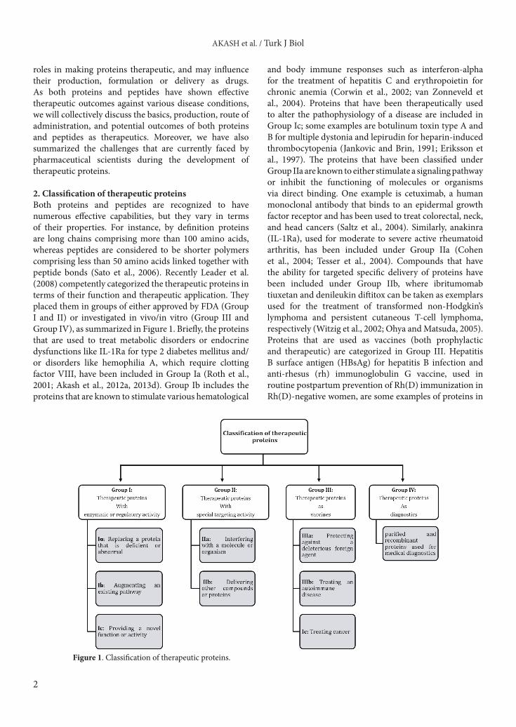
2. Classification of therapeutic proteins Both proteins and peptides are recognized to have numerous effective capabilities, but they vary in terms of their properties. For instance, by definition proteins are long chains comprising more than 100 amino acids, whereas peptides are considered to be shorter polymers comprising less than 50 amino acids linked together with peptide bonds (Sato et al., 2006). Recently Leader et al. (2008) competently categorized the therapeutic proteins in terms of their function and therapeutic application. They placed them in groups of either approved by FDA (Group I and II) or investigated in vivo/in vitro (Group III and Group IV), as summarized in Figure 1. Briefly, the proteins that are used to treat metabolic disorders or endocrine dysfunctions like IL-1Ra for type 2 diabetes mellitus and/or disorders like hemophilia A, which require clotting factor VIII, have been included in Group Ia (Roth et al., 2001; Akash et al., 2012a, 2013d). Group Ib includes the proteins that are known to stimulate various hematological
and body immune responses such as interferon-alpha for the treatment of hepatitis C and erythropoietin for chronic anemia (Corwin et al., 2002; van Zonneveld et al., 2004). Proteins that have been therapeutically used to alter the pathophysiology of a disease are included in Group Ic; some examples are botulinum toxin type A and B for multiple dystonia and lepirudin for heparin-induced thrombocytopenia (Jankovic and Brin, 1991; Eriksson et al., 1997). The proteins that have been classified under Group IIa are known to either stimulate a signaling pathway or inhibit the functioning of molecules or organisms via direct binding. One example is cetuximab, a human monoclonal antibody that binds to an epidermal growth factor receptor and has been used to treat colorectal, neck, and head cancers (Saltz et al., 2004). Similarly, anakinra (IL-1Ra), used for moderate to severe active rheumatoid arthritis, has been included under Group IIa (Cohen et al., 2004; Tesser et al., 2004). Compounds that have the ability for targeted specific delivery of proteins have been included under Group IIb, where ibritumomab tiuxetan and denileukin diftitox can be taken as exemplars used for the treatment of transformed non-Hodgkin’s lymphoma and persistent cutaneous T-cell lymphoma, respectively (Witzig et al., 2002; Ohya and Matsuda, 2005). Proteins that are used as vaccines (both prophylactic and therapeutic) are categorized in Group III. Hepatitis B surface antigen (HBsAg) for hepatitis B infection and anti-rhesus (rh) immunoglobulin G vaccine, used in routine postpartum prevention of Rh(D) immunization in Rh(D)-negative women, are some examples of proteins in
Figure 1. Classification of therapeutic proteins.

AKASH et al. / Turk J Biol
3
Group III (Crosnier et al., 1981; MacKenzie et al., 2004). Lastly, proteins that are members of Group IV are used for diagnostic purposes such as infectious disease diagnostics and imaging agents for cancer detection (Sodee et al., 2000; Campos-Neto et al., 2001). In short, all proteins, and particularly recombinant human proteins that have the advantage of being approved by the FDA, have their special place among the biomedical products with potential utility in every field related to biologics.
In addition to the eukaryotic-based therapeutic proteins mentioned above, some prokaryotic-based therapeutic proteins have also been established. For instance, L-asparaginase (a chemotherapeutic enzyme), methionine gamma-lyase (a possible antitumor agent), and L-glutaminase (an antileukemia enzyme) are the most commonly used therapeutic proteins in the treatment of different diseases (Prakash et al., 2009; Sato and Nozaki, 2009; Ebrahiminezhad et al., 2011; Sharma et al., 2014).
3. Production and purification of therapeutic proteins The source and methodology for the production and purification of therapeutic proteins have gone through major developments. Usually, proteins with low molecular weight are produced chemically, while those with a large number of amino acids are produced using living cells (Mrsny, 2004). Recombinant DNA technology is usually used for the synthesis of many recombinant therapeutic proteins (for instance using mammalian Chinese hamster ovary cells (CHO)) (Jayapal et al., 2007). Examples of therapeutic proteins that have utilized CHO for their production include β-interferon, factor
VIII, and erythropoietin (Kelley, 2001). Escherichia coli (E.coli) as a microbial host has been also employed for the production of different types of proteins (Kamionka, 2011; Gökbulut and Arslanoğlu, 2013; Ahmad et al., 2014; Wang et al., 2014). S. cerevisiae and P. pastoris are also known to be good microbial hosts for better expression and production of therapeutic proteins. For the recombinant expression of therapeutic proteins, recently a cell-free in vitro system has been used where a one-pot reaction was utilized for the transcription and translation of DNA fragments (Martemyanov et al., 2001; Ahmed et al., 2013). Some of the important therapeutic proteins obtained from microorganisms and their potential applications are summarized in Table 1.
Animal cell cultures have also been widely used for the production of various human therapeutic proteins. Unlike the microbial production system, animal cell cultures can carry out posttranslational modifications and may produce biologically active protein. The animal cell bioreactors are quite expensive because of the rich culture media required for the process. However, the transgenic technology is gaining more attention for the production of various therapeutic proteins using transgenic animals. For instance, the mammary glands of different animals like sheep and goats are being used for the expression of the transgene of interest, which is ultimately secreted in the animal’s milk (Janne et al., 1992).
Besides using microbial hosts, many transgenic plants have also been used for the production of different types of therapeutic proteins (Table 2). The DNA of the desired protein is introduced into the plant genome for the purpose
Table 1. Overview of expression cells that produce a variety of therapeutic proteins.
Expression cells Therapeutic proteins Therapeutic application
Chinese hamster ovary cellFactor VIII Hemophilia
Interferon beta Sclerosis
Escherichia coli
Interleukin-1 receptor antagonist Auto-immune diseases
Insulin Diabetes mellitus
Human growth factor Hypopituitary dwarfism
Interferon alpha Leukemia, hepatitis‐B, cancers
Interferon beta Sclerosis
Interferon gamma Chronic granulomatous disease
Streptokinase Acute myocardial infarction
Interleukin‐2 Renal cell carcinoma
Saccharomyces cerevisiae Hepatitis‐B vaccine Hepatitis‐B
Escherichia coli, Erwinia sp., Bacillus sp. L-asparaginase Lymphoma, mast cell tumor
Pseudomonas sp., Pseudomonas putida Methionine gamma-lyase Infectious diseases, cancers
Bacillus sp., Pseudomonas sp., Micrococcus sp. L-glutaminase Leukemia
AKASH et al. / Turk J Biol
4
of obtaining a large volume of that protein (Lis and Sharon, 1993). After production, these therapeutic proteins are purified from proteinaceous and nonproteinaceous compounds. Preparation of genetically modified systems is another technique widely used for targeted therapeutic proteins. Genetic modification of proteins is done to improve the plasma half-life of various proteins with a short biological half-life. For instance, different proteins/peptides such as glucagon-like peptide 1 and thioredoxin have been genetically fused with albumins (Yamashita and Hashida, 2013; Sun et al., 2014). Several methodologies have been adopted for the purification of therapeutic proteins including chromatography, precipitation, differential solubilization, extraction, and ultracentrifugation.
Improper purification of therapeutic proteins and peptides can have a major influence on the immune responses of patients as proteinaceous contaminants can cause allergic reactions (Nayak, 2010). The purification processes are known to majorly affect the structural integrity and subsequent functionality of proteins;
therefore appropriate strategies for purification of therapeutic proteins and peptides should be considered. The major points that should be considered in the selection of an appropriate purification method for a specific protein include yield, structural integrity, purity, immunogenicity, and functionality of the purified protein. For instance, reverse-phase chromatography (RPC) is a highly selective technique for separation and purification of therapeutic proteins; however, the use of organic solvents during the process can denature some proteins, affecting their functionality. Therefore, it is not equally suitable for all therapeutic proteins.
4. Formulation of therapeutic proteinsThe formulation of therapeutic proteins is considered to be more complicated as compared with the formulation of other conventional therapeutic agents. For this reason, product preformulation is recognized as a critical stage in the formulation of proteins and peptides as therapeutics. The three-dimensional structure of proteins is considered
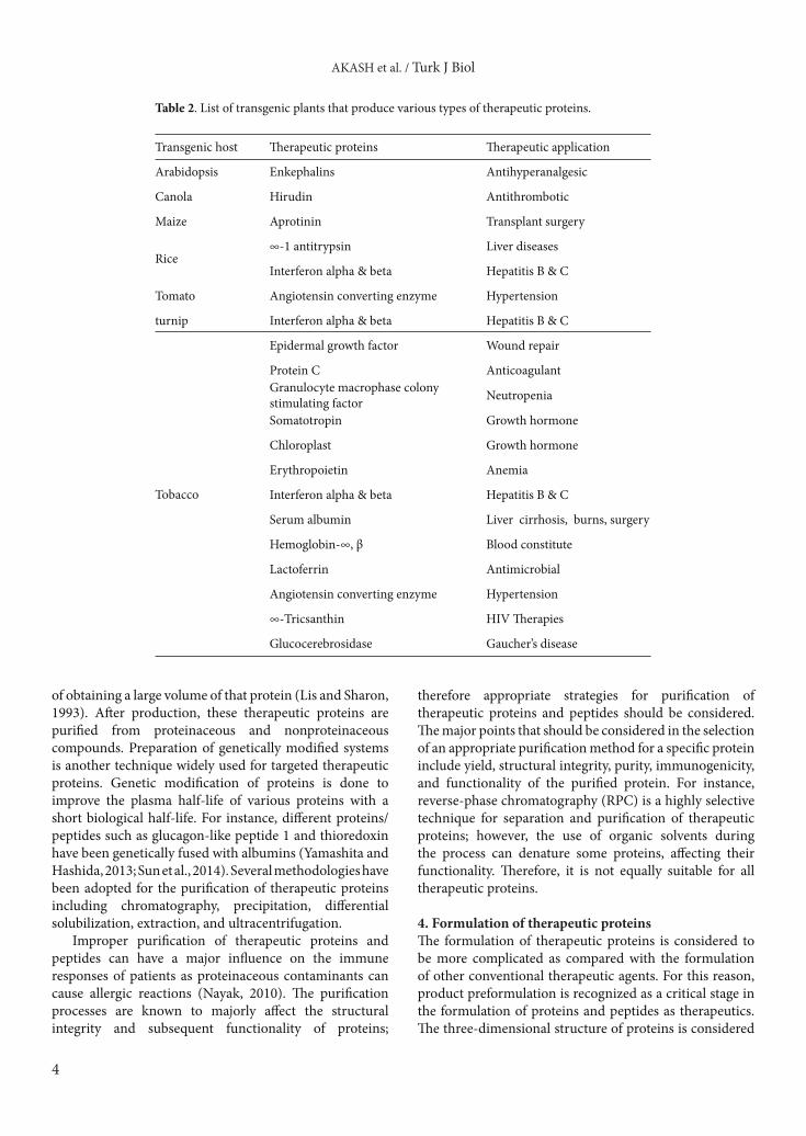
Table 2. List of transgenic plants that produce various types of therapeutic proteins.
Transgenic host Therapeutic proteins Therapeutic application
Arabidopsis Enkephalins Antihyperanalgesic
Canola Hirudin Antithrombotic
Maize Aprotinin Transplant surgery
Rice∞‐1 antitrypsin Liver diseases
Interferon alpha & beta Hepatitis B & C
Tomato Angiotensin converting enzyme Hypertension
turnip Interferon alpha & beta Hepatitis B & C
Tobacco
Epidermal growth factor Wound repair
Protein C AnticoagulantGranulocyte macrophase colony stimulating factor Neutropenia
Somatotropin Growth hormone
Chloroplast Growth hormone
Erythropoietin Anemia
Interferon alpha & beta Hepatitis B & C
Serum albumin Liver cirrhosis, burns, surgery
Hemoglobin‐∞, β Blood constitute
Lactoferrin Antimicrobial
Angiotensin converting enzyme Hypertension
∞‐Tricsanthin HIV Therapies
Glucocerebrosidase Gaucher’s disease

AKASH et al. / Turk J Biol
5
to be the major determinant of their proper functioning. Moreover, changes in external and/or internal variables including temperature, pH, and chemical interaction/modification or mutation may lead to processes like denaturation, aggregation, or precipitation, causing destabilization of the protein structure and affecting its functioning (Chi et al., 2003). Protein aggregation is known to lead to probable immunogenicity in patients and is considered one of the major factors that need to be monitored carefully from the production until the storage stage of therapeutic protein development. Another degradation pathway that may influence protein stability during the formulation of therapeutic proteins is protein oxidation (Torosantucci et al., 2014). Like protein aggregation, the oxidation of therapeutic proteins can result in an alteration of the structure of these proteins, which may further lead to modifications in their secondary, tertiary, and quaternary structure (Torosantucci et al., 2011, 2012). These factors are controlled by adopting suitable and appropriate formulations and protein carrier particulate systems (Akash et al., 2015a, 2015b). Most of the therapeutic proteins are temperature- and pH-sensitive and they are rapidly degraded when given orally. These shortcomings have been overcome by various chemical and physical modifications and using compatible polymers.
5. Pharmacokinetics of therapeutic proteins A great deal of attention to pharmacokinetic (PK) factors is needed for the appropriate development of therapeutic proteins. Therapeutic protein stability is a foremost consideration that needs to be technically confronted. Both chemical and physical instabilities of proteins should be recognized as critical issues because both may lead to degradation via various processes like aggregation, denaturation, hydrolysis, oxidation, and/or racemization. Therefore, various factors such as physical and chemical properties of the proteins and the use of excipients in the formulations of proteins and peptides should be considered in order to increase product efficacy and minimize any drug incompatibility and/or instability. Although the assessment of PK parameters of therapeutic proteins is considered to be complicated because of their physiochemical properties like protein folding and/or instability, it is a critical step for the appropriate formulation and delivery of these proteinaceous drugs. When it comes to the administration of exogenous proteins, they may interfere with the normal physiological functioning of endogenous proteins (Braeckman, 2000).
Besides chemical instability, other major complexities associated with the development of therapeutic proteins include hydrophobic nature, large molecular size, enzymatic degradation, and rapid elimination. Moreover,
the route of administration of therapeutic proteins cannot be neglected because different routes may have different advantages and disadvantages. For instance, parenteral routes, including intravenous (IV), subcutaneous (SC), and intramuscular (IM), are known to bypass the gastrointestinal enzymatic degradation; however, the bioavailability of these therapeutic proteins is known to be reduced after SC and IM administration as compared with the IV route because the SC and IM routes may face minor presystemic degradation (Meibohm and Braeckman, 2008; Zhao et al., 2013).5.1. Absorption and distribution of therapeutic proteins As stated above, owing to their large size and other factors like hydrophilicity and enzymatic degradation, these therapeutic macromolecules are mostly administered via parenteral routes. Following SC injection, the time required for therapeutic proteins to reach maximum systemic circulation (Tmax) is known to be a few hours, while the time for monoclonal antibodies is measured in days (Mannaerts et al., 1998; Montagna et al., 2011). The major factor that determines the absorption and/or bioavailability is the size of the subsequent therapeutic protein administered via a parenteral route. Though the SC route is most widely used for the administration of therapeutic proteins, the bioavailability of these therapeutic proteins following the SC and/or IM route is known to face a large variety of differences in terms of lymph and blood flow/perfusion at the site of administration (Lin, 2009). In other words, the size of therapeutic proteins and peptides as well as the lymphatic and blood supply at the site of drug administration both play pivotal roles in determining the absorption and percentage of bioavailability. The protein and peptide therapeutics with smaller molecular weight usually tend to get absorbed via the blood circulatory system, whereas the lymphatic system is known to absorb therapeutic proteins of a greater molecular size (Porter et al., 2001).
The administration of a few therapeutic proteins such as insulin and glucagon-like peptide-1 (GLP-1), specifically meant for targeting liver and intestinal cells, needs to be done orally. However, large molecular weight, hydrophilicity, and poor intestinal permeation are the main hurdles for the oral route of administration of biologics, including proteins (Chien and Ho, 2008). Currently the use of absorption enhancers is suggested for increasing the permeability of protein drugs across the intestinal epithelium either by enhancing paracellular pathways by opening epithelial junctions or via transcellular pathways by slightly perturbing the mucosal surface (Swenson and Curatolo, 1992; Maher and Brayden, 2012). Additionally, significant advances have been made for the proper delivery and absorption of therapeutic proteins and peptides via the transdermal route (Cleland et al., 2001).

AKASH et al. / Turk J Biol
6
The lungs are known to have a large surface area that provides a closer interaction between alveoli and circulation, which facilitates the absorption of these therapeutic macromolecules. Therefore, the pulmonary route has gained much attention for the delivery of therapeutic proteins. The major advantage of this route is rapid absorption and it bypasses the first-pass effect (Tang et al., 2004).
Besides the routes through which protein drugs are administered, there are a number of other issues to be considered. For example, these drugs may undergo the phase of enzymatic cleavage via proteolytic enzymes found in the host’s gastrointestinal tract (GIT) (Hamman and Steenekamp, 2011). Moreover, the acidic environment of the GIT and the presystemic elimination of protein drugs via the liver may hinder their normal absorption rate (Orive et al., 2004).
The distribution of proteins is equally important to allow these drugs to impart their therapeutic effects. Molecules of smaller weight can be readily distributed to tissues via efficient diffusion through blood capillaries. Other factors that determine the rate and extent of drug distribution are size, lipophilicity, and carrier mediated transportation of that molecule. For small molecules, the transportation is not a complicated process as they (depending on the concentration gradient) may move through passive diffusion. However, for proteins of a large molecular size, an active and/or connective transport is required for movement.
One of the major determinants for the bioavailability and distribution of these molecules is protein binding. It has been recognized that a drug that is free from plasma protein binding can be better distributed in the body and produce its therapeutic effects. Moreover, the metabolism of the drug is also known to be affected by its protein binding. The protein drugs that have long been known to be targets for protein binding include various growth hormones and insulin (Mohler et al., 1992). Some steps have been taken in order to improve the absorption and distribution of protein drugs; the penetration ability of biologics can be altered by evaluating the influence of drug molecular size and binding efficiency on tumors, for instance human IL-2 analogue (Thurber et al., 2008; Liu et al., 2009; Schmidt and Wittrup, 2009). In addition, the existence of receptors may also affect drug distribution in tissues (Braeckman, 2000).5.2. Metabolism and elimination of therapeutic proteins These therapeutic proteins and peptides are usually excreted after being biotransformed and/or degraded via similar pathways involved in the degradation of endogenous proteins. They are broken into fragments of amino acids that might get reutilized for de novo protein/peptide synthesis via the endogenous amino acid pool
(Tang et al., 2004; Baumann, 2006). Various enzymes like proteolysis are mostly responsible for the metabolism of protein drugs and are extensively present throughout the body (Braeckman, 2000). They can be found in the blood, liver, kidneys, small intestine, and various other tissues containing excess of proteases and peptidases for proteolytic degradation of these therapeutic proteins (Tang and Meibohm, 2006). Among these routes, the hepatic pathway is recognized as a major one for protein metabolism and elimination. Therefore, therapeutic proteins that may undergo extensive hepatic metabolism should be given with caution to patients with hepatic impairments (LoRusso et al., 2012).
Many proteins have been recognized to affect different metabolizing enzymes, one of them being the cytochrome p450 (CYP) enzyme. It has been proposed that IL-1 β, IL-6, and TNF-α can efficiently inhibit the activity of many CYP enzymes (Abdel-Razzak et al., 1993). Moreover, several pathways (other than proteolysis) are recognized as responsible for the removal of these therapeutic proteins from the blood, including nonspecific endocytosis and formation of immune-complexes (ICs) followed by Fcγ receptor-mediated clearance. Monoclonal antibodies and fusion proteins may bind to Fcγ receptors, where they get degraded via lysosomes following their internalization into monocytes and macrophages (Raghavan and Bjorkman, 1996; Mould and Green, 2010). Other factors that may influence the metabolism of these therapeutic proteins include their size, charge, structure, distribution, and hydro- or lipophilicity (Tang et al., 2004). As far as the elimination of therapeutic proteins is concerned, the nonmetabolic elimination routes (renal or biliary) are considered to have a minimal contribution. Normally the proteins with a low molecular weight are easily filtered through the renal system, where they go under hydrolytic degradation via enzymes of the renal tubular cells (Meibohm and Zhou, 2012). Those peptides that are resistant to proteolysis tend to be eliminated via the renal system. However, it has been suggested that a few small polypeptides may undergo hydrolysis in the renal brush border, while some may be reabsorbed after being filtered into the renal tubules (Tang et al., 2004).
Conversely, target-mediated drug disposition has been known to influence the distribution and elimination of therapeutic peptides where a large fraction of the drug is bound to pharmacological targets followed by drug–target complex elimination (Mager, 2006). However, the therapeutic drugs that bind to particular receptors are known to be digested intracellularly after being taken up via the receptor.5.3. Transport of therapeutic proteins There are several important pathways involved in the delivery of therapeutic proteins in the gastrointestinal tract
AKASH et al. / Turk J Biol
7
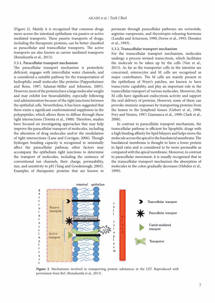
(Figure 2). Mainly it is recognized that common drugs move across the intestinal epithelium via passive or active mediated transports. These passive transports of drugs, including the therapeutic proteins, can be better classified as paracellular and transcellular transports. The active transports are also known as carrier mediated transports (Renukuntla et al., 2013). 5.3.1. Paracellular transport mechanismThe paracellular transport mechanism is proteolytic deficient, engages with intercellular water channels, and is considered a suitable pathway for the transportation of hydrophilic small molecules like proteins (Pappenheimer and Reiss, 1987; Salamat-Miller and Johnston, 2005). However, most of the proteins have a large molecular weight and may exhibit low bioavailability, especially following oral administration because of the tight junctions between the epithelial cells. Nevertheless, it has been suggested that there exists a significant conformational suppleness in the polypeptides, which allows them to diffuse through these tight intersections (Tomita et al., 1988). Therefore, studies have focused on investigating approaches that may help improve the paracellular transport of molecules, including the alteration of drug molecules and/or the modulation of tight intersections (Lane and Corrigan, 2006). Though hydrogen bonding capacity is recognized to minimally affect the paracellular pathway, other factors may accompany the epithelium tight junctions to determine the transport of molecules, including the existence of conventional ion channels, their charge, permeability, size, and sensitivity to pH (Tang and Goodenough, 2003). Examples of therapeutic proteins that are known to
permeate through paracellular pathways are octreotide, arginine vasopressin, and thyrotropin-releasing hormone (Lundin and Artursson, 1990; Drewe et al., 1993; Thwaites et al., 1993).5.3.2. Transcellular transport mechanismFor the transcellular transport mechanism, molecules undergo a process termed transcytosis, which facilitates the molecule to be taken up by the cells (Yun et al., 2013). As far as the transporter cells in the intestine are concerned, enterocytes and M cells are recognized as major contributors. The M cells are mainly present in the epithelium of Peyer’s patches, are known to have transcytotic capability, and play an important role in the transcellular transport of various molecules. Moreover, the M cells have significant endocytosis activity and support the oral delivery of proteins. However, some of them can provoke immune responses by transporting proteins from the lumen to the lymphoid tissues (Gebert et al., 1996; Frey and Neutra, 1997; Giannasca et al., 1999; Clark et al., 2000).
In contrast to paracellular transport mechanism, the transcellular pathway is efficient for lipophilic drugs with a high binding affinity for lipid bilayers and helps move the molecule across the apical to the basolateral membrane. The basolateral membrane is thought to have a lower protein to lipid ratio and is considered to be more permeable as compared with the apical membrane. Moreover, in contrast to paracellular movement, it is usually recognized that in the transcellular transport mechanism the absorption of molecules in the colon gradually decreases (Hebden et al., 1999).
Figure 2. Mechanisms involved in transporting protein substances in the GIT. Reproduced with permission from Ref. (Renukuntla et al., 2013).

AKASH et al. / Turk J Biol
8
The major features that are known to influence this route of transport include hydrophobicity and hydrogen binding potential (Burton et al., 1991; Florence, 2004). It has been observed that the bulk of particle translocation occurs mainly through the epithelium of Peyer’s patches (O’Hagan, 1990; Jani et al., 1992).5.3.3. Carrier mediated transport mechanismMembrane transporters are mainly known to be involved in this transport phenomenon. This pathway usually supports the transportation of di- and tripeptides across the intestinal epithelium and is considered appropriate for the transport of hydrophilic molecules (Barthe et al., 1999). Carrier mediated transport is also referred to as a facilitated diffusion or active transport route. Through this route, the molecule is released into circulation from the basal membrane of the cell after being transferred across the cell membrane (Russell-Jones, 1999). This phenomenon has gained a tremendous amount of attention for being a significant way for transporting various therapeutic proteins and peptides of a small molecular size (Walter et al., 1995). This type of facilitated absorption is energy-dependent where the carriers involved in this transport mechanism utilize membrane receptors for recognizing and transporting the target molecules across the epithelium of the GIT. Unlike the other transport mechanisms, this pathway is independent of concentration
gradient. Examples include the transportation of small di/tripeptides including β-lactam antibiotics and angiotensin converting enzyme inhibitors (Bai and Amidon, 1992). Important factors that can influence the transport of molecules via the carrier-mediated mechanism are pH and temperature. It has been observed that the cellular uptake of cephalexin was more prominent at 37 °C as compared with its cellular uptake at 4 °C. Similarly, it was found that at a pH of 7.0 to 7.4 the transport of cephalexin from the apical membrane to the basolateral membrane was more efficient than at a pH of 5.6 to 6.5 (Hidalgo et al., 1993). On observing the carrier mediated transport of insulin using Caco-2 cell monolayers, it was noted that transferrin receptors (rather than insulin receptors) promoted the transport of conjugated insulin, which was found to be greater than the transport of free insulin (Shah and Shen, 1996). Moreover, some oligopeptide transporters are recognized for the absorption of certain peptidomimetic agents, including rennin and angiotensin converting enzyme inhibitors (Bai and Amidon, 1992).
We have discussed the important transport mechanisms that are probably involved in the transportation of various therapeutic proteins. For instance, the transport of polymeric protein nanoparticles and micelles following oral delivery is known to be facilitated by one pathway, but declined by another (Figure 3) (Plapied et al., 2011).
Figure 3. A proposed mechanism representing the fate of polymeric nanoparticles and micelles for oral drug delivery. Different pathways of transport of nanocarriers or drugs through enterocytes or M cells are represented by orange (for nanoparticles) and blue (for micelles) arrows. (1) Receptor mediated endocytosis; (2) nonspecific transcellular transport; (3) paracellular transport; (4) M cell mediated transport. The size of arrows represents contributions of each type of transport. Reproduced with permission from Ref. (Plapied et al., 2011).

AKASH et al. / Turk J Biol
9
6. Effect of therapeutic proteins on health careTherapeutic proteins have been shown to significantly benefit the health care system by acting as efficient agents for the treatment of various potentially fatal diseases, notably various types of cancers, diabetes mellitus, and auto-immune diseases (Akash et al., 2014a, 2014b; Chen et al., 2014; Rehman et al., 2014a, 2014b; Ünu et al., 2014; Vejselova et al., 2014). The growing field of research focusing on the effects and appropriate delivery of these therapeutic proteins seems to hold a promise of providing historic breakthroughs against various disorders. It seems that the use of therapeutic proteins will ultimately lead to victory over various fatal diseases in the future.
7. ChallengesIn the past several decades, advances and developments in DNA-based genetic engineering technologies have made it possible to develop and synthesize a large number of therapeutic proteins to combat a corresponding variety of life-threatening diseases and syndromes.
It is a fact that pharmaceutical biotechnology and genetic engineering have had a major impact on the health care system and seem likely to attain significant importance in the future, but still there are many current and future challenges that need to be addressed. In the following subsections, we discuss these challenges and ways to make therapeutic proteins the ideal therapeutics.7.1. Development challengesThe overall budget and timelines for the development of therapeutic proteins have been increased dramatically, imposing pharmaceutical scientists to focus only on those molecules that have maximum chances for success in clinical trial programs. From the discovery of new molecules to their entrance into the pharmaceutical market, approximately 10 years are required, but the probability of success (POS) is not a guarantee (Shih, 2012; Strohl and Knight, 2009). For small molecules, the POS is 6%–7%, whereas for monoclonal antibodies and fusion proteins the POS is about 17% (Strohl, 2009). Therefore, it is important that molecules that have a maximum POS be selected.7.2. Safety and immunogenicity issuesMultiple therapeutic products have been developed to modulate the same therapeutic target and/or pathway, which has resulted in tough competition and made it difficult to differentiate the most effective therapeutic product among similar products. For instance, to date, there are five anti-TNF-α therapeutic proteins that are available in the market, including Enbrel, Cimzia, Remicade, Humira, and Simponi. All these agents are administered SC with the help of an autoinjector on a monthly schedule (Strohl and Knight, 2009). The most important feature of these therapeutic proteins is their safety and clinical
efficacy, which can be achieved by a combination of several factors, including disease state, target biology, potency, safety margin, dosing, and selection of patient population (Carter, 2006; Presta, 2008; Strohl, 2009). The safety and clinical efficacy of the administered therapeutic substance can differentiate the best therapeutic agent from the others.
The following are some factors that should be considered (Carter, 2006; Presta, 2008; Shire, 2009; Stebbings et al., 2009; Strohl, 2009) in terms of safety and immunogenicity: 1) molecules that provide maximum margin of safety; 2) the delivery route; 3) prolonged half-life; 4) tissue distribution; 5) stability and enzymatic degradation; and 6) solubility of molecule underdevelopment.
Immunogenicity has become a major challenge for drug efficacy and disease management (Dasgupta et al., 2008; Roger and Goldsmith, 2008). Even a small amount of particulate material in a therapeutic protein formulation, such as protein aggregates, is considered to cause immunogenicity (Rosenberg, 2006; Carpenter et al., 2009). Almost all therapeutic proteins, even those having only human sequences, may induce neutralizing and/or nonneutralizing antibodies in some patients (Dasgupta et al., 2008; Anjum et al., 2013). For these patients, the increased neutralizing antibodies to specific therapeutic proteins and peptides are being used as treatment. However, a reduction in the efficacy of the administered protein therapeutic is a major issue with these antibodies. It can occur anytime due to the production of neutralizing antibodies, the rapid clearance of the protein therapeutic and/or its modified tissue distribution. The clinical consequences of immunogenicity in patients treated with therapeutic proteins are life-threatening conditions. Administered therapeutic proteins neutralize endogenous proteins in patients, causing long-term undesirable toxicities (Schellekens, 2005a). Other immune reactions include inflammation, hypersensitivity, and mild skin reactions to severe anaphylaxis. Some of these immune reactions are clinically manageable either by co-administration of corticosteroids to suppress the inflammation, or by revising the dosing regimen.
Several factors influence the immunogenicity of patients, including: 1) structural features (sequence variation and glycosylation); 2) contaminants or impurities during preparation; 3) storage conditions (denaturation, or aggregation caused by oxidation); 4) dosage and length of treatment; 5) route of administration; 6) type of formulation; and 7) genetic characteristics of patients (Schellekens, 2005b). Improvements in the clinical safety of protein therapeutics are direct results of recent advancements in pharmaceutical biotechnology and genetic engineering. For instance, immunogenic reactions resulting from the introduction of nonhuman antibodies have now been largely circumvented via the generation of

AKASH et al. / Turk J Biol
10
fully human antibody therapeutics (Coenen et al., 2007) and the humanization of rodent antibodies (Easthope and Jarvis, 2001). Pharmaceutical scientists are continuously trying to identify T-cell epitopes in protein therapeutics and boost the immune tolerance via the activation of Treg cells. These two have the ability to dampen the unwanted immunogenic responses (De Groot et al., 2008); however, it is still unclear whether these strategies will help minimize the chances of immunogenicity observed with antibody-based therapeutics. Using established analytical methods (Bilal et al., 2013; Lei et al., 2013a, 2013b, 2013c, 2014a, 2014b, 2015), immunogenicity associated with protein therapeutics cannot be accurately predicted. Pharmaceutical scientists are continuously trying their best to assess the potential of immunogenicity during the discovery phase and ultimately select a molecule with a minimal immunogenic profile as a clinical therapeutic candidate.7.3. Protein stabilityTherapeutic proteins belong to the most widely growing class of pharmaceutical active ingredients that are being used in diverse clinical settings. However, their stability is a major drawback to making them ideal clinical therapeutics (Akash et al., 2013d). Therapeutic proteins tend to aggregate when stored under high concentration conditions as required for their usage (Jiskoot et al., 2012; Shire et al., 2004). Aggregation of therapeutic proteins tends to decrease their overall activity and sometimes elicit immunological reactions (Hermeling et al., 2004; Chennamsetty et al., 2009; Jiskoot et al., 2012). Spatial aggregation propensity identifies the location and size of aggregation prone regions in protein formulations and allows performing target mutations of those regions to engineer antibodies for stability (Chennamsetty et al., 2009; Voynov et al., 2009). Another factor that may influence the stability of therapeutic proteins is temperature. Most often, extracted proteins are stored for an extended period of time to maintain their activity and original structural integrity. Usually, proteins are best stored at ≤4 °C. Storage at room temperature often leads to the degradation of therapeutic proteins. Furthermore, protein instability during sustained delivery can result in the formation of protein particles during in vivo sustained release, which is considered another factor of protein instability. This may induce an immune response in patients treated with sustained release formulations of protein therapeutics (as compared with immunogenicity of conventional protein therapeutics) (Jiskoot et al., 2009).
Several strategies are being applied to increase the stability of therapeutic proteins (Akash et al., 2015a, 2015b). The first approach is to alter the amino acid sequences in the protein structure (Strickler et al., 2006; Lawrence et al., 2007). The second one is to optimize the
formulation of therapeutic proteins (Baynes and Trout, 2004; Baynes et al., 2005; Schneider and Trout, 2009; Ali et al., 2014). Thermosensitive polymers have also shown to increase the stability of therapeutic proteins (Akash et al., 2012b, 2013b, 2013c, 2013d, 2014b, 2014d). Biodegradable polymers have been intensively evaluated for the successful delivery of a variety of therapeutic substances. Protein stability can also be increased using nontoxic nano-structured materials (Domach and Walker, 2010).7.4. Protein degradationProtein degradation is another challenge that occurs at different stages throughout the entire process of development and delivery of the drug to its desired sites. During formulation and storage of therapeutic proteins, aggregation is a major degradation pathway. Proteins are often unstable when they are not in their native environments. Conditions vary considerably among cell compartments and extracellular fluids. If certain conditions are not maintained during their storage, therapeutic proteins may not function properly. Proteins can lose therapeutic activity as a result of proteolysis, aggregation, and suboptimal buffer conditions. It is important to understand and quantify various routes of chemical degradation such as oxidation, deamidation, chemical cross-linking, disulfide modifications, and fragmentation (Jiskoot et al., 2012). Several strategies are being used to prevent the degradation of therapeutic proteins. The most common one is the encapsulation of therapeutic proteins with inert and biocompatible polymers (Akash et al., 2014b, 2015a, 2015b). Protein degradation can also be prevented by co-administration of enzyme inhibitors with therapeutic proteins.7.5. Protein–excipient interactionsImmunogenicity and instability of therapeutic proteins can be stopped by modifying the protein structure via alteration of amino acid sequences and/or optimizing the formulation of therapeutic proteins using different types of polymers. The polymers, used for the incorporation of therapeutic proteins, must be inert, biocompatible, and most preferentially biodegradable in nature. During the formulation development of therapeutic proteins, protein–excipient interactions must be intensively evaluated to enhance the overall stability of protein therapeutics and minimize the chances of any kind of immunogenicity. Different types of techniques are being used by pharmaceutical scientists to evaluate the protein–excipient interactions. These techniques have been critically reviewed by Kamerzell et al. (2011)7.6. Metabolism and eliminationHepatic metabolism and rapid elimination is a major roadblock to the clinical application of therapeutic proteins. Pharmaceutical scientists are continuously struggling to prevent the hepatic first-pass metabolism

AKASH et al. / Turk J Biol
11
and rapid elimination of therapeutic proteins. To date, several efforts have been made to circumvent these hurdles. These efforts include the noninvasive delivery of therapeutic proteins through routes (other than the oral route) that bypass the hepatic metabolism of the administered therapeutic proteins. Moreover, invasive delivery of therapeutic proteins is also an alternative tool to prevent the hepatic first-pass metabolism. Furthermore, the majority of therapeutic proteins have a short biological half-life. This problem might be overcome by encapsulating and/or conjugating desired therapeutic proteins with biocompatible polymers, and using fusion protein technology to extend the half-life of therapeutic proteins. Recently, IL-1Ra, a naturally occurring anti-inflammatory antagonist of pro-inflammatory cytokines, has been successfully encapsulated in PF127. In vitro and in vivo studies have shown that PF127 significantly prolongs the sustained release of IL-1Ra, and maintains in vitro stability, in vivo bioactivity, and therapeutic potentials (Akash et al., 2012b, 2013c, 2014d).
8. ConclusionWe have made an attempt to discuss all the possible factors that are linked with the basis of therapeutic proteins, including their introduction, classification, production, purification, pharmacokinetic parameters, their importance in the health care system, and possible challenges that are currently being faced by scientists during the development of protein-based therapeutics.
In the past couple of decades, therapeutic proteins have gained significance as therapy for a wide range of diseases. Moreover, together with the therapeutic aspects of a growing number of therapeutic proteins, the processes for their production and purification have also gained a wide range of importance, making these valuable agents available in
the pharmaceutical market. However, there are still many factors that need to be considered critically during the production, purification, and formulation of therapeutic proteins for a better quality, safety, and efficacy of these agents. This means that to use these therapeutic proteins for the treatment of various diseases, a proper and rational formulation of protein-based therapeutics is required. Moreover, a better understanding of the pharmacokinetic properties of therapeutic proteins along with the relation of these properties to the pharmacodynamic effects of these agents will further help advance the development and appropriate delivery of therapeutic proteins and peptides. A complete evaluation of the pharmacokinetic parameters and factors that can affect these parameters is required for an appropriate prediction of the biodisposition of these agents both in clinical and nonclinical settings.
Advancements in the field of biotechnology have increased and facilitated the production of therapeutically significant proteins to combat various potentially fatal diseases. However, there are still a few factors that hinder the efficient use of these valuable therapeutics. For instance, the oral route of administration faces proteolysis and/or hydrolysis in the GIT, whereas some drugs go through the hepatic first pass effect or show poor distribution. Therefore, a better insight into the routes of administration and the drug absorption mechanisms (paracellular, transcellular, and carrier mediated) is essential.
AcknowledgmentsThe authors are thankful to the Science and Technology Development of the Ministry of Science and Technology of China (Grant # 2012ZX09506001-004) for its financial support. The first two authors also acknowledge the CSC, China, for providing the scholarships for PhD studies and HEC, Pakistan, for partial support for their PhD studies.
References
Abdel-Razzak Z, Loyer P, Fautrel A, Gautier JC, Corcos L, Turlin B, Beaune P, Guillouzo A (1993). Cytokines down-regulate expression of major cytochrome P-450 enzymes in adult human hepatocytes in primary culture. Mol Pharmacol 44: 707–715.
Ahmad I, Rubbab T, Deeba F, Naqvi SMS (2014). Optimization of E. coli culture conditions for efficient DNA uptake by electroporation. Turk J Biol 38: 568–573.
Ahmed H, HA EZ, Alswiai G (2013). Purification of antioxidant protein isolated from Peganum harmala and its protective effect against CCl4 toxicity in rats. Turk J Biol 37: 39–48.
Akash MSH,Shen Q, Rehman K, Chen S (2012a). Interleukin-1 receptor antagonist: a new therapy for type 2 diabetes mellitus. J Pharm Sci 101: 1647–1658.
Akash MSH, Rehman K, Li N, Gao JQ, Sun H, Chen S (2012b). Sustained delivery of IL-1Ra from pluronic F127-based thermosensitive gel prolongs its therapeutic potentials. Pharm Res 29: 3475–3485.
Akash MSH, Rehman K, Chen S (2013a). Role of inflammatory mechanisms in pathogenesis of type 2 diabetes mellitus. J Cell Biochem 114: 525–531.
Akash MSH, Rehman K, Sun H, Chen S (2013b). Interleukin-1 receptor antagonist improves normoglycemia and insulin sensitivity in diabetic Goto-Kakizaki-rats. Eur J Pharmacol 701: 87–95.
Akash MSH, Rehman K, Sun H, Chen S (2013c). Sustained delivery of IL-1Ra from PF127-gel reduces hyperglycemia in diabetic GK-rats. PLoS One 8:e55925.

AKASH et al. / Turk J Biol
12
Akash MSH, Rehman K, Chen S (2013d). IL-1Ra and its delivery strategies: inserting the association in perspective. Pharm Res 30: 2951–2966.
Akash MSH, Rehman K, Chen S (2014a). Effects of coffee on type 2 diabetes mellitus. Nutrition 30: 755–763.
Akash MSH, Rehman K, Chen S (2014b). Pluronic F127-based thermosensitive gels for delivery of therapeutic proteins and peptides. Polym Rev 54: 573–597.
Akash MSH, Rehman K, Chen S (2014c). Spice plant Allium cepa: dietary supplement for treatment of type 2 diabetes mellitus. Nutrition 30: 1128–1137.
Akash MSH, Rehman K, Sun H, Chen S (2014d). Assessment of release kinetics, stability and polymer interaction of poloxamer 407-based thermosensitive gel of interleukin-1 receptor antagonist. Pharm Dev Technol 19: 278–284.
Akash MSH, Rehman K, Chen S (2015a). Natural and synthetic polymers as drug carriers for delivery of therapeutic proteins. Polym Rev: DOI:10.1080/15583724.2014.995806.
Akash MSH, Rehman K, Chen S (2015b). Polymeric-based particulate systems for delivery of therapeutic proteins. Pharm Dev Technol: DOI: 10.3109/10837450.2014.999785.
Ali B, Ibrahim M, Hussain I, Hussain N, Imran M, Nawaz H, Jan S, Khalid M, Ghous T, Akash MSH (2014). Pakistamide C, a new sphingolipid from Abutilon pakistanicum. Rev Bras Farmacogn 24: 277–281.
Anjum R, Zahra N, Rehman K, Alam R, Parveen A, Tariq M, Akash MSH (2013). Comparative analysis of serum lipid profile between normotensive and hypertensive Pakistani pregnant women. J Mol Genet Med 7: 64.
Bai JP, Amidon GL (1992). Structural specificity of mucosal-cell transport and metabolism of peptide drugs: implication for oral peptide drug delivery. Pharm Res 9: 969–978.
Barthe L, Woodley J, Houin G (1999). Gastrointestinal absorption of drugs: methods and studies. Fundam Clin Pharmacol 13: 154–168.
Baumann A (2006). Early development of therapeutic biologics—pharmacokinetics. Curr Drug Metab 7: 15–21.
Baynes BM, Trout BL (2004). Rational design of solution additives for the prevention of protein aggregation. Biophys J 87: 1631–1639.
Baynes BM, Wang DI, Trout BL (2005). Role of arginine in the stabilization of proteins against aggregation. Biochemistry 44: 4919–4925.
Bilal A, Rehman K, Akash MSH, Hussain K, Ibrahim M, Hussan SS (2013). Development and validation of analytical method for qualitative and quantitative determination of Glibenclamide in different brands of tablet dosage form using UV-visible spectroscopy. J Mol Genet Med 7: 80.
Braeckman R (2000). Pharmacokinetics and pharmacodynamics of protein therapeutics. In Reid ER, editor. Peptides and Protein Drug Analysis. New York, NY, USA: Marcel Dekker Inc, pp. 633–669.
Burton PS, Conradi RA, Hilgers AR (1991). (B) Mechanisms of peptide and protein absorption: (2) Transcellular mechanism of peptide and protein absorption: passive aspects. Adv Drug Deliv Rev 7: 365–385.
Campos-Neto A, Rodrigues-Junior V, Pedral-Sampaio DB, Netto EM, Ovendale PJ, Coler RN, Skeiky YA, Badaro R, Reed SG (2001). Evaluation of DPPD, a single recombinant Mycobacterium tuberculosis protein as an alternative antigen for the Mantoux test. Tuberculosis (Edinb) 81: 353–358.
Carpenter JF, Randolph TW, Jiskoot W, Crommelin DJ, Middaugh CR, Winter G, Fan YX, Kirshner S, Verthelyi D, Kozlowski S et al. (2009). Overlooking subvisible particles in therapeutic protein products: gaps that may compromise product quality. J Pharm Sci 98: 1201–1205.
Carter PJ (2006). Potent antibody therapeutics by design. Nat Rev Immunol 6: 343–357.
Chen B, Akash MSH, Rehman K, Sun H, Chen S (2014). Expression and bioactivity analysis of staphylococcal enterotoxin G and staphylococcal enterotoxin I. Pharm Biol 52: 8–13.
Chennamsetty N, Voynov V, Kayser V, Helk B, Trout BL (2009). Design of therapeutic proteins with enhanced stability. Proc Natl Acad Sci USA 106: 11937–11942.
Chi EY, Krishnan S, Randolph TW, Carpenter JF (2003). Physical stability of proteins in aqueous solution: mechanism and driving forces in nonnative protein aggregation. Pharm Res 20: 1325–1336.
Chien JY, Ho RJ (2008). Drug delivery trends in clinical trials and translational medicine. J Pharm Sci 97: 2543–2547.
Clark MA, Hirst BH, Jepson MA (2000). Lectin-mediated mucosal delivery of drugs and microparticles. Adv Drug Deliv Rev 43: 207–223.
Cleland JL, Daugherty A, Mrsny R (2001). Emerging protein delivery methods. Curr Opin Biotechnol 12: 212–219.
Coenen MJ, Toonen EJ, Scheffer H, Radstake TR, Barrera P, Franke B (2007). Pharmacogenetics of anti-TNF treatment in patients with rheumatoid arthritis. Pharmacogenomics 8: 761–773.
Cohen SB, Moreland LW, Cush JJ, Greenwald MW, Block S, Shergy WJ, Hanrahan PS, Kraishi MM, Patel A, Sun G, Bear MB (2004). A multicentre, double blind, randomised, placebo controlled trial of anakinra (Kineret), a recombinant interleukin 1 receptor antagonist, in patients with rheumatoid arthritis treated with background methotrexate. Ann Rheum Dis 63: 1062–1068.
Corwin HL, Gettinger A, Pearl RG, Fink MP, Levy MM, Shapiro MJ, Corwin MJ, Colton T (2002). Efficacy of recombinant human erythropoietin in critically ill patients: a randomized controlled trial. JAMA 288: 2827–2835.
Craik DJ, Fairlie DP, Liras S, Price D (2013). The future of peptide-based drugs. Chem Biol Drug Des 81: 136–147.
Crosnier J, Jungers P, Courouce AM, Laplanche A, Benhamou E, Degos F, Lacour B, Prunet P, Cerisier Y, Guesry P (1981). Randomised placebo-controlled trial of hepatitis B surface antigen vaccine in French haemodialysis units: I, Medical staff. Lancet 1: 455–459.

AKASH et al. / Turk J Biol
13
Dasgupta S, Bayry J, Andre S, Dimitrov JD, Kaveri SV, Lacroix-Desmazes S (2008). Auditing protein therapeutics management by professional APCs: toward prevention of immune responses against therapeutic proteins. J Immunol 181: 1609–1615.
De Groot AS, Moise L, McMurry JA, Wambre E, Van Overtvelt L, Moingeon P, Scott DW, Martin W (2008). Activation of natural regulatory T cells by IgG Fc-derived peptide “Tregitopes”. Blood 112: 3303–3311.
Domach MM, Walker LM (2010). Stabilizing biomacromolecules in nontoxic nano-structured materials. J Assoc Lab Autom 15: 136–144.
Drewe J, Fricker G, Vonderscher J, Beglinger C (1993). Enteral absorption of octreotide: absorption enhancement by polyoxyethylene-24-cholesterol ether. Br J Pharmacol 108: 298–303.
Easthope S, Jarvis B (2001). Omalizumab. Drugs 61: 253–260; discussion 261.
Ebrahiminezhad A, Rasoul-Amini S, Ghasemi Y (2011). l-Asparaginase production by moderate halophilic bacteria isolated from Maharloo Salt Lake. Indian J Microbiol 51: 307–311.
Eriksson BI, Wille-Jorgensen P, Kalebo P, Mouret P, Rosencher N, Bosch P, Baur M, Ekman S, Bach D, Lindbratt S, Close P (1997). A comparison of recombinant hirudin with a low-molecular-weight heparin to prevent thromboembolic complications after total hip replacement. N Engl J Med 337: 1329–1335.
Florence AT (2004). Issues in oral nanoparticle drug carrier uptake and targeting. J Drug Target 12: 65–70.
Frey A, Neutra MR (1997). Targeting of mucosal vaccines to Peyer’s patch M cells. Behring Inst Mitt: 376–389.
Gebert A, Rothkötter H-J, Pabst R (1996). M cells in Peyer’s patches of the intestine. In Kwang WJ, editors. International Review of Cytology. San Diego, CA, USA: Academic Press, pp. 91–159.
Giannasca PJ, Giannasca KT, Leichtner AM, Neutra MR (1999). Human intestinal M cells display the sialyl Lewis A antigen. Infect Immun 67: 946–953.
Gökbulut AA, Arslanoğlu A (2013). Purification and biochemical characterization of an extracellular lipase from psychrotolerant Pseudomonas fluorescens KE38. Turk J Biol 37: 538–546.
Hamman JH, Steenekamp JH (2011). Oral peptide drug delivery: strategies to overcome challenges. Hoboken, NJ, USA: Wiley-VCH. pp. 71–90.
Hebden JM, Wilson CG, Spiller RC, Gilchrist PJ, Blackshaw E, Frier ME, Perkins AC (1999). Regional differences in quinine absorption from the undisturbed human colon assessed using a timed release delivery system. Pharm Res 16: 1087–1092.
Hermeling S, Crommelin DJ, Schellekens H, Jiskoot W (2004). Structure-immunogenicity relationships of therapeutic proteins. Pharm Res 21: 897–903.
Hidalgo IJ, Ryan FM, Marks GJ, Smith PL (1993). pH-dependent transepithelial transport of cephalexin in rabbit intestinal mucosa. Int J Pharm 98: 83–92.
Ibrahim M, Farooq T, Hussain N, Hussain A, Gulzar T, Hussain I, Akash MSH, Rehmani FS (2013). Acetyl and butyryl cholinesterase inhibitory sesquiterpene lactones from Amberboa ramosa. Chem Cent J 7: 116.
Jani PU, Florence AT, McCarthy DE (1992). Further histological evidence of the gastrointestinal absorption of polystyrene nanospheres in the rat. Int J Pharm 84: 245–252.
Jankovic J, Brin MF (1991). Therapeutic uses of botulinum toxin. N Engl J Med 324: 1186–1194.
Janne J, Hyttinen JM, Peura T, Tolvanen M, Alhonen L, Halmekyto M (1992). Transgenic animals as bioproducers of therapeutic proteins. Ann Med 24: 273–280.
Jayapal KP, Wlaschin KF, Hu W, Yap MG (2007). Recombinant protein therapeutics from CHO cells-20 years and counting. Chem Eng Prog 103: 40.
Jiskoot W, Randolph TW, Volkin DB, Middaugh CR, Schoneich C, Winter G, Friess W, Crommelin DJ, Carpenter JF (2012). Protein instability and immunogenicity: roadblocks to clinical application of injectable protein delivery systems for sustained release. J Pharm Sci 101: 946–954.
Jiskoot W, van Schie RM, Carstens MG, Schellekens H (2009). Immunological risk of injectable drug delivery systems. Pharm Res 26: 1303–1314.
Kamerzell TJ, Esfandiary R, Joshi SB, Middaugh CR, Volkin DB (2011). Protein-excipient interactions: mechanisms and biophysical characterization applied to protein formulation development. Adv Drug Deliv Rev 63: 1118–1159.
Kamionka M (2011). Engineering of therapeutic proteins production in Escherichia coli. Curr Pharm Biotechnol 12: 268–274.
Karacali S, İzzetoğlu S, Deveci R (2014). Glycosylation changes leading to the increase in size on the common core of N-glycans, required enzymes, and related cancer-associated proteins. Turk J Biol 38: 754–771.
Karvar S (2014). The role of ABC transporters in anticancer drug transport. Turk J Biol 38: 800–805.
Kelley BD (2001). Biochemical engineering: bioprocessing of therapeutic proteins. Curr Opin Biotechnol 12: 173–174.
Lane ME, Corrigan OI (2006). Paracellular and transcellular pathways facilitate insulin permeability in rat gut. J Pharm Pharmacol 58: 271–275.
Lawrence MS, Phillips KJ, Liu DR (2007). Supercharging proteins can impart unusual resilience. J Am Chem Soc 129: 10110–10112.
Leader B, Baca QJ, Golan DE (2008). Protein therapeutics: a summary and pharmacological classification. Nat Rev Drug Discov 7: 21–39.
Lei Y, Fang L, Akash MS, Rehman K, Liu Z, Shi W, Chen S (2013a). Estimation of urinary concentration of aflatoxin M1 in Chinese pregnant women. J Food Sci 78: T1835–T1838.
Lei Y, Fang L, Akash MSH, Liu Z, Shi W, Chen S (2013b). Development and comparison of two competitive ELISAs for the detection of bisphenol A in human urine. Anal Methods 5: 6106–6113.
AKASH et al. / Turk J Biol
14
Lei Y, Li X, Akash MS, Zhou L, Tang X, Shi W, Liu Z, Chen S (2015). Development of analytical method for ultrasensitive detection of salbutamol utilizing DNA labeled-immunoprobe. J Pharm Biomed Anal 107c: 204–208.
Lei Y, Liu W, Fang L, Akash MSH, Rehman K, Narenmandura H, Shi W, Lu W, Xu Y, Chen S (2014a). Assessment of urinary concentration of cotinine in Chinese pregnant women exposed to environmental tobacco smoke. Chinese Science Bulletin 59: 1386–1391.
Lei Y, Zhang Q, Fang L, Akash MS, Rehman K, Liu Z, Shi W, Chen S (2014b). Development and comparison of two competitive ELISAs for estimation of cotinine in human exposed to environmental tobacco smoke. Drug Test Anal: 6:1020-1027.
Lei Y, Zhang S, Fang L, Akash MSH, Shi W, Sun K, Xu Y, Chen S (2013c). A sensitive and specific enzyme immunoassay for detecting tartrazine in human urinary samples. Anal Methods 5: 925–930.
Lin JH (2009). Pharmacokinetics of biotech drugs: peptides, proteins, and monoclonal antibodies. Curr Drug Metab 10: 661–691.
Lis H, Sharon N (1993). Protein glycosylation. Structural and functional aspects. Eur J Biochem 218: 1–27.
Liu DV, Maier LM, Hafler DA, Wittrup KD (2009). Engineered interleukin-2 antagonists for the inhibition of regulatory T cells. J Immunother 32: 887–894.
LoRusso PM, Venkatakrishnan K, Ramanathan RK, Sarantopoulos J, Mulkerin D, Shibata SI, Hamilton A, Dowlati A, Mani S, Rudek MA et al. (2012). Pharmacokinetics and safety of bortezomib in patients with advanced malignancies and varying degrees of liver dysfunction: phase I NCI Organ Dysfunction Working Group Study NCI-6432. Clin Cancer Res 18: 2954–2963.
Lundin S, Artursson P (1990). Absorption of a vasopressin analogue, 1-deamino-8-d-arginine-vasopressin (dDAVP), in a human intestinal epithelial cell line, CaCO-2. Int J Pharm 64: 181–186.
MacKenzie IZ, Bichler J, Mason GC, Lunan CB, Stewart P, Al-Azzawi F, De Bono M, Watson N, Andresen I (2004). Efficacy and safety of a new, chromatographically purified rhesus (D) immunoglobulin. Eur J Obstet Gynecol Reprod Biol 117: 154–161.
Mager DE (2006). Target-mediated drug disposition and dynamics. Biochem Pharmacol 72: 1–10.
Maher S, Brayden DJ (2012). Overcoming poor permeability: translating permeation enhancers for oral peptide delivery. Drug Discov Today Technol 9: e113–e119.
Mannaerts BM, Geurts TB, Odink J (1998). A randomized three-way cross-over study in healthy pituitary-suppressed women to compare the bioavailability of human chorionic gonadotrophin (Pregnyl) after intramuscular and subcutaneous administration. Hum Reprod 13: 1461–1464.
Martemyanov KA, Shirokov VA, Kurnasov OV, Gudkov AT, Spirin AS (2001). Cell-free production of biologically active polypeptides: application to the synthesis of antibacterial peptide cecropin. Protein Expr Purif 21: 456–461.
Meibohm B, Braeckman R (2008). Pharmacokinetics and pharmacodynamics of peptide and protein drugs. In Crommelin DJA, Sindelar RD, Meibohm B, editors. Pharmaceutical biotechnology. New York: Informa Healthcare, pp. 95–123.
Meibohm B, Zhou H (2012). Characterizing the impact of renal impairment on the clinical pharmacology of biologics. J Clin Pharmacol 52: 54S–62S.
Mohler MA, Cook JE, Baumann G (1992). Binding proteins of protein therapeutics. In erraiolo BL, Mohler MA, Gloff CA, editors. Protein Pharmacokinetics and Metabolism. New York, NY, USA: Springer, pp. 35–71.
Montagna M, Montillo M, Avanzini MA, Tinelli C, Tedeschi A, Visai L, Ricci F, Vismara E, Morra E, Regazzi M (2011). Relationship between pharmacokinetic profile of subcutaneously administered alemtuzumab and clinical response in patients with chronic lymphocytic leukemia. Haematologica 96: 932–936.
Mould DR, Green B (2010). Pharmacokinetics and pharmacodynamics of monoclonal antibodies: concepts and lessons for drug development. BioDrugs 24: 23–39.
Mrsny RJ (2004). Strategies for targeting protein therapeutics to selected tissues and cells. Expert Opin Biol Ther 4: 65–73.
Nayak AK (2010). Advances in therapeutic protein production and delivery. Int J Pharm Pharm Sci 2: 1–5.
O’Hagan D (1990). Intestinal translocation of particulates—implications for drug and antigen delivery. Adv Drug Deliv Rev 5: 265–285.
Ohya S, Matsuda T (2005). Poly (N-isopropylacrylamide)(PNIPAM)-grafted gelatin as thermoresponsive three-dimensional artificial extracellular matrix: molecular and formulation parameters vs. cell proliferation potential. J Biomater Sci Polym Ed 16: 809–827.
Orive G, Gascon AR, Hernandez RM, Dominguez-Gil A, Pedraz JL (2004). Techniques: new approaches to the delivery of biopharmaceuticals. Trends Pharmacol Sci 25: 382–387.
Pappenheimer JR, Reiss KZ (1987). Contribution of solvent drag through intercellular junctions to absorption of nutrients by the small intestine of the rat. J Membr Biol 100: 123–136.
Plapied L, Duhem N, des Rieux A, Préat V (2011). Fate of polymeric nanocarriers for oral drug delivery. Curr Opin Colloid In 16: 228–237.
Porter CJ, Edwards GA, Charman SA (2001). Lymphatic transport of proteins after s.c. injection: implications of animal model selection. Adv Drug Deliv Rev 50: 157–171.
Prakash PJ, Poorani E, Anantharaman P, Balasubramaniam T (2009). L-Glutaminase production and the growth of marine bacteria. Res J Microbiol 4: 168–172.
Presta LG (2008). Molecular engineering and design of therapeutic antibodies. Curr Opin Immunol 20: 460–470.
Raghavan M, Bjorkman PJ (1996). Fc receptors and their interactions with immunoglobulins. Annu Rev Cell Dev Biol 12: 181–220.
AKASH et al. / Turk J Biol
15
Regan L, Jackson SE (2003). Engineering and design: protein design: theory and practice. Curr Opin Str Biol 13: 479–481.
Rehman K, Iqbal MJ, Zahra N, Akash MSH (2014a). Liver stem cells: from preface to advancements. Curr Stem Cell Res Ther 9: 10–21.
Rehman K, Tariq M, Akash MSH, Gillani Z, Qazi MH (2014b). Effect of HA14-1 on apoptosis-regulating proteins in HeLa cells. Chem Biol Drug Des 83: 317–323.
Renukuntla J, Vadlapudi AD, Patel A, Boddu SH, Mitra AK (2013). Approaches for enhancing oral bioavailability of peptides and proteins. Int J Pharm 447: 75–93.
Roger SD, Goldsmith D (2008). Biosimilars: it’s not as simple as cost alone. J Clin Pharm Ther 33: 459–464.
Rosenberg AS (2006). Effects of protein aggregates: an immunologic perspective. AAPS J 8: E501–E507.
Roth DA, Kessler CM, Pasi KJ, Rup B, Courter SG, Tubridy KL (2001). Human recombinant factor IX: safety and efficacy studies in hemophilia B patients previously treated with plasma-derived factor IX concentrates. Blood 98: 3600–3606.
Russell-Jones GJ (1999). Carrier-mediated transport, oral drug delivery. In Mathiowitz E, editors. Encyclopedia of Controlled Drug Delivery. New York, NY, USA: John Wiley & Sons, Inc., pp. 173–184.
Salamat-Miller N, Johnston TP (2005). Current strategies used to enhance the paracellular transport of therapeutic polypeptides across the intestinal epithelium. Int J Pharm 294: 201–216.
Saltz LB, Meropol NJ, Loehrer PJ, Sr., Needle MN, Kopit J, Mayer RJ (2004). Phase II trial of cetuximab in patients with refractory colorectal cancer that expresses the epidermal growth factor receptor. J Clin Oncol 22: 1201–1208.
Sato AK, Viswanathan M, Kent RB, Wood CR (2006). Therapeutic peptides: technological advances driving peptides into development. Curr Opin Biotechnol 17: 638–642.
Sato D, Nozaki T (2009). Methionine gamma-lyase: the unique reaction mechanism, physiological roles, and therapeutic applications against infectious diseases and cancers. IUBMB Life 61: 1019–1028.
Schellekens H (2005a). Factors influencing the immunogenicity of therapeutic proteins. Nephrol Dial Transplant 20: vi3–vi9.
Schellekens H (2005b). Immunologic mechanisms of EPO-associated pure red cell aplasia. Best Pract Res Clin Haematol 18: 473–480.
Schmidt MM, Wittrup KD (2009). A modeling analysis of the effects of molecular size and binding affinity on tumor targeting. Mol Cancer Ther 8: 2861–2871.
Schneider CP, Trout BL (2009). Investigation of cosolute-protein preferential interaction coefficients: new insight into the mechanism by which arginine inhibits aggregation. J Phys Chem B 113: 2050–2058.
Shah D, Shen WC (1996). Transcellular delivery of an insulin-transferrin conjugate in enterocyte-like Caco-2 cells. J Pharm Sci 85: 1306–1311.
Sharma B, Singh S, Kanwar SS (2014). L-methionase: a therapeutic enzyme to treat malignancies. Biomed Res Int 2014: 506287.
Shih HH (2012). Discovery process for antibody-based therapeutics. editor. Development of antibody-based therapeutics. New York, NY, USA: Springer, pp. 9–32.
Shire SJ (2009). Formulation and manufacturability of biologics. Curr Opin Biotechnol 20: 708–714.
Shire SJ, Shahrokh Z, Liu J (2004). Challenges in the development of high protein concentration formulations. J Pharm Sci 93: 1390–1402.
Sodee DB, Malguria N, Faulhaber P, Resnick MI, Albert J, Bakale G (2000). Multicenter ProstaScint imaging findings in 2154 patients with prostate cancer. The ProstaScint Imaging Centers. Urology 56: 988–993.
Stebbings R, Poole S, Thorpe R (2009). Safety of biologics, lessons learnt from TGN1412. Curr Opin Biotechnol 20: 673–677.
Strickler SS, Gribenko AV, Gribenko AV, Keiffer TR, Tomlinson J, Reihle T, Loladze VV, Makhatadze GI (2006). Protein stability and surface electrostatics: a charged relationship. Biochemistry 45: 2761–2766.
Strohl WR (2009). Therapeutic monoclonal antibodies: past, present, and future. In An Z, editor. Therapeutic Monoclonal Antibodies: From Bench to Clinic. New York, NY, USA: John Wiley & Sons, pp. 4–50.
Strohl WR, Knight DM (2009). Discovery and development of biopharmaceuticals: current issues. Curr Opin Biotechnol 20: 668–672.
Sun R, Liu S, Gao J-L, Tang Z-Z, Chen H, Li C-L, Wu Q (2014). Cloning and expression analysis of 1-deoxy-D-xylulose-5-phosphate synthase gene from the medicinal plant Conyza blinii H. Lév. Turk J Biol 38: 664–670.
Swenson ES, Curatolo WJ (1992). (C) Means to enhance penetration:(2) Intestinal permeability enhancement for proteins, peptides, and other polar drugs: mechanisms and potential toxicity. Adv Drug Deliv 8: 39–92.
Tang L, Meibohm B (2006). Pharmacokinetics of peptides and proteins. In Meibohm B, editors. Pharmacokinetics and Pharmacodynamics of Biotech Drugs: Principles and Case Studies in Drug Development. Weinheim, Germany: Wiley-VCH. pp. 17–44.
Tang L, Persky AM, Hochhaus G, Meibohm B (2004). Pharmacokinetic aspects of biotechnology products. J Pharm Sci 93: 2184–2204.
Tang VW, Goodenough DA (2003). Paracellular ion channel at the tight junction. Biophys J 84: 1660–1673.
Tesser J, Fleischmann R, Dore R, Bennett R, Solinger A, Joh T, Modafferi D, Schechtman J (2004). Concomitant medication use in a large, international, multicenter, placebo controlled trial of anakinra, a recombinant interleukin 1 receptor antagonist, in patients with rheumatoid arthritis. J Rheumatol 31: 649–654.

AKASH et al. / Turk J Biol
16
Thurber GM, Schmidt MM, Wittrup KD (2008). Antibody tumor penetration: transport opposed by systemic and antigen-mediated clearance. Adv Drug Deliv Rev 60: 1421–1434.
Thwaites DT, Hirst BH, Simmons NL (1993). Passive transepithelial absorption of thyrotropin-releasing hormone (TRH) via a paracellular route in cultured intestinal and renal epithelial cell lines. Pharm Res 10: 674–681.
Tomita M, Shiga M, Hayashi M, Awazu S (1988). Enhancement of colonic drug absorption by the paracellular permeation route. Pharm Res 5: 341–346.
Torosantucci R, Kukrer B, Mero A, Van Winsen M, Tantipolphan R, Jiskoot W (2011). Plain and mono-pegylated recombinant human insulin exhibit similar stress-induced aggregation profiles. J Pharm Sci 100: 2574–2585.
Torosantucci R, Mozziconacci O, Sharov V, Schoneich C, Jiskoot W (2012). Chemical modifications in aggregates of recombinant human insulin induced by metal-catalyzed oxidation: covalent cross-linking via michael addition to tyrosine oxidation products. Pharm Res 29: 2276–2293.
Torosantucci R, Schoneich C, Jiskoot W (2014). Oxidation of therapeutic proteins and peptides: structural and biological consequences. Pharm Res 31: 541–553.
Ünu M, Kiraz Y, Kaci FN, Özcan MA, Baran Y (2014). Multidrug resistance in chronic myeloid leukemia. Turk J Biol 38: 806–816.
van Zonneveld M, Honkoop P, Hansen BE, Niesters HG, Darwish Murad S, de Man RA, Schalm SW, Janssen HL (2004). Long-term follow-up of alpha-interferon treatment of patients with chronic hepatitis B. Hepatology 39: 804–810.
Vejselova D, Kutlu HM, Kuş G, Kabadere S, Uyar R (2014). Cytotoxic and apoptotic effects of ceranib-2 offering potential for a new antineoplastic agent in the treatment of cancer cells. Turk J Biol 38: 916–921.
Voynov V, Chennamsetty N, Kayser V, Helk B, Trout BL (2009). Predictive tools for stabilization of therapeutic proteins. MAbs 1: 580–582.
Walter E, Kissel T, Reers M, Dickneite G, Hoffmann D, Stuber W (1995). Transepithelial transport properties of peptidomimetic thrombin inhibitors in monolayers of a human intestinal cell line (Caco-2) and their correlation to in vivo data. Pharm Res 12: 360–365.
Wang H, Chen X, Huang Z, Zhou B, Jia G, Liu G, Zhao H (2014). Expression and purification of porcine PID1 gene in Escherichia coli. Turk J Biol 38: 523–527.
Witzig TE, Gordon LI, Cabanillas F, Czuczman MS, Emmanouilides C, Joyce R, Pohlman BL, Bartlett NL, Wiseman GA, Padre N et al. (2002). Randomized controlled trial of yttrium-90-labeled ibritumomab tiuxetan radioimmunotherapy versus rituximab immunotherapy for patients with relapsed or refractory low-grade, follicular, or transformed B-cell non-Hodgkin’s lymphoma. J Clin Oncol 20: 2453–2463.
Yamashita F, Hashida M (2013). Pharmacokinetic considerations for targeted drug delivery. Adv Drug Deliv Rev 65: 139–147.
Yun Y, Cho YW, Park K (2013). Nanoparticles for oral delivery: targeted nanoparticles with peptidic ligands for oral protein delivery. Adv Drug Deliv Rev 65: 822–832.
Zhao L, Ji P, Li Z, Roy P, Sahajwalla CG (2013). The antibody drug absorption following subcutaneous or intramuscular administration and its mathematical description by coupling physiologically based absorption process with the conventional compartment pharmacokinetic model. J Clin Pharmacol 53: 314–325.