Embed Size (px)
Citation preview
Developmental Design of Synthetic Bacterial Architectures byMorphogenetic EngineeringJonathan Pascalie,*,†,‡,¶ Martin Potier,‡ Taras Kowaliw,† Jean-Louis Giavitto,§ Olivier Michel,‡
Antoine Spicher,‡ and Rene Doursat*,∥,†
†Complex Systems Institute, Paris Ile-de-France (ISC-PIF), CNRS UPS3611, Paris, France‡Algorithmic, Complexity and Logic Laboratory (LACL), Universite Paris-Est Creteil, Creteil, France¶Computer Science Research Institute (IRIT), CNRS UMR5505, Universite de Toulouse, Toulouse, France§Institute for Research and Coordination Acoustic/Music (IRCAM), CNRS UMR9912, Paris, France∥Informatics Research Centre (IRC), Manchester Metropolitan University, Manchester M1 5GD, U.K.
*S Supporting Information
ABSTRACT: Synthetic biology is an emerging scientific fieldthat promotes the standardized manufacturing of biologicalcomponents without natural equivalents. Its goal is to createartificial living systems that can meet various needs in healthcare or energy domains. While most works are focused on theindividual bacterium as a chemical reactor, our project,SynBioTIC, addresses a novel and more complex challenge:shape engineering; that is, the redesign of natural morpho-genesis toward a new kind of developmental 3D printing.Potential applications include organ growth, natural computingin biocircuits, or future vegetal houses. To create in silicomulticellular organisms that exhibit specific shapes, weconstrue their development as an iterative process combining fundamental collective phenomena such as homeostasis,patterning, segmentation, and limb growth. Our numerical experiments rely on the existing Escherichia coli simulator Gro, aphysicochemical computation platform offering reaction-diffusion and collision dynamics solvers. The synthetic bioware of ourmodel executes a set of rules, or genome, in each cell. Cells can differentiate into several predefined types associated with specificactions (divide, emit signal, detect signal, die). Transitions between types are triggered by conditions involving internal andexternal sensors that detect various protein levels inside and around the cell. Indirect communication between bacteria is relayedby morphogen diffusion and the mechanical constraints of 2D packing. Starting from a single bacterium, the overall architectureemerges in a purely endogenous fashion through a series of developmental stages, inlcuding proliferation, differentiation,morphogen diffusion, and synchronization. The genome can be parametrized to control the growth and features of appendagesindividually. As exemplified by the L and T shapes that we obtain, certain precursor cells can be inhibited while others can createlimbs of varying size (divergence of the homology). Such morphogenetic phenotypes open the way to more complex shapesmade of a recursive array of core bodies and limbs and, most importantly, to an evolutionary developmental exploration ofunplanned functional forms.
KEYWORDS: morphogenetic engineering, artificial life, multicellular self-organization, agent-based modeling,physicochemical simulation, developmental 3D printing
Synthetic biology is currently in search of design principlesto achieve a reliable and secure level of functionality from
reusable biological parts, as exemplified by the BioBricksframework.1 The goal is to create artificial living systems thatcan meet various needs in application domains such as healthcare, nanotechnology, energy, and chemistry. So far, most ofthe studies in this field have focused on the low level, seeking tocharacterize and validate the elementary properties of anindividual bacterium. However, beyond genetic engineeringproblems and bioinformatics tools, computer scientists alsoview synthetic biology as a systems design endeavor, likened tolarge software systems and electronic circuits.
In this context, our project, SynBioTIC, is positionedupstream and addresses a novel and more complex challengeat the cell population level: shape engineering, that is, theredesign of natural morphogenesis toward a new kind ofdevelopmental 3D printing. Potential applications includeorgan growth, natural computing in biocircuits, or futurevegetal houses2 and self-repairing buildings.3 To this aim, we
Special Issue: Programmable Biology
Received: November 21, 2015Published: May 31, 2016
Research Article
pubs.acs.org/synthbio
© 2016 American Chemical Society 842 DOI: 10.1021/acssynbio.5b00246ACS Synth. Biol. 2016, 5, 842−861
use realistic agent-based simulations of bacterial mats toexperiment with mechanisms allowing cell assemblies tocollectively self-repair and develop complex structures.From the bioware viewpoint, the motivation is to exploit the
nontrivial collective properties of bacteria. From the softwareviewpoint, SynBioTIC proposes to design and developformalisms and computer tools to literally compile (as inprogramming languages) the overall behavior of a population ofcells into processes local to each cell. It relies on thespecification of a global spatial behavior and its descriptionacross a tower of languages. Each language at a given leveladdresses distinct features. Its set of instructions can becompiled into the lower level and ultimately down to the finalbioware into a cellular regulation network (gene network,signaling and metabolic pathways). This soft-to-wet approach,similar to a classical soft-to-hard compiler, aims to fill the gapbetween the high-level description of a biosystem and its low-level physical requirements.This long-term core research project is part of the broader
unconventional/natural computing family,4 which promotesnon-Turing, in materio architectures at the interface betweencomputer science and biological engineering. It relies on thedevelopment of new approaches such as spatially explicitbacterial modeling with the Gro language,5 or more abstractspatial computing or amorphous computing with the MGSlanguage6 and Proto language,7 to deal with new classes ofapplications characterized by the emergence of a globalbehavior in a large population of cells that are irregularlylocated and dynamically interconnected.Background and Motivation. Cameron et al.8 propose a
brief history of synthetic biology across three major periods,covering important milestones from the 1960s to this day. Theauthors trace the origins of the field to a publication by Jacoband Monod9 that postulated the existence of genetic circuitsinvolved in the cell’s response to its environment, and alreadyenvisioned the design of new regulatory elements. However, adeeper understanding of the gene regulation machinery was stilllacking in order to view synthetic biology as a true engineeringchallenge. With the advent of the genomic era and the rise ofsystems biology in the 1990s, genome sequencing and analysisbecame commonplace and opened the way to solving thedifficult challenge of reverse-engineering gene regulatorynetworks (GRNs).10 By that time, it was a widely acceptednotion that molecular constituents could be considered units ofcomputation.11
From the Individual Cell to Patterns. Initially, syntheticbiology focused on the individual behavior of cells. The maingoal was to design GRNs that could behave functionally in wayssimilar to electronic circuits.12 For example, Gardner et al.13
proposed a model of a toggle switch driving the production oftwo mutually inhibitory repressors, that is, in which a cell couldexpress either one of two proteins in response to externalstimuli. Another example, the repressilator by Elowitz et al.,14
consists of a triple negative-feedback loop that leads the GRNto periodically induce the synthesis of green fluorescent proteinas a readout of its state in individual cells. Overall, thebeginnings of modern synthetic biology were guided byanalogies between the fabrication of organisms and computerengineering. Notable achievements within this paradigmcomprised the implementation of logic gates15,16 and a formof memory.17 These approaches, however, essentially studiedthe behavior of local genetic circuits in single cells withoutaiming toward the design of collective multigene and/or
multicellular function. As pointed out by Palsson,18 in silicobiology needs to move from a reductionist paradigm to onethat views cells as systems and agents in interaction.An important step toward the development of actual
multicellular behavior was the engineering of cell−cellcommunication modules in E. coli.19 With this, a firstachievement of collective function design was the creation ofa homeostatic bacterial population by coupling gene expressionand cell death through “quorum sensing”.20 Attention alsoturned to the spatial extension of cell populations. In a seminalwork on static pattern formation by Basu et al.,21 sender andreceiver bacteria evolve together on the proliferation medium.Cellular differentiation is based on a chemical gradientsynthesized by sender cells, while receiver cells respond to arange of chemical concentrations and form ring-like patterns.Tabor et al.22 proposed an edge-detection algorithm geneticallyencoded into an isogenic community of E. coli sensing an imageof light. Communication among bacteria allows them toidentify the light−dark transition edges and present the resultof the computation visually. More recently, Liu et al.23 built asynthetic genetic circuit that couples density and motility andenables the sequential self-formation of periodic stripes of highand low E. coli cell densities.As argued by Amos,24 synthetic biology will be undergoing a
“third wave”, marking its progression from a single-cellapproach to a population approacha trend he likens to thepast expansion from individual computers to the Internet.Following this direction, the present work aims to push theexploration of spatial self-assembly further by bridging the gapbetween synthetic biology and artif icial morphogenesis. Gen-erally, a clear distinction can be drawn between two major typesof form-creating complex systems: ones that display simplerepetitive patterns (spots and stripes), and ones that producesophisticated functional forms (bodies and constructions). Atthe time of his famous paper on the chemical basis ofmorphogenesis, Turing25 was already well aware of thisqualitative difference, as he is said to have quipped: “Thestripes are easy, it’s the horse part that troubles me”. To pursueTuring’s “zebra” challenge in synthetic biology, we present herea methodology based on realistic in silico simulations ofstructured self-organization in bacterial mats. This proof ofconcept should constitute the basis for a future real-worldimplementation in bioware.
The Perspective from Artificial Development. Doursatet al.26,27 propose a four-part classification of the field ofMorphogenetic Engineering, based on the type of self-assemblyprocess that produces a top-level architecture or organism:“constructing” systems, in which a few agents build a precise,relatively sparse structure (as in modular robotics); “coalescing”systems, in which large flocks or swarms of agents create certainpatterns or adopt global shapes (mostly in simulation);“developing” systems, in which agents are recursively addedby division or aggregation to an initial seed or group; and“generating” systems, in which parts are rewritten, that is,replaced by others based on a grammar (as in L-systems). Thework presented here belongs to the third category, which refersto a recent avenue of bioinspired works such as “artificialontogeny”,28 “artificial embryogeny”,29,30 “embryomorphicengineering,”31,32 or “in silico evo-devo” (evolutionary develop-ment),33 all taking multicellular development as a model andaiming to grow artificial structures starting from a single cell ora few cells.
ACS Synthetic Biology Research Article
DOI: 10.1021/acssynbio.5b00246ACS Synth. Biol. 2016, 5, 842−861
843
From the viewpoint of evolutionary computation (geneticalgorithms, genetic programming), developing systems do notrely on a “direct encoding” of their morphology in the genome,but an indirect encoding in the parameters of the growth process.In this first version of our work, the genotype-to-phenotypemapping is fixed and parameters contain the necessaryinformation to achieve a specific shape. In a later stage, weintend to reintroduce evolution and combine it with rationaldesign to create new shapes. Beyond computer simulations,ideas about developing systems were also partially realized inhardware, or “roboware”, such as Rubenstein’s Kilobotswarm.34 Ultimately, our goal here is also to return to inmaterio computation in the biological substrate and, in afeedback loop, re-engineer morphogenesis into multicellularassemblieswhether prokaryotic species not usually forming
complex structures, or eukaryotic species that could beprogrammed differently, such as plant cells to grow buildingsor animal cells to grow organs. In sum, morphogeneticsynthetic biology could be described as as a form of bio-inspired bioware.To set up the reverse-engineering chain going from a high-
level specification of a structure all the way down to thegeneration of local cellular components, we need to solvefundamental questions pertaining to the representation of aphenotype by a genotype, which are also shared by swarmrobotics and spatial/amorphous computing systems. Definingthe morphology of an artificial creature or robot through itsbuilding blocks has been a classical approach for the past twodecades. In that category, Komosinski’s Framsticks35 were anearly attempt to specify complex organisms from simple sticks.
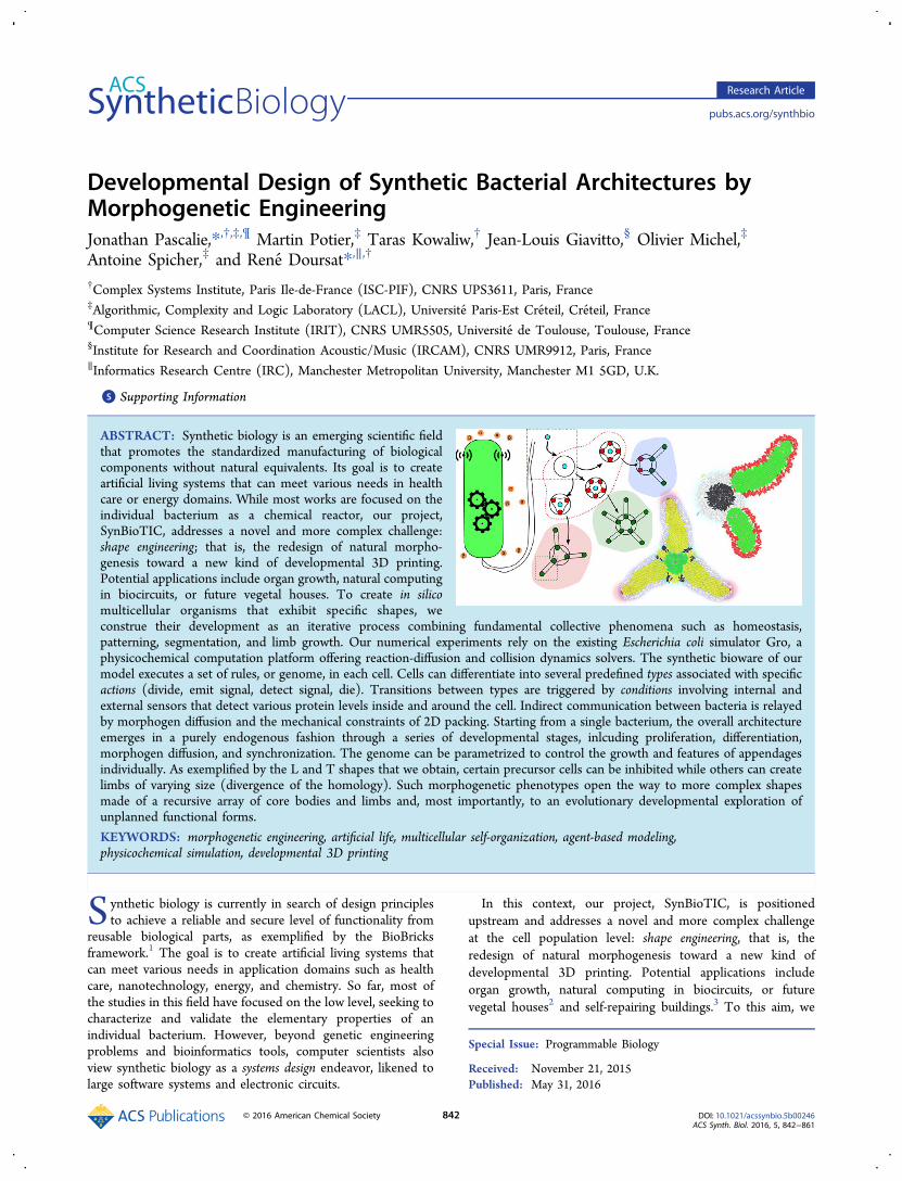
Figure 1. Targeted wheel-and-stick shapes. (A) Five-class taxonomy in increasing developmental complexity. Class I, radially symmetrical degree-1shapes: a single wheel is fitted with identical sticks positioned at regular angles. Class II, asymmetrical degree-1 shapes: heterogeneous sticks areattached to a single polarized wheel. Class III, unaligned degree-n shapes: two or more wheels are linked by sticks, while other sticks grow atunrelated angles. Class IV, aligned degree-n shapes: sticks grow at correlated angles. Class V, cyclic degree-n shapes: sticks from different wheels canmerge. (B) The three elementary “MorphoBricks” whose composition supports this variety of shapes: homeostatic core (wheel), limb growth(stick), and precursor cell (wheel-stick joint). The examples given here belong to Class I (pink and green domains) and Class IV (blue domain).Their implementation in the multicellular medium is explained in the Results section.
ACS Synthetic Biology Research Article
DOI: 10.1021/acssynbio.5b00246ACS Synth. Biol. 2016, 5, 842−861
844
In these examples, however, the specification of the structurerelied on direct encoding in a text filewhich made it easy fora human operator to modify, or possibly for evolution to createhigh-level mutations, but not for any developmental process totake place. On that basis, Lipson and Pollack’s Golem project36
showed that it was possible to cross the reality gap: actualrobots were created following the techniques tested andoptimized in simulation. First, morphologies and controllerswere evolved in a virtual environment, then the best individualswere realized mechanically and immersed in the real world.Together, these works proved the suitability of in silicoexperiments as an “engineering wizard”, even though theirbuilding blocks were not self-made via a growth process.Artificial development is now of great interest to robot design
and mostly studied in simulation,27,37,38 since physical 3D self-assembling systems are still rare. In these works, multicellulardevelopment is used as a means to produce fine-grainedmorphologies and improve them by evolution. From the firststeps made by Eggenberger39 to the latest production of Dissetet al.,40 which uses continuous cell shapes in a continuousmedium, morphological complexity has greatly increased.Nevertheless, three main components remain: stigmergicinteractions via chemical diffusion and gradient-based chemo-taxis; gene regulation triggering differentiation into types; andcell division, motion, and death.
■ MORPHOGENETIC SYNTHETIC BIOLOGYCurrent applications of synthetic biology, which focus on theindividual bacterium as a chemical reactor, come frombiochemistry and cellular biology. The main types ofapplications are the production of useful molecules andmaterials (drugs, biofuels, bioplastics) and the detection ofspecific conditions in the environment, for example by changingcolor (biosensors). The shape engineering challenge ofSynBioTIC, for its part, belongs to “morphogenetic engineer-ing” (ME), a field founded by Doursat et al.26,27 which studiesthe transfer of natural morphogenesis to the design of the self-organizing abilities of the elements of complex systems.Generally, natural pattern formation (stripes, spots, waves,branches) is stochastic and repetitive, in the sense that thecharacteristic scale of the motifs can be known but not theirexact position, whereas elaborate devices are the deterministicproduct of human design. Yet, multicellular biologicalorganisms are striking examples of complex systems that areboth entirely self-organized and strongly architectural, as theydisplay a precise arrangement of parts.41 Accordingly, MEestablishes a new object of research at the intersection betweentraditionally disconnected domains: it stresses the programm-ability of self-organization, underappreciated in complexsystems science, and, conversely, the benefits of self-organization, which are underappreciated in engineering.To support a wide variety of shapes in bioware, the growth
process needs to be sufficiently generic and parametric at thesame time. Our purpose is not to produce a few particularoutcomes but provide a developmental framework capable ofgenerating a whole family of them. In this study, we choose ahigh-level morphological specification based on two types ofelementary multicellular structures: core bodies, also called“wheels”, and limbs, also called “sticks” (Figure 1). Networkarchitectures made of nodes and links are a universal form oforganization observable at all levels of life: gene regulationnetworks, brain, skeleton, branching systems (vascular,respiratory and peripheral nervous systems; virtually all plants).
It embodies a building-block game that can produce rich andcomplex morphologies from simple primitives, especially byisotropic (wheel) and unidirectional (stick) development.Sticks can grow out of wheels at various angles and withvarious lengths, while wheels of various diameters can burgeonat the extremities of newly grown sticks. The set of shapesobtained by combining these building blocks can generally bedescribed by planar graphs or “circuits”, in which neighboringnodes are connected or not by an edge. Here our purpose is tocontrol the exact geometric features of the overall phenotype,by programming them indirectly in the genotype, not just letany mesh grow randomly. We are interested in architectures,not textures.To this aim, we propose a five-class taxonomy of wheel-and-
stick shapes sorted in order of increasing morphological anddevelopmental complexity, that is, difficulty of design (Figure1A):Class I: Radially symmetrical degree-1 shapes. In this first class,
shapes are based on a single isotropic wheel (the core body ofthe organism) fitted with identical sticks positioned at regularangles (its limbs), in the style of a “cross sign” or “starfish”.Development proceeds in two stages: isotropic proliferation ofthe initial cell into the wheel of the organism, then growth of afew sticks (typically 2 to 8) from precursor cells that havedifferentiated around the crown. These precursor cells are ofthe same type, giving rise to individuals that exhibit “serialhomology”. The number, length, and thickness of the sticks canbe tuned in the genome.Class II: Asymmetrical degree-1 shapes. Here precursor cells
adopt different types, hence sticks attached to a single wheelmay have diverse characteristics. In some of them, stick growthcan be inhibited altogether, creating a gap compared to aregular cross or star shape. Examples include letter shapes suchas “T”, “L”, “V”, and “Y”. The main feature of this class is thatthe wheel is polarized, allowing cells to acquire positionalinformation with respect to two main axes, anterioposterior(AP) and dorsoventral (DV) supported by four poles (north,south, east, west). More axes based on intermediate poles(northwest, etc.) can also be introduced.Class III: Unaligned degree-n shapes. This class represents the
first level of a recursive developmental process. It comprisesshapes with two or more wheels linked by sticks. Stickextremities lead to the development of wheels that initiate newsets of sticks. Although wheels can be locally polarized, they donot maintain relative orientation with respect to each other.Attached wheels only share one axis along their common stick.Typical shapes can look like articulated “snakes” (simple chainsof stick segments) or “centipedes”, where segments alternatewith pairs of legs, possibly of different lengths.Class IV: Aligned degree-n shapes. Next, an additional
requirement is that sticks growing on connected wheels doso at specific relative angles, for example parallel orperpendicular to each other. This is where we can designmore controlled geometries such as letters with two joints (“Z”,“F”) and three joints (“W”, “E”).Class V: Cyclic degree-n shapes. Finally, the most complex
shapes of the wheel-and-stick family involve sticks of differentorigins crossing paths and merging into the same wheel,thereby creating cycles in the graph structure. They includecharacters “A” and “4”. Once the convergent growth of twosticks is under control, any mesh figure or circuit architecturecan potentially be reached by carefully designing the genetic
ACS Synthetic Biology Research Article
DOI: 10.1021/acssynbio.5b00246ACS Synth. Biol. 2016, 5, 842−861
845
program. Before that level, however, the previous four classesmust also be mastered and issues of computing speed resolved.The present study does not cover this full taxonomy but
explores its different levels in increasing order of complexity bymodel and simulation. It presents original results about thetheoretical and practical possibility of (re)programmingbacteria to create certain multibacterial shapes in a controlledway. Starting with Classes I and II, we progressively build theelementary components needed for higher classes: single wheel,single stick, undifferentiated then differentiated precursor cells,wheel with sticks, stick with a wheel, and so on. However, sinceeach one of these components may include hundreds of cells,we also quickly face computational limitations that prevent usfrom calculating truly complex organisms. This is why our
numerical experiments stop for now at the entrance of Class III,and we only describe principles and mechanisms for the rest.The remainder of the article is organized as follows. In the
Results section, we describe how the mapping from an abstractgeometry to the multicellular medium can be realized inprinciple, via in silico numerical experiments. We show thedevelopment of three “MorphoBricks” (wheel, stick, and wheel-stick joint) and their composition into more complex Class-Iand Class-II organismsall starting from the controlledproliferation of a single cell. Then, in the Discussion section,we review the simplifications made in the model and theirshortcomings, particularly from the perspective of crossing the“reality gap” toward in vivo implementations. (We use “in vivo”in the synthetic-biological sense of genetic material (DNA,plasmids) implemented inside live cells, as opposed to being
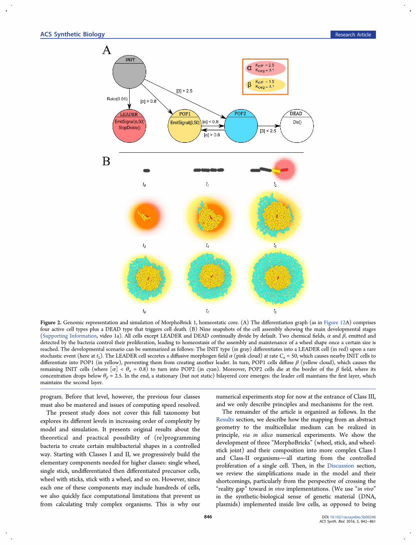
Figure 2. Genomic representation and simulation of MorphoBrick 1, homeostatic core. (A) The differentiation graph (as in Figure 12A) comprisesfour active cell types plus a DEAD type that triggers cell death. (B) Nine snapshots of the cell assembly showing the main developmental stages(Supporting Information, video 1a). All cells except LEADER and DEAD continually divide by default. Two chemical fields, α and β, emitted anddetected by the bacteria control their proliferation, leading to homeostasis of the assembly and maintenance of a wheel shape once a certain size isreached. The developmental scenario can be summarized as follows: The INIT type (in gray) differentiates into a LEADER cell (in red) upon a rarestochastic event (here at t2). The LEADER cell secretes a diffusive morphogen field α (pink cloud) at rate Cα = 50, which causes nearby INIT cells todifferentiate into POP1 (in yellow), preventing them from creating another leader. In turn, POP1 cells diffuse β (yellow cloud), which causes theremaining INIT cells (where [α] < θα = 0.8) to turn into POP2 (in cyan). Moreover, POP2 cells die at the border of the β field, where itsconcentration drops below θβ = 2.5. In the end, a stationary (but not static) bilayered core emerges: the leader cell maintains the first layer, whichmaintains the second layer.
ACS Synthetic Biology Research Article
DOI: 10.1021/acssynbio.5b00246ACS Synth. Biol. 2016, 5, 842−861
846
isolated in a test tube. Although the bacterial colony may becontained in a Petri dish, it is not considered in vitro because itconstitutes a living organism in itself.) In Future Work, wemention possible alternatives and new avenues of researchworth exploring. Finally, in Methods, we present the model ofbacterial behavior ontology at the basis of the simulations, andthe programming language that we designed to encode itsassociated genomic representations.
■ RESULTSHomeostatic Core: MorphoBrick 1. As presented above,
the two morphological components of our shape specificationsare wheels and sticks, which can be combined in a variety ofways to generate increasingly complex forms. In the next fewparagraphs, we describe how these two components are able toarise from the controlled proliferation of a single cell, that is,how the mapping from abstract geometries to the multicellularmedium can be realized. In a third stage, we provide thecomposition mechanism between the wheel and the stick in theform of precursor cells, a targeted differentiation mechanism onthe outer rim of the wheel body by which sticks are affixed towheels. Precursor cell positioning will be the cornerstone of thedevelopmental process as it is responsible for building the jointsbetween the core body and the limbs at locations specified bythe morphogenetic program.Altogether, we call MorphoBricks this set of three self-made,
self-assembling components, in a reference to the concept ofBioBricks1 translated from the molecular, genotypic level to themorphological, phenotypic level (Figure 1B). In each case, wewill pay special attention to the reproducibility andprogrammability of the virtual growth experiments throughsensitivity analysis and parametric exploration. This will alsooffer a glimpse of the great diversity of shapes that couldtheoretically be engineered through the proposed method. Tothis aim, we used OpenMOLE,42 a middleware platformfacilitating massive experimental parametric search of complexsystems models on a computing cluster, leveraging the power ofthe European Grid Infrastructure (EGI).MorphoBrick-1 Homeostatic Development. Homeo-
stasis, the process by which the internal state of a systemremains constant through self-regulation, is a major character-istic of life. Therefore, homeostatic properties should be aprime concern of synthetic biology efforts aiming to buildcomplex structures. Here our first goal is to create a coremorphological component that maintains itself in place. Thisrequires finding a means to maintain a stable population size. Inthe shape engineering challenge, the ability to control the sizeof a colony of bacteria over time is crucial to ensure thesustainability of their collective behavior.The mechanism that we propose for the development and
maintenance of a homeostatic core starting from a single cellrelies on a morphogenetic f ield emitted by a leader cell, whichcould be the initial cell or not (Figure 2B, t2). There are twodesign strategies to ensure the unicity of the leader: either byinhibiting its mitosis, or by making it divide asymmetrically;43
that is, one of its two daughter cells immediately differentiatesinto another type, keeping the leader role in the other daughter.The problem with the latter scenario is that it creates acontinual random-walk displacement of the leader at eachgeneration, which produces a lopsided wheel shape due to themorphogen trail left by the leader’s displacement (a similareffect will be in fact exploited, not suppressed, in MorphoBrick2). Thus, we opt for a nondividing leader cell, instead. A
consequence is that it cannot be the initial cell and must appearlater (but soon enough) via differentiation of another cellwhile proliferation is ensured by the presence of a sufficientnumber of nonleader cells.The genome designed for this purpose is represented
graphically in Figure 2A (see explanation in Methods section,in particular Genomic Representation; the full program can befound in Supporting Information, Code 1a). Initialization andleader cell generation are key mechanisms for the success of thedevelopment of this MorphoBrick and the others. The singlecell of origin is in a neutral INIT state, which can differentiateinto any of three active types composing the assembly:LEADER, POP1, and POP2. The emergence of the first oneis a rare stochastic event; the other two depend on localmorphogen concentrations, themselves resulting from emissionby the cells and passive diffusion. The LEADER cellcontinuously secretes a chemical α by accumulating a fixedconcentration amount Cα = 50, which we refer to as the rate ofemission, in the source location at each time step (the actualconcentration [α] reaches a plateau due to degradation anddiffusion perpetually at work in every point). This triggers thedifferentiation of nearby cells into POP1 and creates a firstcircular layer of this type until a certain concentration θα = 0.8,called dif ferentiation threshold (controlling three transitions).Meanwhile, POP1 cells emit chemical β, which creates a secondcircular layer of POP2 until θβ = 2.5, called survival threshold(controlling two transitions). The net result is the establish-ment of two stationary (but not static) rings of cell populationsaround the LEADER cell, each one responsible for the next.They are characterized by fountain-like collective motion, asbacteria are being pushed out by their own proliferation,becoming alternatively (and reversibly) POP1 or POP2, theneventually dying, depending on the underlying morphogeneticfields that they collectively generate.The final size of the organism and the thickness of the layers
depend on several parameters. In a given chemical environ-ment, morphological characteristics vary with the differentiationand survival thresholds, which are features of the genome:clearly, the smaller is θα or θβ, the thicker is the POP1 or POP2layer, respectively. Conversely, given a certain genome, thetrend would be similar if bacteria were immersed in a differentchemical environment where α or β had a higher diffusion rateκdiff or a smaller degradation rate κdeg. In the next paragraphs,we study quantitatively these phenotypic variations as afunction of the genetic parameters and environmentalconditions, starting with robustness with respect to randominitialization.
MorphoBrick-1 Sensitivity Analysis. First, the invarianceof the homeostatic core behavior with respect to experimentalconditions is studied by repeating many times the samesimulation under the same parameters, only using differentrandom generator seeds. The main consequence is felt at thelevel of the INIT → LEADER transition, as the stochasticdifferentiation event may affect a different cell than the one inFigure 2B at t2 and/or occur at an earlier or later time step.Other, subtler effects are caused by the fact that daughter cellsare not exact clones of the mother. The Gro simulator ascribesslightly different properties to bacteria when they divide, suchas their growth rate, by drawing them from Gaussiandistributions (it also models fluctuations through a variable dtobeying a Gamma distribution), which produces bacteriacolonies of variable size and layout.
ACS Synthetic Biology Research Article
DOI: 10.1021/acssynbio.5b00246ACS Synth. Biol. 2016, 5, 842−861
847
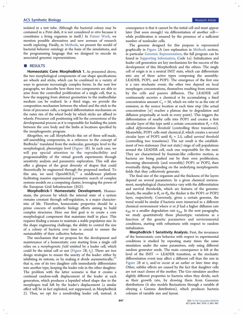
In any case, we analyze the outcome of the developmentalprocess based on three main geometric measures: the totalradius of the whole organism, the inner radius of the inner layer,and their difference, the crown thickness (outer layer’s radius).The global radius is simply the average of the four distancesthat separate the LEADER cell from the left-most, right-most,top-most, and bottom-most bacteria (all cell center coordinatesbeing known). The inner radius is obtained in the same way,counting only the POP1 cells. Figure 3A shows thedistributions of these three quantities for 300 trials under theparametric conditions of Figure 2. Only about 10% of thesimulations were rejected because two or more LEADER cellsemerged and compromised the development of the core. Thenarrow bell-curve aspect of these distributions shows that oursimulated developmental process produces robust morpholo-gies and legitimates the study of morphological variationsthrough numerical experiments.In sum, these results highlight the reliability of the
morphogenetic engineering approach, based here on chemicalfields. The whole assembly is capable of maintaining a wheel
shape of a certain size at the same time all its cells (except theleader) are constantly renewed. On the other hand, thishappens only as long as there exists a LEADER cell that emitsthe proper morphogen. Naturally, the dependency on asingularity of this kind is a much less desirable feature since,as soon as the leader dies, the bilayered-core organizationdisappears through uncontrolled proliferation, then massivedeath of POP1 and POP2 cells deprived of α and βmorphogens. Additional mechanisms are required for a morerealistic biological implementation of this scenario, either bytriggering the spontaneous replacement of the leader (e.g.,through self-election) or by allowing a clump of multipleLEADER cells, or by dispensing with the need of leadersentirely (see Discussion for a review of alternatives).
MorphoBrick-1 Genomic Variations: TransitionThresholds. We now analyze the influence of the variationof thresholds θα and θβ, starting with the latter. One of the rolesof the survival threshold is to induce death in POP2 cells whenthey detect a low β concentration. Therefore, θβ should have adirect influence on the thickness of the POP2 crown. This is
Figure 3. Sensitivity analysis and parametric exploration of MorphoBrick 1, homeostatic core. (A) Distributions of three morphologicalcharacteristics, left to right: outer radius, inner radius, and their difference, the crown thickness. All radial sizes were measured directly in the Groenvironment based on minimum and maximum cell coordinates. These distributions were obtained by repeating the same simulation 300 times withthe parameters of Figure 2 (out of which 30 failed on average and were discarded), only changing the random seed every time. The profiles areessentially bell-shaped, with low variance, showing that our approach based on morphogenetic fields is sufficiently robust to support themorphogenetic engineering of targeted shapes. (B) Morphological characteristics as a function of the survival threshold θβ, varied in [0.2, 20] byincrements of 0.2 (all other parameters as in Figure 2). Since θβ induces death in the POP2 cells (cyan bacteria) which make up the crown, then thelower is θβ, the larger is the crown thickness (cyan curve) and outer radius (black curve) because the inner radius is not affected (yellow curve). Allradial sizes were measured directly in the Gro environment based on minimum and maximum cell coordinates. The gray area indicates the standarddeviation of the total radius, calculated over 100 simulations in each point (10% discarded on average). (a−d) Four examples of wheel shapesobtained under different θβ values (final snapshots at homeostatic equilibrium corresponding to t8 in Figure 2), indicated by pins on the horizontalaxis. (C) Influence of the diffusion rate κdiff (α) varied in [0.03, 3.0] by increments of 0.03. Since chemical signal α is emitted by the LEADER celland affects the differentiation boundary between the POP1 and POP2 layers, then the higher is its diffusion rate, the wider is the inner radius and thetotal radius while the crown thickness remains constant. (a−d) Four final snapshots of wheel shapes at different rate values.
ACS Synthetic Biology Research Article
DOI: 10.1021/acssynbio.5b00246ACS Synth. Biol. 2016, 5, 842−861
848
verified in the statistical analysis of Figure 3B: increasing θβresults in an average decrease of the crown thickness. On theother hand, the inner radius of the POP1 layer remainsconstant, therefore the outer radius of the whole assembly,which is the sum of both, also decreases. These curves wereobtained by simulating 100 specimens for each one of 100different θβ values, with the other parameters set as in Figure 2.Note that the thickness decrease is not linear, since at highsurvival thresholds the border cells are in a steep region of [β]gradient, therefore threshold variations incur only relativelysmall thickness variations; whereas at low survival threshold theborder cells are in the flatter tail region of the gradient, henceslight threshold variations can cause larger size differences.A similar analysis was conducted over the differentiation
threshold θα, which controls the POP1 ↔ POP2 reversibletransition. In this case, as expected, the effect was a decrease of
the inner radius (hence the total radius) with higher values ofθα (curve not shown). Unlike the previous scenario, however,θα variations end up affecting both geometric measures, not justone of them. When the inner radius becomes too small, theouter crown also collapses, since there are not enough POP1cells to create a morphogenetic field of β suitable to POP2 cellsfor their development.
MorphoBrick-1 Environmental Variations: ChemicalRates. Phenotypic variations can also be elicited by modifyingthe properties of the surrounding chemicals, instead of thebacteria. This is a case of “polyphenism”, where the samegenotype immersed in different environments can produce adifferent phenotypes. Here, by varying the diffusion anddegradation rates κdiff and κdeg, the main homeostasis propertyof the core shape can be preserved while its morphologicalcharacteristics are modified. Figure 3C displays the results of a
Figure 4. Extra type and chemical in the implementation of MorphoBrick 1, homeostatic core (Supporting Information, video 1b). (A) Compared toFigure 2, this new genome design contains a third type, POP3, inserted in POP2’s path to cell death. Instead of dying at the edge of the β gradient,the POP2 bacteria (in yellow) first differentiate into POP3 (in green), then disappear at the edge of a new γ gradient emitted by POP2 (withthreshold θγ = 2.5). (B) As a consequence, the growing organism gains a third layer of cells. Notice the spontaneous spatial rearrangement in the laststages of the homeostatic convergence (from t6 to t8), as the initially asymmetric layer disposition caused by the linear, upward growth of populations1, 2, and 3 (from t2 to t5) becomes centered again.
ACS Synthetic Biology Research Article
DOI: 10.1021/acssynbio.5b00246ACS Synth. Biol. 2016, 5, 842−861
849
statistical analysis over a range of diffusion rates of morphogenα. As before, 100 simulations were carried out in each one ofthese 100 points. Unsurprisingly, the curve shows that when thediffusion rate increases, the inner radius and total radius alsoincrease.We also analyzed the organism size as a function of the
degradation rate κdeg(α), which describes how fast morphogenα disappears. In this case, also reassuringly, the higher thedegradation rate is, the smaller is the inner radius (curve notshown). When the degradation rate becomes too high, [α]drops faster than it can be replenished by the LEADER cell’s
signal emission. This results in a small area of concentrationaround the leader, hence a small inner layer, which in turn has adamaging influence on the crown thickness as explained above.
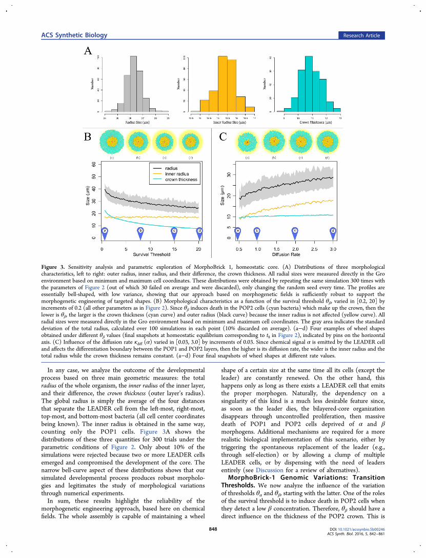
MorphoBrick-1 Genomic Mutation: Number of Types.In a last part of this MorphoBrick-1 study, we look at a hand-mutated genome structure, in which a third cell type POP3 iscreated and new conditional transitions introduced betweenPOP2 and DEAD (Figure 4A; see full program in SupportingInformation, Code 1b). Moreover, POP2 now emits anothermorphogen, γ, which sustains the emergence of POP3 cells andalso regulates their disappearance on the outer rim. In essence,
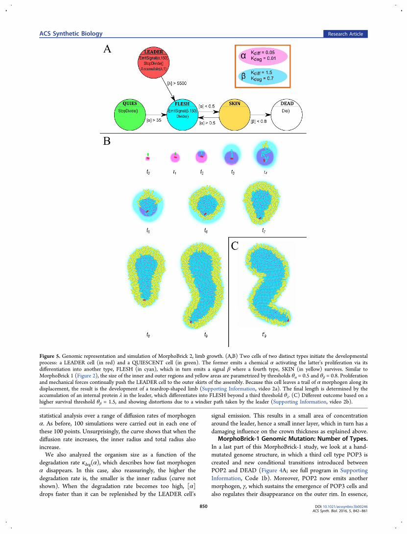
Figure 5. Genomic representation and simulation of MorphoBrick 2, limb growth. (A,B) Two cells of two distinct types initiate the developmentalprocess: a LEADER cell (in red) and a QUIESCENT cell (in green). The former emits a chemical α activating the latter’s proliferation via itsdifferentiation into another type, FLESH (in cyan), which in turn emits a signal β where a fourth type, SKIN (in yellow) survives. Similar toMorphoBrick 1 (Figure 2), the size of the inner and outer regions and yellow areas are parametrized by thresholds θα = 0.5 and θβ = 0.8. Proliferationand mechanical forces continually push the LEADER cell to the outer skirts of the assembly. Because this cell leaves a trail of α morphogen along itsdisplacement, the result is the development of a teardrop-shaped limb (Supporting Information, video 2a). The final length is determined by theaccumulation of an internal protein λ in the leader, which differentiates into FLESH beyond a third threshold θλ. (C) Different outcome based on ahigher survival threshold θβ = 1.5, and showing distortions due to a windier path taken by the leader (Supporting Information, video 2b).
ACS Synthetic Biology Research Article
DOI: 10.1021/acssynbio.5b00246ACS Synth. Biol. 2016, 5, 842−861
850
the INIT → POP1 → POP2 differentiation chain is augmentedwith a third link, POP2 → POP3, and the consequence is theappearance of a third layer in the collective phenotype (in greenin Figure 4B). As before, the thickness of the layers and theglobal radius of the wheel-shaped assembly are parametrized bythe various threshold and rate values. Theoretically, thisapproach can be easily generalized to N POP types: given a
desired number of layers N, a generative approach couldautomatically produce the corresponding SBGP genome, thenthe Gro script.The advantage of increasing the number of types and layers is
to improve positional information, hence positioning diversityand accuracy for the future appendages in later stages (seeMorphoBrick 3 paragraphs below). This is because more
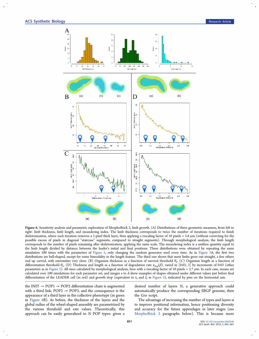
Figure 6. Sensitivity analysis and parametric exploration of MorphoBrick 2, limb growth. (A) Distributions of three geometric measures, from left toright: limb thickness, limb length, and meandering index. The limb thickness corresponds to twice the number of iterations required to finishskeletonization, where each iteration removes a 1-pixel thick layer, then applying a rescaling factor of 10 pixels = 5.8 μm (without correcting for thepossible excess of pixels in diagonal “staircase” segments, compared to straight segments). Through morphological analysis, the limb lengthcorresponds to the number of pixels remaining after skeletonization, applying the same scale. The meandering index is a unitless quantity equal tothe limb length divided by distance between the leader’s initial and final positions. These distributions were obtained by repeating the samesimulation 100 times with the parameters of Figure 5, only changing the random generator seed every time. As in Figure 3A, the first twodistributions are bell-shaped, except for some bimodality in the length feature. The third one shows that most limbs grow out straight, a few othersend up curved, with extremities very close. (B) Organism thickness as a function of survival threshold θβ. (C) Organism length as a function ofdifferentiation threshold θα. (D) Thickness and length as a function of degradation rate κdeg(β), varied in [0.03, 3] by increments of 0.03 (otherparameters as in Figure 5). All sizes calculated by morphological analysis, here with a rescaling factor of 10 pixels = 2.7 μm. In each case, means arecalculated over 100 simulations for each parameter set, and images a to d show examples of shapes obtained under different values just before finaldifferentiation of the LEADER cell (in red) and growth stop (equivalent to t9 and t9′ in Figure 5), indicated by pins on the horizontal axis.
ACS Synthetic Biology Research Article
DOI: 10.1021/acssynbio.5b00246ACS Synth. Biol. 2016, 5, 842−861
851
signals can be interpreted more locally over shorter distances,where gradients are steeper and less sensitive to noise, that is,can provide better control than flat ones. On the other hand,the problem is the complexity of handling multiple chemicalsignals and the difficulty of implementing them in the lab (seeDiscussion section).Limb Growth: MorphoBrick 2. The second component of
our morphogenetic experiments is a stick, or “limb”. Here wedescribe an implementation of standalone limb development inthe multicellular medium. In the next section, we show how itcan grow out of the body, using MorphoBrick-3 precursor cellsas joint structures. The limb’s differentiation graph is based onfour active cell types: LEADER, QUIESCENT, FLESH, andSKIN (Figure 5; see full program in Supporting Information,Code 2a). Initially, only initial cells of the first two types areneeded to start the process, which unfolds in a way similar tomeristem offshoot in plants. The LEADER plays the role of aprecursor cell, while the QUIESCENT cell, upon stimulationby the leader cell’s signal α, turns into a FLESH cell and entersa proliferative cycle. Overall, the net effect is that the leader iscontinually pushed away, leaving a diffusive plume of cells in itspath. FLESH cells spread out in the region [α] > θα = 0.8 andalso emit β, surrounding themselves with an outer layer ofSKIN cells (in the same way POP1 cells surround themselveswith POP2 in MorphoBrick 1), to the extent determined by alower threshold: [β] > θβ = 0.8.The slow diffusion (κdiff = 0.05) and low degradation rates
(κdeg = 0.01) of morphogen α emitted by the LEADER cellbuild a kind of “chemical shelter” for FLESH cells, as it allowsthe concentration to stay high and FLESH cells to proliferatewhile the source cell has time to move away, which fuels thelengthening and widening of the limb. In fact, this is the mostimportant difference with the development of the core (forwhich κdiff = 2.5 and κdeg = 0.1). As in MorphoBrick 1, however,the success of limb development rests upon the survival of asingle LEADER cell. Here its unicity is guaranteed since theleader is already present at the start and does not need a rarestochastic event to appear later in the process (it will be in factprovided by MorphoBrick 3 in the next stage). Yet again, thesame questions as before arise (see Discussion section): Canthe design be modified to accommodate the regeneration of theLEADER cell in case of its untimely disappearance? or couldthere be a clump of multiple leadersor none at all?In any case, a specificity of the limb structure is that its
extension depends on the life duration of the LEADER cell.Here homeostatis is only partial, in the sense that the SKINlayer adopts a certain thickness while still proliferating, whereasno limit length is reached and maintained. Therefore, to controlthe length of the limb, we introduce a clock mechanism in thesource. Once it starts emitting the proliferation activator α, italso accumulates an internal protein λ, and when thisconcentration reaches a given threshold θλ, then LEADERdifferentiates into FLESH, mingles with the crowd, and thegrowth process stops. The limb length can therefore beparametrized in two ways: by varying θλ or the accumulationrate Cλ of the protein. The sensitivity to noise and the influenceof the genomic and chemical parameters on the geometricalfeatures are studied next.MorphoBrick-2 Sensitivity Analysis. As in the previous
section, it is important to analyze the robustness of limbgrowth, if we want to use it as a reliable developmental buildingblock. To this aim, we performed again 100 runs under thedefault parametric conditions of Figure 5 and measured the
average thickness and length of the obtained morphology(Figure 6). Both were obtained by extracting the topologicalskeleton from the limb shape, that is, the pixelized domaincovered by the bacteria after smoothing to avoid too manyspurious branches (see algorithm in Supporting Information,Morphological Analysis). The thickness corresponds to thenumber of iterations required to eat out the flesh and convergeto the skeleton, while the path length is given roughly by thenumber of pixels that constitute the final skeleton (seeconversion to micrometers in the figure caption). As with thehomeostatic core structure, these distributions appear essen-tially bell-shaped with a relatively narrow variance, except for aslight tendency of the length distribution to bimodality,showing a relative lack of midrange values. A third measure,the meandering index, which is the limb length divided bydistance between the initial and final positions of the LEADERcell, indicates that the great majority of phenotypes are linear(value close to 1)although a significant number of them arealso sufficiently curved or convoluted to produce indices of 2 or3 (e.g., hook-shaped limbs whose extremities are close to eachother).
MorphoBrick-2 Genomic Variations: TransitionThresholds. Next, we need again to understand the effect ofgenomic parameters and environmental conditions on thephenotype to be able to target specific morphologicalcharacteristics. Since morphogens α and β play a role equivalentto the MorphoBrick-1 study, we also call θα the differentiationthreshold and θβ the survival threshold here. For reasons similarto the previous study, the results shown in Figure 6B,C confirmthat an increase in θβ provokes a decrease in the thickness ofthe SKIN layer (formerly POP2), hence the whole limb, whilean increase in θα provokes a decrease in the FLESH populationsize (formerly POP1), hence the overall length since the leaderis pushed away slower by less active proliferation and packingforces.
MorphoBrick-2 Environmental Variations: ChemicalRates. External conditions also influence the developmentalprocess. Here we display the influence of the degradation rateκdeg of the survival morphogen β (Figure 6D). A higher ratecauses a decrease in both dimensions of the limb, thickness, andlength, although in different ways. Clearly, the more degradableβ, the more restricted the area where [β] > θβ, hence the lessprolific the SKIN cells. If κdeg is too high, the morphogen neverreaches a level suitable for SKIN to develop and survive. TheFLESH cells, for their part, are not sensitive to β, yet limblength still indirectly depends on the degradation rate. Thereason, again, is that the trajectory and speed of displacement ofthe LEADER cell along the major axis are a result of mechanicalinteractions and spatial constraints exerted by the SKIN layerpressing laterally on the FLESH core. In the case of a morevolatile chemical, there is less density and these forces areweaker. Note that, in any case, cleavage planes are orientedrandomly and do not contribute to the direction taken by theelongation; only the leader’s displacement does.
Precursor Cells and Assembly: MorphoBrick 3. Thethird and last component we present in this study consists ofplacing at the periphery of a wheel a precursor cell that will giverise to a stick. For this, we need a spatiotemporal differentiationmechanism by which certain POP2 cells around the core canself-elect at regular intervals and become the new LEADERcells of limb growth. This is a form of segmentation, a pervasivedevelopmental episode across multicellular species (e.g.,somitogenesis in vertebrates), which will be based here on
ACS Synthetic Biology Research Article
DOI: 10.1021/acssynbio.5b00246ACS Synth. Biol. 2016, 5, 842−861
852
the combination of two low-level mechanisms: wave prop-agation and oscillations.MorphoBrick-3 Segmentation. Wave propagation is
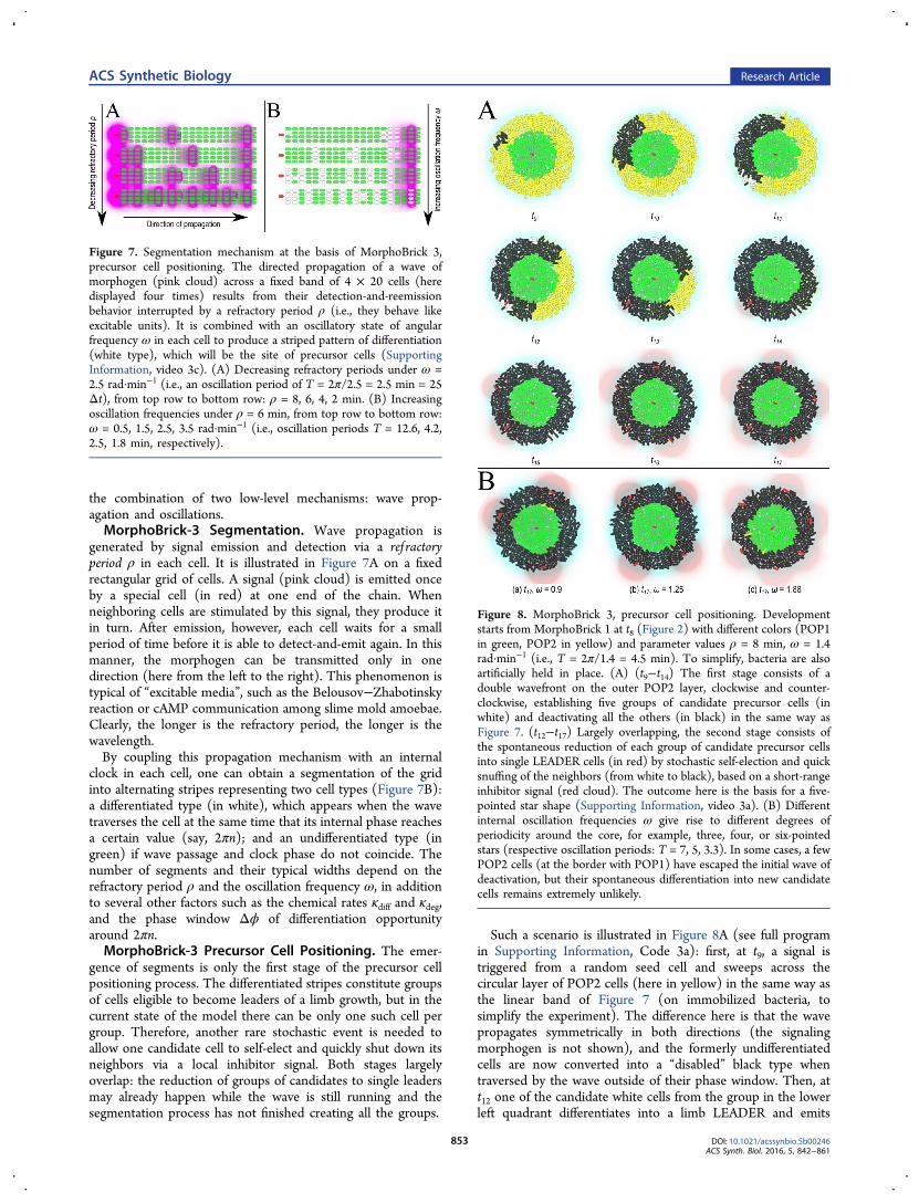
generated by signal emission and detection via a ref ractoryperiod ρ in each cell. It is illustrated in Figure 7A on a fixedrectangular grid of cells. A signal (pink cloud) is emitted onceby a special cell (in red) at one end of the chain. Whenneighboring cells are stimulated by this signal, they produce itin turn. After emission, however, each cell waits for a smallperiod of time before it is able to detect-and-emit again. In thismanner, the morphogen can be transmitted only in onedirection (here from the left to the right). This phenomenon istypical of “excitable media”, such as the Belousov−Zhabotinskyreaction or cAMP communication among slime mold amoebae.Clearly, the longer is the refractory period, the longer is thewavelength.By coupling this propagation mechanism with an internal
clock in each cell, one can obtain a segmentation of the gridinto alternating stripes representing two cell types (Figure 7B):a differentiated type (in white), which appears when the wavetraverses the cell at the same time that its internal phase reachesa certain value (say, 2πn); and an undifferentiated type (ingreen) if wave passage and clock phase do not coincide. Thenumber of segments and their typical widths depend on therefractory period ρ and the oscillation frequency ω, in additionto several other factors such as the chemical rates κdiff and κdeg,and the phase window Δϕ of differentiation opportunityaround 2πn.MorphoBrick-3 Precursor Cell Positioning. The emer-
gence of segments is only the first stage of the precursor cellpositioning process. The differentiated stripes constitute groupsof cells eligible to become leaders of a limb growth, but in thecurrent state of the model there can be only one such cell pergroup. Therefore, another rare stochastic event is needed toallow one candidate cell to self-elect and quickly shut down itsneighbors via a local inhibitor signal. Both stages largelyoverlap: the reduction of groups of candidates to single leadersmay already happen while the wave is still running and thesegmentation process has not finished creating all the groups.
Such a scenario is illustrated in Figure 8A (see full programin Supporting Information, Code 3a): first, at t9, a signal istriggered from a random seed cell and sweeps across thecircular layer of POP2 cells (here in yellow) in the same way asthe linear band of Figure 7 (on immobilized bacteria, tosimplify the experiment). The difference here is that the wavepropagates symmetrically in both directions (the signalingmorphogen is not shown), and the formerly undifferentiatedcells are now converted into a “disabled” black type whentraversed by the wave outside of their phase window. Then, att12 one of the candidate white cells from the group in the lowerleft quadrant differentiates into a limb LEADER and emits
Figure 7. Segmentation mechanism at the basis of MorphoBrick 3,precursor cell positioning. The directed propagation of a wave ofmorphogen (pink cloud) across a fixed band of 4 × 20 cells (heredisplayed four times) results from their detection-and-reemissionbehavior interrupted by a refractory period ρ (i.e., they behave likeexcitable units). It is combined with an oscillatory state of angularfrequency ω in each cell to produce a striped pattern of differentiation(white type), which will be the site of precursor cells (SupportingInformation, video 3c). (A) Decreasing refractory periods under ω =2.5 rad·min−1 (i.e., an oscillation period of T = 2π/2.5 = 2.5 min = 25Δt), from top row to bottom row: ρ = 8, 6, 4, 2 min. (B) Increasingoscillation frequencies under ρ = 6 min, from top row to bottom row:ω = 0.5, 1.5, 2.5, 3.5 rad·min−1 (i.e., oscillation periods T = 12.6, 4.2,2.5, 1.8 min, respectively).
Figure 8. MorphoBrick 3, precursor cell positioning. Developmentstarts from MorphoBrick 1 at t8 (Figure 2) with different colors (POP1in green, POP2 in yellow) and parameter values ρ = 8 min, ω = 1.4rad·min−1 (i.e., T = 2π/1.4 = 4.5 min). To simplify, bacteria are alsoartificially held in place. (A) (t9−t14) The first stage consists of adouble wavefront on the outer POP2 layer, clockwise and counter-clockwise, establishing five groups of candidate precursor cells (inwhite) and deactivating all the others (in black) in the same way asFigure 7. (t12−t17) Largely overlapping, the second stage consists ofthe spontaneous reduction of each group of candidate precursor cellsinto single LEADER cells (in red) by stochastic self-election and quicksnuffing of the neighbors (from white to black), based on a short-rangeinhibitor signal (red cloud). The outcome here is the basis for a five-pointed star shape (Supporting Information, video 3a). (B) Differentinternal oscillation frequencies ω give rise to different degrees ofperiodicity around the core, for example, three, four, or six-pointedstars (respective oscillation periods: T = 7, 5, 3.3). In some cases, a fewPOP2 cells (at the border with POP1) have escaped the initial wave ofdeactivation, but their spontaneous differentiation into new candidatecells remains extremely unlikely.
ACS Synthetic Biology Research Article
DOI: 10.1021/acssynbio.5b00246ACS Synth. Biol. 2016, 5, 842−861
853
another short-range morphogen (pink clouds), which has theeffect of deactivating the other white cells from this group(making them black). Meanwhile, the wave has finishedcovering the whole outer layer at t14: in this case, the ρ andω values are such that a total of five radial stripes (whitegroups) are created. Short thereafter, the other four groupsreduce themselves to single LEADER cells, too (in red). Theresult at t17 is an approximately regular pentagonal structure. Inthis case, the final layout of the five precursors will always be apentagon; only its absolute orientation (between 0 and 72degrees) may vary depending on the location of the first seedcell. This is not a problem for Classes I to III, but in Classes IVand V additional staging mechanisms (possibly involvingexternal stimuli such as light) must be introduced to align orcorrelate limbs between distant bodies. Figure 8B showsdifferent degrees of periodicity (triangle, square, and hexagon)arising from different oscillation frequencies ω.Class-I Shapes: Radially Symmetric Wheels. Putting all
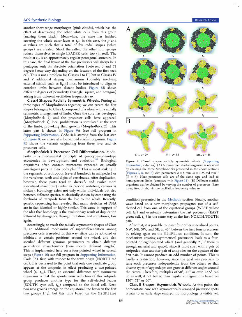
three types of MorphoBricks together, we can create the firstshapes belonging to Class I, composed of a wheel with a radiallysymmetric arrangement of limbs. Once the core has developed(MorphoBrick 1) and the precursor cells have appeared(MorphoBrick 3), local proliferation is stimulated at the rootof the limbs, provoking their growth (MorphoBrick 2). Thislatter part is shown in Figure 9A (see full program inSupporting Information, Code 4a): starting from the last stepof Figure 8, we arrive at a four-armed starfish organism. Figure9B shows the variants originating from three, five, and sixprecursor cells.MorphoBrick-3 Precursor Cell Differentiation. Modu-
larity is a fundamental principle of genotype−phenotypeeconomics in development and evolution.44 Biologicalorganisms often contain numerous repeated or seriallyhomologous parts in their body plan.45 This is most striking inthe segments of arthropods (several hundreds in millipedes) orthe vertebrae, teeth and digits of vertebrates. After duplication,however, these parts tend to diversify and evolve morespecialized structures (lumbar vs cervical vertebrae, canines vsmolars). Homology exists not only within individuals but alsobetween different species, as classically shown by comparing theforelimbs of tetrapods from the bat to the whale. Recently,genetic sequencing has revealed that many stretches of DNAare in fact identical or highly similar. This came in support ofthe idea that homology is the evolutionary result of duplicationfollowed by divergence through mutation, and sometimes, lossagain.Accordingly, to reach the radially asymmetric shapes of Class
II, an additional mechanism of superdifferentiation amongprecursor cells is needed. In this way, sticks can be activated orinhibited at certain positions around the wheel, and alsoascribed different genomic parameters to obtain differentgeometrical characteristics (here mostly different lengths).This is implemented here on a four-pointed wheel in severalsteps (Figure 10; see full program in Supporting Information,Code 3b): first, with respect to the wave origin (NORTH redcell), ω is decreased to the point that only one candidate groupappears at the antipode, in effect producing a two-pointedwheel (t9−t12). Then, an essential difference with symmetricorganisms is that the spontaneous reduction of this antipodegroup produces another type for the self-elected leader(SOUTH cyan cell, t13) compared to the initial cell. Next,two new groups emerge on the equatorial line between the firsttwo groups (t14), but this time based on the MidPlane
condition presented in the Methods section. Finally, anotherwave based on a new morphogen propagates out of a self-elected cell from one of the equatorial groups (WEST yellowcell, t15) and eventually determines the last precursor (EASTgreen cell, t17) in the same way as the first NORTH/SOUTHpair.After that, it is possible to insert four other specialized points,
NW, NE, SW, and SE, at 45° betwen the first four precursorsby relying again on the MidPlane condition. In sum, themechanism creating asymmetrical precursors leads to a four-pointed or eight-pointed wheel (and generally 2n, if there isenough material and space), since it must start with a pair ofantipodes, then another pair of antipodes on the equator of thefirst pair. It cannot produce an odd number of points. This ishardly a restriction, however, since the goal was precisely totune each precursor independently from the others so thatdiverse types of appendages can grow at different angles aroundthe crown. Therefore, multiples of 90°, 45° or even 22.5° cando as well, if not better, than regular configurations based on120°, 72° or 60°.
Class-II Shapes: Asymmetric Wheels. At this point, thehomeostatic core with asymmetrically arranged precursor spotsis akin to an early stage embryo: no morphology is visible yet,
Figure 9. Class-I shapes: radially symmetric wheels (SupportingInformation, video 4a). (A) A four-armed starfish organism is obtainedby chaining the three MorphoBricks presented in the above sections(Figures 2, 8, and 5) with parameters ρ = 8 min, ω = 1.25 rad·min−1
(T = 5). Here precursor cells are of the same type and lead tohomogeneous limbs (compare with Figure 11). (B) Different starfishorganisms can be obtained by varying the number of precursors (herethree, five, or six) via the oscillation frequency value ω.
ACS Synthetic Biology Research Article
DOI: 10.1021/acssynbio.5b00246ACS Synth. Biol. 2016, 5, 842−861
854
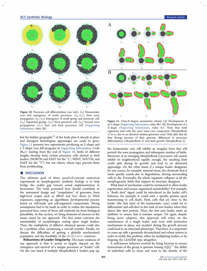
but the hidden geography46 of the body plan is already in placeand divergent homologous appendages are ready to grow.Figure 11 presents two experiments producing an L-shape anda T-shape (see full programs in Supporting Information, Code4b,c): starting from the end of Figure 10, limbs of differentlengths develop from certain precursor cells elected as limbleaders (NORTH and EAST for the “L”; WEST, SOUTH, andEAST for the “T”) but not others, whose type prevent themfrom proliferating.
■ DISCUSSIONThe ultimate goal of these proof-of-concept numericalexperiments in morphogenetic synthetic biology is to helpbridge the reality gap toward actual implementation inbiosystems. The work presented here should contribute tothe automated design and “compilation” of genomes, fromhigh-level scripts such as SBGP and Gro down to DNAsequences, supporting an algorithmic developmental processbased on self-made and self-organized components. Strongassumptions had to be made in order to realize the simulationspresented here, some of them still relatively far from biologicalplausibility. In this section, we bring elements of answers to theissues raised by our approach. The first point concerns thesustainability of morphologies resting upon single leaders.Then, the multiplicity of diffusive chemical species could alsobe a problem when envisioning a wet-lab transfer. Finally, wediscuss the difficulties of getting a globally synchronizedpopulation and the feasibility of the MidPlane construct.Robustness of Leader Singularity. The main weakness of
our approach is that it seems to largely depend on theemergence and survival of a unique precursor or “leader” cell.On the one hand, if multiple MorphoBrick-1 leaders pop up,
the homeostatic core will exhibit an irregular form that willperturb the wave propagation and subsequent number of limbs.Moreover, if an emerging MorphoBrick-3 precursor cell cannotinhibit its neighborhood rapidly enough, the resulting limbcould split during its growth and lead to an abnormalappendage. On the other hand, if a unique leader disappearsfor any reason, for example, external stress, the chemicals that itemits quickly vanish due to degradation, driving surroundingcells to die. Eventually, the whole organism collapses as all themorphogenetic fields that support its structure disappear.What kind of mechanism could be envisioned to allow leader
regeneration and ensure organismal sustainability? For example,a “death alert” signal could be introduced in the leader cell’sbehavior, for example, it could emit a specific protein upontransitioning to cell death. Next, cells that are close to theleader (the first layer of the homeostatic core) could try todifferentiate and self-elect to the rank of new leaders when theydetect this alert protein. Then, the first new leader emits aninhibitor to ensure that it remains unique. Yet again, despitebeing more adaptive, this approach still relies on themaintenance of a single leader and, even with protectionmechanisms in place, any incident during the growth processcould lead to an abnormal phenotype. Therefore, it is importantto come up with a genuinely decentralized and robust system inorder to tackle this problem, either by multiplying leaders or byforgoing the LEADER type altogether.A well-known behavior evolved by living bacteria to ensure
homeostasis of the group is quorum sensing (QS),47 the abilityof individual cells to sense and react to the density of the
Figure 10. Precursor cell differentiation (see text). (t9) Homeostaticcore and emergence of north precursor. (t10−t11) First wavepropagation. (t12−t13) Emergence of south group and precursor cell.(t14) Equatorial groups. (t15) West precursor cell. (t16) Second wavepropagation. (t17) East and final precursor cell (SupportingInformation, video 3b).
Figure 11. Class-II shapes: asymmetric wheels. (A) Development ofan L-shape (Supporting Information, video 4b). (B) Development of aT-shape (Supporting Information, video 4c). Note that bothorganisms start with the exact same core component (MorphoBrick1) in t17 due to an identical random generator seed. Only after that dothey diverge because of their genomic differences in precursordifferentiation (MorphoBrick 3) and limb growth (MorphoBrick 2).
ACS Synthetic Biology Research Article
DOI: 10.1021/acssynbio.5b00246ACS Synth. Biol. 2016, 5, 842−861
855
colony. Under stress, bacteria can emit a chemical that triggerscell death above a certain concentration, resulting in self-regulated population size. Within the great diversity of QSsignals and behaviors, an example is S. pneumoniae which candifferentiate into a “competent” state, that is, one where the cellis more prone to genetic mutation but also, at the same time, togrowth arrest and autolysis based on a QS-dependent on/offswitch.48 Thus, a possible way to solve the dependency on oneor several leader cells would be to use a QS mechanism to limitproliferation. In MorphoBrick 1, starting with all initial cellsalready in the POP1 type, a proper balance between diffusion,degradation, and emission rates could probably be found inorder to create a stationary concentration of morphogens,hence a stable POP1 cluster. It is also likely that this clusterwould stabilize into a roughly circular shape, hence that thecore would correctly develop from there as planned.Alternatively, it should also be possible to reinforce thetendency to form a circle by using a small cluster of LEADERcells in lieu of a single one. Thus, rather than installing QSamong the POP1 cells, it would be practiced by the LEADERcells and POP1 would then simply grow around them. Morelayers could also be added (as in Figure 4) to improve theroundness of the shape even further.Multiplicity of Chemical Species. Another aspect that
could make a direct application to in vivo experimentsproblematic is the multiplicity of diffusive chemical specieshypothesized in the environment. For example, the Class-IIorganisms presented here (the letter shapes) consume no lessthan seven different and highly specific signals. At higher levelsof biological complexity (Class III and beyond), the number ofchemical species involved in bacterial communication will beeven greater according to the MorphoBrick compositionaldesign. Moreover, in the current framework, no chemicalreactions are modeled, that is, molecular species are notsupposed to react with each other if we want to avoid side-effects during development. These conditions will be achallenge to maintain in the real world. A more realisticmodel will need to allow reuse of morphogens in differentlocations and at different stages of the organismal growth. Itmust also take reaction kinetics into account and exploit thenew compounds thus produced to serve as new signalingmolecules. On the other hand, the added complexity of anetwork of chemical reactions will also require fine-tuning of agreat number of parameters, which can ultimately be onlyachieved by evolutionary computation (such as geneticalgorithms), or why not actual biological evolution, by lettingthe artificially engineered bacteria readapt to new conditions ontheir own.Synchrony of Bacterial Population. So far, to ensure the
success of MorphoBrick 3, the bacterial population needs to besynchronized. The coordinated polarization of the initial core ofthe organism is necessary to allow conditional differentiation ofcandidate precursor cells at regular intervals on the outercrown. Yet, this is the only stage where global synchrony isrequired: at any other point during development, bacteria’sinternal clocks or oscillatory behavior are not part of thedynamics. Therefore, it would be more realistic to devise asolution relying on local synchrony only. This said, the methodof forcing cell synchronization, then measuring how theygradually get out of phase during relaxation is commonlyemployed to analyze cell cycles, and in silico modeling andsimulation of this phenomenon were also proposed.49 This iswhy we assumed that the global synchrony stage was a natural
and relatively easily reproducible phenomenon, and we coulddispense with a more accurate depiction of it in our presentnumerical framework. At the synthetic GRN level, we canbriefly mention the work of Danino et al.,50 who proposeengineered genetic circuits able to produce and maintainsynchronized oscillations in a growing population of cells. Thiscould for example constitute a suitable component in a futureimplementation of MorphoBrick 3.
Midplane Feasibility. Finally, growing organisms of ClassII and above depends on the MidPlane mechanism, whichallows a cell to recognize whether it is approximately midwaybetween two different sources of chemical gradients. In theabstract, this is achieved by comparing local productconcentrations and assessing whether they are sufficientlyclose. In reality, the question of how the required combinationof “subtracter” and “zero-tester” logic is implemented in theGRN can be answered by signal-mediated toggle-switchsubcircuits. For example, two repressible promoters can bearranged in a mutually inhibitory loop13 or concurrentpromotion/repression can be exerted by the same transcriptionfactor in conjunction with two different signals.51 In any case,there are several possibilities of making the regulation dynamicsexhibit bistability, such that a cell can fall into one of two statesdepending on the relative levels of two ligand concentrations itreceives.
■ FUTURE WORKToward Higher-Level Classes. This paper covered Class-I
and Class-II organisms. To reach higher-level classes, otherissues need to be solved. First, simulations should be run at alower computing cost and/or time, since for now almost anentire hour is needed to calculate the development of one T-shape or L-shape (even as a batch job, without onlinevisualization). This could be done by parallelizing execution,for instance on graphics processing units (GPUs), andeventually using a more powerful platform than Gro, such asCellModeller developed by Rudge et al.52 or our own TB underconstruction.53 The next question is the ability to grow andpolarize secondary cores. It should be relatively easy to make anew wheel sprout at the extremity of a stick, simply by lettingthe limb’s LEADER cell at the tip (the red cell pushed away inall simulations) become the new central leader of a homeostaticcore, while the nearby FLESH cells become POP1 and POP2and proliferate around it. Then, precursor positioning and limbgrowth would proceed as usual, leading to Class-III organisms(Figure 1).Developmental complexity increases sharply with Class IV,
however, as these organisms require long-range correlatedpositional information across multiple cores to be able todisplay limbs that are globally parallel or aligned. This wouldrequire an additional component, MorphoBrick 4, dedicated tocommunication among crown cells in order to allow them todetect their bearing (N, S, W, or E) with respect to the rootlimb that gave rise to the core, and control the start of the waveof differentiation so that the N/S polarization axis is notrandom. Finally, the gap between Class IV and Class V seemsrelatively less challenging as it would only require a new abilityfor two limbs growing toward each other to meet at their tipsand merge into one.
Polymorphism, Polyphenism, and Evolution. The basisof the morphogenetic engineering effort consists of adevelopmental process executed by a morphogenetic engine(here a bacterial simulator, or a Petri dish), whose purpose is to
ACS Synthetic Biology Research Article
DOI: 10.1021/acssynbio.5b00246ACS Synth. Biol. 2016, 5, 842−861
856
take in input an agent-level genotype G and transform it into acollective phenotype P in output. It does this via a swarm of G-carrying agents that coordinate their behavior and differentiate(by direct peer-to-peer signaling and/or indirect stigmergiccues deposited in the environment), move, and self-assemble(by division and/or aggregation), and collectively construct anarchitecture. We believe that our rational-design methodologybased on compositional building blocks such as MorphoBricksis particularly appropriate when targeting a generative family ofshapes or classes of shapes.To achieve polymorphism inside a given species (here a
“class”), a crafted G |→ P mapping like ours must offer thepossibility of internal parametrization of G (regulatoryparameters such as thresholds, rates, and frequencies) so thatit can give rise to different traits in P (here different limbs).This is similar to the classical laws of population genetics,schematically corresponding to the concepts of alleles or SNPsin the DNA. When grown freely, in absence of environmentalperturbation, the effect of genotypic parametrization is to offera family of different breeds within the same species, as in
Mendel’s peas or Darwin’s pigeons (here the various star shapesof Class I, and letter shapes of Class II).Moreover, under a given (possibly parametrized) G |→ P
mapping, the development process itself should also besensitive to, and modifiable by environmental conditions.External conditions or stimuli encountered by one individualduring its growth, whether mechanical or signaling (hereobstacles and chemicals), should be able to influence theoutcome, giving rise to polyphenism. This is the level of P, forwhich natural analogies can be found more readily in plantsthan animals. Trees can be spruced, pruned, bent, arranged,sculpted, etc., whether intentionally by humans (bonsais,espaliers, topiaries) or spontaneously when faced with adverseor favorable conditions (wind, rocks, light, humidity). The samegardening metaphor could apply here to morphogeneticsynthetic biology.Ultimately, however, the rigidity of pure top-down genome
design, parametrized or not, is incompatible with the highlyadaptive living material that is supposed to implement it. This iswhy beyond the quantitative variations underlying poly-
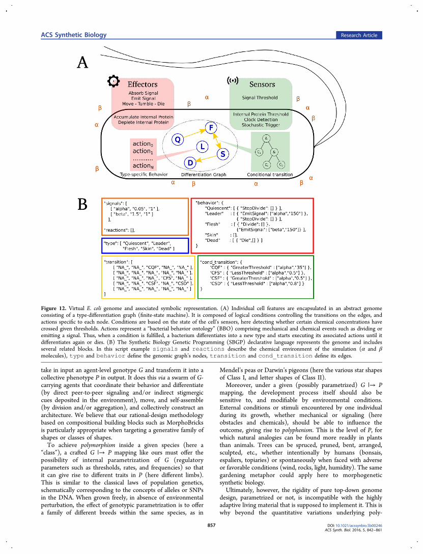
Figure 12. Virtual E. coli genome and associated symbolic representation. (A) Individual cell features are encapsulated in an abstract genomeconsisting of a type-differentiation graph (finite-state machine). It is composed of logical conditions controlling the transitions on the edges, andactions specific to each node. Conditions are based on the state of the cell’s sensors, here detecting whether certain chemical concentrations havecrossed given thresholds. Actions represent a “bacterial behavior ontology” (BBO) comprising mechanical and chemical events such as dividing oremitting a signal. Thus, when a condition is fulfilled, a bacterium differentiates into a new type and starts executing its associated actions until itdifferentiates again or dies. (B) The Synthetic Biology Genetic Programming (SBGP) declarative language represents the genome and includesseveral related blocks. In this script example signals and reactions describe the chemical environment of the simulation (α and βmolecules), type and behavior define the genomic graph’s nodes, transition and cond_transition define its edges.
ACS Synthetic Biology Research Article
DOI: 10.1021/acssynbio.5b00246ACS Synth. Biol. 2016, 5, 842−861
857
morphism and polyphenism, we also need to rely on the mostobvious and powerful force of qualitative variations and trueinnovation in biology: evolutionwhich consists of randommutations and nonrandom selection. In simulation, it meansthe use of evolutionary computation methods, such as geneticprogramming, accompanied by a diversity-preserving fitnessfunction to encourage novelty search. In a wet-lab setup, it mayalso mean letting the bacteria re-evolve by themselves in vivo.This should be the topic of a forthcoming article.
■ METHODSModel of Bacterial Behavior Ontology. Because
prokaryotic cells generally possess a simpler organization andgenome than eukaryotic cells, they are the favorite object ofsynthetic biology experiments and applications. Our first stepwas to define an abstract model of virtual prokaryote based onE. coli. This bacterium contains embedded sensors and effectorsthat allow it to assess and modify its internal state andenvironmental conditions (Figure 12). Through the sensors, itcan detect whether concentrations of specific internal proteinsor external ligands cross certain thresholds, including signalsemitted by its internal clock. For simplicity, we chose an explicitclocking scheme, assuming that its realization in terms ofbiochemical oscillators is well-known. Through the effectors, itis able to release or absorb external signals, and accumulate ordeplete internal proteins. Naturally, our bacterium must also beable to divide, rest, and disappear. Division is not instantaneousbut the outcome of a proliferating state in which the cell enters:in order to divide, it needs to elongate and double its size byexecuting an internal growth cycle. Death, however, is anatomic action directly triggered by the cell itself. Besidesdivision and death, a bacterium can also move forward and/ortumble, but these abilities are not be exploited in the presentwork. Finally, there is no direct molecular signaling betweentwo neighboring bacteria, only indirect communication viamorphogen diffusion (quorum sensing) and the mechanicalconstraints of 2D packing. In any case, the overall architectureemerges in an endogenous fashion.Bacterial dynamics, consisting of differentiation and behavior,
is encapsulated in a finite state machine (FSM) whose nodesrepresent discrete cell types, and edges represent conditionaltransitions between types (Figure 12A). Each type, denoted inthe example of Figure 12 by D, F, Q, ... corresponds to abehavioral repertoire of possible actions [a1, a2, ..., an].Transitions between types, that is, differentiation events, aretriggered by Boolean expressions composed of conditions [c1,c2, ..., cm] pertaining to protein concentrations and time. Inreality, the FSM formalism with its explicit type nodes anddifferentiation edges is a convenient abstraction of a complexunderlying GRN: types are a shortcut for attractors, cliques ofgenes promoting and repressing each other in a cycle or a fixedpoint; while differentiation events represent trajectories inphase space between basins of attraction, followed whenreceiving certain signals. The present model, however, is agrammar of integer types and logical transitions that does notinclude the finer scale of genetic dynamics.Genomic Representation. This model is represented by a
declarative language of our design, Synthetic Biology GeneticProgramming (SBGP), which supports the description of abacterial behavior ontology (BBO) based on internal andexternal conditions (Figure 12B). In an SBGP script, blocks ofsignals and reactions describe the chemical environ-ment of the simulation, while the other blocks describe the
bacteria’s behavior. The type and transition blocksspecify the nodes and edges of the FSM graph. Respectively,the behavior block describes the type-specific outputactions, while the cond_transition block codes for theinput Boolean expressions that trigger the transitions (see fullspecifications in Supporting Information, “SBGP language”).The latter two blocks are defined on the basis of two groups
of commands: effector primitives calling the cell’s actions, andsensor primitives allowing the designer to interface with thestate of internal and external sensors (both tuned byparameters). More precisely:
• Type-specific actions: when a cell belongs to a giventype, it executes behaviors associated with this type untilit differentiates again or dies. These belong to threecategories: (1) external interactions, by which cells canabsorb or diffuse morphogens in the environment(typically, EmitSignal); (2) internal interactions,by which cells can accumulate or deplete internalproteins; and (3) mechanical rules, making cells tumble,proliferate, rest, etc.and die (typically, Divide,StopDivide).
• Type-differentiation conditions: a transition to a newtype is triggered when certain conditions are fulfilled.These also belong to three categories: (1) chemicalconditions, monitoring whether the concentration levelof an external signal or internal protein is above or belowa threshold parameter (typically, GreaterThres-hold and LessThreshold); (2) stochastic con-ditions, introducing probabilities to model rare events orstochastic noise (based on the rate keyword); and (3)“first-or-second-daughter” conditions, allowing asymmet-ric divisions by letting only one of the daughter cells(chosen at random) differentiate after mitosis.
For the first implementation our model, as presented in thispaper, we chose the Gro platform by Klavins et al.,5 aconvenient tool offering a simple procedural language and 2Dgraphical simulator to model the development and behavior ofmicrocolonies of microorganisms. In parallel, we are alsocurrently preparing our own “synthetic biology engine” andvisualization software, called Traveling Bacteria (TB),53 whichwe plan on using later in replacement of Gro. Other, lessphysically realistic, agent-based modeling and simulationplatforms, such as BacSim54 and INDISIM,55 have also beencreated in the past, some of them used for example in thepredictive microbiology of wastewater and food safety.56
For now, the high-level SBGP specifications are translatedinto Gro commands. Note that, in this framework, division isnot implemented as an abstract temporal event, whether regularor probabilistic, but is triggered by the physical growth of therod-shape bacterium when it reaches a certain length. Spatialelongation is modeled in Gro by a differential equation on thevolume (not detailed here).5 The end effect is that divisionhappens on average every 200 time steps, that is, 20 min with atime step Δt representing 0.1 min. In our simulations, all cellsgrow-and-divide in the same way, except when they execute thecustom command StopDivide, which sets their growth rateto zero.
Higher-Level Biological Functions. The SBGP scriptinglanguage was also designed to handle more complex cellularmechanisms by providing useful shortcut keywords compiledinto lengthier sets of Gro instructions. In addition to the low-level sensor and effector primitives, four higher-level functions
ACS Synthetic Biology Research Article
DOI: 10.1021/acssynbio.5b00246ACS Synth. Biol. 2016, 5, 842−861
858

are currently available to the modeler: self-election of leaders,refractory periods, internal oscillatory clocks, and centerlinesbetween gradients. An essential feature supporting theproduction of complex morphologies is the ability for seedcells to pop up spontaneously without the need to predefinethem. This property is implemented here by a rare stochasticevent generator inside each bacterium, handled via the Groinstruction rate. Differentiation events are analogous tochemical reactions at low rate constants, which are very slow onaverage and give rise to discrete occurrences with highvariability over time and populations. Using this mechanism,the probability to observe two bacteria differentiate (nearly)simultaneously is very small.The second feature is based on Gro’s edge detection
example,5 in which the detection or emission of a signal isfollowed by a refractory period in which the signal cannot besensed or released again. This results in wave propagationtypical of excitable media, which is another form ofcommunication among bacteria in an assembly. A travelingwave can carry a message fast across neighboring cells, whichcan react and handle it as needed. Overall, the main advantageof a refraction mechanism is better temporal and spatialaccuracy of interbacterial communication than mere concen-tration gradients, especially when used in conjunction with aninternal oscillatory mechanism (see third feature below), as isthe case in the MorphoBrick-3 stage of our model. Refraction ineach bacterium relies on a counter marking the elapsed timesince the last detection or emission event occurred. Once thecounter reaches a specific threshold, the disabled function isenabled again. Biologically, this internal counter variablerepresents a simple model of temporary saturation ofmembrane receptors or blocking of molecular transport andexocytosis. For example, the refractory period may representthe time it takes for receptors or vesicles to be replenished. InSBGP, a typical refraction command is LessThreshol-dRefracted.Another important high-level mechanism handled by an
SBGP command is the ability to monitor the state of aninternal oscillator in each cell. Here we also made the strongassumption that cellular oscillators are already globallysynchronized at the population level, instead of modelingeach of them independently, coupling them and waiting forphase-locking to emerge. This “clock-on-the-wall” approach isjustified by the fact that global synchrony and quorum sensingare rather common phenomena. Naturally, the advantage ofthis shortcut assumption is to greatly decrease the dimension-ality of parameter space, the behavioral complexity of themodel, and the computing time of the simulation, in order toreach morphogenetic outputs more quickly.Finally, to reach Class II and higher classes of programmable
multibacterial shapes in SBGP, cells need to be able to self-identify as belonging to an equatorial boundary between twoemitting sources of gradients. This mechanism is a well-knownmorphogenetic “design pattern” used in amorphous/spatialcomputing7,57 and morphogenetic engineering.26 It is handledhere by a Boolean function MidPlane (A, B, θ) where A, Bare the two emitting sources of α and β, and θ is a thresholdtuning the width of the centerline, which translates to a doubleinequality 0 ≤ | [α] − [β] | ≤ θ in the script language.Hierarchical Composition. Ultimately, the compositional
aspect of the MorphoBrick framework could be implemented inhierarchical gene circuits, as outlined by Doursat.41 Taking afterthe early regulatory cascade of Drosophila, which goes from
global protein gradients to the precise positioning ofappendages, the idea is to rely on “identity genes” expressedin specific regions of the multicellular colony (similar to“imaginal discs”). Identity genes switch on dedicated subcircuitsthat further subdivide regions into subregions and shape theirexpansion by differential proliferation. New positionalinformation also arises inside regions via the propagation oflocal gradients, and so on. In sum, the morphogenesis of acomplex articulated structure can ultimately proceed byrecursive, multiscale refinement of detailsexcept that theabsolute scale remains the same (the individual bacterium), it isonly the relative scale that decreases while the multicellularorganism grows, as pointed out by Coen.46
Chemical Environment. The bacterial population isimmersed in a diffusive chemical environment represented bya discrete diffusion grid in Gro, which relies on Euler’s methodto solve the associated partial differential equations. The Groenvironment is nontoroidal and its size typically set to 160 μm× 160 μm in our experiments. The underlying diffusion gridconsists of square locations of size (0.5 μm)2 each, that is, 320× 320 of them. Cell coordinates are real-valued and not boundto a discrete lattice. A typical E. coli rod in Gro simulationsmeasures 2 μm × 1 μm (real E. coli bacteria range between 0.5and 3 μm), thus covers 8 diffusion grid squares on average. Asurface area of (160 μm)2 can contain about 12800 cells. Thereare no specific boundary conditions: signals diffusing outsidethe grid are lost. Although a “no-flux” boundary conditionwould have been more realistic, the accumulation of signalalong the borders is negligible and will not disturb themorphogenetic process as long as the organism remainssignificantly smaller than the virtual square Petri dish. Chemicalspecies α, β, γ, ... are declared in the environment by specifyingboth their diffusion and degradation rates (signals block inFigure 12B). Molecules can be emitted, detected, and absorbedby the bacteria. They can also react with each other viachemical equations classically expressed by reactants, products,and reaction rates (athough no reaction was specified here).
■ ASSOCIATED CONTENT
*S Supporting InformationThe Supporting Information is available free of charge on theACS Publications website at DOI: 10.1021/acssynbio.5b00246.
SBGP language specifications; compilation notes;morphological analysis; videos; SBGP and GRO sourcecodes (ZIP)
■ AUTHOR INFORMATION
Corresponding Authors*E-mail: [email protected].*E-mail: [email protected].
NotesThe authors declare no competing financial interest.
■ ACKNOWLEDGMENTS
J.P. was supported by a postdoctoral fellowship from RegionParis Ile-de-France: Complex Systems Institute (ISC-PIF),Appel a projets (AAP) 2013. T.K. was supported by theSynBioTIC project grant funded by the French AgenceNationale de la Recherche (ANR-BLAN-SIMI10-LS-100618-12-01), which also funded the computer equipment.
ACS Synthetic Biology Research Article
DOI: 10.1021/acssynbio.5b00246ACS Synth. Biol. 2016, 5, 842−861
859
■ REFERENCES(1) Knight, T. (2003) Idempotent vector design for standard assembly ofbiobricks, MIT Artificial Intelligence Laboratory; MIT SyntheticBiology Working Group.(2) Stepney, S., Diaconescu, A., Doursat, R., Giavitto, J.-L., Kowaliw,T., Leyser, O., MacLennan, B., Michel, O., Miller, J. F., Nikolic, I.,Spicher, A., Teuscher, C., Tufte, G., Vico, F. J., and Yamamoto, L.(2012) Gardening cyber-physical systems. UCNC ’12: Proceedings ofthe 11th International Conference on Unconventional Computationand Natural Computation, pp 237−238. .(3) Armstrong, R., and Spiller, N. (2010) Synthetic biology: Livingquarters. Nature 467, 916−918.(4) Amos, M., Stepney, S., Doursat, R., Vico, F. J., and Rasmussen, S.(2012) TRUCE: A coordination action for unconventional computa-tion. Int. J. Uncon. Comp. 8, 333−337.(5) Jang, S. S., Oishi, K. T., Egbert, R. G., and Klavins, E. (2012)Specification and simulation of synthetic multicelled behaviors. ACSSynth. Biol. 1, 365−374.(6) Giavitto, J.-L., and Michel, O. (2002) The topological structuresof membrane computing. Fundam. Informaticae 49, 123−145.(7) Beal, J., and Bachrach, J. (2006) Infrastructure for engineeredemergence on sensor/actuator networks. IEEE Intell. Syst. 21, 10−19.(8) Cameron, D. E., Bashor, C. J., and Collins, J. J. (2014) A briefhistory of synthetic biology. Nat. Rev. Microbiol. 12, 381−390.(9) Monod, J., and Jacob, F. (1961) Cold Spring Harbor Symp. Quant.Biol. 26, 389−401.(10) Westerhoff, H. V., and Palsson, B. O. (2004) The evolution ofmolecular biology into systems biology. Nat. Biotechnol. 22, 1249−1252.(11) Bray, D. (1995) Protein molecules as computational elements inliving cells. Nature 376, 307−312.(12) McAdams, H. H., and Shapiro, L. (1995) Circuit simulation ofgenetic networks. Science 269, 650−656.(13) Gardner, T. S., Cantor, C. R., and Collins, J. J. (2000)Construction of a genetic toggle switch in Escherichia coli. Nature 403,339−342.(14) Elowitz, M. B., and Leibler, S. (2000) A synthetic oscillatorynetwork of transcriptional regulators. Nature 403, 335−338.(15) Weiss, R., and Basu, S. (2002) The device physics of cellularlogic gates. NSC-1: The First Workshop of Non-Silicon Computing.Boston, Massachusetts.(16) Rackham, O., and Chin, J. W. (2005) A network of orthogonalribosome· mRNA pairs. Nat. Chem. Biol. 1, 159−166.(17) Ham, T. S., Lee, S. K., Keasling, J. D., and Arkin, A. P. (2006) Atightly regulated inducible expression system utilizing the fim inversionrecombination switch. Biotechnol. Bioeng. 94, 1−4.(18) Palsson, B. (2000) The challenges of in silico biology. Nat.Biotechnol. 18, 1147−1150.(19) Weiss, R., and Knight, T. F., Jr (2001) DNA Computing, pp 1−16, Springer-Verlag.(20) You, L., Cox, R. S., Weiss, R., and Arnold, F. H. (2004)Programmed population control by cell-cell communication andregulated killing. Nature 428, 868−871.(21) Basu, S., Gerchman, Y., Collins, C. H., Arnold, F. H., and Weiss,R. (2005) A synthetic multicellular system for programmed patternformation. Nature 434, 1130−1134.(22) Tabor, J. J., Salis, H. M., Simpson, Z. B., Chevalier, A. A.,Levskaya, A., Marcotte, E. M., Voigt, C. A., and Ellington, A. D. (2009)A synthetic genetic edge detection program. Cell 137, 1272−1281.(23) Liu, C., Fu, X., Liu, L., Ren, X., Chau, C. K., Li, S., Xiang, L.,Zeng, H., Chen, G., Tang, L.-H., Lenz, P., Cui, X., Huang, W., Hwa, T.,and Huang, J.-D. (2011) Sequential establishment of stripe patterns inan expanding cell population. Science 334, 238−241.(24) Amos, M. (2014) Population-based microbial computing: athird wave of synthetic biology? Int. J. Gen. Syst. 43, 770−782.(25) Turing, A. M. (1952) The chemical basis of morphogenesis.Philos. Trans. R. Soc., B 237, 37−72.(26) Doursat, R., Sayama, H., and Michel, O. (2013) A review ofmorphogenetic engineering. Nat. Comput. 12, 517−535.
(27) Doursat, R., and Sanchez, C. (2014) Growing fine-grainedmulticellular robots. Soft Robot. 1, 110−121.(28) Bongard, J. C., and Pfeifer, R. (2003) Morpho-functionalMachines: The New Species, pp 237−258, Springer-Verlag.(29) Stanley, K. O., and Miikkulainen, R. (2003) A taxonomy forartificial embryogeny. Artif. Life 9, 93−130.(30) Kowaliw, T., Grogono, P., and Kharma, N. (2004) Bluenome: ANovel Developmental Model of Artificial Morphogenesis. Genetic andEvolutionary Computation (GECCO) 3102, 93−104.(31) Doursat, R. (2009) Facilitating evolutionary innovation bydevelopmental modularity and variability. Proceedings of the 11thannual conference on genetic and evolutionary computation(GECCO), pp 683−690.(32) Doursat, R., Sanchez, C., Dordea, R., Fourquet, D., andKowaliw, T. (2012) in Morphogenetic Engineering: Toward Program-mable Complex Systems, Understanding Complex Systems, (Doursat,R., Sayama, H., and Michel, O., Eds.) pp 275−311, Springer-Verlag.(33) Joachimczak, M., Kowaliw, T., Doursat, R., and Wrobel, B.(2013) Evolutionary design of soft-bodied animats with decentralizedcontrol. Artif. Life Robot. 18, 152−160.(34) Rubenstein, M., Cornejo, A., and Nagpal, R. (2014) Program-mable self-assembly in a thousand-robot swarm. Science 345, 795−799.(35) Komosinski, M. (2000) Virtual Worlds, pp 214−224, Springer-Verlag.(36) Lipson, H., and Pollack, J. B. (2000) Automatic design andmanufacture of robotic lifeforms. Nature 406, 974−978.(37) Cussat-Blanc, S., Pascalie, J., Mazac, S., Luga, H., and Duthen, Y.(2012) Morphogenetic Engineering, pp 353−381, Springer-Verlag.(38) Cussat-Blanc, S., and Pollack, J. (2014) Cracking the egg:Virtual embryogenesis of real robots. Artif. Life 20, 361−383.(39) Eggenberger, P. (1997) Evolving morphologies of simulated 3Dorganisms based on differential gene expression. ECAL ’97: Proceedingsof the Fourth European Conference on Artificial Life, pp 205−213.(40) Disset, J., Cussat-Blanc, S., and Duthen, Y. (2014) Self-organization of Symbiotic Multicellular Structures. ALIFE XIV:Proceedings of the 14th International Conference on the Simulationand Synthesis of Living Systems, pp 3−5.(41) Doursat, R. (2006) The growing canavas of biologicaldevelopment: multiscale pattern generation on an expanding latticeof gene regulatory networks. InterJournal 1809, 205.(42) Reuillon, R., Leclaire, M., and Rey-Coyrehourcq, S. (2013)OpenMOLE, a workflow engine specifically tailored for the distributedexploration of simulation models. Future Gener. Comp. Sy. 29, 1981−1990.(43) Rudge, T. J., Federici, F., Steiner, P. J., Kan, A., and Haseloff, J.(2013) Cell polarity-driven instability generates self-organized, fractalpatterning of cell layers. ACS Synth. Biol. 2, 705−714.(44) Schlosser, G., and Wagner, G. P. (2004) Modularity indevelopment and evolution, University of Chicago Press.(45) Carroll, S. B., Grenier, J. K., and Weatherbee, S. D. (2013) FromDNA to diversity: molecular genetics and the evolution of animal design,John Wiley & Sons.(46) Coen, E. (1999) The Art of Genes: How Organisms MakeThemselves: How Organisms Make Themselves, Oxford University Press.(47) Waters, C. M., and Bassler, B. L. (2005) Quorum sensing: cell-to-cell communication in bacteria. Annu. Rev. Cell Dev. Biol. 21, 319−346.(48) Dagkessamanskaia, A., Moscoso, M., Henard, V., Guiral, S.,Overweg, K., Reuter, M., Martin, B., Wells, J., and Claverys, J.-P.(2004) Interconnection of competence, stress and CiaR regulons inStreptococcus pneumoniae: competence triggers stationary phaseautolysis of ciaR mutant cells. Mol. Microbiol. 51, 1071−1086.(49) Pascalie, J., Lobjois, V., Luga, H., Ducommun, B., and Duthen,Y. (2012) Checkpoint Orientated Cell Cycle Modeling Issues in Simulationof Synchronized Situation. ALIFE XIII: Proceedings of the 13thInternational Conference on the Simulation and Synthesis of LivingSystems, pp 465−472.
ACS Synthetic Biology Research Article
DOI: 10.1021/acssynbio.5b00246ACS Synth. Biol. 2016, 5, 842−861
860
(50) Danino, T., Mondragon-Palomino, O., Tsimring, L., and Hasty,J. (2010) A synchronized quorum of genetic clocks. Nature 463, 326−330.(51) Peter, I. S., and Davidson, E. H. (2009) Modularity and designprinciples in the sea urchin embryo gene regulatory network. FEBSLett. 583, 3948−3958.(52) Rudge, T. J., Steiner, P. J., Phillips, A., and Haseloff, J. (2012)Computational modeling of synthetic microbial biofilms. ACS Synth.Biol. 1, 345−352.(53) Potier, M. Integration of levels of organization in a languagededicated to the modeling of complex systems. Chapter 3: TravelingBacteria. Ph.D. thesis, Universite Paris-Est Creteil(UPEC), France,2016.(54) Kreft, J.-U., Booth, G., and Wimpenny, J. W. (1998) BacSim, asimulator for individual-based modelling of bacterial colony growth.Microbiology 144, 3275−3287.(55) Ginovart, M., Lopez, D., and Valls, J. (2002) INDISIM, anindividual-based discrete simulation model to study bacterial cultures.J. Theor. Biol. 214, 305−319.(56) Esser, D. S., Leveau, J. H., and Meyer, K. M. (2015) Modelingmicrobial growth and dynamics. Appl. Microbiol. Biotechnol. 99, 8831−8846.(57) Nagpal, R. (2006) Complex Engineered Systems, pp 291−306,Springer-Verlag.
ACS Synthetic Biology Research Article
DOI: 10.1021/acssynbio.5b00246ACS Synth. Biol. 2016, 5, 842−861
861