Embed Size (px)
Citation preview
Diabetic eye screening (DES)Medicine > Endocrinology > Diabetic eye screening (DES)
Published: 18-Mar-2015 Valid until: 31-Mar-2016 Printed on: 26-Jun-2015 © Map of Medicine Ltd This care map was published by England. A printed version of this document is not controlled so may not be up-to-date with the latest clinicalinformation.
Page 1 of 5
R
R
Primary grading
Photographnot taken
Disagreement onimage grading
Arbitration grading
Image ungradable,suspected urgent orreferrable non-diabeticretinopathy
Secondarygrading
Agreement on imagegrading
Referral OutcomeGrade
Referable diseasepresent and/orungradable
Routine digitalscreening test
Invite for routine digitalscreening
Digital photographobtained
Patient reinvited if notattended/responded
Referable non-DR lesions with noreferable DR
Referral to GP
Negative (no disease)primary grading result:R0M0
Primary grading result:R1, R2, M1 ornon-DR lesions
Non-referable grade
Diabetic EyeScreening Programme
Suspend patientfrom routine digitalscreening
Requires monitoringmore frequently thanannually
Requires Slit LampBiomicroscopy (SLB)
Requires referral toHospital Eye Services
Surveillance SLB Digital Surveillance Hospital Eye Services
This document is excluded from the Open Government Licence. Images and content are subject to copyright; All rights reserved ©Map of Medicine Limited 2015 and © Public Health England 2015.
Diabetic eye screening (DES)Medicine > Endocrinology > Diabetic eye screening (DES)
Published: 18-Mar-2015 Valid until: 31-Mar-2016 Printed on: 26-Jun-2015 © Map of Medicine Ltd This care map was published by England. A printed version of this document is not controlled so may not be up-to-date with the latest clinicalinformation.
Page 2 of 5
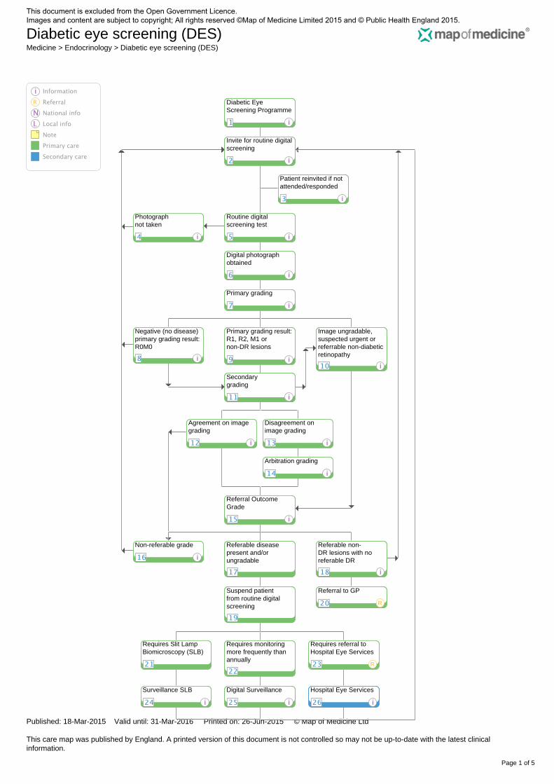
1 Diabetic Eye Screening Programme
Quick info:Patients aged 12 and over who have a diagnosis of Diabetes Mellitus should be referred to the NHS Diabetic Eye ScreeningProgramme (NDESP) for diabetic retinopathy screening. NDESP will consider all these patients eligible except in cases where thereis No Perception of Light (NPL) in both eyes. Exclusions based on Patient Opt-Out or Medically Unfit determinations can be made;however these patients should still be referred to NDESP and will be excluded within the programme's central call/recall system.Local Diabetic Eye Screening Programmes hold collated lists of people with diabetes who are registered with GP Practices in theirarea. Lists include:
• demographics, including contact information
• consent status for additional data transfer
• eligibility for screening in opinion of GP.
2 Invite for routine digital screening
Quick info:Patients are invited to Routine Digital Screening on an annual basis. Invitations may be for a fixed appointment time, or as aninvitation for the patient to phone in and schedule an appointment at their convenience.
3 Patient reinvited if not attended/responded
Quick info:Patients who do not attend or respond to their screening invitation are reinvited. Local protocol will determine how many additionalinvitations are sent to the patient before their GP is notified of their non-attendance; patients will then be recalled on the next annualscreening due date.
4 Photograph not taken
Quick info:Situations where a patient attends routine digital screening appointment but no photograph can be taken include:
• equipment failure (e.g. digital camera)
• patient leaves clinic prior to photography
• evacuation of clinic required.
Note this does not apply to situations where the patient is unable to be photographed due to a medical condition.Patients are reinvited to routine digital screening appointment via the same process as previous invitation.
5 Routine digital screening test
Quick info:After arrival at the eye clinic and confirmation of demographic details with the clinic clerk, consent is taken and the followingobservations will usually be recorded:
• brief eye history
• visual acuities (mandatory).
Prior to consultation and examination, pupils of both eyes will typically be dilated using mydriatic eye drops.The screener or screener/grader then performs the screening test by taking two digital images of the retina in each eye.
This document is excluded from the Open Government Licence. Images and content are subject to copyright; All rights reserved ©Map of Medicine Limited 2015 and © Public Health England 2015.
Diabetic eye screening (DES)Medicine > Endocrinology > Diabetic eye screening (DES)
Published: 18-Mar-2015 Valid until: 31-Mar-2016 Printed on: 26-Jun-2015 © Map of Medicine Ltd This care map was published by England. A printed version of this document is not controlled so may not be up-to-date with the latest clinicalinformation.
Page 3 of 5
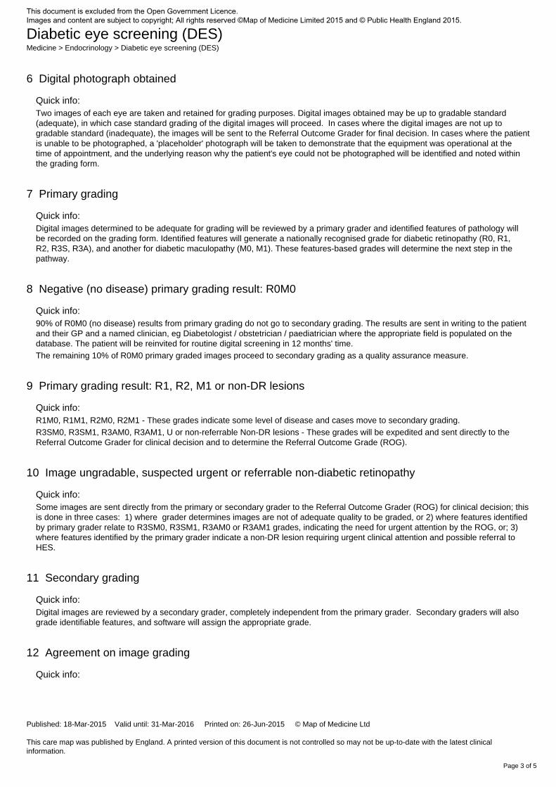
6 Digital photograph obtained
Quick info:Two images of each eye are taken and retained for grading purposes. Digital images obtained may be up to gradable standard(adequate), in which case standard grading of the digital images will proceed. In cases where the digital images are not up togradable standard (inadequate), the images will be sent to the Referral Outcome Grader for final decision. In cases where the patientis unable to be photographed, a 'placeholder' photograph will be taken to demonstrate that the equipment was operational at thetime of appointment, and the underlying reason why the patient's eye could not be photographed will be identified and noted withinthe grading form.
7 Primary grading
Quick info:Digital images determined to be adequate for grading will be reviewed by a primary grader and identified features of pathology willbe recorded on the grading form. Identified features will generate a nationally recognised grade for diabetic retinopathy (R0, R1,R2, R3S, R3A), and another for diabetic maculopathy (M0, M1). These features-based grades will determine the next step in thepathway.
8 Negative (no disease) primary grading result: R0M0
Quick info:90% of R0M0 (no disease) results from primary grading do not go to secondary grading. The results are sent in writing to the patientand their GP and a named clinician, eg Diabetologist / obstetrician / paediatrician where the appropriate field is populated on thedatabase. The patient will be reinvited for routine digital screening in 12 months' time.The remaining 10% of R0M0 primary graded images proceed to secondary grading as a quality assurance measure.
9 Primary grading result: R1, R2, M1 or non-DR lesions
Quick info:R1M0, R1M1, R2M0, R2M1 - These grades indicate some level of disease and cases move to secondary grading. R3SM0, R3SM1, R3AM0, R3AM1, U or non-referrable Non-DR lesions - These grades will be expedited and sent directly to theReferral Outcome Grader for clinical decision and to determine the Referral Outcome Grade (ROG).
10 Image ungradable, suspected urgent or referrable non-diabetic retinopathy
Quick info:Some images are sent directly from the primary or secondary grader to the Referral Outcome Grader (ROG) for clinical decision; thisis done in three cases: 1) where grader determines images are not of adequate quality to be graded, or 2) where features identifiedby primary grader relate to R3SM0, R3SM1, R3AM0 or R3AM1 grades, indicating the need for urgent attention by the ROG, or; 3)where features identified by the primary grader indicate a non-DR lesion requiring urgent clinical attention and possible referral toHES.
11 Secondary grading
Quick info:Digital images are reviewed by a secondary grader, completely independent from the primary grader. Secondary graders will alsograde identifiable features, and software will assign the appropriate grade.
12 Agreement on image grading
Quick info:
This document is excluded from the Open Government Licence. Images and content are subject to copyright; All rights reserved ©Map of Medicine Limited 2015 and © Public Health England 2015.
Diabetic eye screening (DES)Medicine > Endocrinology > Diabetic eye screening (DES)
Published: 18-Mar-2015 Valid until: 31-Mar-2016 Printed on: 26-Jun-2015 © Map of Medicine Ltd This care map was published by England. A printed version of this document is not controlled so may not be up-to-date with the latest clinicalinformation.
Page 4 of 5
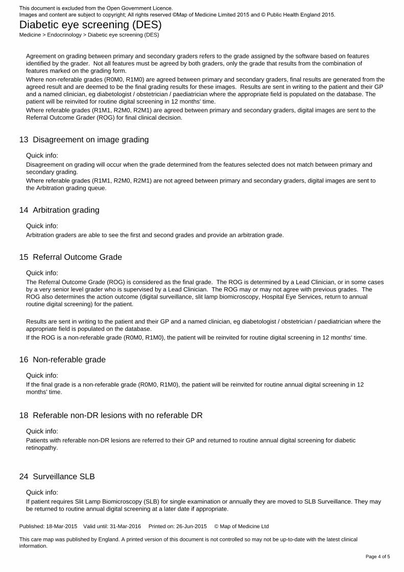
Agreement on grading between primary and secondary graders refers to the grade assigned by the software based on featuresidentified by the grader. Not all features must be agreed by both graders, only the grade that results from the combination offeatures marked on the grading form.Where non-referable grades (R0M0, R1M0) are agreed between primary and secondary graders, final results are generated from theagreed result and are deemed to be the final grading results for these images. Results are sent in writing to the patient and their GPand a named clinician, eg diabetologist / obstetrician / paediatrician where the appropriate field is populated on the database. Thepatient will be reinvited for routine digital screening in 12 months' time.Where referable grades (R1M1, R2M0, R2M1) are agreed between primary and secondary graders, digital images are sent to theReferral Outcome Grader (ROG) for final clinical decision.
13 Disagreement on image grading
Quick info:Disagreement on grading will occur when the grade determined from the features selected does not match between primary andsecondary grading. Where referable grades (R1M1, R2M0, R2M1) are not agreed between primary and secondary graders, digital images are sent tothe Arbitration grading queue.
14 Arbitration grading
Quick info:Arbitration graders are able to see the first and second grades and provide an arbitration grade.
15 Referral Outcome Grade
Quick info:The Referral Outcome Grade (ROG) is considered as the final grade. The ROG is determined by a Lead Clinician, or in some casesby a very senior level grader who is supervised by a Lead Clinician. The ROG may or may not agree with previous grades. TheROG also determines the action outcome (digital surveillance, slit lamp biomicroscopy, Hospital Eye Services, return to annualroutine digital screening) for the patient. Results are sent in writing to the patient and their GP and a named clinician, eg diabetologist / obstetrician / paediatrician where theappropriate field is populated on the database.If the ROG is a non-referable grade (R0M0, R1M0), the patient will be reinvited for routine digital screening in 12 months' time.
16 Non-referable grade
Quick info:If the final grade is a non-referable grade (R0M0, R1M0), the patient will be reinvited for routine annual digital screening in 12months' time.
18 Referable non-DR lesions with no referable DR
Quick info:Patients with referable non-DR lesions are referred to their GP and returned to routine annual digital screening for diabeticretinopathy.
24 Surveillance SLB
Quick info:If patient requires Slit Lamp Biomicroscopy (SLB) for single examination or annually they are moved to SLB Surveillance. They maybe returned to routine annual digital screening at a later date if appropriate.
This document is excluded from the Open Government Licence. Images and content are subject to copyright; All rights reserved ©Map of Medicine Limited 2015 and © Public Health England 2015.
Diabetic eye screening (DES)Medicine > Endocrinology > Diabetic eye screening (DES)
Published: 18-Mar-2015 Valid until: 31-Mar-2016 Printed on: 26-Jun-2015 © Map of Medicine Ltd This care map was published by England. A printed version of this document is not controlled so may not be up-to-date with the latest clinicalinformation.
Page 5 of 5
Patients in SLB Surveillance should be invited, graded and informed of results in the same way as those patients in routine annualdigital screening.
25 Digital Surveillance
Quick info:If patient requires monitoring by digital photography more frequently than annually they are moved to Digital Surveillance. They maybe returned to routine annual digital screening at a later date if appropriate.Some stable patients with R2 and M1 may only require surveillance at 12-month intervals but they must be kept in the digitalsurveillance service. No patient with R2 and M1 grades can be returned to annual digital screening.Patients in Digital Surveillance should be invited, graded and informed of results in the same way as those patients in routine annualdigital screening.
26 Hospital Eye Services
Quick info:After assessment and/or treatment by Hospital Eye Services, unless appropriately excluded or suspended, patients return to routinedigital screening, digital surveillance or SLB surveillance as clinically indicated.
This document is excluded from the Open Government Licence. Images and content are subject to copyright; All rights reserved ©Map of Medicine Limited 2015 and © Public Health England 2015.
Quality Assurance
Provenance: Diabetic Eye Screening
Provenance
It is important that each care map is referenced in line with Map of Medicine guidelines.
Classification
When creating or updating each care map you will need to use the following classification,
please note not all classification may be applicable;
[G] – guideline [M] – meta – analysis [S] – systematic review [A] – randomised controlled trail [B] – non-randomised prospective study [C] – retrospective study [Q] – cost or decision analysis [P] – performance measures or policy documents [E] – practice based information (expert opinion)
Rohan TE, Frost CD, Wald NJ. Prevention of blindness by screening for diabetic retinopathy: a quantitative
assessment. BMJ, 1989; 299:1198-201[Q]
Diabetes care and research in Europe: the Saint Vincent declaration. Diabet Med 1990; 7:360 [G]
Kristinsson JK, Gudmundsson JR, Stefansson E, Jonasson F, Gislason I, Thorsson AV. Screening for diabetic
retinopathy. Initiation and frequency, Acta Ophthalmol Scand 1995; 73:525-8 [E]
Bachmann M, Nelson SJ. Screening for Diabetic Retinopathy: A quantitative overview of the evidence, applied to
the populations of health authorities and boards. Report Bristol: Health Care Evaluation Unit, University of Bristol;
1996 1996 – December [Q]
Stefansson E, Bek T, Porta M, Larsen N, Kristinsson JK, Agardh E. Screening and prevention of diabetic
blindness. Acta Ophthalmol Scand 2000; 78:374-85 [E]
This document is excluded from the Open Government Licence. Images and content are subject to copyright; All rights reserved ©Map of Medicine Limited 2015 and © Public Health England 2015.

D H. National Service Framework for Diabetes: Delivery Strategy - Department of Health. London2002 [G]
Scanlon PH, Foy C, Chen FK. Visual acuity measurement and ocular co-morbidity in diabetic retinopathy
screening, Br J Ophthalmol 2008 Jun; 92(6):775-8 [B]
Garvican L. Issues regarding quality assurance in the English National Screening Programme for Sight-
threatening Diabetic Retinopathy: response to paper by C. Arun et al., 'Establishing on-going quality assurance in
a retinal screening programme'. Diabet Med 2007; 24:688-90; author reply 90-1 [E]
Complications of diabetes: Screening for retinopathy, Management of foot ulcers: NHS Centre for Reviews and
Dissemination, University of York; 1999 August 1999 [S]
Garvican L, Clowes J, Gillow T. Preservation of sight in diabetes: developing a national risk reduction
programme. Diabet Med 2000; 17: 627-34 [E]
Gillow JT, Gray JA. The National Screening Committee review of diabetic retinopathy screening Eye 2001; 15:1-
2 [E]
Management of Type 2 diabetes - retinopathy screening and early management NICE 2002 [G]
Type 1 diabetes: diagnosis and management of type 1 diabetes in adults. NICE 2004 [G]
Scanlon PH, Malhotra R, Greenwood RH, et al. Comparison of two reference standards in validating two field
mydriatic digital photography as a method of screening for diabetic retinopathy. Br J Ophthalmol 2003; 87:1258-
63 [B]
Garvican L, Scanlon PH. A pilot quality assurance scheme for diabetic retinopathy risk reduction programmes.
Diabet Med 2004; 21:1066-74 [B]
Scanlon PH, Carter SC, Foy C, Husband RF, Abbas J, Bachmann MO. Diabetic retinopathy and socioeconomic
deprivation in Gloucestershire. J Med Screen 2008; 15:118-21.54 [B]
Leese GP, Boyle P, Feng Z, Emslie-Smith A, Ellis JD, Screening uptake in a well-established diabetic retinopathy
screening program: the role of geographical access and deprivation. Diabetes Care 2008; 31:2131-5 [C]
Hutchinson A, McIntosh A, Peters J, et al. Effectiveness of screening and monitoring tests for diabetic
retinopathy--a systematic review. Diabet Med 2000; 17:495-506 [S]
Sharp PF, Olson J, Strachan F, et al The value of digital imaging in diabetic retinopathy, Health Technol Assess
2003; 7:1-119 [S]
Harding SP, Broadbent DM, Neoh C, White MC, Vora J. Sensitivity and specificity of photography and direct
ophthalmoscopy in screening for sight threatening eye disease: the Liverpool Diabetic Eye Study. BMJ 1995;
311:1131-5 [B]
Taylor DJ, Fisher J, Jacob J, Tooke JE, The use of digital cameras in a mobile retinal screening environment.
Diabet Med 1999; 16: 680-6 [B]
This document is excluded from the Open Government Licence. Images and content are subject to copyright; All rights reserved ©Map of Medicine Limited 2015 and © Public Health England 2015.
Pandit RJ, Taylor R. Quality assurance in screening for sight-threatening diabetic retinopathy, Diabet Med 2002;
19: 285-91 [B]
Scanlon PH, Malhotra R, Thomas G, et al. The effectiveness of screening for diabetic retinopathy by digital
imaging photography and technician ophthalmoscopy, Diabet Med 2003; 20:467-74 [B]
Harding S, Greenwood R, Aldington S, et al. Grading and disease management in national screening for diabetic
retinopathy in England and Wales. Diabet Med 2003; 20:965-71[E]
Scanlon PH, Stratton IM, Histed M, Chave SJ, Aldington SJ. The influence of background diabetic retinopathy in
the second eye on rates of progression of diabetic retinopathy between 2005 and 2010, Acta Ophthalmol 2013;
91:e335-9 [C]
Murgatroyd H, Cox A, Ellingford A, Ellis JD, MacEwen CJ, Leese GP. Can we predict which patients are at risk of
having an ungradable digital image for screening for diabetic retinopathy? Eye 2008; 22:344-8 [B]
Scanlon PH, Foy C, Malhotra R, Aldington SJ. The influence of age, duration of diabetes, cataract, and pupil size
on image quality in digital photographic retinal screening, Diabetes Care 2005;28:2448-53 [B]
Healy R, Sallam A, Jones V, et al. Agreement between photographic screening and hospital biomicroscopy
grading of diabetic retinopathy and maculopathy, Eur J Ophthalmol 2014;24:550-8 [C]
Sallam A, Scanlon PH, Stratton IM, et al. Agreement and reasons for disagreement between photographic and
hospital biomicroscopy grading of diabetic retinopathy. Diabet Med 2011; 28:741-6 [C]
Prasad S, Kamath GG, Jones K, Clearkin LG, Phillips RP. Effectiveness of optometrist screening for diabetic
retinopathy using slit-lamp biomicroscopy, Eye 2001; 15:595-601[C]
Olson JA, Strachan FM, Hipwell JH, et al. A comparative evaluation of digital imaging, retinal photography and
optometrist examination in screening for diabetic retinopathy, Diabet Med 2003; 20:528-34 [B]
Warburton TJ, Hale PJ, Dewhurst JA. Evaluation of a local optometric diabetic retinopathy screening service,
Diabet Med 2004; 21:632-5 [C]
Mackenzie S, Schmermer C, Charnley A, et al. SDOCT Imaging to Identify Macular Pathology in Patients
Diagnosed with Diabetic Maculopathy by a Digital Photographic Retinal Screening Programme. PLoS One 2011;
6:e14811 [B]
This document is excluded from the Open Government Licence. Images and content are subject to copyright; All rights reserved ©Map of Medicine Limited 2015 and © Public Health England 2015.

Contributors
The following individuals have contributed to this care map:
Name of Screening Programme: Diabetic Eye Screening Programme
Contributor Name
Job Title Conflicts of Interest
Lynne Lacey
Programme Manager - NHS
Diabetic Eye Screening
Programme
Young Person and Adult
Screening Programmes
NHS Screening Programmes
None declared
Peter Scanlon
Consultant Ophthalmologist,
Gloucestershire Hospitals
NHS FT
None declared
David Taylor
National Quality Assurance
Manager, NHS Screening
Programmes
None declared
Hazel Rudge-Pickard Project Lead - YPA
Screening Programmes
NHS Screening Programmes
None declared
This document is excluded from the Open Government Licence. Images and content are subject to copyright; All rights reserved ©Map of Medicine Limited 2015 and © Public Health England 2015.