Embed Size (px)
Citation preview
HE blood supply to the region of the cavernous sinusis provided by interconnecting branches of the ICAand ECA, and it is from these vessels that dural
CCFs—often called dural arteriovenous fistulas—arise.Such fistulas usually are separated anatomically into threetypes: 1) shunts between meningeal branches of the ICAand the cavernous sinus; 2) shunts between meningealbranches of the ECA and the cavernous sinus; and 3)shunts between meningeal branches of both the ICA andECA and the cavernous sinus (Figs. 1–3).7,20 Of these threetypes, the third type is by far the most common.
Pathogenesis
Dural CCFs usually become symptomatic spontaneous-ly. The pathogenesis of these fistulas is controversial.21 Itwas once speculated that spontaneous dural CCFs formafter rupture of one or more of the thin-walled dural arter-ies that normally traverse the cavernous sinus.79 Extensive,preformed, dural arterial anastomoses not directly involvedin the fistula may dilate and contribute collateral blood sup-
ply after rupture, resulting in an angiographic appearanceindistinguishable from that of a congenital vascular mal-formation. Sequential arteriography demonstrates that thefeeder vessels of dural CCFs change with time as the ves-sels spontaneously open and close.100 Although this theoryis favored by some investigators,7 it fails to explain whyspontaneous dural CCFs are more common in elderlywomen than in men. A second theory for the origin of duralCCFs is that most of these lesions develop in response tospontaneous venous thrombosis in the cavernous sinus andrepresent an attempt to provide a pathway for collateralvenous outflow.40 This theory is favored by most investiga-tors because it also explains the pathogenesis of arteriove-nous fistulas that develop in the sigmoid and other duralsinuses.21
Certain factors may predispose patients to the develop-ment of symptomatic dural CCFs. These factors includepregnancy, systemic hypertension, atherosclerotic vasculardisease, connective tissue disease (such as Ehlers–Danlossyndrome), and minor trauma (Fig. 4).75,91,102 In addition,iatrogenic dural CCFs occasionally occur.14
Clinical Manifestations
Dural CCFs usually occur in middle-aged or elderlywomen, but they may produce symptoms in either sex at
Neurosurg. Focus / Volume 23 / November, 2007
Neurosurg Focus 23 (5):E13, 2007
Diagnosis and management of dural carotid–cavernoussinus fistulas
NEIL R. MILLER, M.D.
Departments of Ophthalmology, Neurology, and Neurosurgery, Johns Hopkins Medical Institutions,Baltimore, Maryland
PA carotid–cavernous sinus fistula (CCF) is an abnormal communication between the cavernous sinus and the carotidarterial system. Some fistulas are characterized by a direct connection between the cavernous segment of the internalcarotid artery (ICA) and the cavernous sinus, whereas others consist of a communication between the cavernous sinusand one or more meningeal branches of the ICA, external carotid artery, or both. These dural fistulas usually have lowrates of arterial blood flow and until recently were difficult to diagnose and treat. In this paper, the author discusses theanatomy, pathogenesis, clinical manifestations, diagnosis, treatment, and prognosis of dural CCFs. (DOI: 10.3171/FOC-07/11/E13)
KEY WORDS • carotid–cavernous sinus fistula • internal carotid artery •external carotid artery • superior ophthalmic vein
T
1
Abbreviations used in this paper: CCF = carotid–cavernous fistu-la; CN = cranial nerve; CT = computed tomography; ECA = exter-nal carotid artery; ICA = internal carotid artery; MR = magnetic res-onance; SOV = superior ophthalmic vein.
Unauthenticated | Downloaded 05/31/20 12:20 PM UTC
any age, even in childhood or infancy.95 The symptoms andsigns produced by these lesions are influenced by a numberof factors, including the size of the fistula, the locationwithin the cavernous sinus, the rate of blood flow, anddrainage route, especially if the drainage route is posterior,anterior, or both.98,99 The drainage route of the fistula isprobably related to its basic anatomical configuration, al-though Grove30 postulated that many, if not all, fistulas ini-tially drain posteriorly into the inferior petrosal sinus, basi-lar venous plexus, or both. Grove believed that when thisnormal pathway for drainage becomes thrombosed, the fis-tula begins to drain anteriorly, producing visual symptomsand signs. I and others have examined patients whose clin-ical course suggests that this theory is correct.38 Such
patients initially may experience an acute, isolated, ocularmotor nerve paresis, the evaluation of which reveals a pos-teriorly draining fistula (Fig. 5, upper). Shortly thereafter,these patients develop typical signs of an anteriorly drain-ing fistula (Fig. 5, lower).
Posteriorly Draining Fistulas
When dural CCFs drain posteriorly into the superior andinferior petrosal sinuses, they are usually asymptomatic. Insome cases, however, such fistulas produce a cranial neu-ropathy, such as a trigeminal neuropathy,87 facial nerveparesis,77 or an ocular motor nerve paresis.24 In most ofthese cases, there is no evidence of orbital congestion.
N. R. Miller
2 Neurosurg. Focus / Volume 23 / November, 2007
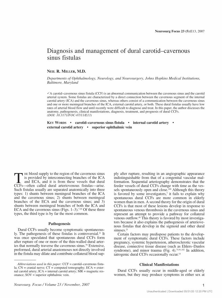
FIG. 1. Images showing the appearance of a dural CCF in whichthe only contribution is from extradural branches of the ICA.Upper: A selective left ICA arteriogram shows a fistula at the pos-terior portion of the cavernous carotid artery (arrow). The left ECAarteriogram was normal. Lower: Artist’s drawing shows that thistype of fistula is fed only by extradural branches of the ICA withno contribution from the extradural branches of the ipsilateral ECA(Type B according to Barrow and colleagues, 1985).
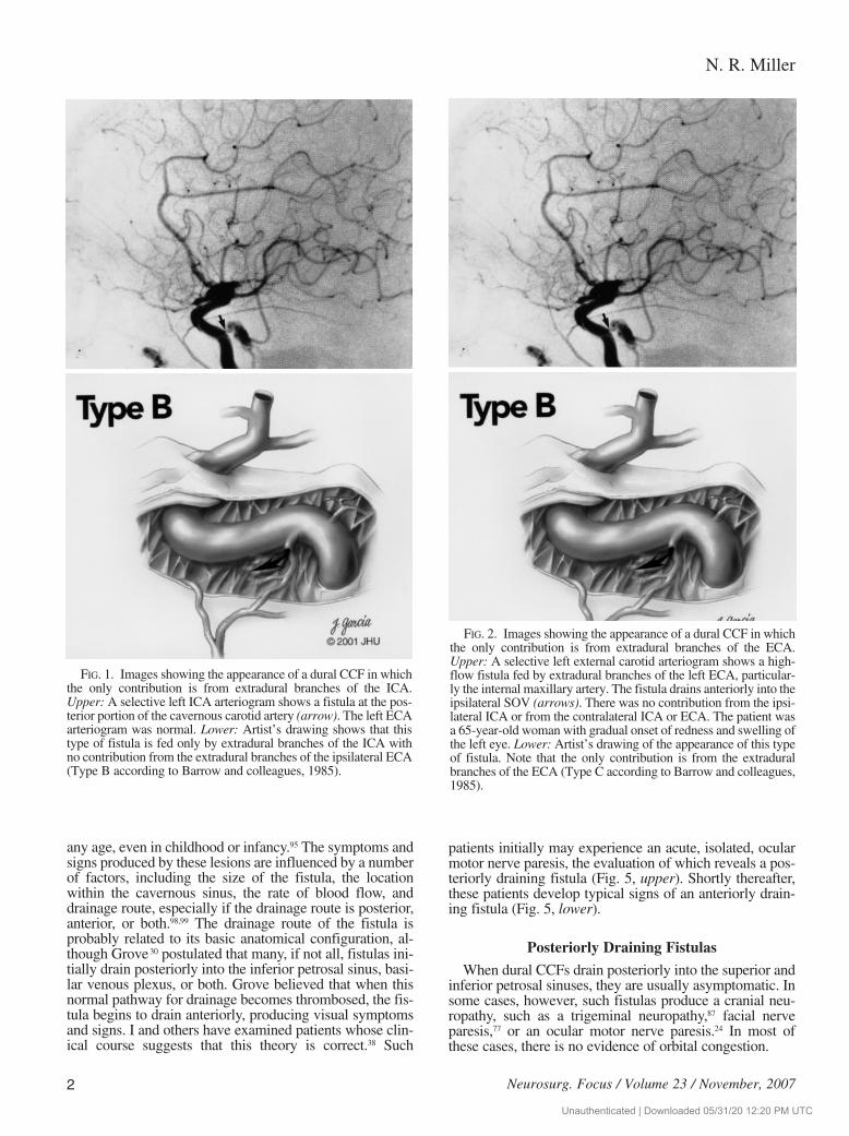
FIG. 2. Images showing the appearance of a dural CCF in whichthe only contribution is from extradural branches of the ECA.Upper: A selective left external carotid arteriogram shows a high-flow fistula fed by extradural branches of the left ECA, particular-ly the internal maxillary artery. The fistula drains anteriorly into theipsilateral SOV (arrows). There was no contribution from the ipsi-lateral ICA or from the contralateral ICA or ECA. The patient wasa 65-year-old woman with gradual onset of redness and swelling ofthe left eye. Lower: Artist’s drawing of the appearance of this typeof fistula. Note that the only contribution is from the extraduralbranches of the ECA (Type C according to Barrow and colleagues,1985).
Unauthenticated | Downloaded 05/31/20 12:20 PM UTC
In most cases of ocular motor nerve paresis caused by aposteriorly draining dural CCF, the onset of the paresis issudden, and only one of the ocular motor nerves is affect-ed. The oculomotor nerve (CN III) is most often affected,and the resulting paresis may be complete with involve-ment of the pupil, incomplete with pupil involvement, orincomplete with pupil sparing. I have never seen a patientwith a complete, pupil-sparing oculomotor nerve paresis inthis setting. In almost all cases, the paresis is associatedwith ipsilateral orbital or ocular pain, a presentation thatinitially suggests an intracranial aneurysm (Fig. 6).38,65 Thecorrect diagnosis in such cases is not evident until cerebralangiography is performed. In other cases, the posteriorlydraining fistula produces an abducent (CN VI) or trochlear(CN IV) nerve paresis, again usually associated with ocu-lar or orbital pain (Fig. 7).24,52,79,92
The cranial neuropathies that are caused by a posteriorlydraining dural CCF are usually the initial sign of the fistu-la. In many of these cases, failure to diagnose and treat thefistula leads eventually to a change in the direction of theflow of blood in the fistula. The flow becomes anterior, andthe patient develops evidence of orbital congestion. Inother cases, the blood flow in the fistula initially is anteri-or, producing orbital manifestations. With time, however,the anterior drainage ceases, and posterior flow is associat-ed with the development of the cranial neuropathy.
Dural fistulas that drain posteriorly sometimes causebrainstem congestion that may be associated with neuro-logical deficits.101 In addition, such fistulas rarely may pro-duce intracranial hemorrhage.35
Anteriorly Draining Fistulas
Similarly to their direct counterparts, dural CCFs usual-ly produce visual symptoms and signs when they drainanteriorly into the superior and inferior ophthalmic veins.The clinical manifestations of patients with dural CCFs thatdrain anteriorly are therefore similar to, but usually muchless severe than, those of patients with direct fistulas, be-cause most dural fistulas contain blood flowing at a lowrate. Dural fistulas usually produce an important and rathercharacteristic syndrome that, nevertheless, often is misdi-agnosed.12,30,47,48,60,79,84,98,99 Unlike direct fistulas, there oftenis no objective or subjective bruit with dural fistulas; evenwhen a subjective bruit is present, the patient may not men-tion it, either because it is mild or because the patient doesnot associate the sound with his or her ocular symptomsand signs. In the mildest cases, there is redness of one, orrarely, both eyes, caused by dilation and arterialization of
Neurosurg. Focus / Volume 23 / November, 2007
Diagnosis and treatment of dural carotid–cavernous sinus fistulas
3
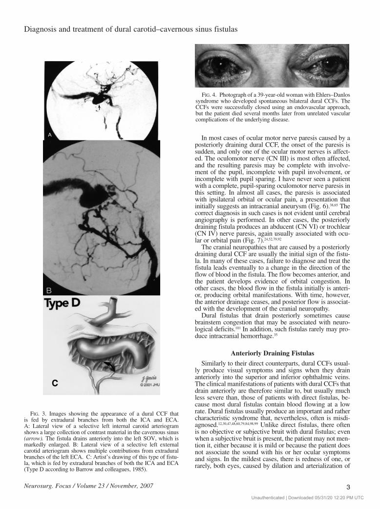
FIG. 3. Images showing the appearance of a dural CCF thatis fed by extradural branches from both the ICA and ECA.A: Lateral view of a selective left internal carotid arteriogramshows a large collection of contrast material in the cavernous sinus(arrow). The fistula drains anteriorly into the left SOV, which ismarkedly enlarged. B: Lateral view of a selective left externalcarotid arteriogram shows multiple contributions from extraduralbranches of the left ECA. C: Artist’s drawing of this type of fistu-la, which is fed by extradural branches of both the ICA and ECA(Type D according to Barrow and colleagues, 1985).
FIG. 4. Photograph of a 39-year-old woman with Ehlers–Danlossyndrome who developed spontaneous bilateral dural CCFs. TheCCFs were successfully closed using an endovascular approach,but the patient died several months later from unrelated vascularcomplications of the underlying disease.
Unauthenticated | Downloaded 05/31/20 12:20 PM UTC
both conjunctival and episcleral veins (Fig. 8). The appear-ance of the eye in these cases may suggest conjunctivitis,episcleritis, or thyroid eye disease; however, a careful
examination of the dilated vessels usually demonstrates atypical tortuous corkscrew appearance that is virtuallypathognomonic of a dural CCF (Fig. 9). There also may beminimal eyelid swelling, conjunctival chemosis, proptosis,or a combination of these symptoms. Diplopia from abdu-cent nerve paresis may be present (Fig. 10). Ophthalmo-scopy results may be normal, or there may be mild dilationof retinal veins.
In more advanced dural CCFs, particularly those with ahigh flow rate, the symptoms and signs are identical tothose in patients with direct CCFs.41,71,98,99 In these cases,proptosis, chemosis, and dilation of conjunctival vesselsare obvious (Fig. 11). Diplopia may result from ophthal-moparesis caused by ocular motor nerve pareses, orbitalcongestion, or both mechanisms, and there may be signifi-cant periorbital or retroocular discomfort or pain, initiallysuggesting an inflammatory process or even the Tolo-sa–Hunt syndrome.15,85 Some patients develop facial pain,facial weakness, or both.43 Increased episcleral venouspressure may produce increased intraocular pressure thatoccasionally is quite high.41,47,84,108 Angle–closure glaucomamay develop from elevated orbital venous pressure, con-gestion of the iris and choroid, and forward displacement ofthe iris–lens diaphragm.103 In other cases, chronic ischemiaproduces neovascular glaucoma. Ophthalmoscopic abnor-malities include venous stasis retinopathy with intraretinalhemorrhages, central retinal vein occlusion, proliferativeretinopathy, retinal detachment, vitreous hemorrhage, cho-roidal folds, choroidal effusion, choroidal detachment, oroptic disc swelling (Fig. 12).25,29,55,61,76,90
N. R. Miller
4 Neurosurg. Focus / Volume 23 / November, 2007
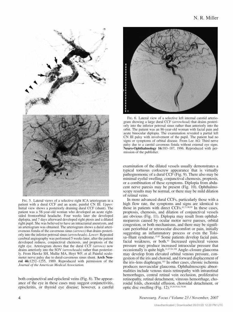
FIG. 5. Lateral views of a selective right ICA arteriogram in apatient with a dural CCF and an acute, painful CN III. Upper:Initial view shows a posteriorly draining dural CCF (shunt). Thepatient was a 58-year-old woman who developed an acute right-sided frontoorbital headache. Four weeks later she developeddiplopia, and 7 days afterward developed right ptosis and a dilatedright pupil. She was believed to have an intracranial aneurysm, andan arteriogram was obtained. The arteriogram shows a dural arteri-ovenous fistula of the cavernous sinus (arrows) that drains posteri-orly into the inferior petrosal sinus (arrowheads). Lower: Repeatedcerebral angiography was performed 5 weeks later, after the patientdeveloped redness, conjunctival chemosis, and proptosis of theright eye. Arteriogram shows that the dural CCF (arrows) nowdrains anteriorly into the SOV (arrowheads) rather than posterior-ly. From Hawke SH, Mullie MA, Hoyt WF, et al: Painful oculo-moter nerve palsy due to dural-cavernous sinus shunt. Arch Neu-rol 46:1252–1255, 1989. Reproduced with permission of theJournal of the American Medical Association.
FIG. 6. Lateral view of a selective left internal carotid arterio-gram showing a large dural CCF (arrowhead) that drains posteri-orly into the inferior petrosal sinus rather than anteriorly into theorbit. The patient was an 86-year-old woman with facial pain andacute binocular diplopia. The examination revealed a partial leftCN III palsy with involvement of the pupil. The patient had nosigns or symptoms of orbital disease. From Lee AG: Third nervepalsy due to a carotid cavernous fistula without external eye signs.Neuro-Ophthalmology 16:183–187, 1996. Reproduced with per-mission of the publisher.
Unauthenticated | Downloaded 05/31/20 12:20 PM UTC
Visual loss, although less frequent than in patients withdirect CCFs, occurs in 20–30% of patients with duralCCFs.30,79 This visual loss may be caused by ischemic opticneuropathy, chorioretinal dysfunction, or uncontrolledglaucoma.61,84
The ocular manifestations of unilateral dural CCFs al-most always are ipsilateral to the fistula, but they may besolely contralateral or bilateral (Fig. 13).98,99 When unilater-al fistulas cause bilateral manifestations, there is a highprobability that the fistula is draining into cortical veins(Fig. 14).98
Although most dural fistulas are unilateral, bilateralspontaneous dural fistulas have been described.37 Patientswith bilateral dural CCFs often have severe systemic hy-pertension, atherosclerosis, or some type of systemic con-nective tissue disease such as Ehlers–Danlos syndromeType IV. Most patients with bilateral dural CCFs have bilat-eral manifestations; however, I once examined a patientwith bilateral dural fistulas who demonstrated only left-sided ocular manifestations: the left-sided fistula drainedanteriorly into the left orbit via the left SOV, and the right-
sided fistula drained across the intercavernous sinus andthen anteriorly into the left orbit.
In some instances, dural CCFs drain both anteriorly andposteriorly. In most of these cases, the only manifestationsare those related to the anterior drainage; however, somepatients develop manifestations from the posteriordrainage, such as facial nerve paresis or acute hemiparesisassociated with neuroimaging evidence of brainstem con-gestion.77,94
Differential Diagnosis
Because the symptoms and signs of a dural CCF oftenare mild, usually developing spontaneously and ratherslowly, this lesion is often misdiagnosed initially. When thepatient simply has a red eye, perhaps with minimal eyelidswelling, it may be believed that he or she has a chronicconjunctivitis or blepharoconjunctivitis that is refractory totopical therapy (Fig. 15). In patients who develop diplopiafrom abducent nerve paresis, the significance of a slightlyred eye may be missed (Fig. 16).
In patients with evidence of orbital congestion, red eye,conjunctival chemosis, and other symptoms, diagnosesother than a spontaneous dural CCF, such as dysthyroidorbitopathy, orbital pseudotumor, orbital cellulitis, episcle-ritis, sphenoorbital meningioma, or Tolosa–Hunt syn-drome, may be considered.15,30,79,81,84,85 The correct diagnosisin such cases may not be able to be made until symptomsand signs worsen, new symptoms and signs develop, orappropriate diagnostic studies are performed. In addition,trauma to the posterior orbit in the region of the superiororbital fissure may produce such manifestations,70 and I andothers have examined patients with congenital or acquired
Neurosurg. Focus / Volume 23 / November, 2007
Diagnosis and treatment of dural carotid–cavernous sinus fistulas
5
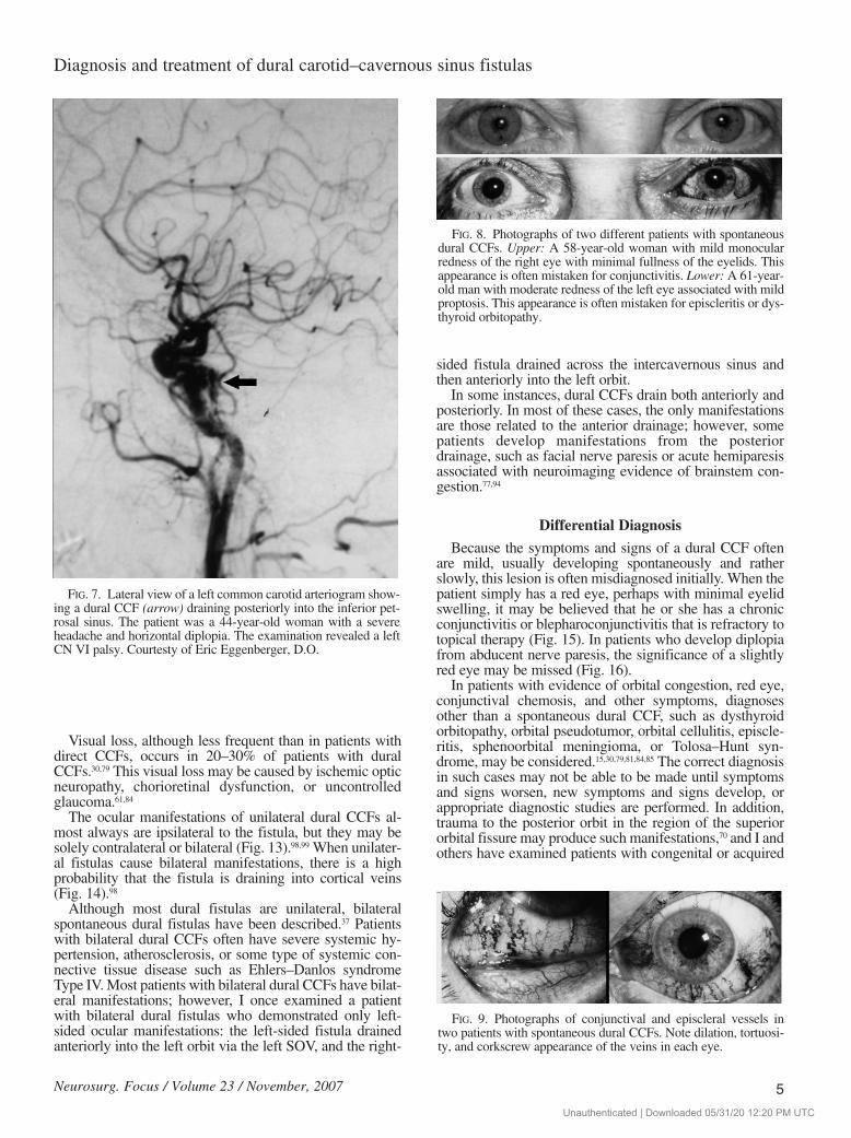
FIG. 7. Lateral view of a left common carotid arteriogram show-ing a dural CCF (arrow) draining posteriorly into the inferior pet-rosal sinus. The patient was a 44-year-old woman with a severeheadache and horizontal diplopia. The examination revealed a leftCN VI palsy. Courtesty of Eric Eggenberger, D.O.
FIG. 8. Photographs of two different patients with spontaneousdural CCFs. Upper: A 58-year-old woman with mild monocularredness of the right eye with minimal fullness of the eyelids. Thisappearance is often mistaken for conjunctivitis. Lower: A 61-year-old man with moderate redness of the left eye associated with mildproptosis. This appearance is often mistaken for episcleritis or dys-thyroid orbitopathy.
FIG. 9. Photographs of conjunctival and episcleral vessels intwo patients with spontaneous dural CCFs. Note dilation, tortuosi-ty, and corkscrew appearance of the veins in each eye.
Unauthenticated | Downloaded 05/31/20 12:20 PM UTC
anomalous intracranial venous drainage who developedclinical manifestations suggesting a dural CCF.105
Diagnosis
It should be clear from the previous discussion that thediagnosis of a dural CCF should be considered in any pa-tient who spontaneously develops a red eye, chemosis ofthe conjunctiva, abducent nerve paresis, or mild orbitalcongestion with proptosis. Auscultation of the orbit maydisclose a bruit, but this is relatively uncommon. Tonom-etry, however, usually shows asymmetry of the ocular pulsewith greater pulse amplitude on the side of the lesion. Theasymmetry in the amplitude of the ocular pulse can be dis-covered using any tonometer, although I prefer to use apneumotonometer that provides both a direct measurementand an objective record of the ocular pulse amplitude (Fig.17).28
When a dural CCF is suspected, CT scanning, CTangiography, MR imaging, MR angiography, orbital ultra-sonography, transorbital and transcranial color Dopplerimaging, or a combination of these tests may be beneficialin confirming the diagnosis (Figs. 18–21).5,17,46,50,88 The goldstandard diagnostic test, however, as in the case of thedirect CCF, is a catheter angiogram (Figs. 1–3).19,21 Becausemany dural CCFs are fed either by meningeal branches ofthe ECA or by meningeal branches of both the ICA andECA and others are fed by arteries from both sides or arefed by unilateral arteries but produce bilateral symptomsand signs, selective angiography of both the ICA and ECAon both sides should always be performed.19 When per-formed by an experienced neuroradiologist, catheter angi-ography has a morbidity rate of less than 1% and virtuallyno mortality rate, except in patients with connective tissuedisorders such as Ehlers–Danlos syndrome, in whom the
risks are much greater (because of excessive fragility of theextracranial and intracranial vessels).91
Natural History of Dural CCFs
The majority of patients with a dural CCF have no dif-ference in mortality rates from those of the normal popula-tion because the lesion usually affects only the eyes. Spon-taneous intracranial hemorrhage is exceptionally rare.35
Thus, when one considers the natural history of a duralCCF, one is usually concerned with ocular morbidity.
Regardless of whether they drain anteriorly or posterior-ly, 20–50% of dural CCFs close spontaneously, after an-giography, or after air flight travel (Figs. 10 and 22).69 In
N. R. Miller
6 Neurosurg. Focus / Volume 23 / November, 2007
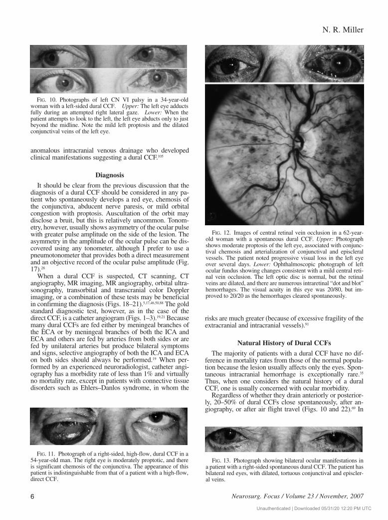
FIG. 10. Photographs of left CN VI palsy in a 34-year-oldwoman with a left-sided dural CCF. Upper: The left eye adductsfully during an attempted right lateral gaze. Lower: When thepatient attempts to look to the left, the left eye abducts only to justbeyond the midline. Note the mild left proptosis and the dilatedconjunctival veins of the left eye.
FIG. 11. Photograph of a right-sided, high-flow, dural CCF in a54-year-old man. The right eye is moderately proptotic, and thereis significant chemosis of the conjunctiva. The appearance of thispatient is indistinguishable from that of a patient with a high-flow,direct CCF.
FIG. 12. Images of central retinal vein occlusion in a 62-year-old woman with a spontaneous dural CCF. Upper: Photographshows moderate proptosis of the left eye, associated with conjunc-tival chemosis and arterialization of conjunctival and episcleralvessels. The patient noted progressive visual loss in the left eyeover several days. Lower: Ophthalmoscopic photograph of leftocular fundus showing changes consistent with a mild central reti-nal vein occlusion. The left optic disc is normal, but the retinalveins are dilated, and there are numerous intraretinal “dot and blot”hemorrhages. The visual acuity in this eye was 20/80, but im-proved to 20/20 as the hemorrhages cleared spontaneously.
FIG. 13. Photograph showing bilateral ocular manifestations ina patient with a right-sided spontaneous dural CCF. The patient hasbilateral red eyes, with dilated, tortuous conjunctival and episcler-al veins.
Unauthenticated | Downloaded 05/31/20 12:20 PM UTC
some cases, the symptoms and signs begin to resolve with-in days to weeks after angiography. In others, they do notresolve until months to years after the fistula has becomesymptomatic.
I believe it is appropriate to follow up clinically patientswho have mild ocular manifestations to determine if the fis-tula will close spontaneously. During the waiting period,patients do not need to alter their lifestyle. They should,however, be examined at regular intervals so that their visu-al function, intraocular pressure, and ophthalmoscopic ap-pearance can be monitored.89 During this time, exposurekeratopathy caused by proptosis can be treated using ocu-lar lubrication, and persistent bothersome diplopia can betreated using prism therapy or occlusion of one eye. In-creased intraocular pressure rarely is so severe that itrequires treatment.41 If intraocular pressure is substantiallyelevated, one can attempt to lower it with one of the manytopical agents that reduce the production of aqueous humor.Because in most cases the cause of the elevated intraocularpressure is raised episcleral venous pressure, such agentsmay not be helpful, however, and even agents such aslatanoprost, a prostaglandin receptor agonist that increasesuveoscleral outflow of aqueous humor, may not be effec-tive in patients with a dural CCF because of the significantbackup of arterial blood in the orbital veins. Nevertheless,
it is still worthwhile to administer these drugs for a fewweeks because even a small reduction in intraocular pres-sure may protect the patient’s vision. In the final analysis,however, the best treatment for severely increased intraoc-ular pressure is closure of the fistula.
Patients with a dural CCF may experience acute worsen-ing of ocular manifestations. This clinical deteriorationresults from an increase in blood flow through the fistula insome cases, but in others, it is caused by spontaneousthrombosis of the SOV.28,93 Patients in whom clinical wors-ening is caused by spontaneous progressive thrombosis ofthe SOV usually begin to improve within several weeks,and most eventually experience complete resolution ofsymptoms and signs (Fig. 23). Systemic corticosteroids ad-ministered when deterioration occurs may lessen the sever-ity of symptoms and signs and perhaps reduce the length oftime until recovery occurs.93
Patients in whom a dural CCF persists or in whom sucha fistula is not recognized may experience major hemor-rhagic and other complications when they undergo intraoc-ular or orbital surgery performed for other reasons, such asfor cataracts or strabismus.84 Others may undergo uncom-
Neurosurg. Focus / Volume 23 / November, 2007
Diagnosis and treatment of dural carotid–cavernous sinus fistulas
7
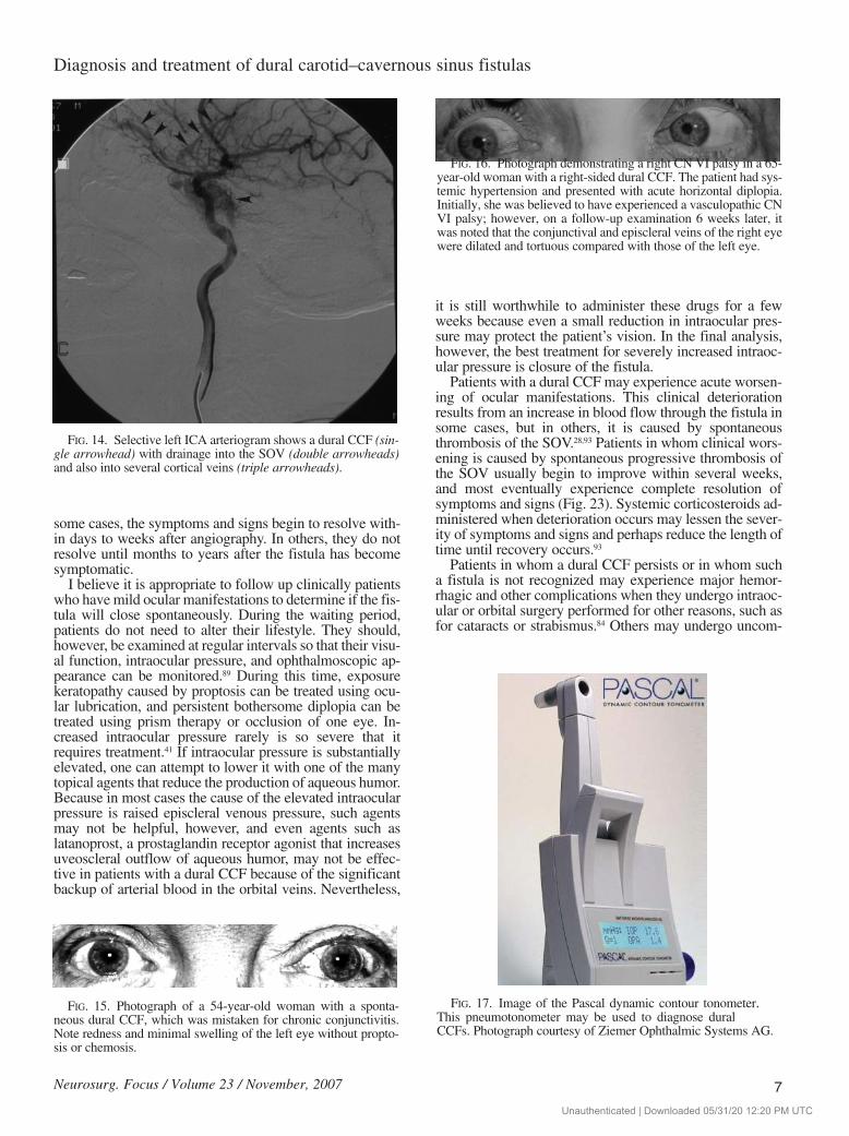
FIG. 14. Selective left ICA arteriogram shows a dural CCF (sin-gle arrowhead) with drainage into the SOV (double arrowheads)and also into several cortical veins (triple arrowheads).
FIG. 15. Photograph of a 54-year-old woman with a sponta-neous dural CCF, which was mistaken for chronic conjunctivitis.Note redness and minimal swelling of the left eye without propto-sis or chemosis.
FIG. 17. Image of the Pascal dynamic contour tonometer.This pneumotonometer may be used to diagnose duralCCFs. Photograph courtesy of Ziemer Ophthalmic Systems AG.
FIG. 16. Photograph demonstrating a right CN VI palsy in a 65-year-old woman with a right-sided dural CCF. The patient had sys-temic hypertension and presented with acute horizontal diplopia.Initially, she was believed to have experienced a vasculopathic CNVI palsy; however, on a follow-up examination 6 weeks later, itwas noted that the conjunctival and episcleral veins of the right eyewere dilated and tortuous compared with those of the left eye.
Unauthenticated | Downloaded 05/31/20 12:20 PM UTC
plicated surgery, only to lose vision subsequently fromischemic complications of the fistula.96
Treatment
The visual manifestations of a dural CCF usually do notrequire local treatment. Occasionally, increased intraocularpressure requires treatment with topical or oral pressure-lowering agents. Although pressure-lowering ocular sur-gery has been advocated for patients in whom medicaltherapy does not reduce the intraocular pressure to an ac-ceptable level,48,84 I believe that if intraocular pressureremains unacceptably elevated despite maximum medicaltherapy, definitive treatment of the fistula should be per-formed instead of ocular surgery. Only if treatment of thefistula cannot be performed or is unsuccessful, or if theintraocular pressure remains elevated despite closure of thefistula, should ocular surgery be considered.25 Similarly, al-though the proliferative retinopathy that may occasionallyaccompany a severe, high-flow dural CCF can be treated
successfully with photocoagulation,25,36 it is best to treat thefistula producing the retinopathy whenever possible. Again,if the fistula cannot be treated or treatment is unsuccessful,photocoagulation may be needed to preserve vision.
Dural CCFs may be treated using direct surgery,18,21,39,60,80
conventional radiation therapy,107 stereotactic radiosur-gery,32 intermittent manual self-compression of the affectedICA with the contralateral hand,44 or even occlusion of theipsilateral ICA.60 In general, however, endovascular em-bolization is the optimum treatment for those lesions thatproduce progressive or unacceptable symptoms and signsincluding visual loss, diplopia, an intolerable bruit, severeproptosis, and, most importantly, cortical venous drain-age.4,16,20,21,39,53,60,71,102 A number of synthetic and naturalmaterials can be used for embolization, including absor-bable gelatin (Gelfoam); Silastic; platinum coils; low-vis-cosity silicone rubber; autogenous clot, muscle, or dura;tetradecyl sulfate (a sclerosing agent); polyvinyl alcoholparticles (Ivalon); ethanol; oxidized cellulose (Oxycel); andisobutyl-2-cyanoacrylate glue (Bucrylate).21,31,49,51,53,54,60,106
In patients with a fistula fed only by meningeal branch-es of the ECA or by meningeal branches from both theECA and ICA, the embolization material is introduced viaa microcatheter placed in the ECA and passed into the spe-
N. R. Miller
8 Neurosurg. Focus / Volume 23 / November, 2007
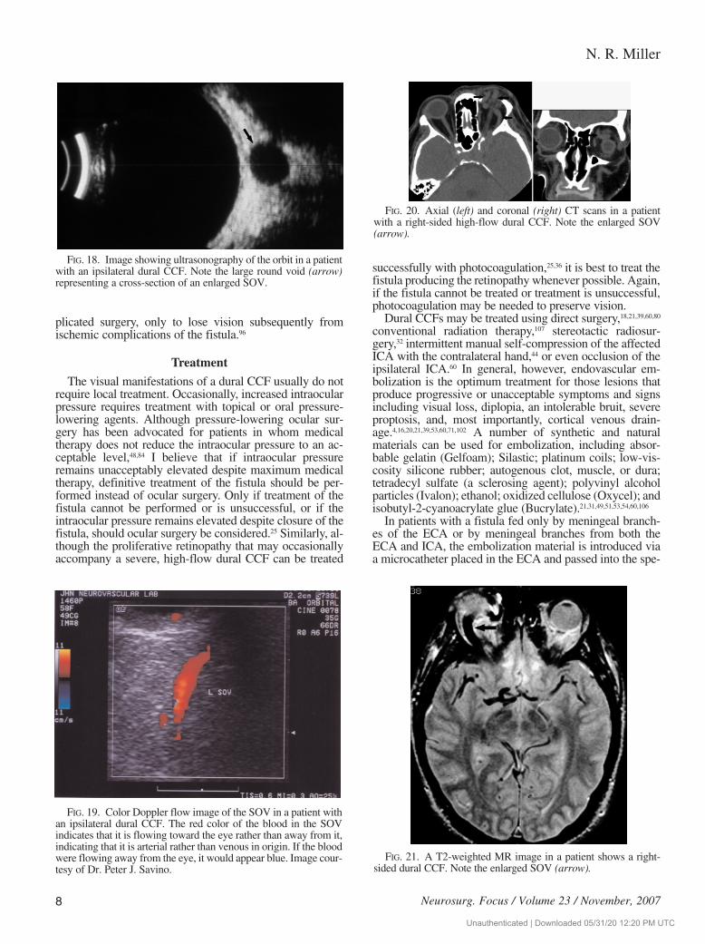
FIG. 18. Image showing ultrasonography of the orbit in a patientwith an ipsilateral dural CCF. Note the large round void (arrow)representing a cross-section of an enlarged SOV.
FIG. 21. A T2-weighted MR image in a patient shows a right-sided dural CCF. Note the enlarged SOV (arrow).
FIG. 19. Color Doppler flow image of the SOV in a patient withan ipsilateral dural CCF. The red color of the blood in the SOVindicates that it is flowing toward the eye rather than away from it,indicating that it is arterial rather than venous in origin. If the bloodwere flowing away from the eye, it would appear blue. Image cour-tesy of Dr. Peter J. Savino.
FIG. 20. Axial (left) and coronal (right) CT scans in a patientwith a right-sided high-flow dural CCF. Note the enlarged SOV(arrow).
Unauthenticated | Downloaded 05/31/20 12:20 PM UTC
cific branch or branches that feed the fistula.21,60,68,106 TheICA usually is not embolized unless the interventionalistcan successfully catheterize the meningohypophyseal trunkor other meningeal feeders from the artery.14 When the fis-tula is fed only by branches from the ECA, successful clo-sure of these branches is often possible and associated withrapid resolution of all ocular symptoms and signs.7,60
When a dural fistula is fed by branches from both theECA and ICA, embolization of the feeders from the ECAusing various agents such as polyvinyl alcohol or glue mayreduce the blood flow in the fistula sufficiently such thatnonembolized feeders from the ICA will thrombose spon-taneously.54,69 Embolization of feeders from the ICA isalmost never appropriate because of the significant poten-tial neurological morbidity from distal embolization.60 Ifthrombosis does not occur with this technique, the fistulacan be treated by placement of detachable platinum coils ordetachable balloons within the cavernous sinus using atransvenous route. The favored approach usually is via thefemoral or internal jugular vein into the inferior or superiorpetrosal sinus, and from there into the cavernous sinus, butif this approach fails, a variety of other approaches may beused, most of which involve cannulation of the superior orinferior ophthalmic vein.1,5,7–11,13,16,20–22,26,27,34,42,49,51,53,56,57,59,63,67,
73,78,83,97,102 In some cases, more than one session and morethan one approach are needed.11,57
At my institution, we prefer the direct SOV approachperformed in most cases by surgical exposure of the vessel(Fig. 24 and accompanying video). All procedures are per-formed in a neurosurgical operating room under fluoro-scopic guidance. With the patient in a state of general anes-thesia, a sheath is placed in a common femoral artery topermit intraoperative angiography. Following appropriatepreparation and draping of the affected eye and orbitalregions, a curvilinear skin incision is made at the level ofthe superior lid crease or the superior sulcus of the uppereyelid nasally, using magnification provided either by an
operating microscope or magnifying loupes (Figs. 24A and24B). The incision is continued down through the orbicu-laris oculi muscle, with careful attention to hemostasis. Theorbital septum is identified and opened with sharp spring-action scissors, exposing the retroseptal orbital fat. TheSOV is identified using blunt dissection. The vein appearsas a reddish-blue vessel that varies in size from 3 to 8 mmin diameter (Fig. 24C). The vein is carefully cleaned fromits attachments to surrounding orbital fat until a segmentmeasuring 10–20 mm long is exposed (Fig. 24D). Two lig-atures, consisting of 2–0 black silk sutures for small veinsand silicone vascular loops 1 mm in diameter for largeveins, are passed underneath the vessel using a Kelly orright-angled clamp (Fig. 24E), and the two ends of each lig-ature are then passed through a piece of tubing that variesin size from a pediatric feeding tube to a French catheter,depending on the diameter of the ligatures (Fig. 24F). Thetwo ligatures are then placed as far apart as possible to iso-late a segment of the vein.
Once the ligatures are in place around the vein and aretightened down to prevent bleeding (Fig. 24G), a small in-cision is made in the wall of the portion of the vein betweenthe ligatures using sharp spring-action iris scissors (Fig.24H). Brisk arterial bleeding indicates that a full-thicknessopening has been achieved. The ligatures are then tightenedusing the feeding tubes or catheters. A microcatheter, thesize of which is determined by the diameter of the vein, isplaced into the vein opening, using a jewelers’ forceps tosteady and direct it (Fig. 24I). The placement is facilitatedusing a two-person technique, with one person threadingthe catheter and the other manipulating the ligatures toallow passage of the catheter while limiting bleeding. Thecatheter is threaded posteriorly under fluoroscopic control
Neurosurg. Focus / Volume 23 / November, 2007
Diagnosis and treatment of dural carotid–cavernous sinus fistulas
9
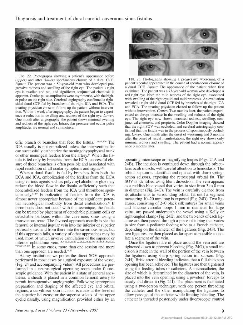
FIG. 22. Photographs showing a patient’s appearance before(upper) and after (lower) spontaneous closure of a dural CCF.Upper: The patient was a 59-year-old man who developed pro-gressive redness and swelling of the right eye. The patient’s righteye is swollen and red, and significant conjunctival chemosis isapparent. Ocular pulse amplitudes were asymmetric, with the high-er pulse on the right side. Cerebral angiography confirmed a right-sided dural CCF fed by branches of the right ICA and ECA. Thetreating physician chose to follow up the patient without interven-tion. Within 1 week after angiography, the patient began to experi-ence a reduction in swelling and redness of the right eye. Lower:One month after angiography, the patient shows minimal swellingand redness of the right eye. Intraocular pressure and ocular pulseamplitudes are normal and symmetrical.
FIG. 23. Photographs showing a progressive worsening of apatient’s ocular appearance in the course of spontaneous closure ofa dural CCF. Upper: The appearance of the patient when firstexamined. The patient was a 73-year-old woman who developed ared right eye. Note the mild redness of the right eye, associatedwith swelling of the right eyelid and mild proptosis. An evaluationrevealed a right-sided dural CCF fed by branches of the right ICAand ECA. The treating physician elected to follow up the patientwithout intervention. Center: Two months later, the patient experi-enced an abrupt increase in the swelling and redness of the righteye. The right eye now shows increased redness, swelling, con-junctival chemosis, and proptosis. Color Doppler imaging showedthat the right SOV was occluded, and cerebral arteriography con-firmed that the fistula was in the process of spontaneously occlud-ing. Lower: One month after the onset of worsening and 3 monthsafter the onset of visual manifestations, the right eye shows onlyminimal redness and swelling. The patient had a normal appear-ance 3 months later.
Unauthenticated | Downloaded 05/31/20 12:20 PM UTC
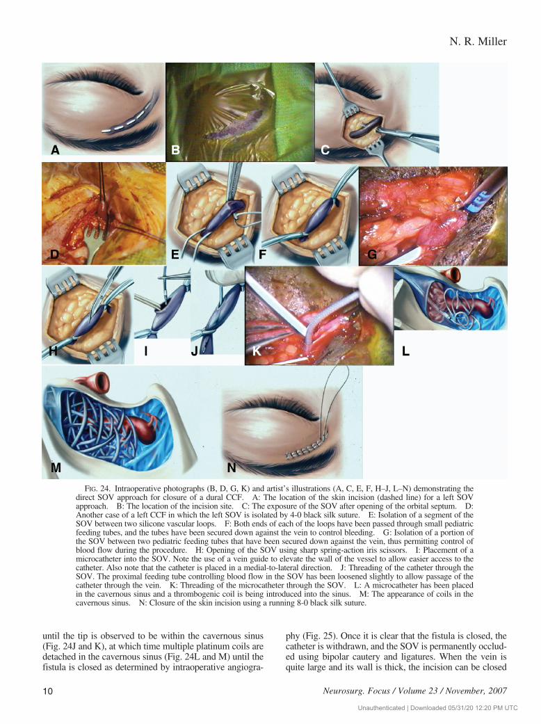
until the tip is observed to be within the cavernous sinus(Fig. 24J and K), at which time multiple platinum coils aredetached in the cavernous sinus (Fig. 24L and M) until thefistula is closed as determined by intraoperative angiogra-
phy (Fig. 25). Once it is clear that the fistula is closed, thecatheter is withdrawn, and the SOV is permanently occlud-ed using bipolar cautery and ligatures. When the vein isquite large and its wall is thick, the incision can be closed
N. R. Miller
10 Neurosurg. Focus / Volume 23 / November, 2007
FIG. 24. Intraoperative photographs (B, D, G, K) and artist’s illustrations (A, C, E, F, H–J, L–N) demonstrating thedirect SOV approach for closure of a dural CCF. A: The location of the skin incision (dashed line) for a left SOVapproach. B: The location of the incision site. C: The exposure of the SOV after opening of the orbital septum. D:Another case of a left CCF in which the left SOV is isolated by 4-0 black silk suture. E: Isolation of a segment of theSOV between two silicone vascular loops. F: Both ends of each of the loops have been passed through small pediatricfeeding tubes, and the tubes have been secured down against the vein to control bleeding. G: Isolation of a portion ofthe SOV between two pediatric feeding tubes that have been secured down against the vein, thus permitting control ofblood flow during the procedure. H: Opening of the SOV using sharp spring-action iris scissors. I: Placement of amicrocatheter into the SOV. Note the use of a vein guide to elevate the wall of the vessel to allow easier access to thecatheter. Also note that the catheter is placed in a medial-to-lateral direction. J: Threading of the catheter through theSOV. The proximal feeding tube controlling blood flow in the SOV has been loosened slightly to allow passage of thecatheter through the vein. K: Threading of the microcatheter through the SOV. L: A microcatheter has been placedin the cavernous sinus and a thrombogenic coil is being introduced into the sinus. M: The appearance of coils in thecavernous sinus. N: Closure of the skin incision using a running 8-0 black silk suture.
Unauthenticated | Downloaded 05/31/20 12:20 PM UTC
using a 10-0 nylon suture. The orbit is then irrigated withan antibiotic solution, and the skin incision is closed usinga running 8-0 black nylon suture (Fig. 24N). No attempt ismade to close the orbital septum in most cases. A Xeroformgauze pad is placed over the incision site, and a light eyepatch is placed over the pad for 24 hours. The incision siteis then treated with a topical antibiotic ointment, such aserythromycin or bacitracin, and the skin suture is removedin 5–7 days. When performed by an experienced team, this
approach is successful in the majority of cases and, in fact,may be the best initial treatment for all dural CCFs.27,60,73,74,86
It is important to be aware, however, that should an attemptat closing a dural CCF transvenously via the SOV beunsuccessful and the SOV sacrificed or ligated during theprocedure, there is a significant risk of increased venouspressure in the orbit with subsequent neovascular glaucomaand severe visual loss.33 For this reason, physicians at-tempting this form of treatment should always have analternative available, such as a transvenous approachthrough the inferior petrosal sinus, endovascular occlusionof the artery or arteries supplying the fistula, or direct sur-gery on the fistula.66,72
Successful closure of dural CCFs by standard particulateor glue embolization is possible in 70–95% of allcases.3,39,60,62,69 When transvenous coil or balloon occlusionof the fistula is used, the rate of successful closure is 90–100%.71,73
Complications from endovascular treatment of duralCCFs are rare except in patients with connective tissue dis-orders such as Ehlers–Danlos syndrome.45,91 Nevertheless,significant complications may occur, including hemorrhageat the catheter site or in the orbit, local infection, sepsis,ophthalmic artery occlusion,104 and both transient and per-manent neurologic deficits, particularly ocular motor nervepareses.2,60,82 Devoto and associates23 reported the develop-ment of increasing proptosis, chemosis, and markedly ele-vated intraocular pressure associated with a mid-dilatedand poorly reactive pupil during attempted transvenousclosure of a dural CCF. The patient was treated with intra-venous mannitol and acetazolamide, as well as topical tim-olol maleate and apraclonidine. At the same time, the inter-ventionalist introduced larger coils into the anterior portionof the cavernous sinus. Within several minutes, the condi-tion had resolved, and the patient had a successful resultwith vision of 20/20 the next day.
Prognosis After Treatment
It is not unusual for dural CCFs to recanalize or formnew abnormal vessels after transarterial embolization withparticles or other material.64 Recurrence of ocular symp-toms and signs indicate the recurrence of the fistula, andpatients in whom manifestations recur require repeatangiography and consideration of further treatment. I amless concerned about incomplete closure or recurrence ifthe fistula has been closed transvenously using detachablecoils or balloons. Symptoms and signs usually begin toimprove within hours to days after successful closure of adural CCF (Fig. 26).27,60,73 Any preexisting bruit immediate-
Neurosurg. Focus / Volume 23 / November, 2007
Diagnosis and treatment of dural carotid–cavernous sinus fistulas
11
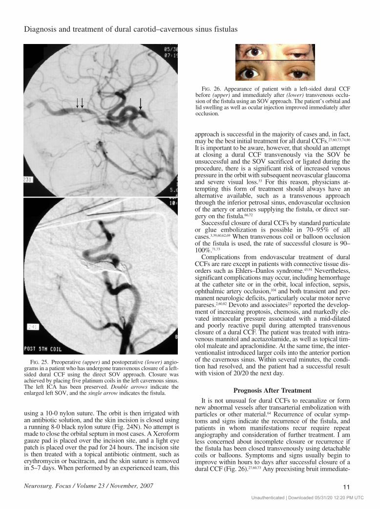
FIG. 25. Preoperative (upper) and postoperative (lower) angio-grams in a patient who has undergone transvenous closure of a left-sided dural CCF using the direct SOV approach. Closure wasachieved by placing five platinum coils in the left cavernous sinus.The left ICA has been preserved. Double arrows indicate theenlarged left SOV, and the single arrow indicates the fistula.
FIG. 26. Appearance of patient with a left-sided dural CCFbefore (upper) and immediately after (lower) transvenous occlu-sion of the fistula using an SOV approach. The patient’s orbital andlid swelling as well as ocular injection improved immediately afterocclusion.
Unauthenticated | Downloaded 05/31/20 12:20 PM UTC
ly disappears, and intraocular pressure immediately returnsto normal. Proptosis, conjunctival chemosis, redness of theeye, and ophthalmoparesis (whether caused by orbital con-gestion or an ocular motor nerve paresis) usually resolvecompletely within weeks to months, and most patients havea normal or near-normal external appearance within 6months (Fig. 27). At the same time, patients with visualloss caused by choroidal effusion or detachment usuallyexperience substantial if not complete recovery of visualfunction.64 Unfortunately, patients with visual loss causedby retinal damage (for example, central retinal vein occlu-sion) usually have persistently poor visual function.61
Patients whose dural CCFs are treated using techniques
other than endovascular closure, such as stereotactic radio-surgery, often take longer to improve than patients whosefistulas are closed using endovascular techniques.6,58 Nev-ertheless, these techniques may provide excellent resultsover time.
Conclusions
The diagnosis and management of dural CCFs have dra-matically improved in recent years. The widespread avail-ability of noninvasive imaging techniques, combined withimprovements in catheter angiography, permit rapid andaccurate diagnosis in most cases, and new endovascular
N. R. Miller
12 Neurosurg. Focus / Volume 23 / November, 2007
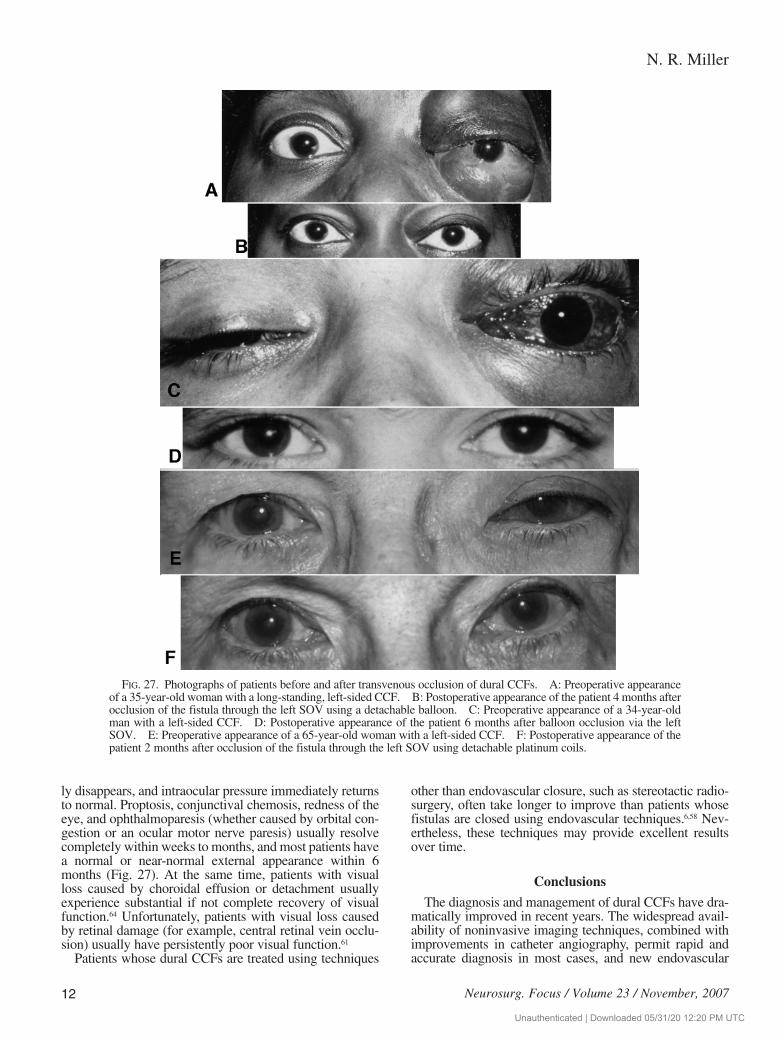
FIG. 27. Photographs of patients before and after transvenous occlusion of dural CCFs. A: Preoperative appearanceof a 35-year-old woman with a long-standing, left-sided CCF. B: Postoperative appearance of the patient 4 months afterocclusion of the fistula through the left SOV using a detachable balloon. C: Preoperative appearance of a 34-year-oldman with a left-sided CCF. D: Postoperative appearance of the patient 6 months after balloon occlusion via the leftSOV. E: Preoperative appearance of a 65-year-old woman with a left-sided CCF. F: Postoperative appearance of thepatient 2 months after occlusion of the fistula through the left SOV using detachable platinum coils.
Unauthenticated | Downloaded 05/31/20 12:20 PM UTC
therapeutic techniques allow most patients with these le-sions to be treated successfully with little or no morbidityor death and with resolution of most, if not all, clinicalmanifestations.
References
1. Agid R, Willinsky RA, Haw C, Souza MP, Vanek IJ, terBruggeKG: Targeted compartmental embolization of cavernous sinusdural arteriovenous fistulae using transfemoral medial and lateralfacial vein approaches. Neuroradiology 46:156–160, 2004
2. Aihara N, Mase M, Yamada K, Banno T, Watanabe K,Kamiya K, et al: Deterioration of ocular motor dysfunctionafter transvenous embolization of dural arteriovenous fistulainvolving the cavernous sinus. Acta Neurochir (Wien) 141:707–710, 1999
3. Albuquerque FC, Heinz GW, McDougall CG: Reversal of blind-ness after transvenous embolization of a carotid-cavernous fistu-la: case report. Neurosurgery 52:233–237, 2003
4. Annesley-Williams DJ, Goddard AJ, Brennan RP, Gholkar A: En-dovascular approach to treatment of indirect carotico-cavernousfistulae. Br J Neurosurg 15:228–233, 2001
5. Balayre S, Boissonnot M, Gicquel JJ, Drouineau J: [Endovasculartreatment of sinus dural fistulas using vein catheterism.] J FrOphtalmol 25:621–626, 2002 (Fr)
6. Barcia-Salorio JL, Soler F, Barcia JA, Hernández G: Stereotacticradiosurgery for the treatment of low-flow carotid-cavernous fis-tulae: results in a series of 25 cases. Stereotact Funct Neurosurg63:266–270, 1994
7. Barrow DL, Spector RH, Braun IF, Landman JA, Tindall SC,Tindall GT: Classification and treatment of spontaneous carotid-cavernous sinus fistulas. J Neurosurg 62:248–256, 1985
8. Benndorf G, Bender A, Campi A, Menneking H, Lanksch WR:Treatment of a cavernous sinus dural arteriovenous fistula by deeporbital puncture of the superior ophthalmic vein. Neuroradiology43:499–502, 2001
9. Benndorf G, Bender A, Lehmann R, Lanksch W: Transvenous oc-clusion of dural cavernous sinus fistulas through the thrombosedinferior petrosal sinus: report of four cases and review of the liter-ature. Surg Neurol 54:42–54, 2000
10. Berkmen T, Troffkin NA, Wakhloo AK: Transvenous sonograph-ically guided percutaneous access for treatment of an indirectcarotid cavernous fistula. AJNR Am J Neuroradiol 24:1548–1551, 2003
11. Berlis A, Klisch J, Spetzger U, Faist M, Schumacher M: Carotidcavernous fistula: embolization via a bilateral superior oph-thalmic vein approach. AJNR Am J Neuroradiol 23:1736–1738, 2002
12. Bhatti MT, Peters KR: A red eye and then a really red eye. SurvOphthalmol 48:224–229, 2003
13. Biondi A, Milea D, Cognard C, Ricciardi GK, Bonneville F, vanEffenterre R: Cavernous sinus dural fistulae treated by transve-nous approach through the facial vein: report of seven cases andreview of the literature. AJNR Am J Neuroradiol 24:1240–1246, 2003
14. Borden NM, Liebman KM: Endovascular access to the meningo-hypophyseal trunk. AJNR Am J Neuroradiol 22:725–727, 2001
15. Brazis PW, Capobianco DJ, Chang FL, McLeish WM, Earnest FIV: Low flow dural arteriovenous shunt: another cause of “sinis-ter” Tolosa-Hunt syndrome. Headache 34:523–525, 1994
16. Cheng KM, Chan CM, Cheung YL: Transvenous embolization ofdural carotid-cavernous fistulas by multiple venous routes: aseries of 27 cases. Acta Neurochir 145:17–29, 2003
17. Coskun O, Hamon M, Catroux G, Gosme L, Courthéoux P, Thér-on J: Carotid-cavernous fistulas: diagnosis with spiral CT angiog-raphy. AJNR Am J Neuroradiol 21:712–716, 2000
18. Day JD, Fukushima T: Direct microsurgery of dural arteriovenousmalformation type carotid-cavernous sinus fistulas: indications,technique, and results. Neurosurgery 41:1119–1126, 1997
19. Debrun GM: Angiographic workup of a carotid cavernous sinusfistula (CCF), or what information does the interventionalist needfor treatment? Surg Neurol 44:75–79, 1995
20. Debrun GM, Nauta HJ, Miller NR, Drake CG, Heros RC, AhnHS: Combining the detachable balloon technique and surgery inimaging carotid cavernous fistulae. Surg Neurol 32:3–10, 1989
21. Debrun GM, Viñuela F, Fox AJ, Davis KR, Ahn HS: Indicationsfor treatment and classification of 132 carotid-cavernous fistulas.Neurosurgery 22:285–289, 1988
22. Derang J, Ying H, Long Y, Reifa S, Qiming W, Yimu F, et al:Treatment of carotid-cavernous sinus fistulas retrograde via thesuperior ophthalmic vein (SOV). Surg Neurol 52:286–293, 1999
23. Devoto MH, Egbert JE, Tomsick TA, Kulwin DR: Acute exoph-thalmos during treatment of a cavernous sinus-dural fistulathrough the superior ophthalmic vein. Arch Ophthalmol 115:823–824, 1997
24. Eggenberger E: A bruital headache and double vision. SurvOphthalmol 45:147–153, 2000
25. Fiore PM, Latina MA, Shingleton BJ, Rizzo JF, Ebert E, BellowsAR: The dural shunt syndrome. I. Management of glaucoma.Ophthalmology 97:56–62, 1990
26. Gioulekas J, Mitchell P, Tress B, McNabb AA: Embolization ofcarotid cavernous fistulas via the superior ophthalmic vein. AustN Z J Ophthalmol 25:47–53, 1997
27. Goldberg RA, Goldey SH, Duckwiler G, Viñuela F: Managementof cavernous sinus-dural fistulas. Indications and techniques forprimary embolization via the superior ophthalmic vein. ArchOphthalmol 114:707–714, 1996
28. Golnik KC, Miller NR: Diagnosis of cavernous sinus arteriove-nous fistula by measurement of ocular pulse amplitude. Oph-thalmology 99:1146–1152, 1992
29. Gonshor LG, Kline LB: Choroidal folds and dural cavernous sinusfistula. Arch Ophthalmol 109:1065–1066, 1991
30. Grove AS Jr: The dural shunt syndrome. Pathophysiology andclinical course. Ophthalmology 91:31–44, 1984
31. Guglielmi G, Viñuela F, Briganti F, Duckwiler G: Carotid-cav-ernous fistula caused by a ruptured intracavernous aneurysm: en-dovascular treatment by electrothrombosis with detachable coils.Neurosurgery 31:591–597, 1992
32. Guo WY, Pan DHC, Wu HM, Chung WY, Shiau CY, Wang LW,et al: Radiosurgery as a treatment alternative for dural arteriove-nous fistulas of the cavernous sinus. AJNR Am J Neuroradiol19:1081–1087, 1998
33. Gupta N, Kikkawa DO, Levi L, Weinreb RN: Severe vision lossand neovascular glaucoma complicating superior ophthalmic veinapproach to carotid-cavernous sinus fistula. Am J Ophthalmol124:853–854, 1997
34. Hara T, Hamada J, Kai Y, Ushio Y: Surgical transvenous emboli-zation of a carotid-cavernous dural fistula with cortical drainagevia a petrosal vein: two technical reports. Neurosurgery 50:1380–1384, 2002
35. Harding AE, Kendall B, Leonard TJ, Johnson MH: Intracerebralhaemorrhage complicating dural arteriovenous fistula: a reportof two cases. J Neurol Neurosurg Psychiatry 47:905–911,1984
36. Harris MJ, Fine SL, Miller NR: Photocoagulation treatment ofproliferative retinopathy secondary to a carotid-cavernous sinusfistula. Am J Ophthalmol 90:515–518, 1980
37. Haugen OH, Sletteberg O, Thomassen L, Kråkenes J: Bilateralnon-traumatic carotid cavernous sinus fistula with spontaneousclosure. Acta Ophthalmol (Copenh) 68:743–747, 1990
38. Hawke SH, Mullie MA, Hoyt WF, Hallinan JM, Halmagyi GM:Painful oculomotor nerve palsy due to dural-cavernous sinusshunt. Arch Neurol 46:1252–1255, 1989
39. Higashida RT, Hieshima GB, Halbach VV: Advances in the treat-ment of complex cerebrovascular disorders by interventional neu-rovascular techniques. Circulation 83 (2 Suppl):1196–1206,1991
40. Houser OW, Campbell JK, Campbell RJ, Sundt TM Jr: Arterio-
Neurosurg. Focus / Volume 23 / November, 2007
Diagnosis and treatment of dural carotid–cavernous sinus fistulas
13
Unauthenticated | Downloaded 05/31/20 12:20 PM UTC
venous malformation affecting the transverse dural sinus-anacquired lesion. Mayo Clin Proc 54:651–661, 1979
41. Ishijima K, Kashiwagi K, Nakano K, Shibuya T, Tsumura T,Tsukahara S: Ocular manifestations and prognosis of secondaryglaucoma in patients with carotid-cavernous fistula. Jpn J Oph-thalmol 47:603–608, 2003
42. Jahan R, Gobin YP, Glenn B, Duckwiler GR, Viñuela F: Trans-venous embolization of a dural arteriovenous fistula of the cav-ernous sinus through the contralateral pterygoid plexus. Neuro-radiology 40:189–193, 1998
43. Jensen RW, Chuman H, Trobe JD, Deveikis JP: Facial and trigem-inal neuropathies in cavernous sinus fistulas. J Neuroophthalmol24:34–38, 2004
44. Kai Y, Hamada J, Morioka M, Yano S, Kuratsu J: Treatment ofcavernous sinus dural arteriovenous fistulae by external manualcarotid compression. Neurosurgery 60:253–258, 2007
45. Kashiwagi S, Tsuchida E, Goto K, Shiroyama Y, Yamashita T,Takahasi M, et al: Balloon occlusion of a spontaneous carotid-cavernous fistula in Ehlers-Danlos syndrome Type IV. Surg Neu-rol 39:187–190, 1993
46. Kawaguchi S, Sakaki T, Uranishi R: Color Doppler flow imagingof the superior ophthalmic vein in dural arteriovenous fistulas.Stroke 33:2009–2013, 2002
47. Keltner JL, Gittinger JW Jr, Miller NR, Burder RM: A red eye andhigh intraocular pressure. Surv Ophthalmol 31:328–336, 1987
48. Keltner JL, Satterfield D, Dublin AB, Lee BC: Dural and carotidcavernous sinus fistulas. Diagnosis, management, and complica-tions. Ophthalmology 94:1585–1600, 1987
49. Khayata MH, Dean BL, Spetzler RF: Materials and embolicagents for endovascular treatment. Neurosurg Clin N Am 5:-475–484, 1994
50. Kiliç T, Elmaci I, Bayri Y, Pamir MN, Erzen C: Value of tran-scranial Doppler ultrasonography in the diagnosis and follow-upof carotid-cavernous fistulae. Acta Neurochir (Wien) 143:1257–1265, 2001
51. Kinugasa K, Tokunaga K, Kamata I, Mandai S, Sugui K, HandaA, et al: Selection and combination of techniques for treatingspontaneous carotid-cavernous sinus fistulas. Neurol Med Chir34:597–606, 1994
52. Kishi S, Sawada A, Mori T, Yasuoka M: [Three cases of carotidcavernous sinus fistulas where the main ocular manifestation wasrestricted ocular motility.] Nippon Ganka Gakkai Zasshi 103:597–603, 1999 (Jpn)
53. Kobayashi N, Miyachi S, Negoro M, Suzuki O, Hattori K,Kojima T, et al: Endovascular treatment strategy for directcarotid-cavernous fistulas resulting from rupture of intracav-ernous carotid aneurysms. AJNR Am J Neuroradiol 24:1789–1796, 2003
54. Koebbe CJ, Horowitz M, Jungreis C, Levy E, Pless M: Alcoholembolization of carotid-cavernous indirect fistulae. Neurosur-gery 52:1111–1116, 2003
55. Kojima H, Urakawa Y, Sato Y, Lee Y, Segawa K: [Central retinalvein occlusion associated with spontaneous carotid cavernous fis-tula.] Folia Ophthalmol Jpn 42:1869–1874, 1991 (Jpn)
56. Komiyama M, Morikawa K, Fu Y, Yagura H, Yasui T, BabaM: Indirect carotid-cavernous sinus fistula: transvenousembolization from the external jugular vein using a superiorophthalmic vein approach. A case report. Surg Neurol 33:57–63, 1990
57. Krisht AF, Burson T: Combined pretemporal and endovascularapproach to the cavernous sinus for the treatment of carotid-cav-ernous dural fistulae: technical case report. Neurosurgery 44:415–418, 1999
58. Kubota Y, Tochikubo T, Mori T, Komoto M, Nishikawa H: [Vari-ous ocular symptoms in carotid-cavernous fistula after radio-surgery: A case report.] Folia Ophthalmol Jpn 44:219–222,1993 (Jpn)
59. Kupersmith M: Techniques and surgical approach for transvenousembolization. Arch Ophthalmol 114:750, 1996
60. Kupersmith MJ, Berenstein A, Choi IS, Warren F, Flamm E:Management of nontraumatic vascular shunts involving the cav-ernous sinus. Ophthalmology 95:121–130, 1988
61. Kupersmith MJ, Vargas EM, Warren F, Berenstein A: Venousobstruction as the cause of retinal/choroidal dysfunction associat-ed with arteriovenous shunts in the cavernous sinus. J Neuro-ophthalmol 16:1–6, 1996
62. Kurata A, Miyasaka Y, Kunii M, Nagai S, Ohmomo T, MorishimaH, et al: The value of long-term clinical follow-up for cases ofspontaneous carotid cavernous fistula. Acta Neurochir 140:65–72, 1998
63. Kwan E, Hieshima GB, Higashida RT, Halbach VV, Wolpert SM:Interventional neuroradiology in neuro-ophthalmology. J ClinNeuroophthalmol 9:83–97, 1989
64. Lasjaunias P: Surgical Neuroangiography: EndovascularTreatment of Craniofacial Lesions. Heidelberg: Springer-Ver-lag, 1987
65. Lee AG: Third nerve palsy due to a carotid cavernous fistula with-out external eye signs. Neuro-Ophthalmology 16:183–187,1996
66. Leibovitch I, Modjtahedi S, Duckwiler GR, Goldberg RA: Les-sons learned from difficult or unsuccessful cannulations of thesuperior ophthalmic vein in the treatment of cavernous sinus duralfistulas. Ophthalmology 113:1220–1226, 2006
67. Liang CC, Michon JJ, Cheng KM, Chan CM, Cheung YL: Oph-thalmologic outcome of transvenous embolization of spontaneouscarotid-cavernous fistulas: a preliminary report. Int Ophthalmol23:43–47, 1999
68. Liu HM, Huang YC, Wang YH, Tu YK: Transarterial emboliza-tion of complex cavernous sinus dural arteriovenous fistulae withlow-concentration cyanoacrylate. Neuroradiology 42:766–770,2000
69. Liu HM, Wang YH, Chen YF, Cheng JS, Yip PK, Tu YK: Long-term clinical outcome of spontaneous carotid cavernous sinus fis-tulae supplied by dural branches of the internal carotid artery.Neuroradiology 43:1007–1014, 2001
70. Llorente Pendás S, Albertos Castro JM: Traumatic superior orbitalfissure syndrome: report of case. J Oral Maxillofac Surg 53:934–936, 1995
71. Meyers PM, Halbach VV, Dowd CF, Lempert TE, Malek AM,Phatouros CC, et al: Dural carotid cavernous fistula: definitiveendovascular management and long-term follow-up. Am JOphthalmol 134:85–92, 2002
72. Miller NR: Severe vision loss and neovascular glaucoma compli-cating superior ophthalmic vein approach to carotid-cavernoussinus fistula. Am J Ophthalmol 125:883–884, 1998
73. Miller NR, Monsein LH, Debrun GM, Tamargo RJ, Nauta HJ:Treatment of carotid-cavernous sinus fistulas using a superiorophthalmic vein approach. J Neurosurg 83:838–842, 1995
74. Miller NR, Monsein LH, Tamargo RJ: Treatment of carotid-cav-ernous sinus fistulas using a superior ophthalmic vein approach, inRengachary SS, Wilkins RH (eds): Neurosurgical OperativeAtlas. Park Ridge, Ill: AANS, 1997, Vol 6, pp 1–4
75. Mironov A: Classification of spontaneous dural arteriovenous fis-tulas with regard to their pathogenesis. Acta Radiol 36:582–592,1995
76. Moldovan SM, Borderie V, Francais-Maury C, Laroche L: [Duralcarotid-cavernous fistula with uveal effusion syndrome.] J FrOphtalmol 20:217–220, 1997 (Fr)
77. Moster ML, Sergott RC, Grossman RI: Dural carotid-cavernoussinus vascular malformation with facial nerve paresis. Can JOphthalmol 23:27–29, 1988
78. Mounayer C, Piotin M, Spelle L, Moret J: Superior petrosal sinuscatheterization for transvenous embolization of a dural carotidcavernous sinus fistula. AJNR Am J Neuroradiol 23:1153–1155, 2002
79. Newton TH, Hoyt WF: Dural arteriovenous shunts in the region ofthe cavernous sinus. Neuroradiology 1:71–81, 1970
80. Nishijima M, Kamiyama K, Oka N, Endo S, Takaku A: Electro-
N. R. Miller
14 Neurosurg. Focus / Volume 23 / November, 2007
Unauthenticated | Downloaded 05/31/20 12:20 PM UTC
thrombosis of spontaneous carotid-cavernous fistula by copperneedle insertion. Neurosurgery 14:400–405, 1984
81. Oestreicher JH, Frueh BR: Carotid-cavernous fistula mimickingGraves’ eye disease. Ophthal Plast Reconstr Surg 11:238–244,1995
82. Oishi H, Arai H, Sato K, Iizuka Y: Complications associated withtransvenous embolization of cavernous dural arteriovenous fistu-la. Acta Neurochir 141:1265–1271, 1999
83. Oono S, Matsui Y, Saito I, Nakamatsu T, Katou A: Dural carotid-cavernous fistula successfully treated by embolization via inferiorophthalmic vein. Case report. Neuro-ophthalmology 20:69–74,1998
84. Phelps CD, Thompson HS, Ossoinig KC: The diagnosis and prog-nosis of atypical carotid-cavernous fistula (red-eyed shunt syn-drome). Am J Ophthalmol 93:423–426, 1982
85. Procope JA, Kidwell EDR Jr, Copeland RA Jr, Perry AF: Duralcavernous sinus fistula: An unusual presentation. J Natl MedAssoc 86:363–364, 1994
86. Quiñones D, Duckwiler G, Gobin PY, Goldberg RA, Viñuela F:Embolization of dural cavernous fistulas via superior ophthalmicvein approach. AJNR Am J Neuroradiol 18:921–928, 1997
87. Rizzo M, Bosch EP, Gross CE: Trigeminal sensory neuropathydue to dural external carotid cavernous sinus fistula. Neurology32:89–91, 1982
88. Rucker JC, Newman NJ: Diffuse dural enhancement in cavernoussinus dural arteriovenous fistula. Neuroradiology 45:88–89,2003
89. Sacks JG, Gerson MC: Elevated intraocular pressure in dural shuntsyndrome: a judgment on exercise. J Clin Neuroophthalmol10:305–306, 1990
90. Saitou M, Matsuhashi H, Yoshimoto H, Mikami T: [Central reti-nal vein occlusion in a patient with spontaneous carotid cavernoussinus fistula.] Nippon Ganka Kiyo 49:470–473, 1998 (Jpn)
91. Schievink WI, Piepgras DG, Earnest F IV, Gordon H:Spontaneous carotid-cavernous fistulae in Ehlers-Danlos syn-drome Type IV. Case report. J Neurosurg 74:991–998, 1991
92. Selky AK, Purvin VA: Isolated trochlear nerve palsy secondary todural carotid-cavernous sinus fistula. J Neuroophthalmol 14:52–54, 1994
93. Sergott RC, Grossman RI, Savino PJ, Bosley TM, Schatz NJ: Thesyndrome of paradoxical worsening of dural-cavernous sinus arte-riovenous malformations. Ophthalmology 94:205–212, 1987
94. Shintani S, Tsuruoka S, Shiigai T: Carotid-cavernous fistula withbrainstem congestion mimicking tumor on MRI. Neurology 55:1929–1931, 2000
95. Skolnick KA, McDonnell JF: Spontaneous dural cavernous sinusfistula in a child. J AAPOS 4:383–385, 2000
96. Slochower D, Dowhan TP: Cataract extraction in a patient withcarotid cavernous sinus fistula. Ophthalmic Surg 22:474–477,1991
97. Spinelli HM, Falcone S, Lee G: Orbital venous approach to the
cavernous sinus: an analysis of the facial and orbital venous sys-tem. Ann Plast Surg 33:377–384, 1994
98. Stiebel-Kalish H, Setton A, Berenstein A, Kalish Y, Nimii Y,Kupersmith MJ: Bilateral orbital signs predict cortical venousdrainage in cavernous sinus dural AVMs. Neurology 58:1521–1524, 2002
99. Stiebel-Kalish H, Setton A, Nimii Y, Kalish Y, Hartman J, HunaBar-On R, et al: Cavernous sinus dural arteriovenous malforma-tions: patterns of venous drainage are related to clinical signs andsymptoms. Ophthalmology 109:1685–1691, 2002
100. Takahashi M, Nakano Y: Magnification angiography of duralcarotid-cavernous fistulae, with emphasis on clinical andangiographic evolution. Neuroradiology 19:249–256, 1980
101. Takahashi S, Tomura N, Watarai J, Mizoi K, Manabe H:Dural arteriovenous fistula of the cavernous sinus withvenous congestion of the brain stem: report of two cases.AJNR Am J Neuroradiol 20:886–888, 1999
102. Taki W, Nakahara I, Nishi S, Yamashita K, Sadatou A, Mat-sumoto K, et al: Pathogenetic and therapeutic considerationsof carotid-cavernous sinus fistulas. Acta Neurochir127:6–14, 1994
103. Talks SJ, Salmon JF, Elston JS, Bron AJ: Cavernous-duralfistula with secondary angle-closure glaucoma. Am JOphthalmol 124:851–853, 1997
104. Taniguchi I, Kazuo K, Miyazaki D, Okamoto H, KuwayamaY: [Ophthalmic artery occlusion after neuroradiologicalembolization to treat spontaneous carotid-cavernous sinus fis-tula.] Folia Ophthalmol Jpn 45:668–671, 1994 (Jpn)
105. Tech KE, Becker CJ, Lazo A, Slovis TL, Rabinowicz IM:Anomalous intracranial venous drainage mimicking orbital orcavernous arteriovenous fistula. AJNR Am J Neuroradiol16:171–174, 1995
106. Touho H, Furuoka N, Ohnishi H, Komatsu T, Karasawa J:Traumatic arteriovenous fistula treated by superselectiveembolization with microcoils: case report. Neuroradiology37:65–67, 1995
107. Yen MY, Yen SH, Teng MMH, Liu JH: Radiotherapy ofdural carotid-cavernous sinus fistulas. Neuro-Ophthalmology 16:133–142, 1996
108. Zito E, Biton C, Abada S, Bonsch M: [Carotid-cavernous fis-tulas: Regarding a case.] Bull Soc Ophtalmol Fr98:436–441, 1998 (Fr)
Manuscript submitted August 24, 2007. Accepted September 7, 2007. Address reprint requests to: Neil R. Miller, M.D., F.A.C.S., Mau-
menee 127, Wilmer Eye Institute, Johns Hopkins Hospital, 600North Wolfe Street, Baltimore, Maryland 21287. email: [email protected].
Neurosurg. Focus / Volume 23 / November, 2007
Diagnosis and treatment of dural carotid–cavernous sinus fistulas
15
A video accompanies this article. Click HERE to view with Real Player
and HERE to view with Windows Media Player.
Unauthenticated | Downloaded 05/31/20 12:20 PM UTC