Embed Size (px)
Citation preview
1
Diagnosis and Treatment of Catheter-Related Bloodstream Infection:
Clinical Guidelines of the Spanish Society of Clinical Microbiology and
Infectious Diseases (SEIMC) and the Spanish Society of Intensive Care
Medicine and Coronary Units (SEMICYUC).
Coordinadores de la guía (por orden alfabético):
Fernando Chaves (Servicio de Microbiología, Hospital Universitario 12 de
Octubre)
José Garnacho-Montero (Unidad Clínica de Cuidados Intensivos. Hospital
Universitario Virgen Macarena)
José Luis del Pozo (Área de Enfermedades Infecciosas. Servicio de
Microbiología. Clínica Universidad de Navarra, Pamplona)
Autores (por orden alfabético):
-Emilio Bouza. Servicio Microbiología Clínica y Enfermedades Infecciosas,
Hospital General Universitario Gregorio Marañón, Madrid, Spain.
Instituto de Investigación Sanitaria Gregorio Marañón, Madrid, Spain.
CIBER Enfermedades respiratorias, CIBERES.
Departamento de Medicina, Facultad de Medicina, Universidad Complutense
de Madrid, Madrid, Spain.
-José Antonio Capdevila (Servicio de Medicina Interna, Hospital de Mataró)
-Marina de Cueto (Unidad de Enfermedades Infecciosas y Microbiologia
Hospital Universitario Virgen Macarena. Sevilla)
-Mª Ángeles Domínguez Servicio de Microbiología. Hospital Universitari de
Bellvitge. IDIBELL. L'Hospitalet de Llobregat, Barcelona, España
-Jaime Esteban (Departamento de Microbiología Clínica, Fundación Jimenez
Diaz, Universidad Autónoma de Madrid).
-Nuria Fernández-Hidalgo (Servei de Malalties Infeccioses. Hospital Universitari
Vall d'Hebron. Universitat Autònoma de Barcelona. Barcelona, Spain)
-Marta Fernández Sampedro (Valdecilla) Servicio de Enfermedades
Infecciosas, Hospital Universitario Marqués de Valdecilla, Santander, Spain.
2
-Jesús Fortún (Ramón y Cajal) Unidad de Enfermedades Infecciosas, Hoispital
Universitario Ramón y Cajal, Madrid, Spain.
-María Guembe (H Gregorio Marañon) Unidad de Enfermedades Infecciosas y
Microbiología Clínica, Hospital General Universitario Gregorio Marañón,
Instituto de Investigación Sanitaria Gregorio Marañón, Madrid, Spain
-Leonardo Lorente (HU Tenerife) Unidad de Cuidados Intensivos, Hospital
Universitario de Canarias, Santa Cruz de Tenerife, Spain
-Jose Ramón Paño Unidad de Enfermedades Infecciosas, Hospital Clínico
Universitario "Lozano Blesa", Instituto de Investigación Sanitaria Aragón (IIS
Aragón), Zaragoza, Spain
-Paula Ramírez (La Fe) Unidad de Cuidados Intensivos. Hospital Universitario y
Politecnico la Fe. Centro de Investigación Biomedica En Red-Enfermedades
Respiratorias (CibeRes, CB06/06/0028), Instituto de Salud Carlos III.
-Miguel Salavert (La Fe) Unidad de Enfermedades Infecciosas, Hospital
Universitario y Politécnico La Fe, Valencia, España
-Miguel Sánchez (Servicio de Medicina Intensiva, Hospital Clínico San Carlos,
Departamento de Medicina, Facultad de Medicina, Universidad Complutense
de Madrid, Madrid, Spain)
-Jordi Valles (H Sabadell) Unidad de Cuidados Intensivos, Consorci Hospitalari
Universitari Parc Taulí, CIBER Enfermedades Respiratorias, Sabadell, Spain
Patrocinio: SEIMC, SEMICYUC
3
1. Introduction: Justification and aims.
Intravascular devices have become an essential component of modern
medicine for the administration of intravenous fluids, medication, blood products
and parenteral nutrition and for monitoring hemodynamic status and providing
hemodialysis. According to national data supplied by the study of the
prevalence of nosocomial infections in Spain (EPINE), it is estimated that about
70% of patients admitted to Spanish hospitals will wear one of these devices at
some point during their stay.1 Local or systemic infections represent one of the
main associated complications.2 The incidence of catheter-related infections
varies considerably depending on the type and intended use, the insertion site,
the experience and training of the individual who places the catheter, the
frequency with which the catheter is accessed, duration of catheter placement,
the characteristics of the patient, and the use of proven prevention strategies.
Catheter-related bloodstream infections (CRBSIs) are among the most frequent
infections acquired in hospital. Current estimates are that between 15 and 30%
of all nosocomial bacteremias are catheter-related.3 CRBSIs have significant
associated morbidity, incur increased hospital costs,4 estimated at
approximately 18,000 euros per episode, and length of stay.5 Attributable
mortality ranges between 12 and 25%.6 In recent years, there has been a
remarkable increase in our knowledge of the epidemiology of CRBSI and of the
most appropriate methodologies for diagnosis, management and prevention.
The vast amount of information accumulated and the inherent complexity of this
type of infection make it necessary to sort and analyze the available
information. At the same time, there are few current guidelines available on this
topic. The last Spanish catheter-related infections guidelines were published in
2004.7 The aim of this new guide is to update recommendations for the
diagnosis and management of catheter-related bloodstream infections. This
document targets only microbiological diagnosis and antimicrobial therapy;
other aspects of infection management and prevention are therefore excluded.
Only adult patients with these infections are covered.
4
2. Methods
The two participating Societies (Sociedad Española de Enfermedades
Infecciosas y Microbiología Clínica and the Sociedad Española de Medicina
Intensiva, Crítica y Unidades Coronarias) nominated three coordinators for this
project (FC, JGM and JLdP: a microbiologist, an intensivist, and an infectious
disease physician). This coordinating group selected the rest of the members of
the panel, including microbiologists, intensivists, and infectious disease
physicians. The Scientific Committees of both Societies approved their
proposal. The present Statement was written following the SEIMC guidelines for
consensus statements (www.seimc.org) as well as the recommendations of the
AGREE Collaboration (www.agreecollaboration.org) for evaluating the
methodological quality of clinical practice guidelines. The strength of the
recommendations and quality of the evidence were graded in accordance with
ESCMID guidelines (Table 1).
The coordinating group identified 39 key topics that were formulated in
accordance with the PICO format defining the population, intervention,
comparator, and outcome of interest. These key questions were approved by
the Scientific Committees of both Societies and then distributed to the different
members of the panel (2 or 3 questions each) for further development. The
coordinating group wrote the first draft based on the sections submitted by each
participant, which was then sent to the panel for critical review. Before its final
approval, the document was published on the intranet of both Societies and left
open to suggestions and comments from members. All authors and
coordinators of the Statement have agreed the contents of the document and
the final recommendations.
5
3. Catheter-related bloodstream infection diagnosis (Table 2)
3.1. General aspects
When should catheter-related bloodstream infection be suspected?
CRBSI should be clinically suspected if the patient has fever, chills or
hypotension with signs of infection proximal to insertion sites of peripheral
venous cannulae or on the skin overlying the subcutaneous tunnel of a tunneled
catheter.8 Several circumstances should increase suspicion that a given
episode of bacteremia is catheter-related. The most obvious one is a patient
with local signs of infection at the catheter. In addition, bloodstream infections
are often caused by microorganisms that colonize the skin, such as
Staphylococcus aureus, coagulase-negative staphylococci, Corynebacterium
spp, Bacillus spp, Candida spp, among others. CRBSI should also be
considered in settings of persistent or recurrent blood cultures for given
microorganisms.8 Clinical suspicion of CRBSI should also arise in patients with
intravenous catheters who have focal infections known to be caused by the
hematogenous spread of bacteria (i.e. septic emboli); this is the case in
endocarditis or suppurative thrombophlebitis, particularly if caused by
Staphylococcus spp. or Candida spp. in patients with venous catheters. Septic
emboli secondary to a CRBSI are more frequently found in the lungs,9 although
virtually any organ can be affected by septic metastasis arising from an infected
catheter.10,11
Recommendations:
• CRBSI should be suspected in patients with intravenous catheters and
fever, chills or other signs of sepsis, even in the absence of local signs of
infection, and especially if no alternative source is identified (A-III).
• Clinical suspicion of CRBSI should also arise in patients with intravenous
catheters with metastatic infections caused by hematogenous spread of
microorganisms (i.e. septic emboli) (A-III).
• Persistent or recurrent bacteremia caused by microorganisms that
colonize the skin in patients with intravenous catheters should lead to
CRBSI suspicion (A-III).
6
How is complicated catheter-related bloodstream infection defined?
There are several factors associated with worse outcomes in patients
with CRBSI and identifying these risk factors can help in the management of
those patients. There is no universally accepted definition of complicated
CRBSI. Endocarditis is one of the main CRBSI-associated complications with a
prolonged therapy that requires catheter removal. Suppurative thrombophlebitis
also makes CRBSI complicated, as do metastatic foci of infection, which usually
require prolonged therapy and catheter removal. Local complications, such as
tunnel infection or a port abscess, even in the absence of septic
thrombophlebitis, require catheter removal and so complicate a CRBSI.10,11
Systemic severity (septic shock) in patients with suspected CRBSI is another
circumstance that should lead to prompt catheter removal. Non-resolving fever
or bacteremia (≥72 hours) should lead to a detailed reassessment of the patient
in order to rule out local or distant infectious complications and so should be
considered complicated CRBSI. It is very important to closely monitor
immunocompromised hosts with CRBSI for possible treatment failure.
Recommendations:
▪ Patients diagnosed with CRBSI and with endocarditis, suppurative
thrombophlebitis, septic metastasis, extraluminal infections, septic
shock, non-resolving CRBSI, or immunocompromised patients should
be categorized as complicated CRBSI (A-III).
▪ Non-resolving fever or bacteremia (≥72 hours) should lead to a
detailed reassessment of the patient in order to rule out local or
distant infectious complications and so should be considered
complicated CRBSI (A-III).
3.2. Diagnosis without catheter withdrawal (conservative diagnosis)
How should blood cultures be taken?
Because the aim of a blood culture is to detect true bacteremia and avoid
contamination leading to unnecessary treatment, a proper diagnostic
methodology is needed. This is particularly important when catheter-related
7
bacteremia is suspected, because the common etiologic agents are also the
most frequent contaminants.
Conventional blood cultures are currently performed using commercial
systems with automated detection of growth. These systems consist of an
aerobic and an anaerobic bottle, considered as one blood culture set. Some
studies show a sensitivity of <80% for one blood culture set and >99% for 3 or
more culture sets.12–14 To ensure optimal detection of bacteremia, the volume of
blood is the essential factor. The Clinical and Laboratory Standards Institute
(CLSI) recommends therefore that a blood volume of at least 20 ml be
inoculated into each of 2 blood culture sets (two bottles per set) taken from
different venipuncture sites.15
Blood must be obtained using an aseptic methodology to reduce the risk
of contamination16–18 to less than 3% of all blood culture sets,19 which is
considered to be the acceptable range. The venipuncture should be performed
after disinfecting the skin. The three key factors when choosing the antiseptic
are: antimicrobial spectrum, method of application, and duration of antimicrobial
effect. The most commonly used disinfectants are alcohol-, chlorhexidine- and
iodine-based products.20–24 A recent meta-analysis of 6 randomized control
trials concluded that: 1) overall, alcohol-based products seemed to be superior
to non-alcohol-based solutions, and 2) solutions containing a combination of
alcohol and chlorhexidine showed significant reductions in contaminated blood
cultures compared with aqueous povidone-iodine.23 The most widely studied
concentration is 2% chlorhexidine gluconate in isopropyl alcohol. On the other
hand, a recent study showed that choice of antiseptic agent did not impact
contamination rates when the blood cultures were collected by a phlebotomy
team. Perhaps the single most important aspect is the use of proper technique,
which includes time required to perform the procedure and allowing enough
time for the disinfectant to exert its antimicrobial effect. Alcohol and
chlorhexidine products require 30 seconds to dry, whereas povidone iodine
preparations require 1.5-2 minutes. No studies have evaluated the effect of
disinfecting catheter access hubs before drawing the blood samples,16 although
it seems to be a rational intervention aimed at minimizing risk of contamination.
The timing of blood culture collection may vary. Although most blood
culture systems have different methods of minimizing the effect of
8
antibiotics,25,26 the samples should be obtained, if at all possible, before
antibiotic therapy is started.16,25–27 Blood cultures obtained from intravascular
catheters are associated with higher sensitivity and negative predictive values.17
In patients with suspected CRBSI, two sets of blood cultures should be taken,
one from a peripheral vein and the other from the catheter hub. For multiple-
lumen venous catheters, several studies suggest that blood cultures be drawn
from all lumens (i.e. the same volume from each lumen) to establish a diagnosis
of CRBSI. Omitting a culture of samples from one or more lumens is associated
with failing to detect a considerable number of CRBSI episodes.28–30
Once drawn, the blood should be immediately inoculated into the blood
culture bottles, which should then be appropriately marked (peripheral vein,
catheter, etc.) and promptly and simultaneously incubated in the automated
machine, in order to interpret the results on the basis of time to positivity of each
blood culture set. Beause the rubber caps are not sterile, they are usually
disinfected with an alcohol solution, which must be dried before inoculation.
Since the incidence of true anaerobic bacteremia is low,31 it may be preferable
to inoculate the optimal volume of blood into the aerobic bottle first, and then
the remaining volume into the anaerobic bottle.
Recommendation:
▪ Blood cultures should be obtained using an aseptic technique and
before the initiation of antimicrobial therapy (A-I)
▪ Skin preparation for obtaining blood samples drawn percutaneously
should be performed with proper techniques, including the time to
perform the procedure and leaving adequate time for the disinfectant
to take effect (A-I). Alcohol-containing products are associated with
low rates of contamination. Alcohol-chlorhexidine solutions reduce
blood culture contamination more efficiently than aqueous povidone-
iodine (A-I).
▪ Two pairs of blood cultures should be drawn in patients with
suspected CRBSI, one from a peripheral vein and the other from the
catheter (A-I).
▪ For multiple-lumen venous catheters, samples should be obtained
from all lumens (A-II).
9
How should conventional blood cultures be interpreted?
Identification of the microorganism is considered crucial for interpreting
the significance of the result. Propionibacterium spp., Bacillus spp, and most
Corynebacterium spp. almost always mean contamination.16,26,32 Contamination
is defined as the isolation of an organism in a blood culture that is not present in
the patient’s bloodstream.19 Unfortunately, some of the microorganisms that
frequently contaminate blood cultures are also common causes of CRBSI, such
as coagulase-negative staphylococci, which is the leading cause of CRBSI.
Other organisms that cause bacteremia, such as S. aureus and Enterococcus
spp., can also be detected as contaminants, albeit in a low percentage of
cases.33 In the case of skin commensals, at least 2 positive blood cultures with
an identical strain are required for them to be considered a cause of
bacteremia.25
Matrix-assisted laser desorption/ionization time-of-flight mass
spectrometry (MALDI-TOF MS) is one of the most widely evaluated new
technologies for the rapid microbial identification of blood culture isolates.34–40
Although the performance of MALDI-TOF-based identification varies depending
on the enrichment and purification methods used, this technology has shown
high sensitivity and specificity for rapid identification of microbes in positive
blood cultures.34–40 MALDI-TOF has some limitations associated with the
identification of some Gram-positive microorganisms (Streptococcus spp.), non-
fermenting Gram-negatives, and non-albicans Candida species,39 although its
use in the clinical setting could improve time to identification of microorganisms,
time to effective therapy and time to optimal antimicrobial therapy.41
Detecting the actual time to positivity of each blood culture is considered
critical to the diagnosis of CRBSI. Several studies have confirmed that
measuring the differential time to positivity (DTP) of blood cultures obtained
from a central venous catheter and a peripheral vein is highly diagnostic for
suspected CRBSI.42,43 Blot et al.44,45 reported that a DTP cut-off limit of 120
minutes had 94% sensitivity and 94% specificity for catheter-related infection,
and 96.4% sensitivity and 100% specificity for catheter-related sepsis. Other
studies showed similar results for the same cut-off value, with sensitivities
10
ranging from 72% to 96.4% and specificities between 90.3% and 95%.42,43
Raad et al.46 showed that a DTP of ≥120 minutes was associated with a 81%
sensitivity and 92% specificity for short-term catheters (<30 days) and 93%
sensitivity and 75% specificity for long-term catheters (>30 days). Although this
diagnostic test has been implemented in routine clinical practice, some authors
have reported that DTP is not useful for diagnosis of CRBSI in medical surgical
intensive care units.47 These differences can be attributed to the definition of
CRBSI used48 or to the type of microorganism causing the CRBSI.49–51 A recent
report suggested that a DTP of ≥120 minutes was the optimal cut-off point for
diagnosis of Candida spp. CRBSI (85% sensitivity and 82% specificity), except
for Candida glabrata.51 However, in a study of catheter-related candidaemia
(CRC) that included mainly Candida albicans and Candida parapsilosis, Bouza
et al.49 found that a DTP of ≥120 minutes had high sensitivity (94.7%) but low
specificity (40%). In general, the accuracy of the DTP method requires
accurately tracking how long it takes the blood cultures from the source (central
venous catheter vs. peripheral vein) to become positive. The method also relies
on the cultures being placed in the automated machine at the same time.46
For suspected CRBSI, detection of the identical microorganism in blood
cultures obtained via peripheral venipuncture and the suspected catheter was
recently evaluated as a means of diagnosing CRBSI without catheter removal.
Although most laboratories use antimicrobial susceptibility testing and
biochemical identification to establish identity without using molecular
techniques, which seems to be the most practical way to compare isolates, the
possibility of polyclonal infection should always be considered, as several
studies have demonstrated that polyclonal infections are probably more
common than previously suspected.52–54
Recommendation:
• Positivity of blood cultures obtained through the catheter ≥120 minutes
before those obtained from a peripheral vein with the same
microorganism is highly suggestive of CRBSI. An optimal DTP cut-off for
the diagnosis of catheter-related candidemia has not been established.
(A-II).

11
• The interpretation of DTP should consider adherence to the procedural
technique used and the type of microorganism (A-II).
• Rapid microbial identification by MALDI-TOF MS from a positive blood
culture significantly reduces time to identification of microorganisms and
has clinical impact on the management of patients with suspected
bloodstream infection (A-II).
How should quantitative blood cultures be taken and interpreted?
The quantitative methodology is based on lysing red blood cells with
different detergents, centrifugation (i.e. lysis-centrifugation) and inoculating the
sediment into different culture media and in different atmospheres.55,56 This
system has shown better results than conventional methods in terms of
detection times and specificity, but is relatively complex and the sample must be
processed within 20-30 minutes of inoculation of the blood into the tube.26,27
There are no specific guidelines for the procedure of obtaining blood cultures,
so that the recommendations for conventional blood cultures above also apply
to quantitative blood cultures,15,16,25–27,32 except for inoculation into the bottle. In
the lysis-centrifugation system, 10 ml of blood is inoculated into the lysis tube,
which contains the specific amount of detergent for this volume. After
inoculation, the blood and detergent should be gently mixed before
centrifugation is performed. Another currently used method for diagnosing
CRBSI is the pour plate method 57. Briefly, for each quantitative blood culture, 1-
3 mL of blood is mixed with 20 mL of previously melted brain heart infusion agar
at 56ºC in petri plates, then the plates are incubated aerobically for 4 days at
35-37ºC.
The number of blood cultures required is similar to conventional blood
cultures. For diagnosis of CRBSI, several authors have demonstrated that a
differential colony count that is (5 to 10 times) greater for the intravascular
catheter blood culture than the peripheral vein culture is indicative of
CRBSI.42,58–61 In a meta-analysis performed by Safdar et al,62 the differential
quantitative blood culture (DQBC) was the best approach for diagnosing CRBSI
without catheter removal, with a pooled sensitivity of 0.79 (95% CI: 0.74, 0.84),
12
and pooled specificity of 0.99 (95% CI: 0.98, 1.0). There is some controversy
about the cut-off point of DQBC. A study that evaluated different cut-off points
for paired quantitative blood cultures for the diagnosis of CRBSI showed that
the DQBC was not useful with short-term central venous catheters (CVCs),
although in long-term CVCs, DQBCs of 2:1 or greater or 5:1 or greater were
sensitive, but associated with low specificity and positive predictive values.61
Quantitative blood cultures are labor intensive and expensive, which makes
them less practicable for routine use.
Recommendation:
• A quantitative blood culture with a colony count 3 times greater in a
sample drawn through a catheter than from the peripheral vein supports
a diagnosis of CRBSI (A-II).
What particular aspects should be considered for the diagnosis of CRBSI
in patients on hemodialysis?
For patients without a functioning vascular access, central venous
catheters (CVC) have become an acceptable means of vascular access for
hemodialysis (HD), although their clinical usefulness is severely limited by
potential infectious complications.63–65 The relative risk of a CVC causing
CRBSI in HD patients is estimated to be approximately 10 times higher than the
risk of bacteremia in patients with an arteriovenous fistula or graft.63,65,66
In HD patients, particularly in the outpatient setting, it is difficult to meet
the standard microbiological criteria of paired quantitative blood cultures and
differential time to positivity to confirm diagnosis of CRBSI. The limitations of the
standard diagnostic criteria for CRBSI include the following:
1. Obtaining peripheral blood cultures may be impossible in up to 40% of HD
patients, either because their peripheral veins have been exhausted or because
of the need to avoid venipuncture in veins intended for the future creation of a
dialysis fistula or graft.25,66–69
2. If blood cultures are drawn during the dialysis session when systemic blood is
circulating through the catheter, there is no significant difference between
peripheral and catheter blood culture results, so that peripheral sampling can be
omitted.67–69
13
3. In the absence of concurrent blood cultures from the catheter and a
peripheral vein, there is a risk that a positive blood culture corresponds to a
source of infection other than the catheter.67,68
4. In the outpatient setting, longer preincubation due to excessive time for
transportation may lead to a false-negative DTP.25,69
Recommendations:
• Whenever possible, paired blood samples from the CVC and a peripheral
vein should be obtained for CRBSI diagnosis in hemodialysis patients (A-
II).
• Peripheral blood samples should be obtained from veins that are not
intended for future creation of dialysis fistulae or grafts. The veins of the
hand for outpatients and hand or femoral veins for hospital inpatients
should be used to obtain peripheral blood cultures (A-III).
• If a blood sample cannot be drawn from a peripheral vein, two separate
samples should be drawn, 10 to 15 minutes apart, through the CVC or
the dialysis circuit connected to the catheter (B-II).
What other conservative techniques may be used for diagnosis of CRBSI?
Conservative methods for the diagnosis of CRBSI include endoluminal
brushing, superficial cultures of the skin around the insertion site and catheter
hubs, and the Gram stain with acridine orange leukocyte cytospin (AOLC)
test.42,43,70–72 Endoluminal brushing, a method of sampling the internal surface
of the catheter, showed high sensitivity (95% to 100%) and specificity (84% to
89%) in two studies,72,73 although the procedure is impractical and unreliable
and major side-effects have been reported, such as cardiac arrhythmias and
embolization with subsequent bacteremia.56 Superficial cultures
(semiquantitative cultures of skin around the catheter insertion site and catheter
hubs) have also been proposed for the diagnosis of CRBSI,43 based on a
sensitivity and specificity of 78% and 92%, respectively. It has been suggested
that superficial and peripheral blood cultures be combined to screen for CRBSI,
reserving DQBC as a more specific technique for confirmation. Other authors
have also reported on the Gram stain-AOLC test as a rapid method for
14
diagnosis of CRBSI.70 The method requires two 50 μL samples of catheter
blood. After several steps, including the use of cytospin technology, a
monolayer of leukocytes and microorganisms is placed on two slides, then
stained with either acridine orange or Gram stain, and viewed by ultraviolet and
light microscopy, respectively. The authors reported a 96% sensitivity and 92%
specificity.70 In the meta-analysis by Safdar et al.,62 the overall sensitivity and
specificity of the AOLC test were 72% and 91%, respectively. Generally
speaking, these methods have not been validated by other authors and are not
widely used in clinical laboratories. Table 2 gives a brief summary of these
conservative methods and those requiring catheter removal.
Recommendation:
• Endoluminal brushing of the internal surface of the catheter may be
useful for diagnosis of CRBSI. However, the procedure is impractical and
major side-effects have been reported (C-III).
• Semiquantitative cultures of skin around the catheter insertion site and
catheter hubs with ≥15 cfu may be indicative for CRBSI. These
procedures must be combined with peripheral blood culture (B-II).
• Gram stain - acridine orange leukocyte cytospin (AOLC) of catheter
blood may be used as a rapid method for diagnosis of CRBSI. The
presence of any microorganisms in a minimum of 100 high-powered
fields may be indicative of CRBSI (B-II).
What is the value of molecular techniques for the diagnosis of CRBSI?
Most molecular techniques for diagnosis of CRBSI without catheter
withdrawal are performed directly on blood samples drawn through catheters.
Various molecular methods have been applied to different patient populations.
A 16S rDNA analysis of blood drawn through vascular access devices in
patients with hematologic disorders had a 100% positive predictive value for
CRBSI.74,75 Other authors used pulsed-field gel electrophoresis (PFGE) to
confirm CRBSI caused by coagulase-negative staphylococci (CoNS) in patients
with neutropenia.76 Most studies are based on real-time PCR, such as
LightCycler® SeptiFast or Gene Xpert®, which are demonstrated to be a useful
15
complementary diagnostic tool for blood cultures, especially in patients
receiving antibiotics.77–80 There is very little data about the use of molecular
techniques with samples other than blood to confirm a CRBSI episode.81
Although direct molecular detection techniques for detecting
microorganisms in the blood and other samples are a promising approach for
improving patient management and outcome by streamlining the diagnosis of
CRBSI, they are still currently unable to replace the traditional culture and
remain expensive and time-consuming.82,83
Recommendations:
• There is not enough information to recommend implementing molecular
techniques in clinical practice for CRBSI diagnosis (C-II).
3.3. Diagnosis of CRBSI with catheter withdrawal
When should a catheter tip be sent for culture?
Diagnosis of CRBSI requires establishing the presence of a bloodstream
infection (see section 3.2: How should blood cultures be taken?) and
demonstrating that the infection is related to the catheter. As a general
recommendation, a catheter culture should only be obtained when a CRBSI is
suspected,84 thus avoiding unnecessary cultures. Several factors should be
taken into consideration when determining whether the catheter should be
removed: the type of catheter, ease of new catheter insertion, immune status,
the severity of the underlying illness of the patient, and the presence and
severity of sepsis.85–88
Recommendations:
• Catheter cultures should only be obtained when CRBSI is suspected (A
II).
How should a catheter be sent to and processed in the Microbiology
Laboratory?
16
After pulling the catheter, its tip should be cut to a length of 5 cm
approximately, under sterile conditions and avoiding contact with the patient's
skin, and then placed in a dry, sterile container for transport. The catheter tip
should be stored at 4-8ºC27 while transport to the laboratory is arranged.
The most widely used laboratory technique is the semi-quantitative
method described by Maki, in which the catheter segment is rolled across a
blood agar plate using sterile forceps. After overnight incubation, the number of
colony-forming units (CFU) is counted.89 One limitation of this method is that it
mainly detects colonization on the external surface of the catheter. This is more
of a concern with long-term catheters, where luminal colonization more
frequently leads to bloodstream infections.56,90 In 1980, Cleri described a
quantitative culture method to improve the detection of microorganisms
progressing inside the catheter lumen.91 Quantitative cultures of the endolumen
were obtained by immersing the catheter segment in 2-10 ml of tryptic soy broth
(TSB), then flushing it three times with a syringe. The broth was serially diluted
100-fold. 0.1 ml of each dilution was streaked onto sheep blood agar and the
number of CFUs counted after incubation.91
Brun-Bruisson et al92 simplified Cleri’s technique by placing the catheter
segments into a test tube with 1 ml of sterile distilled water. After vortexing for 1
minute, 0.1 ml of the suspension is plated onto blood agar. Other modifications of
quantitative endoluminal cultures include a quantitative sonication technique,93 in
which the catheter tip is placed in 10 ml of TSB and sonicated for 1 min. 0.1 ml of
both the sonicated broth and a 1:100 dilution of the broth are plated onto blood
agar and the number of colony-forming units counted.
In order to distinguish between colonization on the internal and external
surfaces of the catheter, Liñares et al 90 used the semiquantitative method for
culturing the catheters,89 then a modified quantitative technique, flushing each
catheter lumen with 2 ml of TSB, which was then serially diluted and plated.
All quantitative methods are time-consuming, whereas the simplicity of
semiquantitative techniques has contributed to their widespread use in clinical
microbiology laboratories.43,94 Several prospective studies have compared Maki's
semiquantitative technique with quantitative methods (sonication and vortexing)
for detection of CRBSI and concluded that the three methods exhibited similar
reliability, although Maki's semiquantitative technique was simpler to use.95,96
17
The predictive values of quantitative or semiquantitative methods may
vary depending on the type and location of the catheter, the culture
methodology used, and the source of catheter colonization.97 For example,
skin-colonizing microorganisms are more likely to colonize the external surface
of a recently inserted catheter, so that Maki’s semiquantitative method would be
very sensitive for identifying this colonization. By contrast, a catheter that has
been in place for more than a week could become colonized intraluminally via
the hub, rendering the roll plate method less sensitive. In this case, methods
that obtain samples for culture from both internal and external surfaces are
more sensitive.95
Recommendations:
• The most reliable diagnostic methodologies for catheters sent to culture
are the semiquantitative (roll plate) or quantitative (vortex or sonication
methods) (A-II).
• Qualitative cultures (culture of the catheter tip by broth immersion) are
unreliable for distinguishing between contamination and infection, and
are not therefore suitable for the diagnosis of CRBSI (A-II).
How should the results of catheter cultures be interpreted?
A semiquantitative catheter cultures discriminate between catheters as the
cause of infection and non-significant colonization. The catheter is considered to
be the source of infection if growth from a culture of the catheter tip is 15 CFU,
whereas <15 CFU with no associated clinical signs is considered to be catheter
colonization.89 The cut-off point of 15 CFU is significantly associated with clinical
signs and bacteremia, with a 76% specificity.89 Subsequent studies have validated
the semiquantitative culture technique for evaluating catheter-related
infections.98,99 There is no established cut-off point for mycobacteria and fungi.
For quantitative catheter cultures (flushing the internal surface and vortexing), the
cut-off point has been established at 103 CFU/segment, based again on its
association with bacteremia in CRBSI. Colony counts of less than 103 CFU are
considered intermediate, possible contamination, or the early stages of
18
colonization.91,92 For quantitative cultures based on sonication, a cut-off point of
>102 CFU was established to discriminate between catheter infection and catheter
colonization.93 In general, semiquantitative and quantitative cultures give
comparable results, although the semiquantitative procedure is easier and faster
in practice.27,100
Recommendations:
• The presence of more than 14 CFU per plate by semiquantitative culture
(roll-plate) is indicative of significant catheter colonization (A-II).
• A count of 103 CFU/segment or more using quantitative culture methods
based on vortexing or flushing the internal surface reflects significant
catheter colonization (A-II).
• Counts above 102 CFU/segment for quantitative culture methods based on
sonication indicate significant catheter colonization (A-II).
How should a subcutaneous reservoir be processed?
Venous access devices (VADs) are widely used for long-term access to
the vascular system, mainly in cancer patients. The diagnosis and management
of CRBSI also includes a recommendation to perform a qualitative culture of the
port reservoir contents as well as a semiquantitative culture of the catheter tip if
VAD-related bloodstream infection (VAD-RBSI) is suspected. This has been
thoroughly studied in patients with suspected VAD-RBSI by comparing VAD
cultures with blood cultures obtained before removal. In all studies, the catheter
tip cultures failed to detect several VAD-RBSI episodes, whereas cultures of the
endoluminal content (thrombotic material) had better predictive value.101–104
Bouza et al. assessed the validity values of cultures obtained from
multiple sites of 223 VADs that had been withdrawn for some reason and
confirmed that the rate of VAD colonization improved when they not only
obtained cultures from the catheter tip and the inside of the port, but also from
the sonication fluid used to obtain microorganisms from the external surface of
the port.105 In addition, del Pozo et al. assessed the yield from the septum of
240 VAPs after sonication. The latter procedure showed the highest sensitivity
19
and specificity (78% and 93%, respectively) for diagnosing VAD colonization
with a cut-off of 110 CFU/ml.106
These recent findings will probably have an impact on the routine
laboratory processing of pulled VADs, since confirmation of VAD-RBSI requires
performing cultures of the catheter tip, and the inner and outer surfaces of the
port. There is no consensus statement for thresholds for VAD cultures.
Recommendations:
• Venous access devices removed for suspected CRBSI should be sent to
the microbiology laboratory. Routine processing should include a
combination of cultures from different parts of the VAD, including a
culture after septum sonication and semiquantitative catheter tip cultures
(B-II).
What is the present value of molecular techniques for the diagnosis of
CRBSI after catheter removal?
Diagnosis of CRBSI requires confirmation that the microorganisms
isolated from blood and catheter tip cultures are phenotypically identical. A
recent study using quantitative PCR for the detection of CoNS suggested that
the role of the catheter as a source of bacteremia may be overestimated.107
Indeed, the conventional microbiological procedures used to diagnose CoNS
CRBSI performed badly when compared with an evaluation by PFGE of
different morphotypes of CoNS isolated from catheter tip and blood cultures.108
By contrast, using microsatellite markers, the genotypes of Candida isolates
recovered from blood cultures and catheter tips were a match in 91% of patients
studied.109
Due to its low sensitivity, 16S rRNA polymerase chain reaction (PCR)
has not managed to replace the conventional culture and there are at present
no data about the application of molecular methods to non-tunneled catheters.
On the other hand, the application of 16S rRNA PCR using endoluminal
samples increased detection of venous access device-related bloodstream
infection (VAD-RBSI) in patients undergoing antibiotic therapy by 21.1%.110
20
In summary, molecular methods have the potential to improve diagnosis
of CRBSI in patients undergoing antibiotic therapy, although these techniques
have not been standardized.
Recommendation:
• 16S rRNA PCR could be performed with septum sonication fluid to rule
out or confirm VAD-RBSI in patients undergoing antibiotic therapy (C-III).
21
3.4. Diagnosis of local signs of infection
What samples should be taken and how should they be interpreted when
an insertion site infection is suspected?
Insertion site infections are characterized by signs of inflammation,
including induration, erythema, warmth, and pain or tenderness within 2 cm of
the catheter insertion site. They may also be associated with other signs and
symptoms of infection, such as fever or purulent discharge from the insertion
site, with or without a concomitant bloodstream infection.6,111 A microbiologically
documented insertion site infection is defined as exudate with a positive culture
at the catheter insertion site.6,111 The sensitivity and positive predictive value of
local inflammation for the diagnosis of CRBSI is shown to be very low.112 When
catheter infection is suspected and there is exudate at the catheter insertion
site, the exudate should be sent for Gram staining, routine culture, and
additional culture for fungi as indicated when assessing immunocompromised
patients.25 Blood cultures should also be drawn.6,111,112
In the absence of local signs of infection, the results of several studies
suggest that semi-quantitative cultures of swabs of skin taken from around the
insertion site and surface cultures from the internal surface of the catheter hubs
may be useful for ruling out catheter colonization and infection, and so avoiding
unnecessary catheter withdrawals.43,81,113–115 For skin samples, a dry cotton
swab should be rubbed over a 2 cm2 area around the insertion site. For hub
samples a small alginate swab should be introduced into each hub and rubbed
repeatedly against its inner surface.43,113 Semi-quantitative growth of < 15 CFU
from both the insertion site and the catheter hub enables CRBSI to be ruled
out,43,113 although surface cultures show very low specificity and positive
predictive value. Combining a semiquantitative culture of the subcutaneous
tract with a hub swab culture improves specificity and positive predictive
values.116
VAD-related infection should be suspected if a patient exhibits signs of a
local infection, such as pain or erythema at the implant site.104 A local
complicated infection is defined as infection of the tunnel or pocket, with
extended erythema or induration (more than 2 cm), purulent collection, skin
necrosis and spontaneous rupture and drainage. Clinical signs of local infection,
22
such as redness or purulent exudate, have high specificity but low
sensitivity.101,104 A recent study showed that 23% of patients with VAD-related
infection had local signs of infection.117 In such cases, a culture of purulent fluid
and/or necrotic tissue surrounding the port is required. Blood culture from
peripheral veins should also be performed in order to rule out CRBSI.
Recommendations:
• When there is exudate at the catheter insertion site, it should be sent for
Gram staining and culture. Blood cultures should also be drawn (A-III).
• In patients with suspected catheter-related infection but negative
superficial cultures (growth of < 15 CFU from both the insertion site and
catheter hub cultures), the possibility of infection can reasonably be ruled
out (B-II).
4. Catheter related bloodstream infection treatment.
The main antimicrobial drug and dosage regimens that should be used
for CRBSI are shown in Table 3.
When can a catheter be retained until blood cultures are available?
Two studies found no differences in outcome when early CVC removal
was compared with a watchful waiting strategy for suspected CRBSI in
patients with non-tunnelled catheters.118–120 These studies excluded patients
with neutropenia, solid organ or hematologic malignancy, immunosuppressive
drugs or radiation therapy, organ transplants, intravascular foreign bodies,
hemodynamic instability, suppuration or frank erythema/induration at the
insertion site, as well as bacteremia or fungemia. One of these ICU studies
was a randomized single-center clinical trial118 and the other was prospective,
observational, and multicenter.119 In the multicenter study, CRBSI was
confirmed in only 12% of patients and there was no difference in mortality
between immediate and late removal of the CVC. Another randomized trial
demonstrated that, with critically ill patients, the DTP method makes it
23
possible to use a watchful waiting strategy up to definitive diagnosis of
CRBSI.121 It should be noted that catheter exchange is not without its risks,
and severe complications, although fortunately uncommon, can occur.122
Recommendation:
• Immediate removal of the CVC is not routinely recommended when
CRBSI is suspected in patients who are hemodynamically stable,
without immunosuppressive therapy, intravascular foreign bodies or
organ transplantation, no suppuration at the insertion site or
bacteremia/fungemia, (A-I).
When is it safe to perform a catheter exchange over a guidewire?
A CVC replacement can be inserted by percutaneous venipuncture at a
new site or by using the Seldinger over-the-guidewire technique. A meta-
analysis of 12 randomized controlled trials (RCT)123 that evaluated guidewire
exchange versus new-site insertion found non-significant differences between
the two for the prevention of CRBSI. Guidewire exchange was associated with
fewer mechanical complications (8 RCTs, relative risk = 0.48, 95% confidence
interval = 0.12 to 1.91) but also a higher rate of catheter colonization (9 RCTs,
relative risk = 1.26, 95% confidence interval = 0.87 to 1.84), catheter exit-site
infections (5 RCTs, relative risk = 1.52, 95% confidence interval = 0.34 to 6.73)
and catheter-related bacteremia (9 RCTs, relative risk = 1.72, 95% confidence
interval = 0.89 to 3.33).123 A study of 1,598 CVCs in critically ill patients showed
that over-the-guidewire exchange was associated with the development of
CRBSI.124 On the other hand, inserting tunneled hemodialysis catheters using
elective guidewire exchange from non-tunneled catheters was not associated
with a higher incidence of catheter infections, and venous access was
preserved in these high-risk patients.125
Guidewire exchange is not indicated for patients with documented
catheter infections or CRBSI.126 Using guidewire-assisted exchange to replace
a malfunctioning catheter is an option if there is no evidence of infection at the
catheter site and new percutaneous venipuncture is not recommended becase
of a high risk of complications (difficult venous access, bleeding diathesis).
24
Recommendations:
• Routine replacement of a CVC by guidewire exchange is not
recommended because this strategy is associated with a higher risk of
associated infectious complications. (B-II)
• Guidewire exchange of a CVC is contraindicated in patients with
documented catheter related infections. (A-II)
• Guidewire exchange should be restricted to patients with very difficult
venous access (i.e. extensive burns, morbid obesity, or severe
coagulopathy) and without documented catheter infection (B-II). In this
case, a meticulous aseptic technique and a culture of the catheter tip are
mandatory. (A-III)
• If the catheter tip culture is positive, the new line, inserted over a
guidewire, should be re-placed via a new direct venipuncture. (C-III)
What should be done if the catheter tip culture is positive but the blood
cultures are negative?
There is very limited data about the clinical implications of a positive CVC
tip culture with negative blood cultures taken at the time of catheter removal.
Two retrospective studies127,128 concluded that an intravascular catheter
colonized with S. aureus is a risk factor for subsequent S. aureus CRBSI.
Antibiotic therapy initiated within 24 hours of catheter removal significantly
reduced the risk for subsequent S. aureus bacteremia (SAB).
Another retrospective multicenter study showed a lower incidence of
septic complications after the removal of a colonized catheter in patients with
early antibiotic treatment (13% vs. 4%) (OR 4.2; 95% CI 1.1-15.6). In that study,
exit-site infection was also a risk factor for the development of S. aureus CRBSI
(OR 3.39; 95% CI 1.19-9.34).127 A meta-analysis of four retrospective studies
yielded a pooled OR of 5.8 (95% CI, 2.6-13.2) for SAB when antibiotic therapy
was not initiated. The number needed to treat to prevent 1 episode of SAB was
7.4.129 Conversely, a more recent retrospective study concluded that
administration of early antistaphylococcal therapy had no impact on outcome,
25
which was defined as S. aureus infection within 3 months of catheter withdrawal
or death with no obvious cause. The only factor independently associated with
a poor outcome were clinical signs of sepsis at the time the catheter was
removed (OR 20.8; 95% CI 2.0-206.1).130,131
A retrospective study of patients with CVC tips colonized with Candida
spp. observed that the incidence of subsequent candidemia (SC) was only
1.7% and a multivariate analysis of risk factors for poor prognosis showed that
antifungal therapy was not protective in this setting (OR 0.82; 95% CI 0.27-
2.47).132 A more recent study showed that the incidence of SC was 2.5% and
that administration of antifungals was not protective in 55% of patients.133
Another study however showed that the risk of infectious complications
following catheter removal was higher when Candida spp. were involved (7.7%)
than in the case of bacterial infection (1.8%) and initiating antifungal therapy
was suggested for all patients with positive catheter tip cultures and negative
blood cultures 134.
No clear recommendations can be given if the catheter is colonized with
other microorganisms. The decision should be individualized, although
antimicrobial therapy would be justified only in patients with septic shock and no
other obvious explanation for the clinical picture.
Recommendations:
• Antibiotic treatment (i.e. 5-7 days) should be given to patients with
catheter tip cultures positive for S. aureus and negative blood cultures if
the patient shows systemic or local infection. (B- II).
• In non-neutropenic patients or those without valvular heart disease, the
presence of a catheter tip culture positive for Candida spp. and negative
or unavailable blood cultures should be assessed on an individual basis
before starting systematic antifungal treatment. Antifungal treatment
should not be prescribed for patients without systemic signs of infection
(B-II).
• No clear recommendations can be given for catheters colonized with
other microorganisms (C-III).
26
4.2. Empirical antimicrobial therapy
What is the empirical antimicrobial therapy for CRBSI?
The initial choice of antimicrobial should be based on an assessment of
the risk factors for infection, the severity of the clinical picture and the likely
pathogens associated with the specific intravascular device. Figure 1
summarizes the recommended empirical approach for a patient with a high
index of suspicion for CRBSI.
Patients with S. aureus CRBSI are at high risk for hematogenous
metastasis, especially when the catheter cannot be removed and/or antibiotic
treatment is not appropriate.135 As most CoNS are methicillin-resistant, the
choice of empirical therapy should include antibiotics with activity against these
strains. Vancomycin is the most commonly prescribed antimicrobial for CoNS
and methicillin-resistant S. aureus (MRSA) bacteremia in recent decades.
Studies comparing the efficacy and safety of glycopeptides (i.e., vancomycin vs
teicoplanin) for Staphylococcus spp. (including MRSA) bacteremia have not
observed significant differences,136,137 although clinical isolates of
Staphylococcus epidermidis and Staphylococcus haemolyticus have been
reported with reduced susceptibility to teicoplanin.138
Vancomycin is associated with lower clinical success rates for MRSA
bacteremia with MICs ≥ 1.5 mg/L (measured by E-test).139,140 In a case-control
study focusing on cases of MRSA bloodstream infection with a vancomycin MIC
≥ 1.5 mg/L (measured by E-test), a higher survival rate was observed in the
patient group treated with daptomycin.141 Multivariate analysis confirmed that
renal impairment and previous therapy with vancomycin were associated with
significantly higher clinical failure. The impact on the outcome of bacteremia
caused by CoNS with vancomycin MIC ≥ 1.5 mg/L (measured by E-test) is an
unresolved issue.
Previous studies have indicated that vancomycin is inferior to beta-
lactams (i.e. cefazolin or oxacillin) for the treatment of methicillin-susceptible
Staphylococcus aureus (MSSA) bloodstream infections.142–144 This would justify
the inclusion of a beta-lactam in the empirical treatment of any suspected case
of CRBSI. A recent study compared beta-lactams and vancomycin for empirical
27
and definitive therapy of MSSA bloodstream infections among 5787 patients
from 122 hospitals.145 Patients who received definitive therapy with a beta-
lactam had a 35% lower mortality compared with patients who received
vancomycin (HR, 0.65; 95% CI, 0.52–0.80) after controlling for other factors.145
Daptomycin is a lipopeptide antibiotic with in vitro activity against Gram-
positive bacteria and is also more bactericidal than vancomycin.146,147 The only
randomized trial that has compared daptomycin with vancomycin or a β-lactam
concluded that daptomycin was noninferior to vancomycin.148 In a recent cohort
study including 579 episodes of bacteremia caused by MRSA, no significant
differences were observed in the mortality of patients treated with vancomycin
or daptomycin (OR 1.42 [95%CI 0.83-2.44]).149 However, a recent study
analyzing the efficacy of daptomycin in 40 cancer patients treated for Gram-
positive CRBSI (including S. aureus) compared with a historical control group of
40 patients treated with vancomycin confirmed faster bacteriological eradication
and clinical resolution in the daptomycin group.150
In a randomized clinical trial of skin-structure infection and CRBSI with S.
aureus, including MRSA, linezolid and its comparators showed similar efficacy
for CRBSI.151 A meta-analysis of 5 randomized controlled trials of MRSA
bacteremia observed that linezolid was noninferior to vancomycin.152
Recommendations:
• If CRBSI is suspected, antimicrobial therapy should be started as soon
as possible with a bactericidal agent active against S. aureus and CoNS,
especially if associated with sepsis or septic shock (B-II).
• Vancomycin is recommended for empirical therapy in patients with
suspected CRBSI (B-II). Teicoplanin is not recommended as empirical
therapy, given the existence of coagulase-negative staphylococci with
reduced susceptibility to teicoplanin (C-III).
• Daptomycin can be administered for cases of CRBSI with septic shock
(C-III), acute kidney injury (B-III), to patients with recent exposure to
vancomycin (> 1 week in the past 3 months) (C-III) or if the local
prevalence of S. aureus isolates with vancomycin MIC ≥ 1.5 μg/ml is high
(C-III). The local prevalence of S. aureus isolates with vancomycin MIC ≥
1.5 μg/ml supporting routine empirical use of daptomycin remains
28
undefined.
• Linezolid should only be used in patients with contraindications for the
previous agents (B-II).
When should empirical coverage of Gram-negative bacilli or fungi be
added?
The incidence of Gram-negative bacilli (GN)-CRBSI is reported to be
17% to 25% of all episodes of CRBSI.153,154 GN-CRBSI is particularly relevant
during outbreaks and in patients with special conditions, such as spinal cord
injuries, femoral catheters, neutropenia and hematologic malignancy,
gastrointestinal colonization, prolonged ICU stay, post-operative status or
diabetes.155–157 In some centers, the predominance of GN-CRBSI has been
related to an increase in transplants (solid organ or hematologic bone
marrow)157 and the implementation of bundled strategies for the prevention of
CRBSI including the use of chlorhexidine/silver sulphadiazine-impregnated
catheters, which preferentially prevent Gram-positive CRBSI.158 In a recent
report, solid organ transplant, prior use of penicillin and hospital stays of more
than 11 days were independently associated with a significantly higher risk of
GN-CRBSI, whereas, cirrhosis, diabetes and use of quinolones were associated
with a higher risk of Gram-positive CRBSI.154 Femoral catheterization is
associated with a higher incidence of CRBSI due to Gram-negative bacilli than
at other anatomic sites, so that empirical antibiotic coverage for Gram-negative
bacilii has been suggested when CRBSI is suspected in patients with femoral
access.159 No clinical trial has validated the benefits of specific drugs for the
management of GN-CRBSI; empirical coverage should be based on local
antimicrobial susceptibility data and disease severity.158
A prospective study of risk factors for yeast bacteremia found that the
rate of Candida spp. CRBSI was significantly higher in femoral catheters than at
other catheter sites (16.67% vs 1.92%; p =0.035) 159. A recent study, however,
identified only solid tumors (OR, 3.11; 95% CI, 1.75-5.53), total parenteral
nutrition (OR, 2.65; 95% CI, 1.39-5.06) and administration of anti-anaerobic
agents (OR, 2.22; 95% CI, 1.03-4.79) as independent variables for Candida
CRBSI. In that study, the (1,3)-β-D-glucan (BDG) test was positive in 94.6%
29
(35/37) of Candida spp.-CRBSI patients and 9.4% (10/106) of non-candidal
CRBSI cases.160 For ICU patients, multivariate logistic regression analysis
identified severity of illness on the day of candidemia (as measured by the
SOFA score) as the only potential risk factor for CRBSI caused by Candida
spp.161
Recommendations:
• Patients with suspected CRBSI should receive empirical antibiotic
therapy (in addition to coverage for Gram-positive pathogens) to cover
Gram-negative bacilli under any of the following circumstances:
hemodynamic instability (septic shock), neutropenia or hematologic
malignancy, solid organ or bone marrow transplant, femoral catheter in
place, a high index of colonization with Gram-negative bacilli or
prolonged ICU admission (C-III).
• Antimicrobial therapy should be adapted to local epidemiology and must
include an antipseudomonal agent (i.e., piperacillin-tazobactam,
carbapenems, a fourth-generation cephalosporin, aztreonam, quinolones
or aminoglycosides) (A-II). Aztreonam and cephalosporins should be
avoided in patients with colonization or at risk for extended-spectrum β-
lactamase infections (A-I).
• The need for empirical antifungal therapy in a patient with suspected
catheter-related candidemia should be evaluated along with the
possibility of catheter removal (A-III).
• Empirical therapy for suspected catheter-related candidemia should be
considered in patients who are hemodynamically unstable with one or
more of the following conditions: total parenteral nutrition, prolonged use
of broad-spectrum antibiotics, malignancy, femoral catheterization,
colonization due to Candida species at multiple sites or intense previous
anti-anaerobic therapy (C-III).
• The use of biomarkers (such as 1,3-beta-D-glucan) may be useful when
considering initiation of empirical antifungal treatment (B-III).
30
What particular aspects should be considered in the empirical treatment
of CRBSI in patients on hemodialysis?
Vascular catheters are the leading source of bacteremia in HD
patients.162,163 Bacteremia usually develops when the catheter is in use.
Catheter salvage should be a priority in these patients.
Conservative management is associated with a higher success rate
when a combination of systemic antibiotics and catheter antibiotic lock protocol
is used.164–167
The microorganisms that cause CRBSI in hemodialysis patients are
similar to those observed in other patient populations, although usually with a
higher proportion of S. aureus in most series.168–171 S. aureus CRBSI is one of
the most difficult microorganisms to treat while maintaining a catheter in place
due to its propensity to cause septic complications, treatment failure and
relapses.172,173 S. epidermidis CRBSI, however, has shown excellent results
when treated conservatively by combining systemic and local antibiotics during
the interdialytic period.166
Alternatively, if retaining the catheter is not possible, catheter exchange
over a guidewire has been shown to be safe. This approach could lead to
higher cure rates for S. aureus infections than treatment based on antibiotic
lock therapy.166 Systemic antibiotics should be administered taking into
consideration the PK/PD characteristics of each particular drug for patients with
end-stage renal disease or undergoing hemodialysis.
Recommendation
• Conservative management of CRBSI should be attempted with
hemodialysis patients. Combining systemic and local intracatheter
antibiotics is associated with better results when compared to systemic
antibiotics alone (A-I).
• In patients with a tunneled hemodialysis catheter, guidewire exchange is
an alternative, especially when catheter removal is not feasible. (C-III).
4.3. Targeted antimicrobial therapy
Figure 2 summarizes the pathogen-directed management of confirmed CRBSI.
31
What is the recommended directed therapy and optimal duration of
treatment for CRBSI due to Staphylococcus aureus?
Methicillin-susceptible S. aureus (MSSA) CRBSI.
The treatment of choice is high-dose intravenous isoxazolyl penicillin,
(i.e. cloxacillin). Cefazolin is an adequate alternative.174–176 Treatment with other
beta-lactams, including second- and third-generation cephalosporins, has been
associated with increased mortality.176 Likewise, the in vitro activity and clinical
results of vancomycin therapy for MSSA have been repeatedly shown to be
significantly worse.142–144,177 In patients allergic to beta-lactams, the use of
intravenous daptomycin yields comparable results to cloxacillin.148 Infections
caused by methicillin-susceptible S. aureus (MSSA) strains with reduced
susceptibility to vancomycin (MIC ≥ 1.5 mg/L, measured by E-test) have been
associated with worse outcomes, even when treated with cloxacillin.178
Duration of uncomplicated MSSA CRBSI treatment is 14 days, including
for patients with intravenous prosthetic devices and negative transesophageal
echocardiographic (TEE) findings.179 Blood cultures should be obtained after 72
hours of antibiotic therapy.180 The management of patients with persistent
positive blood cultures and/or no clinical improvement after catheter removal is
outlined elsewhere.179 Duration of treatment for these episodes of complicated
CRBSI is 4 to 6 weeks.
Methicillin-resistant S. aureus (MRSA) CRBSI: Vancomycin is the treatment
of choice for MRSA-CRBSI.179 The vancomycin dose should be adjusted to
maintain trough levels of 15-20 mg/L in order to achieve the best predictor of
efficacy for this antibiotic in MRSA bacteremia (i.e. AUC/MIC >400).181
Teicoplanin is a suitable alternative to vancomycin, probably associated with
fewer side effects, although serum level concentrations cannot be measured in
clinical practice and the optimal dose is not well defined.182 If the vancomycin
MIC is ≥1.5 mg/L,183,184 alternative antibiotics such as daptomycin should be
considered, although there are no randomized studies available. Combination
therapies for complicated MRSA bacteremia have been reported, such as
daptomycin with a beta-lactam (i.e. cloxacillin), daptomycin with fosfomycin, and
32
imipenem with fosfomycin). For further information, this panel recommends a
guideline recently released by the SEIMC.179 Duration of treatment for
uncomplicated and complicated MRSA CRBSI is the same as for MSSA.
Recommendations:
• The treatment of choice for an episode of MSSA CRBSI is cloxacillin or
cefazoline (B-I).
• Patients allergic to beta-lactams should be treated with daptomycin (A-I)
or a glycopeptide (B-II).
• The best antimicrobial treatment for episodes caused by MSSA strains
with reduced susceptibility to vancomycin (MIC ≥ 1.5 mg/L measured by
E-test) has not been elucidated. This panel suggests using a
combination of cloxacillin and daptomycin when blood cultures remain
positive and/or there is no obvious clinical improvement after catheter
removal (C-III).
• Vancomycin is the treatment of choice for CRBSI caused by MRSA (B-II).
Teicoplanin may be a valid alternative, especially in cases of serious side
effects associated with the use of vancomycin. (C-III)
• Alternatively, patients may be treated with daptomycin, specifically if the
MIC measured by E-test is ≥1.5 mg/L (A-I).
• Linezolid should only be used in patients when the previous agents are
contraindicated (C-III).
• For both MSSA and MRSA CRBSI, blood cultures should be obtained
after 72 hours of antibiotic therapy (C-III).
33
What is the recommended directed therapy and optimal duration of
treatment for CRBSI due to coagulase-negative Staphylococcus (CoNS)?
CoNS-CRBSI is associated with a significant increase in duration of
hospital stay, although without attributable mortality.185–187 As these infections
may resolve simply by removing the catheter, some authors suggest that
antibiotic therapy is not necessary in immunocompetent patients with no signs
of infection and no foreign bodies. If the catheter is removed, uncomplicated
CRBSI can be treated with a short course of 5 to 7 days of antibiotics. In the
infrequent case of a strain that is susceptible to methicillin, the recommended
antibiotics are a penicillinase-resistant penicillin (i.e. cloxacillin 2 g/4 hours) or
cefazolin. Vancomycin is the treatment of choice for MR-CoNS CRBSI.
Teicoplanin is also a suitable alternative for directed therapy.188
10–14 days of antibiotic therapy is recommended for patients with
intravascular devices, biomedical devices, or persistent markers of inflammation
after catheter removal, although this issue has not been addressed in clinical
studies. If for some reason the catheter needs to be retained, antibiotic lock
therapy is a further reasonable alternative.189
Staphylococcus lugdunensis can cause severe infection, with an
aggressive clinical course similar to Staphylococcus aureus infection. For this
reason, S. lugdunensis CRBSI should be managed as for S. aureus
bloodstream infection.190
Recommendations:
• Cloxacillin or cefazolin are the treatments of choice for episodes of
CRBSI caused by CoNS susceptible to methicillin (B-I).
• For CoNS resistant to methicillin, a glycopeptide is the treatment of
choice for directed therapy (B-II). Teicoplanin is recommended in the
case of serious side effects associated with vancomycin. (C-III).
• The optimal trough concentration of vancomycin for the treatment of
CoNS CRBSI is an unresolved issue and this panel cannot issue a
specific recommendation (C-III).
34
• S. lugdunensis CRBSI should be managed as for S aureus CRBSI (C-
III).
What is the recommended directed therapy and its optimal duration for
CRBSI due to Enterococcus spp.?
Enterococcus spp are becoming an increasingly common cause of
CRBSI and represent the fourth leading cause of these infections.191 For
susceptible isolates, ampicillin is the drug of choice. After adjusting for
confounders, glycopeptide use is associated with increased mortality in patients
with Enterococcus faecalis bacteremia, compared with β-lactam therapy.192
There is no information to support the superiority of combination therapy (a
beta-lactam plus an aminoglycoside) over β-lactam monotherapy for
uncomplicated CRBSI.189 For other species of Enterococcus, particularly E.
faecium, with a high rate of resistance to ampicillin, vancomycin is the drug of
choice. For Enterococcus faecium isolates resistant to vancomycin, linezolid
seems to be superior to daptomycin.193,194 Duration of treatment is an
unresolved issue, but is within the range of 7 to 14 days.
It is worth mentioning that a recent retrospective cohort study of adults
with enterococcal CRBSI showed a lower in-hospital mortality rate for patients
whose CVCs had been removed (18.3% versus 37.9%; p=0.03). In the
multivariate analysis, catheter retention was an independent predictor of
mortality (OR 3.34 [95% CI 1.21 to 9.26]).195
Recommendations:
• Enterococcal CRBSI should be treated with catheter withdrawal and one
active antimicrobial (A-III).
• Ampicillin is the drug of choice for susceptible isolates (A-II).
Vancomycin should be reserved for isolates resistant to ampicillin or
cases of beta-lactam allergy. For vancomycin-resistant isolates or severe
adverse effects, linezolid is preferred to daptomycin (B-III).
• There is no evidence that combination therapy is necessary if IE has
been properly ruled out (A-III).
35
• Despite data suggesting that duration of treatment may be shorter, the
standard 7-14 day regimen continues to be recommended (A-III).
What is the recommended directed therapy and its optimal duration for
CRBSI due to Gram-negative bacilli?
As stated in the section on empirical therapy, no clinical trials have
assessed specific antibiotic drugs in the management of GN-CRBSI. For
targeted therapy, the choice should be based on susceptibility results and
directed at the narrowest spectrum antibiotic. In this clinical scenario, the
principles of antimicrobial stewardship should be applied wisely.196 There are no
studies evaluating the length of antimicrobial therapy for patients with GN-
CRBSI. Duration of therapy should be individualized, taking into account clinical
factors such as resolution of symptoms or immunological status. Recommended
length of treatment is usually no less than 7 days.
Recommendations:
• Directed therapy for GN-CRBSI should be chosen on the basis of the
susceptibility results (C-III).
• The appropriate length of antimicrobial therapy has not been elucidated,
although it is recommended to continue therapy for at least 7 days (C-II).
What is the recommended directed therapy and its optimal duration for
CRBSI due to Candida spp.?
Echinocandins are currently recommended for empirical therapy in
candidemic patients with severe infections.197,198 The decision of whether to
continue with an echinocandin or use a step-down therapy to an agent with a
narrower spectrum (i.e. fluconazole) is based on several factors: a) catheter
removal; b) the strain is fluconazole-susceptible; c) the patient has a good
clinical response and is hemodynamically stable; d) blood cultures have
become negative. An open-label, non-comparative study documented de-
escalation from anidulafungin to fluconazole as a safe strategy for patients with
candidemia.199 In critically ill patients with invasive candidiasis, an observational
36
study confirmed that de-escalation within 5 days is not related to increased day-
28 mortality.200 No study has specifically assessed the impact of de-escalation
of antifungal treatments in CRBSI caused by Candida spp. Combination therapy
is not recommended for Candida-CRBSI.197,198 Removal of an intravenous
catheter is an independent determinant of survival in patients with candidemia,
especially when the catheter is the source of Candida bloodstream infection or
associated with septic shock.120,201–203
Biofilm formation is an important factor in the pathogenesis of CRBSI and
the choice of the most appropriate treatment should be guided by differences in
the activity of antifungals against Candida biofilms. Liposomal amphotericin B
and echinocandins are active against Candida cells in biofilm, while the activity
of amphotericin B deoxycholate and azoles is poor. 204 In the possible situation
with certain types of patient that the catheter cannot be removed for some
reason and must remain in place, it is wise to use an antifungal agent with high
activity against the biofilm.205–208
Based on the study protocol of relevant clinical trials, the recommended
duration of treatment is two weeks (14 days) after the first negative blood
culture, so that follow-up blood cultures every other day until blood cultures
become negative are helpful to establish the appropriate duration of antifungal
therapy.
Recommendations:
• In patients with Candida spp CRBSI, this panel advocates de-escalation
from an echinocandin or a lipid formulation of amphotericin B to
fluconazole for susceptible isolates in clinically stable patients who have
undergone catheter removal (B-II).
• The recommended duration of therapy for candidemia without obvious
metastatic complications is two weeks after the first set of negative blood
cultures (B-III).
• In candidemia, all intravascular catheters should be removed if at all
feasible (B-II), particularly in patients with septic shock and when
Candida CRBSI is suspected (B-III).
37
• If a catheter that is the source of a Candida bloodstream infection cannot
be removed for any reason and remains in place, an antifungal agent
with high activity against biofilms should be used (i.e. an echinocandin or
liposomal amphotericin B) (A-II).
What is the recommended directed therapy and its optimal duration for
CRBSI due to nontuberculous mycobacteria (NTM)?
CRBSI and/or sepsis are the most common healthcare-associated types
of infection due to pathogenic rapidly growing mycobacteria (RGM) in both
immunosuppressed and immunocompetent patients. The organisms may not
only cause mycobacteremia, but can also present as local wound exudate from
an exit site or tunnel infection. The most commonly recovered RGM species or
groups include M. fortuitum, M. abscessus, and the M. mucogenicum group.209–
211 Both short- and long-term catheters should be removed in CRBSI due to
mycobacteria.
The duration of treatment for NTM CRBSI varies, but is usually at least 6
to 12 weeks to prevent relapse.212,213 In leukemic children, recent studies
suggest that systemic infections due to mycobacteria may require up to 2 years
of therapy, even if the catheter is removed. The prognosis is excellent if the
catheter is pulled in addition to systemic antibiotic therapy over an extended
period of time.
Recommendations:
• The treatment for CRBSI caused by NTM involves removal of the
infected catheter (B-II) followed by combination antimicrobial treatment
appropriate for the species involved (B-III).
• The duration of treatment for NTM CRBSI should be 6 to 12 weeks to
prevent recurrence of infection and the development of septic metastases
(B-III).
Should antimicrobials for CRSBI be administered intravenously for the
entire course of treatment?
38
The efficacy of treatment for CRBSI depends on the following factors: a)
early or prompt removal of the catheter; b) documentation of bacteremia,
identification of the causative organism and its susceptibility pattern; c) clinical
response during the first 48-72 hours of empiric therapy; and d) development of
complications. All patients with CRBSI require initial intravenous antimicrobial
therapy. The above factors should determine duration of treatment and whether
to use a sequential treatment or switch to the oral route. A randomized open
trial compared oral combination therapy with a fluoroquinolone plus rifampicin
(iv for 24 h, but switched to the oral route as soon as possible) with standard
parenteral therapy (flucloxacillin or vancomycin) for bacteremia or deep-seated
infections caused by S. aureus or catheter-related bacteremia due to drug-
susceptible CoNS. Approximately 40% of infections were CRBSI: two-thirds due
to S aureus and the rest to CoNS. Clinical and bacteriological cure rates were
similar in both groups, although the median length of hospital stay was
significantly shorter in the oral group.214 A recent study demonstrated that oral
linezolid as monotherapy or combination therapy, mostly with rifampicin, is a
valid alternative to intravenous therapy for patients with Gram-positive
infections, although the number of CRBSI cases was low. Interestingly, none of
the patients with CRBSI needed to be readmitted to hospital due to infection or
to revert to intravenous antibiotic treatment.215
Clinical trials evaluating echinocandins allowed a swift change to oral
fluconazole after 7-10 days of intravenous therapy, although specific analyses
of the outcome of the Candida CRBSI subgroup are not available.216–218 A
recent non-comparative trial of candidemia, in which approximately 50% of the
episodes were CRBSI, showed that an early step-down strategy from
intravenous anidulafungin to oral azole therapy after 5 days was effective and
safe and reduced the length of intravenous treatment.199
No specific information is available about the use of oral therapy for
Gram-negative CRBSI. Sequential oral therapy can be considered for clinically
stable patients without metastatic complications and with negative blood
cultures after onset of treatment and removal of the intravenous line.
Recommendations:
39
• Sequential oral therapy can be considered in clinically stable patients
without metastatic complications and with negative blood cultures after
onset of treatment and removal of the intravenous line, if a therapeutic
option with high oral bioavailability is available (A-II).
• In uncomplicated CRBSI caused by fluoroquinolone-susceptible
staphylococci, initial intravenous antibiotic treatment may be switched to
high-dose oral fluoroquinolones plus rifampicin in order to complete the
course of antibiotic therapy if the patient is clinically stable and clearance
of bacteremia is documented. Linezolid could be an option if the
microorganism involved is fluorquinolone-resistant (A-II).
• In uncomplicated CRBSI caused by fluoroquinolone-susceptible Gram-
negative bacilli, initial intravenous antibiotic treatment may be switched to
high-dose oral fluoroquinolones in order to complete the course of
antibiotic therapy if the patient is clinically stable and clearance of
bacteremia is documented (A-II).
• A step-down from an echinocandin or lipid formulation of amphotericin B
to oral fluconazole is safe and effective (C-III).
4.4. Conservative treatment: Antibiotic Lock Therapy (ALT)
When is conservative management with antibiotic lock therapy
recommended?
Whenever a conservative treatment is chosen, antibiotic lock therapy
should be combined with a systemic antimicrobial. The patient should also be in
a stable condition and the causative microorganism considered of low virulence,
i.e. CoNS. Metastasis or local septic complications should be excluded before
initiating conservative treatment. Table 4 summarizes the indications for
catheter removal that make antibiotic lock therapy impossible. Lock therapy
involves filling the catheter lumen with a mixture of an anticoagulant and a
highly concentrated antibiotic or antiseptic, and temporarily stopping the
catheter from flushing. There is no complete agreement at present about the
choice of drugs, the duration of each lock period or local treatment.219 The first
randomized, placebo-controlled trial220 included only CRBSI from long-term
VADs, whether tunneled or totally implanted, and compared an antibiotic lock

40
solution containing vancomycin and ceftazidime with placebo, in addition to
parenteral antimicrobial treatment in both arms. 174 patients developed
bacteremia, 85 of which were catheter-related and 44 patients met the criteria
for the modified intention-to-treat analysis. Failure to cure CRBSI occurred in
33% of patients in the antibiotic lock arm and 57% in the placebo group (HR
0.55 P=0.10). The study failed to show statistically significant differences and
had to be prematurely stopped due to enrolment difficulties. An open,
retrospective and prospective non-comparative study of antibiotic lock therapy
with vancomycin plus ciprofloxacin or amikacin for 7-16 days showed an 82%
cure rate.172 A prospective non-comparative study of tunneled hemodialysis
catheters causing bacteremia combined systemic antimicrobial therapy with
lock therapy and cured 40 of 79 patients.221 When compared with the author’s
own historical series of patients treated with systemic antibiotics and immediate
catheter withdrawal, salvage therapy was not associated with increased
complications or long-term differences in survival.
Recommendation:
• Conservative treatment should not be prescribed for patients with
metastatic or local septic complications (A-II).
• The use of lock therapy added to systemic antimicrobial agents is
systematically recommended for infected catheters that fulfill the criteria
for catheter retention: the patient is stable and the microorganism
involved is considered to be of low virulence (i.e. CoNS). (A-I)
• In stable patients without local or systemic complications, conservative
treatment may also be attempted for enterococci, corynebacterium
(except Corynebacterium jeikeium) and Gram-negatives (consultation
with an ID expert is suggested in such cases) (C-III)
• The use of an antibiotic lock does not preclude the need for systemic
antimicrobial therapy (A-I).
What antibiotics and concentrations of antibiotic lock solutions are
recommended?
The ideal antibiotic for the conservative treatment of CRBSI should have
the following characteristics: 1) high activity against biofilms (ability to penetrate
41
and disrupt the biofilm); 2) able to achieve high concentrations (100-1,000 times
the MIC of planktonic cells); 3) prolonged stability at room temperature over
several days (enables prepared solutions to be stored and the antibiotic lock to
be replaced every 24h-72h); 4) compatibility with anticoagulants; 5) safety; 6)
low potential for resistance; and 7) cost effective.222–224
There are no randomized studies comparing the effectiveness of different
antibiotics used for antimicrobial lock therapy (ALT). The data derive from very
heterogeneous observational studies. This is a summary of the most commonly
used published evidence.
Vancomycin is probably the most widely used antibiotic for ALT at
concentrations ranging from 2,000 to 20,000 mg/L, with 2000 mg/L being the
most commonly used222,225 since the drug precipitates at 10,000 mg/L.
Vancomycin at 2,000 mg/L is stable at 37ºC,172 and can be combined with
heparin at 20-100 IU/mL and 4% sodium citrate,226,227 as well as with other
antibiotics such as ciprofloxacin, gentamicin, amikacin and ceftazidime, which
facilitates the treatment of polymicrobial infections. In terms of efficacy, 2,000
mg/L vancomycin has been shown to cure 77%-93% of infections caused by
CoNS.172,225,228
Teicoplanin has been used at concentrations between 5,000 and 20,000 mg/L,
the most commonly used being 10,000 mg/L.172 It remains stable for 96 hours,
with and without associated heparin.229 It can combine with 100 IU/mL
heparin,225 and with amikacin and gentamicin for polymicrobial infections.230
Teicoplanin 10,000 mg/L has shown superior efficacy to vancomycin 2,000
mg/L.225
Daptomycin has been used at concentrations of between 3,500 and 5,000
mg/L.225,231 Ringer’s lactate should be added to the solution. The solution
remains stable with and without heparin for 96h,229 and can be combined with
heparin 100, 400 and 5,000 IU/mL and 4% sodium citrate (daptomycin 5,000
mg/L), as well as with 25% ethanol.232 In a study of 13 cases, daptomycin 5,000
mg/L achieved an 85% clinical cure rate.233
Ciprofloxacin at 2,000 mg/L has been used for the treatment of infections
caused by Gram-negative bacilli, including Pseudomonas spp.,172,228,234
reaching success rates of 95% in selected populations.223 The solution remains
42
stable for 10 days at 37ºC. It precipitates with heparin,230 but maintains its
efficacy.172
Amikacin has been widely used at concentrations of between 1,500 and 60,000
mg/L, the most frequently used being 2,000 mg/L.222 It can be administered with
heparin and its efficacy is high, above 90%.222
Other antibiotics used as ALT for the conservative treatment of CRBSI are
gentamicin (2,000-5,000 mg/L), cefazolin (5,000-10,000 mg/L), and ceftazidime
(500-10,000 mg/L).222,223
Recommendation:
• The most frequently used antibiotics for conservative treatment of CRBSI
using ALT are vancomycin 2,000 mg/L, teicoplanin 10,000 mg/L,
daptomycin 5,000 mg/L, ciprofloxacin 2,000 mg/L, and amikacin 2,000
mg/L (B-I).
How should antibiotic lock therapy be performed?
Lock solutions reported in the literature with potential use in clinical
practice are described in Table 5. Although many published studies on the
effectiveness of ALT are available, few describe the technique in detail.104,222–224
ALT preparation and storage. The solution should be prepared under sterile
conditions, ideally in the Pharmacy Service. These solutions have prolonged
stability and can be prepared every 3-7 days and stored at 4ºC until required for
use (Table 6).
Volume of the lock solution. Most studies use between 2 and 3 mL in tunneled
catheters and 3 to 5 mL in totally implantable ports.172,234–239 However,
considering the great variability of catheters used, the exact catheter volume
specified in the instructions provided by the manufacturer should be
instilled.234,236,240
Replacement of ALT solutions. Before using the catheter or replacing the ALT
solution, the previous ALT should be removed172,221,237,240–243 to prevent the risk
43
of adverse events associated with the rapid infusion of antibiotics at high
concentrations and the cleaning of the catheter lumen occurring by entrainment.
Length of ALT. The optimal duration of ALT is not known. In most recent
studies, ALT was given for 10-14 days,172,220,228,233,234,238,242–244 although a
shorter treatment duration may be efficacious, especially for Gram-negative
infections.172,244
Frequency of ALT. The frequency of ALT replacement has not been
established. It is usually performed every 24-72h and adapted to the use and
needs of the infected line. In hemodialysis patients, ALT is replaced after each
hemodialysis session.172,233–235,240,245 If more frequent use of the catheter is
needed, the lock is replaced every 24h.220,228,244
Catheter use. Ideally, the catheter should not be used while the ALT solution is
in place. However, for patients receiving parenteral nutrition or those with few or
no other venous access options, ALT and catheter may be alternated. In such
cases, a minimum of 8-12h a day is recommended.220,228,234,244,246 If the catheter
has more than one lumen, all should be treated.
Systemic treatment. Bacteremic patients should be treated with systemic
antibiotics for a period of 7-14 days.172,228,234,236,242,244,246 This period may be
shorter for CoNS infections.25
Recommendation:
• An ALT solution should be prepared under sterile conditions. It should be
infused after removing the previous dose and the exact volume of the
catheter lumen should be infused. The recommended duration of ALT is
10 to 14 days. The ALT solution must remain in the catheter lumen for a
minimum of 12h a day and should be replaced every 24-72h (B-I).
What non-antibiotic substances could be used for lock therapy?
44
Apart from the antibiotics described above, other non-antibiotic substances
have been used for lock therapy.
Ethanol (with activity against bacteria and fungi) has been used for the
prevention of CRBSI in long-term CVCs. In several therapeutic randomized
trials, a 70% ethanol lock showed a significant decrease in the rate of CRBSI
compared with saline or heparin solutions.167,247–250 It is important to note that
these studies also reported severe adverse events, such as flushes, dizziness,
doubling of liver enzymes, catheter rupture or thrombosis, leading to interruption
of therapy in some patients.250 In two retrospective studies and one randomized
study including more than 100 patients that used 70% ethanol lock therapy for
the treatment of CRBSI, cure rates were reported in 62%-91% of cases with no
significant adverse events.167,251,252
Taurolidine, like 70% ethanol, has been evaluated in several large randomized
studies of the prevention of CRBSI. Taurolidine, mostly compared with heparin,
was associated with significant reductions in the rate of bloodstream infections.
In a retrospective study of 11 cancer patients treated for CRBSI with a
taurolidine lock, only three relapsed, but were eventually cured with another
taurolidine lock.253
EDTA and citrate. These two chelators are able to disrupt biofilm, thus
increasing antimicrobial activity. Several in vitro studies have demonstrated the
proven anti-biofilm effect of EDTA alone or in combination with gentamicin or
minocycline plus 25% ethanol.104,222 Further clinical studies are needed to
establish the role of these two substances.254
Recommendations:
• 70% ethanol and taurolidine locks might be used for the conservative
treatment of CRBSI, however, there is no evidence to advocate for their
routine use. (B-I).
What are the criteria for failure of conservative management?
The criteria for failure of conservative treatment of CRBSI are based on
the patient’s worsening clinical condition, persistence of infection, and catheter
dysfunction or removal.85,166,172,228,255–257
45
It does not seem to be ethical to perform a randomized clinical trial about
retaining infected catheters for certain critical clinical conditions. Catheter
dysfunction requiring replacement is also considered failure of conservative
treatment. In most reports, catheters were removed for ongoing sepsis, defined
as persistent fever or bacteremia after 48-72 hours of adequate therapy, or if
metastatic septic complications like endocarditis or osteomyelitis, or local
complications, such as venous thrombosis, septic phlebitis or tunnelitis,
occurred. Conservative management is contraindicated for some of these
complications, which should be followed by sequential blood cultures drawn
both from a peripheral vein and through the catheter to monitor the clinical
course of CRBSI.228,255
Definitions of efficacy or failure of conservative management in clinical
studies or clinical practice sometimes include late relapse of infection.257
Recommendation:
• Any clinical condition or catheter dysfunction prompting catheter removal
should be considered failure of conservative management (A-I).
4.5. Management of local complications
How should insertion site infection be managed?
Short-term catheters (peripheral venous, non-tunneled CVCs and arterial
catheters) with erythema, pain, warmth, induration and/or purulent drainage
within 2 cm of the catheter exit site should be removed in spite of absence of
concomitant bacteremia.25,258 Any exudate at the insertion site should be
submitted for Gram staining, routine culture, and fungal culture when assessing
immunocompromised patients.25
In uncomplicated infections involving long-term catheters (tunneled
CVCs, hemodialysis), defined as absence of fever, positive blood cultures or
purulence, cultures of any drainage from the exit site should be obtained,
together with peripheral blood cultures.259 Under these circumstances, topical
application of an antibiotic ointment at the insertion site may be considered,
based on exit-site culture results. If the infection does not resolve or purulent
exudate develops, systemic antibiotics should be administered. If clinical signs
46
of infection persist after 48-72 hours of appropriate antimicrobial therapy, the
catheter should be removed.25,259 The topical application of antibiotic ointment
to the insertion site following catheter removal is not recommended.260
Recommendations:
• For peripheral venous catheters, catheter removal is mandatory if there
is local pain, induration, erythema or exudate (A-I).
• For non-tunneled CVCs, the presence of erythema or purulence at the
catheter insertion site requires immediate catheter removal (B-II).
• For uncomplicated exit site infections with long-term catheters, a
conservative approach with topical antimicrobial agents should first be
attempted. In cases of topical treatment failure, systemic antibiotics
should be administered (B-III).
• Persistence of clinical signs of infection beyond 72 hours of conservative
management requires removal of the catheter (B-II).
How should tunnelitis be managed?
Tunnel infection in a long-term catheter, other than a hemodialysis
catheter, should be managed with catheter removal, drainage and incision, if
indicated, and 7-10 days of systemic antibiotic therapy in the absence of
concomitant bacteremia or candidemia.25,261 If systemic antibiotics fail, the
catheter should be removed. In the setting of tunnel infection with fever,
catheter removal is the first option, together with adequate antibiotic therapy.
68,262
Taking a conservative approach, failure rates of more than 50% have
been reported and, in this case, are associated with increased cost and
workload.222,263
Recommendations:
• Patients with tunnelitis not associated with a hemodialysis catheter
require catheter removal, incision and drainage, if indicated, and 7-10

47
days of systemic antimicrobial therapy in the absence of concomitant
bacteremia or candidemia (A-II).
• For tunnelitis without fever in hemodialysis catheters, systemic antibiotic
therapy may be attempted first (A-II). In tunnel infection with fever,
catheter removal is the first therapeutic option together with systemic
antimicrobial therapy (A-II).
• Tunnelitis conservative management is associated with higher failure
rates (B-II).
How should a local infection associated with a port reservoir be
managed?
A complicated local infection of a venous access device is defined as
infection of the tunnel or port pocket with erythema or induration (more than 2
cm), purulent collection, skin necrosis and spontaneous rupture and drainage. A
stitch abscess is a focal area of purulence or redness around a suture. The
single offending stitch can usually be removed without further consequences
and should not be confused with a port infection.263 Management of a port
reservoir infection requires removal of the port, drainage of affected tissues and
administration of antibiotic therapy for 7-10 days in the absence of concomitant
bacteremia or fungemia.25,222,224 Depending on the severity of the infection, the
insertion wound may be sutured following removal of the port, or, if there is
significant drainage, exudate or pus, the wound should be left open and packed
with iodoform gauze to heal by secondary intention.263 Surgical removal of a
venous access port is frequently a challenge to management and is initially
avoided. Alternately, it may be possible to salvage the port with a conservative
treatment by stopping use of the device and initiating a combination of antibiotic
lock therapy and systemic antibiotic treatment.104 Most infections are associated
with intraluminal colonization and it is necessary therefore to administer a high
concentration of antimicrobial solution to try and sterilize the device.264
Recommendations:
• In the presence of signs of local inflammation at the reservoir pocket, the
port must be removed, the affected tissue drained and systemic antibiotic
therapy started (A-II).
48
• If a conservative strategy is the only option, a combination of systemic
antibiotics and antibiotic lock therapy should be prescribed, bearing in
mind that this approach is associated with a high failure rate (B-II).
4.6. Patient follow-up
In which patients and when should a follow-up blood culture be taken?
Persistent bloodstream infection is defined as the presence of viable
pathogens in the blood after 3 days of appropriate antimicrobial treatment.
Persistent bacteremia with certain pathogens has been associated with the
development of complications and worse outcomes.265 Patients with persistent
bacteremia due to Staphylococcus aureus present higher relapse rates and
related mortality within 12 weeks of a bacteremia episode.266 The most robust
predictor of complicated S. aureus bacteremia was positive follow-up blood
cultures at 48 to 96 hours after the first positive blood culture.267 In a study in
which blood cultures were taken every 3 days following a positive blood culture
for S. aureus, the rate of septic metastasis for bacteremia lasting <3 days was
5%, increasing to 25% in patients with 10 days of documented bacteremia.268
Persistent candidemia has also been associated with a high mortality
rate. Kim et al. reported that persistent candidemia increased the risk of
mortality, with adjusted hazard ratio of 2.5 (95% CI 1.33-4.72). As antifungal
therapy should be continued until 14 days after the first negative blood culture,
follow-up blood cultures should be obtained daily until the first negative blood
culture.269
Recommendations:
• Follow-up blood cultures should be taken from all patients with S. aureus
or Candida spp CRBSI (A-II).
49
• In patients with S. aureus CRBSI, we recommend that follow-up blood
cultures should be obtained every 72 hours until the first negative result
(A-II).
• Control blood cultures in CRBSI due to Candida spp should be obtained
every 48 hours until the first negative blood culture (A-II).
• For other causative microorganisms of CRBSI and if catheter salvage is
attempted, follow-up blood cultures should be obtained 72 hours after
starting appropriate antibiotic therapy. If persistent bacteremia is
documented, catheter removal is required (B-II).
• It is not necessary to routinely perform follow-up blood cultures in
patients with CRBSI due to microorganisms other than S. aureus or
Candida spp if the catheter has been withdrawn (A-II).
When should echocardiography be performed?
The risk of underlying infective endocarditis in bacteremic patients
depends mainly on the etiologic agent causing the bacteremia and the
predisposing conditions of the patient. Patients wih S. aureus bacteremia are at
high risk for IE, which is frequently not clinically evident or suspected.
The absence of valvular risk (no valvular disease, either previously or
diagnosed at the moment of SAB), together with a clinical and microbiological
response (negative blood cultures) to therapy within the first 72 h of catheter
removal and initiation of adequate antibiotic therapy were associated with a
favorable outcome (absence of complications or relapse) in more than 95% of
patients who received treatment for at least 14 days after negative blood
cultures. A recent systematic review270 of 9 observational studies with sample
sizes ranging from 98 to 877 patients271,272 reported an incidence of 2% to 14%
detected by transthoracic echocardiography (TTE) and 14% to 25% by
transesophageal echocardiography (TEE). Clinical findings and TTE were
poorly predictive of subsequent TEE findings. In a high proportion of cases, IE
was not suspected on clinical grounds and 15% of cases were reclassified by
TEE.273
Currently, 6 studies274–279 suggest that, due to the very low risk for IE,
TEE can safely be avoided in patients without any of the following risk factors:
50
prolonged bacteremia, hemodialysis, community-acquisition, metastatic foci of
infection, immunologic or embolic phenomena, intravenous drug abuse (IVDA),
implantable CVC, intracardiac device), prosthetic valve, previous IE or cardiac
structural abnormality.
In patients with proven enterococcal CRBSI, the requirement to
systematically rule out endocarditis is currently under discussion. Estimates of
endocardial involvement vary and are not well addressed in the medical
literature. In a recent study of 1515 patients with enterococcal BSI (E-BSI), 65
(4.29%) had enterococcal endocarditis, representing 16.7% of patients with E-
BSI who underwent TTE and 35.5% with E-BSI who underwent TEE. A bedside
score totalling 12 points for predicting enterococcal endocarditis was
developed, the NOVA score, based on the number of positive blood cultures,
origin of the bacteremia, prior valve disease and the auscultation of a heart
murmur. A NOVA score of less than 5 points, which corresponded to 14 to 27%
of patients with enterococcal bacteremia, identified a subgroup at very low risk
for enterococcal endocarditis who could avoid TEE.280
The incidence of endocarditis in patients with candidemia has been
assessed less frequently. In a recent study, endocarditis was detected in 2.9 %
of patients with candidemia using TTE and in 11.5% undergoing TEE.281
Recommendations:
• TEE should be performed in the vast majority of patients with
Staphylococcus aureus bacteremia. TEE is not necessary or can be
delayed in patients without the following risk factors: prolonged
bacteremia, hemodialysis, metastatic foci of infection, IVDA, implantable
CVC, intracardiac device, prosthetic valve, previous IE or cardiac
structural abnormality. (A-II)
• The need for TEE in episodes of CRBSI caused by other pathogens
should be individualized. This panel considers that IE should be ruled out
in all patients with persistent bacteremia (or fungemia) (C-III).
Enterococcus spp and Candida spp pathogens are associated with a
high risk of developing endocarditis.
What is the diagnosis and management for suppurative thrombophlebitis?
51
Suppurative thrombophlebitis refers to venous thrombosis associated
with infection and bacteremia. The pathogenesis of catheter-related thrombosis
results from the activation of coagulation pathways by the foreign material in the
bloodstream, vascular endothelial damage and endothelial cell activation.25,282.
Infection may also stimulate thrombus formation by aggravating coagulation
abnormalities. The presence of a thrombus mass around the catheter increases
the risk for microbial colonization and bacteremia.283 CRBSI and thrombosis
appear therefore to have a bidirectional relationship.
Suppurative thrombophlebitis combines the signs and symptoms of
infection from the thrombosis with the dysfunction of the involved catheter.
Microbiological and radiologic tests are necessary to confirm the diagnosis.
Thrombosis should be confirmed with ultrasonography (70-100% sensitivity and
93% specificity), high-resolution computed tomography or phlebography.
Limited experience with magnetic resonance imaging suggests that it may also
be useful in the diagnosis of thrombophlebitis.25,284 Recent data indicate that a
proactive search for thrombosis in the setting of suspected CRBSI is a safe and
effective strategy that enables the catheter to be preserved in neutropenic
patients if thrombosis is ruled out.284
Management of thrombophlebitis requires catheter removal, prolonged
antimicrobial treatment of at least 4-6 weeks, surgery (drainage of abscess
and/or venous resection) if a collection is detected or the clinical response is not
achieved, and thrombus treatment (anticoagulation or even thrombolytic
therapy).283 Venous resection has not been shown to be superior to
conservative management (including involvement of superficial veins). There is
insufficient clinical evidence available to support the use of systemic
anticoagulation, while systemic thrombolysis has only been used in specific
cases.285–287
Follow-up of thrombophlebitis should include clinical data, sequential
ultrasonography and possibly biomarkers. Procalcitonin (PCT) will probably be
more effective for detecting non-responding CRBSI potentially complicated by
the associated thrombophlebitis, in which case urgent catheter removal would
be required.288
Recommendation
52
• Suppurative thrombophlebitis should be ruled out in all episodes of
CRBSI with persistent bacteremia (A-II).
• Confirmed diagnosis, mainly by ultrasonography, should be followed by
catheter withdrawal, prolonged antibiotic treatment and an individualized
assessment of the need for anticoagulation (A-II).
When can a new catheter be inserted?
There is no scientific evidence indicating how long the patient should wait
before a new catheter can be safely inserted after an episode of CRBSI. The
placement of a new catheter will obviously be determined by the need for
vascular access. Patients with short-term catheters for vital continuous infusion
medications usually require immediate insertion of a new catheter. If it is
feasible to wait, PCT may be useful for monitoring the response to therapy. In a
small prospective study including 26 patients with CRBSI, a serum PCT
concentration of >1.5 ng/ml on day 3 of therapy was associated with lack of
response to therapy (sensitivity 70%, specificity 68.7%; p = 0.028), while a
decrease in serum PCT concentrations of at least 1.00 ng/ml from day 1 to day
2 and of 0.30 ng/ml from day 2 to day 3 indicated response to therapy (p =
0.037 and 0.017, respectively).288
The clinical situation of patients with long-term catheters, implantable
venous access catheters (IVAC) or tunneled catheters may allow for a time
interval before a new catheter is placed. Experts recommend waiting for
resolution of clinical signs or even microbiological eradication (negative blood
cultures). The only available study is a small case-control evaluation, which
showed no differences between removal with simultaneous reimplantation in 13
patients and delayed reimplantation (mean 14 days) in 21. There were two
cases of re-infection in the simultaneous reimplantation group (15.4%) and one
case in the delayed reimplantation group (4.8%).289 Non-randomized studies of
hemodialysis-associated CRBSI have shown heterogeneous results.166
Recommendation
53
• Although there is a clear lack of scientific evidence, it seems advisable to
wait, if feasible, before placing a new catheter after an episode of CRBSI.
The waiting period should be determined by the resolution of signs and
symptoms. If a patient urgently needs vascular access, a catheter should
be inserted without delay (C-III).
• Insertion of a new catheter after a diagnosis of CRBSI is always possible
if the patient´s clinical condition dictates the need for a new vascular
access (A-III).
References
1. (EPINE 2015. INFORME GLOBAL DE ESPAÑA. RESUMEN) - EPINE
2015 INFORME GLOBAL DE ESPAÑA RESUMEN.pdf [Internet]. [citado 19 de
noviembre de 2016]. Disponible en:
http://hws.vhebron.net/epine/Descargas/EPINE%202015%20INFORME%20GL
OBAL%20DE%20ESPA%C3%91A%20RESUMEN.pdf
2. Fortún J. [Infections related to intravascular devices used for infusion
therapy]. Enferm Infecc Microbiol Clin. marzo de 2008;26(3):168-74.
3. Rodríguez-Baño J, López-Prieto MD, Portillo MM, Retamar P, Natera C,
Nuño E, et al. Epidemiology and clinical features of community-acquired,
healthcare-associated and nosocomial bloodstream infections in tertiary-care
and community hospitals. Clin Microbiol Infect. septiembre de 2010;16(9):1408-
13.
4. Riu M, Terradas R, Sala M, Comas M, Knobel H, Grau S, et al. [Costs
associated with nosocomial bacteraemias in a University Hospital]. Enferm
Infecc Microbiol Clin. marzo de 2012;30(3):137-42.
5. Olaechea PM, Palomar M, Álvarez-Lerma F, Otal JJ, Insausti J, López-
Pueyo MJ, et al. Morbidity and mortality associated with primary and catheter-
related bloodstream infections in critically ill patients. Rev Esp Quimioter. marzo
de 2013;26(1):21-9.
54
6. Raad I, Hanna H, Maki D. Intravascular catheter-related infections:
advances in diagnosis, prevention, and management. Lancet Infect Dis. octubre
de 2007;7(10):645-57.
7. León C, Ariza J, SEIMC, SEMICYUC. [Guidelines for the treatment of
short-term intravascular catheter-related infections in adults; SEIMC-
SEMICYUC Consensus Conference]. Enferm Infecc Microbiol Clin. febrero de
2004;22(2):92-101.
8. Mandell, Douglas, and Bennett’s. Infections Caused by Percutaneous
Intravascular Devices. En: Principles and Practice of Infectious Diseases. 8th
ed. Philadelphia: Saunders; Philadelphia;
9. Ye R, Zhao L, Wang C, Wu X, Yan H. Clinical characteristics of septic
pulmonary embolism in adults: a systematic review. Respir Med. enero de
2014;108(1):1-8.
10. Raad I, Narro J, Khan A, Tarrand J, Vartivarian S, Bodey GP. Serious
complications of vascular catheter-related Staphylococcus aureus bacteremia in
cancer patients. Eur J Clin Microbiol Infect Dis. agosto de 1992;11(8):675-82.
11. Ghanem GA, Boktour M, Warneke C, Pham-Williams T, Kassis C, Bahna
P, et al. Catheter-related Staphylococcus aureus bacteremia in cancer patients:
high rate of complications with therapeutic implications. Medicine (Baltimore).
enero de 2007;86(1):54-60.
12. Washington JA. Blood cultures: principles and techniques. Mayo Clin
Proc. febrero de 1975;50(2):91-8.
13. Lee A, Mirrett S, Reller LB, Weinstein MP. Detection of bloodstream
infections in adults: how many blood cultures are needed? J Clin Microbiol.
noviembre de 2007;45(11):3546-8.
14. Cockerill FR, Wilson JW, Vetter EA, Goodman KM, Torgerson CA,
Harmsen WS, et al. Optimal testing parameters for blood cultures. Clin Infect
Dis. 15 de junio de 2004;38(12):1724-30.
15. M47-A: Principles and Procedures for Blood Cultures; Approved
Guideline - M47A_sample.pdf [Internet]. [citado 28 de junio de 2016].
Disponible en: http://shop.clsi.org/site/Sample_pdf/M47A_sample.pdf
16. Garcia RA, Spitzer ED, Beaudry J, Beck C, Diblasi R, Gilleeny-Blabac M,
et al. Multidisciplinary team review of best practices for collection and handling
of blood cultures to determine effective interventions for increasing the yield of
55
true-positive bacteremias, reducing contamination, and eliminating false-
positive central line-associated bloodstream infections. Am J Infect Control. 1
de noviembre de 2015;43(11):1222-37.
17. Falagas ME, Kazantzi MS, Bliziotis IA. Comparison of utility of blood
cultures from intravascular catheters and peripheral veins: a systematic review
and decision analysis. J Med Microbiol. enero de 2008;57(Pt 1):1-8.
18. Boyce JM, Nadeau J, Dumigan D, Miller D, Dubowsky C, Reilly L, et al.
Obtaining blood cultures by venipuncture versus from central lines: impact on
blood culture contamination rates and potential effect on central line-associated
bloodstream infection reporting. Infect Control Hosp Epidemiol. octubre de
2013;34(10):1042-7.
19. Dawson S. Blood culture contaminants. J Hosp Infect. mayo de
2014;87(1):1-10.
20. Mimoz O, Karim A, Mercat A, Cosseron M, Falissard B, Parker F, et al.
Chlorhexidine compared with povidone-iodine as skin preparation before blood
culture. A randomized, controlled trial. Ann Intern Med. 7 de diciembre de
1999;131(11):834-7.
21. Suwanpimolkul G, Pongkumpai M, Suankratay C. A randomized trial of
2% chlorhexidine tincture compared with 10% aqueous povidone-iodine for
venipuncture site disinfection: Effects on blood culture contamination rates. J
Infect. mayo de 2008;56(5):354-9.
22. Kiyoyama T, Tokuda Y, Shiiki S, Hachiman T, Shimasaki T, Endo K.
Isopropyl alcohol compared with isopropyl alcohol plus povidone-iodine as skin
preparation for prevention of blood culture contamination. J Clin Microbiol.
enero de 2009;47(1):54-8.
23. Caldeira D, David C, Sampaio C. Skin antiseptics in venous puncture-
site disinfection for prevention of blood culture contamination: systematic review
with meta-analysis. J Hosp Infect. marzo de 2011;77(3):223-32.
24. Washer LL, Chenoweth C, Kim H-W, Rogers MAM, Malani AN, Riddell J,
et al. Blood culture contamination: a randomized trial evaluating the
comparative effectiveness of 3 skin antiseptic interventions. Infect Control Hosp
Epidemiol. enero de 2013;34(1):15-21.
25. Mermel LA, Allon M, Bouza E, Craven DE, Flynn P, O’Grady NP, et al.
Clinical practice guidelines for the diagnosis and management of intravascular
56
catheter-related infection: 2009 Update by the Infectious Diseases Society of
America. Clin Infect Dis. 1 de julio de 2009;49(1):1-45.
26. Wilson ML, Weinstein MP, Reller LB. Laboratory Detection of Bacteremia
and Fungemia. En: Manual of Clinical Microbiology. 11th ed. Washington, DC:
Jorgensen JH, Pfaller MA editors; p. 15-28.
27. Procedimientos en Microbiología Clínica - seimc-
procedimientomicrobiologia1a.pdf [Internet]. [citado 28 de junio de 2016].
Disponible en:
https://www.seimc.org/contenidos/documentoscientificos/procedimientosmicrobi
ologia/seimc-procedimientomicrobiologia1a.pdf
28. Robinson JL. Sensitivity of a blood culture drawn through a single lumen
of a multilumen, long-term, indwelling, central venous catheter in pediatric
oncology patients. J Pediatr Hematol Oncol. enero de 2002;24(1):72-4.
29. Guembe M, Rodríguez-Créixems M, Sánchez-Carrillo C, Pérez-Parra A,
Martín-Rabadán P, Bouza E. How many lumens should be cultured in the
conservative diagnosis of catheter-related bloodstream infections? Clin Infect
Dis. 15 de junio de 2010;50(12):1575-9.
30. Cuellar-Rodriguez J, Connor D, Murray P, Gea-Banacloche J, National
Institutes of Health (NIH), Bethesda, MD, USA. Discrepant results from
sampling different lumens of multilumen catheters: the case for sampling all
lumens. Eur J Clin Microbiol Infect Dis. mayo de 2014;33(5):831-5.
31. Fenner L, Widmer AF, Straub C, Frei R. Is the incidence of anaerobic
bacteremia decreasing? Analysis of 114,000 blood cultures over a ten-year
period. J Clin Microbiol. julio de 2008;46(7):2432-4.
32. BSI Event Protocol - 4PSC_CLABScurrent.pdf [Internet]. [citado 4 de
julio de 2016]. Disponible en:
http://www.cdc.gov/nhsn/PDFs/pscManual/4PSC_CLABScurrent.pdf
33. Weinstein MP, Towns ML, Quartey SM, Mirrett S, Reimer LG, Parmigiani
G, et al. The clinical significance of positive blood cultures in the 1990s: a
prospective comprehensive evaluation of the microbiology, epidemiology, and
outcome of bacteremia and fungemia in adults. Clin Infect Dis. abril de
1997;24(4):584-602.
57
34. Stevenson LG, Drake SK, Murray PR. Rapid identification of bacteria in
positive blood culture broths by matrix-assisted laser desorption ionization-time
of flight mass spectrometry. J Clin Microbiol. febrero de 2010;48(2):444-7.
35. Ferreira L, Sánchez-Juanes F, Porras-Guerra I, García-García MI,
García-Sánchez JE, González-Buitrago JM, et al. Microorganisms direct
identification from blood culture by matrix-assisted laser desorption/ionization
time-of-flight mass spectrometry. Clin Microbiol Infect. abril de 2011;17(4):546-
51.
36. Chen JHK, Ho P-L, Kwan GSW, She KKK, Siu GKH, Cheng VCC, et al.
Direct bacterial identification in positive blood cultures by use of two commercial
matrix-assisted laser desorption ionization-time of flight mass spectrometry
systems. J Clin Microbiol. junio de 2013;51(6):1733-9.
37. Martiny D, Debaugnies F, Gateff D, Gérard M, Aoun M, Martin C, et al.
Impact of rapid microbial identification directly from positive blood cultures using
matrix-assisted laser desorption/ionization time-of-flight mass spectrometry on
patient management. Clin Microbiol Infect. diciembre de 2013;19(12):E568-581.
38. Rodríguez-Sánchez B, Sánchez-Carrillo C, Ruiz A, Marín M, Cercenado
E, Rodríguez-Créixems M, et al. Direct identification of pathogens from positive
blood cultures using matrix-assisted laser desorption-ionization time-of-flight
mass spectrometry. Clin Microbiol Infect. julio de 2014;20(7):O421-427.
39. Carlos Rodríguez J, Ángel Bratos M, Merino E, Ezpeleta C. [Use of
MALDI-TOF in the rapid diagnosis of sepsis]. Enferm Infecc Microbiol Clin. junio
de 2016;34 Suppl 2:19-25.
40. Scott JS, Sterling SA, To H, Seals SR, Jones AE. Diagnostic
performance of matrix-assisted laser desorption ionisation time-of-flight mass
spectrometry in blood bacterial infections: a systematic review and meta-
analysis. Infect Dis (Lond). julio de 2016;48(7):530-6.
41. Huang AM, Newton D, Kunapuli A, Gandhi TN, Washer LL, Isip J, et al.
Impact of rapid organism identification via matrix-assisted laser
desorption/ionization time-of-flight combined with antimicrobial stewardship
team intervention in adult patients with bacteremia and candidemia. Clin Infect
Dis. noviembre de 2013;57(9):1237-45.
42. Catton JA, Dobbins BM, Kite P, Wood JM, Eastwood K, Sugden S, et al.
In situ diagnosis of intravascular catheter-related bloodstream infection: a
58
comparison of quantitative culture, differential time to positivity, and endoluminal
brushing. Crit Care Med. abril de 2005;33(4):787-91.
43. Bouza E, Alvarado N, Alcalá L, Pérez MJ, Rincón C, Muñoz P. A
randomized and prospective study of 3 procedures for the diagnosis of
catheter-related bloodstream infection without catheter withdrawal. Clin Infect
Dis. 15 de marzo de 2007;44(6):820-6.
44. Blot F, Nitenberg G, Chachaty E, Raynard B, Germann N, Antoun S,
et al. Diagnosis of catheter-related bacteraemia: a prospective comparison of
the time to positivity of hub-blood versus peripheral-blood cultures. Lancet. 25
de septiembre de 1999;354(9184):1071-7.
45. Blot F, Schmidt E, Nitenberg G, Tancrède C, Leclercq B, Laplanche A,
et al. Earlier positivity of central-venous- versus peripheral-blood cultures is
highly predictive of catheter-related sepsis. J Clin Microbiol. enero de
1998;36(1):105-9.
46. Raad I, Hanna HA, Alakech B, Chatzinikolaou I, Johnson MM, Tarrand J.
Differential time to positivity: a useful method for diagnosing catheter-related
bloodstream infections. Ann Intern Med. 6 de enero de 2004;140(1):18-25.
47. Rijnders BJ, Verwaest C, Peetermans WE, Wilmer A, Vandecasteele S,
Van Eldere J, et al. Difference in time to positivity of hub-blood versus nonhub-
blood cultures is not useful for the diagnosis of catheter-related bloodstream
infection in critically ill patients. Crit Care Med. julio de 2001;29(7):1399-403.
48. Blot F. Why should paired blood cultures not be useful for diagnosing
catheter-related bacteremia in critically ill patients? Crit Care Med. junio de
2002;30(6):1402-3.
49. Bouza E, Alcalá L, Muñoz P, Martín-Rabadán P, Guembe M, Rodríguez-
Créixems M, et al. Can microbiologists help to assess catheter involvement in
candidaemic patients before removal? Clin Microbiol Infect. febrero de
2013;19(2):E129-135.
50. Kaasch AJ, Rieg S, Hellmich M, Kern WV, Seifert H. Differential time to
positivity is not predictive for central line-related Staphylococcus aureus
bloodstream infection in routine clinical care. J Infect. enero de 2014;68(1):58-
61.
59
51. Park K-H, Lee MS, Lee S-O, Choi S-H, Sung H, Kim M-N, et al.
Diagnostic usefulness of differential time to positivity for catheter-related
candidemia. J Clin Microbiol. julio de 2014;52(7):2566-72.
52. Hakim A, Deplano A, Maes N, Kentos A, Rossi C, Struelens MJ.
Polyclonal coagulase-negative staphylococcal catheter-related bacteremia
documented by molecular identification and typing. Clin Microbiol Infect. abril de
1999;5(4):224-7.
53. Rijnders BJ, Van Wijngaerden E, Van Eldere J, Peetermans WE.
Polyclonal Staphylococcus epidermidis intravascular catheter-related infections.
Clin Microbiol Infect. julio de 2001;7(7):388-91.
54. García de Viedma D, Martín Rabadán P, Díaz M, Cercenado E, Bouza
E. Heterogeneous antimicrobial resistance patterns in polyclonal populations of
coagulase-negative staphylococci isolated from catheters. J Clin Microbiol. abril
de 2000;38(4):1359-63.
55. Yagupsky P, Nolte FS. Quantitative aspects of septicemia. Clin Microbiol
Rev. julio de 1990;3(3):269-79.
56. Bouza E, Burillo A, Muñoz P. Catheter-related infections: diagnosis and
intravascular treatment. Clin Microbiol Infect. mayo de 2002;8(5):265-74.
57. Planes AM, Calleja R, Bernet A, Campins-Martí M, Almirante B,
Pumarola T, et al. Evaluation of the usefulness of a quantitative blood culture in
the diagnosis of catheter-related bloodstream infection: Comparative analysis of
two periods (2002 and 2012). Enferm Infecc Microbiol Clin. octubre de
2016;34(8):484-9.
58. Flynn PM, Shenep JL, Barrett FF. Differential quantitation with a
commercial blood culture tube for diagnosis of catheter-related infection. J Clin
Microbiol. mayo de 1988;26(5):1045-6.
59. Capdevila JA, Planes AM, Palomar M, Gasser I, Almirante B, Pahissa A,
et al. Value of differential quantitative blood cultures in the diagnosis of
catheter-related sepsis. Eur J Clin Microbiol Infect Dis. mayo de
1992;11(5):403-7.
60. Quilici N, Audibert G, Conroy MC, Bollaert PE, Guillemin F, Welfringer P,
et al. Differential quantitative blood cultures in the diagnosis of catheter-related
sepsis in intensive care units. Clin Infect Dis. noviembre de 1997;25(5):1066-70.
60
61. Chatzinikolaou I, Hanna H, Hachem R, Alakech B, Tarrand J, Raad I.
Differential quantitative blood cultures for the diagnosis of catheter-related
bloodstream infections associated with short- and long-term catheters: a
prospective study. Diagn Microbiol Infect Dis. noviembre de 2004;50(3):167-72.
62. Safdar N, Fine JP, Maki DG. Meta-analysis: methods for diagnosing
intravascular device-related bloodstream infection. Ann Intern Med. 15 de
marzo de 2005;142(6):451-66.
63. Mokrzycki MH, Zhang M, Cohen H, Golestaneh L, Laut JM, Rosenberg
SO. Tunnelled haemodialysis catheter bacteraemia: risk factors for bacteraemia
recurrence, infectious complications and mortality. Nephrol Dial Transplant. abril
de 2006;21(4):1024-31.
64. Taylor G, Gravel D, Johnston L, Embil J, Holton D, Paton S, et al.
Incidence of bloodstream infection in multicenter inception cohorts of
hemodialysis patients. Am J Infect Control. mayo de 2004;32(3):155-60.
65. Oliver MJ, Callery SM, Thorpe KE, Schwab SJ, Churchill DN. Risk of
bacteremia from temporary hemodialysis catheters by site of insertion and
duration of use: a prospective study. Kidney Int. diciembre de 2000;58(6):2543-
5.
66. Lafrance J-P, Rahme E, Lelorier J, Iqbal S. Vascular access-related
infections: definitions, incidence rates, and risk factors. Am J Kidney Dis.
noviembre de 2008;52(5):982-93.
67. Allon M. Dialysis catheter-related bacteremia: treatment and prophylaxis.
Am J Kidney Dis. noviembre de 2004;44(5):779-91.
68. Vanholder R, Canaud B, Fluck R, Jadoul M, Labriola L, Marti-Monros A,
et al. Diagnosis, prevention and treatment of haemodialysis catheter-related
bloodstream infections (CRBSI): a position statement of European Renal Best
Practice (ERBP). NDT Plus. 6 de enero de 2010;3(3):234-46.
69. Allon M. Treatment guidelines for dialysis catheter-related bacteremia: an
update. Am J Kidney Dis. julio de 2009;54(1):13-7.
70. Kite P, Dobbins BM, Wilcox MH, McMahon MJ. Rapid diagnosis of
central-venous-catheter-related bloodstream infection without catheter removal.
Lancet. 30 de octubre de 1999;354(9189):1504-7.
61
71. Dobbins BM, Kite P, Catton JA, Wilcox MH, McMahon MJ. In situ
endoluminal brushing: a safe technique for the diagnosis of catheter-related
bloodstream infection. J Hosp Infect. noviembre de 2004;58(3):233-7.
72. Kite P, Dobbins BM, Wilcox MH, Fawley WN, Kindon AJ, Thomas D,
et al. Evaluation of a novel endoluminal brush method for in situ diagnosis of
catheter related sepsis. J Clin Pathol. abril de 1997;50(4):278-82.
73. Catton JA, Dobbins BM, Kite P, Wood JM, Eastwood K, Sugden S, et al.
In situ diagnosis of intravascular catheter-related bloodstream infection: a
comparison of quantitative culture, differential time to positivity, and endoluminal
brushing. Crit Care Med. abril de 2005;33(4):787-91.
74. Millar MR, Johnson G, Wilks M, Skinner R, Stoneham S, Pizer B, et al.
Molecular diagnosis of vascular access device-associated infection in children
being treated for cancer or leukaemia. Clin Microbiol Infect. marzo de
2008;14(3):213-20.
75. Millar M, Zhou W, Skinner R, Pizer B, Hennessy E, Wilks M, et al.
Accuracy of bacterial DNA testing for central venous catheter-associated
bloodstream infection in children with cancer. Health Technol Assess. febrero
de 2011;15(7):1-114.
76. Müller-Premru M, Cernelc P. Molecular epidemiology of catheter-related
bloodstream infections caused by coagulase-negative staphylococci in
haematological patients with neutropenia. Epidemiol Infect. octubre de
2004;132(5):921-5.
77. Dark P, Wilson C, Blackwood B, McAuley DF, Perkins GD, McMullan R,
et al. Accuracy of LightCycler(R) SeptiFast for the detection and identification of
pathogens in the blood of patients with suspected sepsis: a systematic review
protocol. BMJ Open. 2012;2(1):e000392.
78. Pasqualini L, Mencacci A, Leli C, Montagna P, Cardaccia A, Cenci E,
et al. Diagnostic performance of a multiple real-time PCR assay in patients with
suspected sepsis hospitalized in an internal medicine ward. J Clin Microbiol.
abril de 2012;50(4):1285-8.
79. Torres-Martos E, Pérez-Ruiz M, Pedrosa-Corral I, Peña-Caballero M,
Jiménez-Valera MM, Pérez-Ramírez MD, et al. [Evaluation of the LightCycler®
SeptiFast test in newborns and infants with clinical suspicion of sepsis]. Enferm
Infecc Microbiol Clin. julio de 2013;31(6):375-9.
62
80. Biendo M, Mammeri H, Pluquet E, Guillon H, Rousseau F, Canarelli B,
et al. Value of Xpert MRSA/SA blood culture assay on the Gene Xpert® Dx
System for rapid detection of Staphylococcus aureus and coagulase-negative
staphylococci in patients with staphylococcal bacteremia. Diagn Microbiol Infect
Dis. febrero de 2013;75(2):139-43.
81. Bouza E, Rojas L, Guembe M, Marín M, Anaya F, Luño J, et al.
Predictive value of superficial cultures to anticipate tunneled hemodialysis
catheter-related bloodstream infection. Diagn Microbiol Infect Dis. marzo de
2014;78(3):316-9.
82. Afshari A, Schrenzel J, Ieven M, Harbarth S. Bench-to-bedside review:
Rapid molecular diagnostics for bloodstream infection--a new frontier? Crit
Care. 2012;16(3):222.
83. Janum S, Zingg W, Classen V, Afshari A. Bench-to-bedside review:
Challenges of diagnosis, care and prevention of central catheter-related
bloodstream infections in children. Crit Care. 2013;17(4):238.
84. Pascual A, Cercenado E, Salavert M, Elías García-Sánchez J, Eiros JM,
Liñares J, et al. Update on pathogenesis and diagnosis of intravascular
catheter-related infections. Enferm Infecc Microbiol Clin. marzo de 2011;29
Suppl 4:16-21.
85. Timsit J-F, Dubois Y, Minet C, Bonadona A, Lugosi M, Ara-Somohano C,
et al. New challenges in the diagnosis, management, and prevention of central
venous catheter-related infections. Semin Respir Crit Care Med. abril de
2011;32(2):139-50.
86. Wolf H-H, Leithäuser M, Maschmeyer G, Salwender H, Klein U,
Chaberny I, et al. Central venous catheter-related infections in hematology and
oncology : guidelines of the Infectious Diseases Working Party (AGIHO) of the
German Society of Hematology and Oncology (DGHO). Ann Hematol.
noviembre de 2008;87(11):863-76.
87. Simon A, Bode U, Beutel K. Diagnosis and treatment of catheter-related
infections in paediatric oncology: an update. Clin Microbiol Infect. julio de
2006;12(7):606-20.
88. Leonidou L, Gogos CA. Catheter-related bloodstream infections: catheter
management according to pathogen. Int J Antimicrob Agents. diciembre de
2010;36 Suppl 2:S26-32.
63
89. Maki DG, Weise CE, Sarafin HW. A semiquantitative culture method for
identifying intravenous-catheter-related infection. N Engl J Med. 9 de junio de
1977;296(23):1305-9.
90. Liñares J, Sitges-Serra A, Garau J, Pérez JL, Martín R. Pathogenesis of
catheter sepsis: a prospective study with quantitative and semiquantitative
cultures of catheter hub and segments. J Clin Microbiol. marzo de
1985;21(3):357-60.
91. Cleri DJ, Corrado ML, Seligman SJ. Quantitative culture of intravenous
catheters and other intravascular inserts. J Infect Dis. junio de 1980;141(6):781-
6.
92. Brun-Buisson C, Abrouk F, Legrand P, Huet Y, Larabi S, Rapin M.
Diagnosis of central venous catheter-related sepsis. Critical level of quantitative
tip cultures. Arch Intern Med. mayo de 1987;147(5):873-7.
93. Sherertz RJ, Raad II, Belani A, Koo LC, Rand KH, Pickett DL, et al.
Three-year experience with sonicated vascular catheter cultures in a clinical
microbiology laboratory. J Clin Microbiol. enero de 1990;28(1):76-82.
94. Erb S, Frei R, Schregenberger K, Dangel M, Nogarth D, Widmer AF.
Sonication for diagnosis of catheter-related infection is not better than traditional
roll-plate culture: a prospective cohort study with 975 central venous catheters.
Clin Infect Dis. 15 de agosto de 2014;59(4):541-4.
95. Bouza E, Alvarado N, Alcalá L, Sánchez-Conde M, Pérez MJ, Muñoz P,
et al. A prospective, randomized, and comparative study of 3 different methods
for the diagnosis of intravascular catheter colonization. Clin Infect Dis. 15 de
abril de 2005;40(8):1096-100.
96. Slobbe L, El Barzouhi A, Boersma E, Rijnders BJA. Comparison of the
roll plate method to the sonication method to diagnose catheter colonization
and bacteremia in patients with long-term tunnelled catheters: a randomized
prospective study. J Clin Microbiol. abril de 2009;47(4):885-8.
97. Raad I, Costerton W, Sabharwal U, Sacilowski M, Anaissie E, Bodey GP.
Ultrastructural analysis of indwelling vascular catheters: a quantitative
relationship between luminal colonization and duration of placement. J Infect
Dis. agosto de 1993;168(2):400-7.
64
98. Collignon PJ, Soni N, Pearson IY, Woods WP, Munro R, Sorrell TC. Is
semiquantitative culture of central vein catheter tips useful in the diagnosis of
catheter-associated bacteremia? J Clin Microbiol. octubre de 1986;24(4):532-5.
99. Dooley DP, Garcia A, Kelly JW, Longfield RN, Harrison L. Validation of
catheter semiquantitative culture technique for nonstaphylococcal organisms. J
Clin Microbiol. febrero de 1996;34(2):409-12.
100. de Cueto-López M, Del Pozo-León JL, Franco-Álvarez de Luna F, Marin-
Arriaza M. [Microbiological diagnosis of medical device-associated infections].
Enferm Infecc Microbiol Clin. 27 de marzo de 2015;
101. Douard MC, Arlet G, Longuet P, Troje C, Rouveau M, Ponscarme D,
et al. Diagnosis of venous access port-related infections. Clin Infect Dis.
noviembre de 1999;29(5):1197-202.
102. Longuet P, Douard MC, Arlet G, Molina JM, Benoit C, Leport C. Venous
access port--related bacteremia in patients with acquired immunodeficiency
syndrome or cancer: the reservoir as a diagnostic and therapeutic tool. Clin
Infect Dis. 15 de junio de 2001;32(12):1776-83.
103. Whitman ED, Boatman AM. Comparison of diagnostic specimens and
methods to evaluate infected venous access ports. Am J Surg. diciembre de
1995;170(6):665-669; discussion 669-670.
104. Lebeaux D, Fernández-Hidalgo N, Chauhan A, Lee S, Ghigo J-M,
Almirante B, et al. Management of infections related to totally implantable
venous-access ports: challenges and perspectives. Lancet Infect Dis. febrero
de 2014;14(2):146-59.
105. Bouza E, Martín-Rabadán P, Echenagusia A, Camúñez F, Rodríguez-
Rosales G, Simó G, et al. Diagnosis of venous access port colonization requires
cultures from multiple sites: should guidelines be amended? Diagn Microbiol
Infect Dis. febrero de 2014;78(2):162-7.
106. del Pozo JL, Alonso M, de la Torre M, Aguinaga A. Infections related to
totally implantable venous-access ports. Lancet Infect Dis. agosto de
2014;14(8):676.
107. Lepainteur M, Desroches M, Bourrel AS, Aberrane S, Fihman V,
L’Hériteau F, et al. Role of the central venous catheter in bloodstream infections
caused by coagulase-negative staphylococci in very preterm neonates. Pediatr
Infect Dis J. junio de 2013;32(6):622-8.
65
108. Aldea-Mansilla C, García de Viedma D, Cercenado E, Martín-Rabadán
P, Marín M, Bouza E. Comparison of phenotypic with genotypic procedures for
confirmation of coagulase-negative Staphylococcus catheter-related
bloodstream infections. J Clin Microbiol. octubre de 2006;44(10):3529-32.
109. Escribano P, Guinea J, Marcos-Zambrano L, Recio S, Peláez T,
Rodríguez-Créixems M, et al. Does identification to species level provide
sufficient evidence to confirm catheter-related fungemia caused by Candida
albicans? Med Mycol. octubre de 2013;51(7):769-73.
110. Guembe M, Marín M, Martín-Rabadán P, Echenagusia A, Camúñez F,
Rodríguez-Rosales G, et al. Use of universal 16S rRNA gene PCR as a
diagnostic tool for venous access port-related bloodstream infections. J Clin
Microbiol. marzo de 2013;51(3):799-804.
111. Weber DJ, Rutala WA. Central line-associated bloodstream infections:
prevention and management. Infect Dis Clin North Am. marzo de
2011;25(1):77-102.
112. Safdar N, Maki DG. Inflammation at the insertion site is not predictive of
catheter-related bloodstream infection with short-term, noncuffed central venous
catheters. Crit Care Med. diciembre de 2002;30(12):2632-5.
113. Guembe M, Martín-Rabadán P, Echenagusia A, Camúñez F, Rodríguez-
Rosales G, Simó G, et al. Value of superficial cultures for prediction of catheter-
related bloodstream infection in long-term catheters: a prospective study. J Clin
Microbiol. septiembre de 2013;51(9):3025-30.
114. Bouza E, Muñoz P, Burillo A, López-Rodríguez J, Fernández-Pérez C,
Pérez MJ, et al. The challenge of anticipating catheter tip colonization in major
heart surgery patients in the intensive care unit: are surface cultures useful? Crit
Care Med. septiembre de 2005;33(9):1953-60.
115. León M, García M, Herranz MA, González V, Martínez A, Castillo F, et al.
[Diagnostic value of Gram staining of peri-catheter skin and the connection in
the prediction of intravascular-catheter-related bacteremia]. Enferm Infecc
Microbiol Clin. mayo de 1998;16(5):214-8.
116. Fortún J, Perez-Molina JA, Asensio A, Calderón C, Casado JL, Mir N,
et al. Semiquantitative culture of subcutaneous segment for conservative
diagnosis of intravascular catheter-related infection. JPEN J Parenter Enteral
Nutr. agosto de 2000;24(4):210-4.
66
117. Lebeaux D, Larroque B, Gellen-Dautremer J, Leflon-Guibout V, Dreyer
C, Bialek S, et al. Clinical outcome after a totally implantable venous access
port-related infection in cancer patients: a prospective study and review of the
literature. Medicine (Baltimore). noviembre de 2012;91(6):309-18.
118. Rijnders BJ, Peetermans WE, Verwaest C, Wilmer A, Van Wijngaerden
E. Watchful waiting versus immediate catheter removal in ICU patients with
suspected catheter-related infection: a randomized trial. Intensive Care Med.
junio de 2004;30(6):1073-80.
119. Lorente L, Martín MM, Vidal P, Rebollo S, Ostabal MI, Solé-Violán J,
et al. Should central venous catheter be systematically removed in patients with
suspected catheter related infection? Crit Care. 2014;18(5):564.
120. Janum S, Afshari A. Central venous catheter (CVC) removal for patients
of all ages with candidaemia. Cochrane Database Syst Rev. 11 de julio de
2016;7:CD011195.
121. Sabatier C, García X, Ferrer R, Duarte M, Colomina M, Alcaráz D, et al.
Blood culture differential time to positivity enables safe catheter retention in
suspected catheter-related bloodstream infection: a randomized controlled trial.
Med Intensiva. abril de 2015;39(3):135-41.
122. Parienti J-J, Mongardon N, Mégarbane B, Mira J-P, Kalfon P, Gros A,
et al. Intravascular Complications of Central Venous Catheterization by
Insertion Site. N Engl J Med. 24 de septiembre de 2015;373(13):1220-9.
123. Cook D, Randolph A, Kernerman P, Cupido C, King D, Soukup C, et al.
Central venous catheter replacement strategies: a systematic review of the
literature. Crit Care Med. agosto de 1997;25(8):1417-24.
124. Garnacho-Montero J, Aldabó-Pallás T, Palomar-Martínez M, Vallés J,
Almirante B, Garcés R, et al. Risk factors and prognosis of catheter-related
bloodstream infection in critically ill patients: a multicenter study. Intensive Care
Med. diciembre de 2008;34(12):2185-93.
125. Casey J, Davies J, Balshaw-Greer A, Taylor N, Crowe AV, McClelland P.
Inserting tunnelled hemodialysis catheters using elective guidewire exchange
from nontunnelled catheters: is there a greater risk of infection when compared
with new-site replacement? Hemodial Int. enero de 2008;12(1):52-4.
126. Safdar N, Kluger DM, Maki DG. A review of risk factors for catheter-
related bloodstream infection caused by percutaneously inserted, noncuffed
67
central venous catheters: implications for preventive strategies. Medicine
(Baltimore). noviembre de 2002;81(6):466-79.
127. Ekkelenkamp MB, van der Bruggen T, van de Vijver DAMC, Wolfs TFW,
Bonten MJM. Bacteremic complications of intravascular catheters colonized
with Staphylococcus aureus. Clin Infect Dis. 1 de enero de 2008;46(1):114-8.
128. Ruhe JJ, Menon A. Clinical significance of isolated Staphylococcus
aureus central venous catheter tip cultures. Clin Microbiol Infect. septiembre de
2006;12(9):933-6.
129. Hetem DJ, de Ruiter SC, Buiting AGM, Kluytmans JAJW, Thijsen SF,
Vlaminckx BJM, et al. Preventing Staphylococcus aureus bacteremia and
sepsis in patients with Staphylococcus aureus colonization of intravascular
catheters: a retrospective multicenter study and meta-analysis. Medicine
(Baltimore). julio de 2011;90(4):284-8.
130. Muñoz P, Fernández Cruz A, Usubillaga R, Zorzano A, Rodríguez-
Créixems M, Guembe M, et al. Central venous catheter colonization with
Staphylococcus aureus is not always an indication for antimicrobial therapy.
Clin Microbiol Infect. septiembre de 2012;18(9):877-82.
131. Guembe M, Rodríguez-Créixems M, Martín-Rabadán P, Alcalá L, Muñoz
P, Bouza E. The risk of catheter-related bloodstream infection after withdrawal
of colonized catheters is low. Eur J Clin Microbiol Infect Dis. mayo de
2014;33(5):729-34.
132. Pérez-Parra A, Muñoz P, Guinea J, Martín-Rabadán P, Guembe M,
Bouza E. Is Candida colonization of central vascular catheters in non-
candidemic, non-neutropenic patients an indication for antifungals? Intensive
Care Med 2009;35(4):707-12.
133. López-Medrano F, Fernández-Ruiz M, Origüen J, Belarte-Tornero LC,
Carazo-Medina R, Panizo-Mota F, et al. Clinical significance of Candida
colonization of intravascular catheters in the absence of documented
candidemia. Diagn Microbiol Infect Dis. junio de 2012;73(2):157-61.
134. Park K-H, Kim S-H, Song EH, Jang E-Y, Lee EJ, Chong YP, et al.
Development of bacteraemia or fungaemia after removal of colonized central
venous catheters in patients with negative concomitant blood cultures. Clin
Microbiol Infect 2010;16(6):742-6.
68
135. Naber CK. Staphylococcus aureus bacteremia: epidemiology,
pathophysiology, and management strategies. Clin Infect Dis. 15 de mayo de
2009;48 Suppl 4:S231-237.
136. Svetitsky S, Leibovici L, Paul M. Comparative efficacy and safety of
vancomycin versus teicoplanin: systematic review and meta-analysis.
Antimicrob Agents Chemother. octubre de 2009;53(10):4069-79.
137. Yoon YK, Park DW, Sohn JW, Kim HY, Kim Y-S, Lee C-S, et al.
Multicenter prospective observational study of the comparative efficacy and
safety of vancomycin versus teicoplanin in patients with health care-associated
methicillin-resistant Staphylococcus aureus bacteremia. Antimicrob Agents
Chemother. 2014;58(1):317-24.
138. Rodríguez-Aranda A, Daskalaki M, Villar J, Sanz F, Otero JR, Chaves F.
Nosocomial spread of linezolid-resistant Staphylococcus haemolyticus
infections in an intensive care unit. Diagn Microbiol Infect Dis. abril de
2009;63(4):398-402.
139. Moise PA, Sakoulas G, Forrest A, Schentag JJ. Vancomycin in vitro
bactericidal activity and its relationship to efficacy in clearance of methicillin-
resistant Staphylococcus aureus bacteremia. Antimicrob Agents Chemother.
julio de 2007;51(7):2582-6.
140. Sakoulas G, Moise-Broder PA, Schentag J, Forrest A, Moellering RC,
Eliopoulos GM. Relationship of MIC and bactericidal activity to efficacy of
vancomycin for treatment of methicillin-resistant Staphylococcus aureus
bacteremia. J Clin Microbiol. junio de 2004;42(6):2398-402.
141. Moore CL, Osaki-Kiyan P, Haque NZ, Perri MB, Donabedian S, Zervos
MJ. Daptomycin versus vancomycin for bloodstream infections due to
methicillin-resistant Staphylococcus aureus with a high vancomycin minimum
inhibitory concentration: a case-control study. Clin Infect Dis. 1 de enero de
2012;54(1):51-8.
142. Stryjewski ME, Szczech LA, Benjamin DK, Inrig JK, Kanafani ZA,
Engemann JJ, et al. Use of vancomycin or first-generation cephalosporins for
the treatment of hemodialysis-dependent patients with methicillin-susceptible
Staphylococcus aureus bacteremia. Clin Infect Dis. 15 de enero de
2007;44(2):190-6.
69
143. Kim S-H, Kim K-H, Kim H-B, Kim N-J, Kim E-C, Oh M, et al. Outcome of
vancomycin treatment in patients with methicillin-susceptible Staphylococcus
aureus bacteremia. Antimicrob Agents Chemother. enero de 2008;52(1):192-7.
144. Schweizer ML, Furuno JP, Harris AD, Johnson JK, Shardell MD,
McGregor JC, et al. Comparative effectiveness of nafcillin or cefazolin versus
vancomycin in methicillin-susceptible Staphylococcus aureus bacteremia. BMC
Infect Dis. 2011;11:279.
145. McDanel JS, Perencevich EN, Diekema DJ, Herwaldt LA, Smith TC,
Chrischilles EA, et al. Comparative effectiveness of beta-lactams versus
vancomycin for treatment of methicillin-susceptible Staphylococcus aureus
bloodstream infections among 122 hospitals. Clin Infect Dis. 1 de agosto de
2015;61(3):361-7.
146. Leonard SN, Rybak MJ. Evaluation of vancomycin and daptomycin
against methicillin-resistant Staphylococcus aureus and heterogeneously
vancomycin-intermediate S. aureus in an in vitro
pharmacokinetic/pharmacodynamic model with simulated endocardial
vegetations. J Antimicrob Chemother. enero de 2009;63(1):155-60.
147. Marco F, de la Mària CG, Armero Y, Amat E, Soy D, Moreno A, et al.
Daptomycin is effective in treatment of experimental endocarditis due to
methicillin-resistant and glycopeptide-intermediate Staphylococcus aureus.
Antimicrob Agents Chemother. julio de 2008;52(7):2538-43.
148. Fowler VG, Boucher HW, Corey GR, Abrutyn E, Karchmer AW, Rupp
ME, et al. Daptomycin versus standard therapy for bacteremia and endocarditis
caused by Staphylococcus aureus. N Engl J Med. 17 de agosto de
2006;355(7):653-65.
149. Gasch O, Camoez M, Dominguez MA, Padilla B, Pintado V, Almirante B,
et al. Predictive factors for mortality in patients with methicillin-resistant
Staphylococcus aureus bloodstream infection: impact on outcome of host,
microorganism and therapy. Clin Microbiol Infect. noviembre de
2013;19(11):1049-57.
150. Chaftari A-M, Hachem R, Mulanovich V, Chemaly RF, Adachi J,
Jacobson K, et al. Efficacy and safety of daptomycin in the treatment of Gram-
positive catheter-related bloodstream infections in cancer patients. Int J
Antimicrob Agents. agosto de 2010;36(2):182-6.
70
151. Wilcox MH, Tack KJ, Bouza E, Herr DL, Ruf BR, Ijzerman MM, et al.
Complicated skin and skin-structure infections and catheter-related bloodstream
infections: noninferiority of linezolid in a phase 3 study. Clin Infect Dis. 15 de
enero de 2009;48(2):203-12.
152. Shorr AF, Kunkel MJ, Kollef M. Linezolid versus vancomycin for
Staphylococcus aureus bacteraemia: pooled analysis of randomized studies. J
Antimicrob Chemother. noviembre de 2005;56(5):923-9.
153. Bouza E, Eworo A, Fernández Cruz A, Reigadas E, Rodríguez-Créixems
M, Muñoz P. Catheter-related bloodstream infections caused by Gram-negative
bacteria. J Hosp Infect. diciembre de 2013;85(4):316-20.
154. Marcos M, Soriano A, Iñurrieta A, Martínez JA, Romero A, Cobos N,
et al. Changing epidemiology of central venous catheter-related bloodstream
infections: increasing prevalence of Gram-negative pathogens. J Antimicrob
Chemother. septiembre de 2011;66(9):2119-25.
155. Sreeramoju PV, Tolentino J, Garcia-Houchins S, Weber SG. Predictive
factors for the development of central line-associated bloodstream infection due
to gram-negative bacteria in intensive care unit patients after surgery. Infect
Control Hosp Epidemiol. enero de 2008;29(1):51-6.
156. Boktour M, Hanna H, Ansari S, Bahna B, Hachem R, Tarrand J, et al.
Central venous catheter and Stenotrophomonas maltophilia bacteremia in
cancer patients. Cancer. 1 de mayo de 2006;106(9):1967-73.
157. Bouza E, Burillo A, Guembe M. Managing intravascular catheter-related
infections in heart transplant patients: how far can we apply IDSA guidelines for
immunocompromised patients? Curr Opin Infect Dis. agosto de 2011;24(4):302-
8.
158. Braun E, Hussein K, Geffen Y, Rabino G, Bar-Lavie Y, Paul M.
Predominance of Gram-negative bacilli among patients with catheter-related
bloodstream infections. Clin Microbiol Infect. octubre de 2014;20(10):O627-629.
159. Lorente L, Jiménez A, Santana M, Iribarren JL, Jiménez JJ, Martín MM,
et al. Microorganisms responsible for intravascular catheter-related bloodstream
infection according to the catheter site. Crit Care Med. octubre de
2007;35(10):2424-7.
71
160. Nagao M, Hotta G, Yamamoto M, Matsumura Y, Ito Y, Takakura S, et al.
Predictors of Candida spp. as causative agents of catheter-related bloodstream
infections. Diagn Microbiol Infect Dis. noviembre de 2014;80(3):200-3.
161. Hu B, Du Z, Kang Y, Zang B, Cui W, Qin B, et al. Catheter-related
Candida bloodstream infection in intensive care unit patients: a subgroup
analysis of the China-SCAN study. BMC Infect Dis. 2014;14:594.
162. Powe NR, Jaar B, Furth SL, Hermann J, Briggs W. Septicemia in dialysis
patients: incidence, risk factors, and prognosis. Kidney Int. marzo de
1999;55(3):1081-90.
163. Hoen B, Paul-Dauphin A, Hestin D, Kessler M. EPIBACDIAL: a
multicenter prospective study of risk factors for bacteremia in chronic
hemodialysis patients. J Am Soc Nephrol. mayo de 1998;9(5):869-76.
164. Capdevila JA, Segarra A, Planes AM, Ramírez-Arellano M, Pahissa A,
Piera L, et al. Successful treatment of haemodialysis catheter-related sepsis
without catheter removal. Nephrol Dial Transplant. 1993;8(3):231-4.
165. Lee HR, Lee YK, Song YL, Kim SJ, Joo MH, Kim SG, Oh JE, Seo JW,
Koo JR, Kim HJ, Noh JW, Shin SJ. Treatment of catéter-related bacteremia with
an antibiotic lock protocol in hemodiálisis patients. Korean J Nephrol.
2005;24:903-11.
166. Aslam S, Vaida F, Ritter M, Mehta RL. Systematic review and meta-
analysis on management of hemodialysis catheter-related bacteremia. J Am
Soc Nephrol. diciembre de 2014;25(12):2927-41.
167. Khosroshahi HT, Mahdipur H, Parkhideh S, Basmenji S, Khalilzadeh M,
Tozihi M. The effectiveness of systemic antibiotic therapy with and without
ethanol-locked solution in the treatment of hemodialysis-related catheter
infection. Saudi J Kidney Dis Transpl. junio de 2015;26(3):477-81.
168. Gibson SP, Mosquera D. Five years experience with the Quinton
Permcath for vascular access. Nephrol Dial Transplant. 1991;6(4):269-74.
169. Almirall J, Gonzalez J, Rello J, Campistol JM, Montoliu J, Puig de la
Bellacasa J, et al. Infection of hemodialysis catheters: incidence and
mechanisms. Am J Nephrol. 1989;9(6):454-9.
170. Alexandraki I, Sullivan R, Zaiden R, Bailey C, McCarter Y, Khan A, et al.
Blood culture isolates in hemodialysis vascular catheter-related bacteremia. Am
J Med Sci. octubre de 2008;336(4):297-302.
72
171. Hayes WN, Tennankore K, Battistella M, Chan CT. Vascular access-
related infection in nocturnal home hemodialysis. Hemodial Int. abril de
2014;18(2):481-7.
172. Fernandez-Hidalgo N, Almirante B, Calleja R, Ruiz I, Planes AM,
Rodriguez D, et al. Antibiotic-lock therapy for long-term intravascular catheter-
related bacteraemia: results of an open, non-comparative study. J Antimicrob
Chemother. junio de 2006;57(6):1172-80.
173. Langer JM, Cohen RM, Berns JS, Chittams J, Cooper ET, Trerotola SO.
Staphylococcus-infected tunneled dialysis catheters: is over-the-wire exchange
an appropriate management option? Cardiovasc Intervent Radiol. diciembre de
2011;34(6):1230-5.
174. Lee S, Choe PG, Song K-H, Park S-W, Kim HB, Kim NJ, et al. Is
cefazolin inferior to nafcillin for treatment of methicillin-susceptible
Staphylococcus aureus bacteremia? Antimicrob Agents Chemother. noviembre
de 2011;55(11):5122-6.
175. Li J, Echevarria KL, Hughes DW, Cadena JA, Bowling JE, Lewis JS.
Comparison of cefazolin versus oxacillin for treatment of complicated
bacteremia caused by methicillin-susceptible Staphylococcus aureus.
Antimicrob Agents Chemother. septiembre de 2014;58(9):5117-24.
176. Paul M, Zemer-Wassercug N, Talker O, Lishtzinsky Y, Lev B, Samra Z,
et al. Are all beta-lactams similarly effective in the treatment of methicillin-
sensitive Staphylococcus aureus bacteraemia? Clin Microbiol Infect. octubre de
2011;17(10):1581-6.
177. Chang F-Y, Peacock JE, Musher DM, Triplett P, MacDonald BB, Mylotte
JM, et al. Staphylococcus aureus bacteremia: recurrence and the impact of
antibiotic treatment in a prospective multicenter study. Medicine (Baltimore).
septiembre de 2003;82(5):333-9.
178. Aguado JM, San-Juan R, Lalueza A, Sanz F, Rodríguez-Otero J,
Gómez-Gonzalez C, et al. High vancomycin MIC and complicated methicillin-
susceptible Staphylococcus aureus bacteremia. Emerging Infect Dis. junio de
2011;17(6):1099-102.
179. Gudiol F, Aguado JM, Almirante B, Bouza E, Cercenado E, Domínguez
MÁ, et al. Diagnosis and treatment of bacteremia and endocarditis due to
Staphylococcus aureus. A clinical guideline from the Spanish Society of Clinical
73
Microbiology and Infectious Diseases (SEIMC). Enferm Infecc Microbiol Clin.
noviembre de 2015;33(9):625.e1-625.e23.
180. Chang F-Y, MacDonald BB, Peacock JE, Musher DM, Triplett P, Mylotte
JM, et al. A prospective multicenter study of Staphylococcus aureus
bacteremia: incidence of endocarditis, risk factors for mortality, and clinical
impact of methicillin resistance. Medicine (Baltimore). septiembre de
2003;82(5):322-32.
181. Men P, Li H-B, Zhai S-D, Zhao R-S. Association between the AUC0-
24/MIC Ratio of Vancomycin and Its Clinical Effectiveness: A Systematic
Review and Meta-Analysis. PLoS ONE. 2016;11(1):e0146224.
182. Lee C-H, Tsai C-Y, Li C-C, Chien C-C, Liu J-W. Teicoplanin therapy for
MRSA bacteraemia: a retrospective study emphasizing the importance of
maintenance dosing in improving clinical outcomes. J Antimicrob Chemother.
enero de 2015;70(1):257-63.
183. Soriano A, Marco F, Martínez JA, Pisos E, Almela M, Dimova VP, et al.
Influence of vancomycin minimum inhibitory concentration on the treatment of
methicillin-resistant Staphylococcus aureus bacteremia. Clin Infect Dis. 15 de
enero de 2008;46(2):193-200.
184. Lodise TP, Graves J, Evans A, Graffunder E, Helmecke M, Lomaestro
BM, et al. Relationship between vancomycin MIC and failure among patients
with methicillin-resistant Staphylococcus aureus bacteremia treated with
vancomycin. Antimicrob Agents Chemother. septiembre de 2008;52(9):3315-20.
185. Park SY, Kwon KH, Chung J-W, Huh HJ, Chae SL. Coagulase-negative
staphylococcal bacteremia: risk factors for mortality and impact of initial
appropriate antimicrobial therapy on outcome. Eur J Clin Microbiol Infect Dis.
julio de 2015;34(7):1395-401.
186. Molina J, Peñuela I, Lepe JA, Gutiérrez-Pizarraya A, Gómez MJ, García-
Cabrera E, et al. Mortality and hospital stay related to coagulase-negative
Staphylococci bacteremia in non-critical patients. J Infect. febrero de
2013;66(2):155-62.
187. Olaechea PM, Alvarez-Lerma F, Palomar M, Insausti J, López-Pueyo
MJ, Martínez-Pellús A, et al. [Impact of primary and intravascular catheter-
related bacteremia due to coagulase-negative staphylococci in critically ill
patients]. Med Intensiva. mayo de 2011;35(4):217-25.
74
188. Falcone M, Russo A, Pompeo ME, Vena A, Marruncheddu L, Ciccaglioni
A, et al. Retrospective case-control analysis of patients with staphylococcal
infections receiving daptomycin or glycopeptide therapy. Int J Antimicrob
Agents. enero de 2012;39(1):64-8.
189. Olaechea Astigarraga PM, Garnacho Montero J, Grau Cerrato S,
Rodríguez Colomo O, Palomar Martínez M, Zaragoza Crespo R, et al.
[Summary of the GEIPC-SEIMC and GTEI-SEMICYUC recommendations for
the treatment of infections caused by gram positive cocci in critical patients].
Farm Hosp. diciembre de 2007;31(6):353-69.
190. Choi S-H, Chung J-W, Lee EJ, Kim TH, Lee MS, Kang JM, et al.
Incidence, characteristics, and outcomes of Staphylococcus lugdunensis
bacteremia. J Clin Microbiol. septiembre de 2010;48(9):3346-9.
191. Reigadas E, Rodríguez-Créixems M, Guembe M, Sánchez-Carrillo C,
Martín-Rabadán P, Bouza E. Catheter-related bloodstream infection caused by
Enterococcus spp. Clin Microbiol Infect. mayo de 2013;19(5):457-61.
192. Foo H, Chater M, Maley M, van Hal SJ. Glycopeptide use is associated
with increased mortality in Enterococcus faecalis bacteraemia. J Antimicrob
Chemother. agosto de 2014;69(8):2252-7.
193. Balli EP, Venetis CA, Miyakis S. Systematic review and meta-analysis of
linezolid versus daptomycin for treatment of vancomycin-resistant enterococcal
bacteremia. Antimicrob Agents Chemother. 2014;58(2):734-9.
194. Chuang Y-C, Wang J-T, Lin H-Y, Chang S-C. Daptomycin versus
linezolid for treatment of vancomycin-resistant enterococcal bacteremia:
systematic review and meta-analysis. BMC Infect Dis. 2014;14:687.
195. Marschall J, Piccirillo ML, Fraser VJ, Doherty JA, Warren DK. Catheter
removal versus retention in the management of catheter-associated
enterococcal bloodstream infections. Can J Infect Dis Med Microbiol.
2013;24(3):e83-87.
196. Rodríguez-Baño J, Paño-Pardo JR, Alvarez-Rocha L, Asensio A, Calbo
E, Cercenado E, et al. [Programs for optimizing the use of antibiotics (PROA) in
Spanish hospitals: GEIH-SEIMC, SEFH and SEMPSPH consensus document].
Enferm Infecc Microbiol Clin. enero de 2012;30(1):22.e1-22.e23.
197. Pappas PG, Kauffman CA, Andes DR, Clancy CJ, Marr KA, Ostrosky-
Zeichner L, et al. Clinical Practice Guideline for the Management of Candidiasis:
75
2016 Update by the Infectious Diseases Society of America. Clin Infect Dis. 15
de febrero de 2016;62(4):e1-50.
198. Aguado JM, Ruiz-Camps I, Muñoz P, Mensa J, Almirante B, Vázquez L,
et al. [Guidelines for the treatment of Invasive Candidiasis and other yeasts.
Spanish Society of Infectious Diseases and Clinical Microbiology (SEIMC).
2010 Update]. Enferm Infecc Microbiol Clin. mayo de 2011;29(5):345-61.
199. Vazquez J, Reboli AC, Pappas PG, Patterson TF, Reinhardt J, Chin-
Hong P, et al. Evaluation of an early step-down strategy from intravenous
anidulafungin to oral azole therapy for the treatment of candidemia and other
forms of invasive candidiasis: results from an open-label trial. BMC Infect Dis.
2014;14:97.
200. Bailly S, Leroy O, Montravers P, Constantin J-M, Dupont H, Guillemot D,
et al. Antifungal de-escalation was not associated with adverse outcome in
critically ill patients treated for invasive candidiasis: post hoc analyses of the
AmarCAND2 study data. Intensive Care Med. noviembre de 2015;41(11):1931-
40.
201. Garnacho-Montero J, Díaz-Martín A, García-Cabrera E, Ruiz Pérez de
Pipaón M, Hernández-Caballero C, Lepe-Jiménez JA. Impact on hospital
mortality of catheter removal and adequate antifungal therapy in Candida spp.
bloodstream infections. J Antimicrob Chemother. enero de 2013;68(1):206-13.
202. Andes DR, Safdar N, Baddley JW, Playford G, Reboli AC, Rex JH, et al.
Impact of treatment strategy on outcomes in patients with candidemia and other
forms of invasive candidiasis: a patient-level quantitative review of randomized
trials. Clin Infect Dis. abril de 2012;54(8):1110-22.
203. Bassetti M, Righi E, Ansaldi F, Merelli M, Trucchi C, Cecilia T, et al. A
multicenter study of septic shock due to candidemia: outcomes and predictors
of mortality. Intensive Care Med. junio de 2014;40(6):839-45.
204. Chatzimoschou A, Katragkou A, Simitsopoulou M, Antachopoulos C,
Georgiadou E, Walsh TJ, et al. Activities of triazole-echinocandin combinations
against Candida species in biofilms and as planktonic cells. Antimicrob Agents
Chemother. mayo de 2011;55(5):1968-74.
205. Ramage G, Jose A, Sherry L, Lappin DF, Jones B, Williams C.
Liposomal amphotericin B displays rapid dose-dependent activity against
76
Candida albicans biofilms. Antimicrob Agents Chemother. mayo de
2013;57(5):2369-71.
206. Choi HW, Shin JH, Jung SI, Park KH, Cho D, Kee SJ, et al. Species-
specific differences in the susceptibilities of biofilms formed by Candida
bloodstream isolates to echinocandin antifungals. Antimicrob Agents
Chemother. abril de 2007;51(4):1520-3.
207. Walraven CJ, Lee SA. Antifungal lock therapy. Antimicrob Agents
Chemother. enero de 2013;57(1):1-8.
208. Tobudic S, Kratzer C, Lassnigg A, Graninger W, Presterl E. In vitro
activity of antifungal combinations against Candida albicans biofilms. J
Antimicrob Chemother. febrero de 2010;65(2):271-4.
209. El Helou G, Hachem R, Viola GM, El Zakhem A, Chaftari A-M, Jiang Y,
et al. Management of rapidly growing mycobacterial bacteremia in cancer
patients. Clin Infect Dis. marzo de 2013;56(6):843-6.
210. El Helou G, Viola GM, Hachem R, Han XY, Raad II. Rapidly growing
mycobacterial bloodstream infections. Lancet Infect Dis. febrero de
2013;13(2):166-74.
211. Raad II, Vartivarian S, Khan A, Bodey GP. Catheter-related infections
caused by the Mycobacterium fortuitum complex: 15 cases and review. Rev
Infect Dis. diciembre de 1991;13(6):1120-5.
212. Griffith DE, Aksamit T, Brown-Elliott BA, Catanzaro A, Daley C, Gordin F,
et al. An official ATS/IDSA statement: diagnosis, treatment, and prevention of
nontuberculous mycobacterial diseases. Am J Respir Crit Care Med. 15 de
febrero de 2007;175(4):367-416.
213. Brown-Elliott BA, Wallace RJ, Petti CA, Mann LB, McGlasson M, Chihara
S, et al. Mycobacterium neoaurum and Mycobacterium bacteremicum sp. nov.
as causes of mycobacteremia. J Clin Microbiol. diciembre de 2010;48(12):4377-
85.
214. Schrenzel J, Harbarth S, Schockmel G, Genné D, Bregenzer T,
Flueckiger U, et al. A randomized clinical trial to compare fleroxacin-rifampicin
with flucloxacillin or vancomycin for the treatment of staphylococcal infection.
Clin Infect Dis. 1 de noviembre de 2004;39(9):1285-92.
215. Rodriguez-Pardo D, Pigrau C, Campany D, Diaz-Brito V, Morata L, de
Diego IC, et al. Effectiveness of sequential intravenous-to-oral antibiotic switch
77
therapy in hospitalized patients with gram-positive infection: the SEQUENCE
cohort study. Eur J Clin Microbiol Infect Dis. 14 de mayo de 2016;
216. Mora-Duarte J, Betts R, Rotstein C, Colombo AL, Thompson-Moya L,
Smietana J, et al. Comparison of caspofungin and amphotericin B for invasive
candidiasis. N Engl J Med. 19 de diciembre de 2002;347(25):2020-9.
217. Reboli AC, Rotstein C, Pappas PG, Chapman SW, Kett DH, Kumar D,
et al. Anidulafungin versus fluconazole for invasive candidiasis. N Engl J Med.
14 de junio de 2007;356(24):2472-82.
218. Kuse E-R, Chetchotisakd P, da Cunha CA, Ruhnke M, Barrios C,
Raghunadharao D, et al. Micafungin versus liposomal amphotericin B for
candidaemia and invasive candidosis: a phase III randomised double-blind trial.
Lancet. 5 de mayo de 2007;369(9572):1519-27.
219. Niyyar VD, Lok CE. Pros and cons of catheter lock solutions. Curr Opin
Nephrol Hypertens. noviembre de 2013;22(6):669-74.
220. Rijnders BJ, Van Wijngaerden E, Vandecasteele SJ, Stas M,
Peetermans WE. Treatment of long-term intravascular catheter-related
bacteraemia with antibiotic lock: randomized, placebo-controlled trial. J
Antimicrob Chemother. enero de 2005;55(1):90-4.
221. Krishnasami Z, Carlton D, Bimbo L, Taylor ME, Balkovetz DF, Barker J,
et al. Management of hemodialysis catheter-related bacteremia with an
adjunctive antibiotic lock solution. Kidney Int. marzo de 2002;61(3):1136-42.
222. Fernández-Hidalgo N, Almirante B. Antibiotic-lock therapy: a clinical
viewpoint. Expert Rev Anti Infect Ther. enero de 2014;12(1):117-29.
223. Justo JA, Bookstaver PB. Antibiotic lock therapy: review of technique and
logistical challenges. Infect Drug Resist. 2014;7:343-63.
224. Bustos C, Aguinaga A, Carmona-Torre F, Del Pozo JL. Long-term
catheterization: current approaches in the diagnosis and treatment of port-
related infections. Infect Drug Resist. 2014;7:25-35.
225. del Pozo JL. Role of antibiotic lock therapy for the treatment of catheter-
related bloodstream infections. Int J Artif Organs. septiembre de
2009;32(9):678-88.
226. Dotson B, Lynn S, Savakis K, Churchwell MD. Physical compatibility of
4% sodium citrate with selected antimicrobial agents. Am J Health Syst Pharm.
15 de julio de 2010;67(14):1195-8.
78
227. Battistella M, Walker S, Law S, Lok C. Antibiotic lock: in vitro stability of
vancomycin and four percent sodium citrate stored in dialysis catheters at 37
degrees C. Hemodial Int. julio de 2009;13(3):322-8.
228. Fortún J, Grill F, Martín-Dávila P, Blázquez J, Tato M, Sánchez-Corral J,
et al. Treatment of long-term intravascular catheter-related bacteraemia with
antibiotic-lock therapy. J Antimicrob Chemother. octubre de 2006;58(4):816-21.
229. Bookstaver PB, Rokas KEE, Norris LB, Edwards JM, Sherertz RJ.
Stability and compatibility of antimicrobial lock solutions. Am J Health Syst
Pharm. 15 de diciembre de 2013;70(24):2185-98.
230. Droste JC, Jeraj HA, MacDonald A, Farrington K. Stability and in vitro
efficacy of antibiotic-heparin lock solutions potentially useful for treatment of
central venous catheter-related sepsis. J Antimicrob Chemother. abril de
2003;51(4):849-55.
231. Yılmaz H, Mutlu Yılmaz E, Esen S, Sünbül M, Leblebicioğlu H.
[Treatment of hemodialysis catheter-associated bacteremia due to methicillin-
resistant Staphylococcus aureus by daptomycin lock method]. Mikrobiyol Bul.
julio de 2012;46(3):470-4.
232. Estes R, Theusch J, Beck A, Pitrak D, Mullane KM. Activity of
daptomycin with or without 25 percent ethanol compared to combinations of
minocycline, EDTA, and 25 percent ethanol against methicillin-resistant
Staphylococcus aureus isolates embedded in biofilm. Antimicrob Agents
Chemother. abril de 2013;57(4):1998-2000.
233. Del Pozo JL, Rodil R, Aguinaga A, Yuste JR, Bustos C, Montero A, et al.
Daptomycin lock therapy for grampositive long-term catheter-related
bloodstream infections. Int J Clin Pract. marzo de 2012;66(3):305-8.
234. Funalleras G, Fernández-Hidalgo N, Borrego A, Almirante B, Planes AM,
Rodríguez D, et al. Effectiveness of antibiotic-lock therapy for long-term
catheter-related bacteremia due to Gram-negative bacilli: a prospective
observational study. Clin Infect Dis. noviembre de 2011;53(9):e129-132.
235. Maya ID, Carlton D, Estrada E, Allon M. Treatment of dialysis catheter-
related Staphylococcus aureus bacteremia with an antibiotic lock: a quality
improvement report. Am J Kidney Dis. agosto de 2007;50(2):289-95.
236. Santarpia L, Pasanisi F, Alfonsi L, Violante G, Tiseo D, De Simone G,
et al. Prevention and treatment of implanted central venous catheter (CVC) -
79
related sepsis: a report after six years of home parenteral nutrition (HPN). Clin
Nutr. junio de 2002;21(3):207-11.
237. Domingo P, Fontanet A, Sánchez F, Allende L, Vazquez G. Morbidity
associated with long-term use of totally implantable ports in patients with AIDS.
Clin Infect Dis. agosto de 1999;29(2):346-51.
238. Benoit JL, Carandang G, Sitrin M, Arnow PM. Intraluminal antibiotic
treatment of central venous catheter infections in patients receiving parenteral
nutrition at home. Clin Infect Dis. noviembre de 1995;21(5):1286-8.
239. Messing B, Peitra-Cohen S, Debure A, Beliah M, Bernier JJ. Antibiotic-
lock technique: a new approach to optimal therapy for catheter-related sepsis in
home-parenteral nutrition patients. JPEN J Parenter Enteral Nutr. abril de
1988;12(2):185-9.
240. Moore CL, Besarab A, Ajluni M, Soi V, Peterson EL, Johnson LE, et al.
Comparative effectiveness of two catheter locking solutions to reduce catheter-
related bloodstream infection in hemodialysis patients. Clin J Am Soc Nephrol.
julio de 2014;9(7):1232-9.
241. Poole CV, Carlton D, Bimbo L, Allon M. Treatment of catheter-related
bacteraemia with an antibiotic lock protocol: effect of bacterial pathogen.
Nephrol Dial Transplant. mayo de 2004;19(5):1237-44.
242. Vardhan A, Davies J, Daryanani I, Crowe A, McClelland P. Treatment of
haemodialysis catheter-related infections. Nephrol Dial Transplant. junio de
2002;17(6):1149-50.
243. Bailey E, Berry N, Cheesbrough JS. Antimicrobial lock therapy for
catheter-related bacteraemia among patients on maintenance haemodialysis. J
Antimicrob Chemother. octubre de 2002;50(4):615-7.
244. Del Pozo JL, Alonso M, Serrera A, Hernaez S, Aguinaga A, Leiva J.
Effectiveness of the antibiotic lock therapy for the treatment of port-related
enterococci, Gram-negative, or Gram-positive bacilli bloodstream infections.
Diagn Microbiol Infect Dis. febrero de 2009;63(2):208-12.
245. Joshi AJ, Hart PD. Antibiotic catheter locks in the treatment of tunneled
hemodialysis catheter-related blood stream infection. Semin Dial. abril de
2013;26(2):223-6.
246. Del Pozo JL, García Cenoz M, Hernáez S, Martínez A, Serrera A,
Aguinaga A, et al. Effectiveness of teicoplanin versus vancomycin lock therapy
80
in the treatment of port-related coagulase-negative staphylococci bacteraemia:
a prospective case-series analysis. Int J Antimicrob Agents. noviembre de
2009;34(5):482-5.
247. Schoot RA, van Ommen CH, Stijnen T, Tissing WJE, Michiels E, Abbink
FCH, et al. Prevention of central venous catheter-associated bloodstream
infections in paediatric oncology patients using 70% ethanol locks: A
randomised controlled multi-centre trial. Eur J Cancer. septiembre de
2015;51(14):2031-8.
248. Broom JK, Krishnasamy R, Hawley CM, Playford EG, Johnson DW. A
randomised controlled trial of Heparin versus EthAnol Lock THerapY for the
prevention of Catheter Associated infecTion in Haemodialysis patients--the
HEALTHY-CATH trial. BMC Nephrol. 2012;13:146.
249. Slobbe L, Doorduijn JK, Lugtenburg PJ, El Barzouhi A, Boersma E, van
Leeuwen WB, et al. Prevention of catheter-related bacteremia with a daily
ethanol lock in patients with tunnelled catheters: a randomized, placebo-
controlled trial. PLoS ONE. 2010;5(5):e10840.
250. Pérez-Granda MJ, Barrio JM, Muñoz P, Hortal J, Rincón C, Rabadán
PM, et al. Ethanol lock therapy (E-Lock) in the prevention of catheter-related
bloodstream infections (CR-BSI) after major heart surgery (MHS): a randomized
clinical trial. PLoS ONE. 2014;9(3):e91838.
251. Kubiak DW, Gilmore ET, Buckley MW, Lynch R, Marty FM, Koo S.
Adjunctive management of central line-associated bloodstream infections with
70% ethanol-lock therapy. J Antimicrob Chemother. junio de 2014;69(6):1665-
8.
252. McGrath EJ, Salloum R, Chen X, Jiang Y, Boldt-MacDonald K, Becker C,
et al. Short-dwell ethanol lock therapy in children is associated with increased
clearance of central line-associated bloodstream infections. Clin Pediatr (Phila).
octubre de 2011;50(10):943-51.
253. Koldehoff M, Zakrzewski JL. Taurolidine is effective in the treatment of
central venous catheter-related bloodstream infections in cancer patients. Int J
Antimicrob Agents. noviembre de 2004;24(5):491-5.
254. Reitzel RA, Rosenblatt J, Hirsh-Ginsberg C, Murray K, Chaftari A-M,
Hachem R, et al. In Vitro Assessment of the Antimicrobial Efficacy of Optimized
Nitroglycerin-Citrate-Ethanol as a Nonantibiotic, Antimicrobial Catheter Lock
81
Solution for Prevention of Central Line-Associated Bloodstream Infections.
Antimicrob Agents Chemother. septiembre de 2016;60(9):5175-81.
255. Dibb MJ, Abraham A, Chadwick PR, Shaffer JL, Teubner A, Carlson GL,
et al. Central Venous Catheter Salvage in Home Parenteral Nutrition Catheter-
Related Bloodstream Infections: Long-Term Safety and Efficacy Data. JPEN J
Parenter Enteral Nutr. julio de 2016;40(5):699-704.
256. Wintenberger C, Epaulard O, Hincky-Vitrat V, Brion JP, Recule C,
François P, et al. Outcome of central venous catheter-related bacteraemia
according to compliance with guidelines: experience with 91 episodes. J Hosp
Infect. marzo de 2012;80(3):245-51.
257. Capdevila JA, Segarra A, Planes AM, Gasser I, Gavaldà J, Valverde PR,
et al. Long-term follow-up of patients with catheter-related bacteremia treated
without catheter removal. Clinical Microbiology and Infection. 1 de agosto de
1998;4(8):472-6.
258. Microsoft Word - ebpg-vascular-access -
12_management_of_the_infected_vascular_access.pdf [Internet]. [citado 1 de
julio de 2016]. Disponible en:
http://www.vascularaccesssociety.com/resources/media/Guidelines/12_manage
ment_of_the_infected_vascular_access.pdf
259. Mayhall CG. Diagnosis and management of infections of implantable
devices used for prolonged venous access. Curr Clin Top Infect Dis.
1992;12:83-110.
260. Ferrer C, Almirante B. [Venous catheter-related infections]. Enferm Infecc
Microbiol Clin. febrero de 2014;32(2):115-24.
261. Pittiruti M, Hamilton H, Biffi R, MacFie J, Pertkiewicz M, ESPEN. ESPEN
Guidelines on Parenteral Nutrition: central venous catheters (access, care,
diagnosis and therapy of complications). Clin Nutr. agosto de 2009;28(4):365-
77.
262. 93500NKF_CPG_Cover-R1.indd - 12-50-0210_jag_dcp_guidelines-
hd_oct06_sectiona_ofc.pdf [Internet]. [citado 1 de julio de 2016]. Disponible en:
https://www.kidney.org/sites/default/files/docs/12-50-0210_jag_dcp_guidelines-
hd_oct06_sectiona_ofc.pdf
263. Meek ME. Diagnosis and treatment of central venous access-associated
infections. Tech Vasc Interv Radiol. diciembre de 2011;14(4):212-6.
82
264. Segarra-Newnham M, Martin-Cooper EM. Antibiotic lock technique: a
review of the literature. Ann Pharmacother. febrero de 2005;39(2):311-8.
265. Fowler VG, Justice A, Moore C, Benjamin DK, Woods CW, Campbell S,
et al. Risk factors for hematogenous complications of intravascular catheter-
associated Staphylococcus aureus bacteremia. Clin Infect Dis. 1 de marzo de
2005;40(5):695-703.
266. Chong YP, Moon SM, Bang K-M, Park HJ, Park S-Y, Kim M-N, et al.
Treatment duration for uncomplicated Staphylococcus aureus bacteremia to
prevent relapse: analysis of a prospective observational cohort study.
Antimicrob Agents Chemother. marzo de 2013;57(3):1150-6.
267. Fowler VG, Olsen MK, Corey GR, Woods CW, Cabell CH, Reller LB,
et al. Clinical identifiers of complicated Staphylococcus aureus bacteremia. Arch
Intern Med. 22 de septiembre de 2003;163(17):2066-72.
268. Khatib R, Johnson LB, Fakih MG, Riederer K, Khosrovaneh A, Shamse
Tabriz M, et al. Persistence in Staphylococcus aureus bacteremia: incidence,
characteristics of patients and outcome. Scand J Infect Dis. 2006;38(1):7-14.
269. Kim S-H, Yoon YK, Kim MJ, Sohn JW. Clinical impact of time to positivity
for Candida species on mortality in patients with candidaemia. J Antimicrob
Chemother. diciembre de 2013;68(12):2890-7.
270. Holland TL, Arnold C, Fowler VG. Clinical management of
Staphylococcus aureus bacteremia: a review. JAMA. 1 de octubre de
2014;312(13):1330-41.
271. Fowler VG, Li J, Corey GR, Boley J, Marr KA, Gopal AK, et al. Role of
echocardiography in evaluation of patients with Staphylococcus aureus
bacteremia: experience in 103 patients. J Am Coll Cardiol. octubre de
1997;30(4):1072-8.
272. Sullenberger AL, Avedissian LS, Kent SM. Importance of
transesophageal echocardiography in the evaluation of Staphylococcus aureus
bacteremia. J Heart Valve Dis. enero de 2005;14(1):23-8.
273. Incani A, Hair C, Purnell P, O’Brien DP, Cheng AC, Appelbe A, et al.
Staphylococcus aureus bacteraemia: evaluation of the role of transoesophageal
echocardiography in identifying clinically unsuspected endocarditis. Eur J Clin
Microbiol Infect Dis. agosto de 2013;32(8):1003-8.
83
274. Van Hal SJ, Mathur G, Kelly J, Aronis C, Cranney GB, Jones PD. The
role of transthoracic echocardiography in excluding left sided infective
endocarditis in Staphylococcus aureus bacteraemia. J Infect. octubre de
2005;51(3):218-21.
275. Khatib R, Sharma M. Echocardiography is dispensable in uncomplicated
Staphylococcus aureus bacteremia. Medicine (Baltimore). mayo de
2013;92(3):182-8.
276. Kaasch AJ, Fowler VG, Rieg S, Peyerl-Hoffmann G, Birkholz H, Hellmich
M, et al. Use of a simple criteria set for guiding echocardiography in nosocomial
Staphylococcus aureus bacteremia. Clin Infect Dis. 1 de julio de 2011;53(1):1-9.
277. Rasmussen RV, Høst U, Arpi M, Hassager C, Johansen HK, Korup E,
et al. Prevalence of infective endocarditis in patients with Staphylococcus
aureus bacteraemia: the value of screening with echocardiography. Eur J
Echocardiogr. junio de 2011;12(6):414-20.
278. Joseph JP, Meddows TR, Webster DP, Newton JD, Myerson SG,
Prendergast B, et al. Prioritizing echocardiography in Staphylococcus aureus
bacteraemia. J Antimicrob Chemother. febrero de 2013;68(2):444-9.
279. Buitron de la Vega P, Tandon P, Qureshi W, Nasr Y, Jayaprakash R,
Arshad S, et al. Simplified risk stratification criteria for identification of patients
with MRSA bacteremia at low risk of infective endocarditis: implications for
avoiding routine transesophageal echocardiography in MRSA bacteremia. Eur J
Clin Microbiol Infect Dis. febrero de 2016;35(2):261-8.
280. Bouza E, Kestler M, Beca T, Mariscal G, Rodríguez-Créixems M,
Bermejo J, et al. The NOVA score: a proposal to reduce the need for
transesophageal echocardiography in patients with enterococcal bacteremia.
Clin Infect Dis. 15 de febrero de 2015;60(4):528-35.
281. Fernández-Cruz A, Cruz Menárguez M, Muñoz P, Pedromingo M, Peláez
T, Solís J, et al. The search for endocarditis in patients with candidemia: a
systematic recommendation for echocardiography? A prospective cohort. Eur J
Clin Microbiol Infect Dis. agosto de 2015;34(8):1543-9.
282. Crowley AL, Peterson GE, Benjamin DK, Rimmer SH, Todd C, Cabell
CH, et al. Venous thrombosis in patients with short- and long-term central
venous catheter-associated Staphylococcus aureus bacteremia. Crit Care Med.
febrero de 2008;36(2):385-90.
84
283. van Rooden CJ, Schippers EF, Barge RMY, Rosendaal FR, Guiot HFL,
van der Meer FJM, et al. Infectious complications of central venous catheters
increase the risk of catheter-related thrombosis in hematology patients: a
prospective study. J Clin Oncol. 20 de abril de 2005;23(12):2655-60.
284. Picardi M, Pagliuca S, Chiurazzi F, Iula D, Catania M, Rossano F, et al.
Early ultrasonographic finding of septic thrombophlebitis is the main indicator of
central venous catheter removal to reduce infection-related mortality in
neutropenic patients with bloodstream infection. Ann Oncol. agosto de
2012;23(8):2122-8.
285. Falagas ME, Vardakas KZ, Athanasiou S. Intravenous heparin in
combination with antibiotics for the treatment of deep vein septic
thrombophlebitis: a systematic review. Eur J Pharmacol. 28 de febrero de
2007;557(2-3):93-8.
286. Kniemeyer HW, Grabitz K, Buhl R, Wüst HJ, Sandmann W. Surgical
treatment of septic deep venous thrombosis. Surgery. julio de 1995;118(1):49-
53.
287. Volkow P, Cornejo-Juárez P, Arizpe-Bravo AB, García-Méndez J,
Baltazares-Lipp E, Pérez-Padilla R. Catheter-related septic thrombophlebitis of
the great central veins successfully treated with low-dose streptokinase
thrombolysis and antimicrobials. Thromb J. 22 de agosto de 2005;3:11.
288. Theodorou VP, Papaioannou VE, Tripsianis GA, Panopoulou MK,
Christophoridis EK, Kouliatsis GA, et al. Procalcitonin and procalcitonin kinetics
for diagnosis and prognosis of intravascular catheter-related bloodstream
infections in selected critically ill patients: a prospective observational study.
BMC Infect Dis. 2012;12:247.
289. Simoné G, Piroth L, Lakkis Z, Rat P, Heyd B, Ortega-Deballon P. Delay
before implanting a port-a-cath after removing the previous one because of
infection. Med Mal Infect. julio de 2014;44(7):315-20.
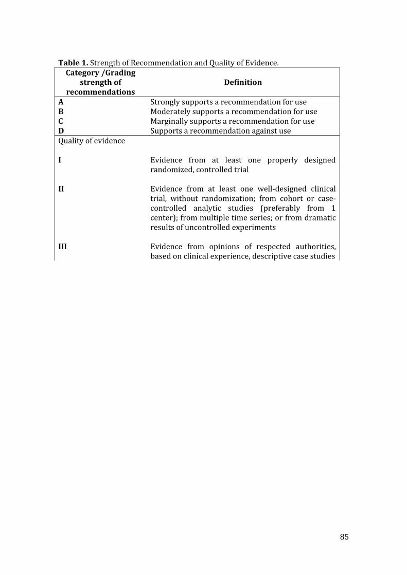
85
Table 1. Strength of Recommendation and Quality of Evidence.
Category /Grading Definition strength of
recommendations A Strongly supports a recommendation for use B Moderately supports a recommendation for use C Marginally supports a recommendation for use D Supports a recommendation against use Quality of evidence
I Evidence from at least one properly designed
randomized, controlled trial
II Evidence from at least one well-designed clinical trial, without randomization; from cohort or case-controlled analytic studies (preferably from 1 center); from multiple time series; or from dramatic results of uncontrolled experiments
III Evidence from opinions of respected authorities, based on clinical experience, descriptive case studies
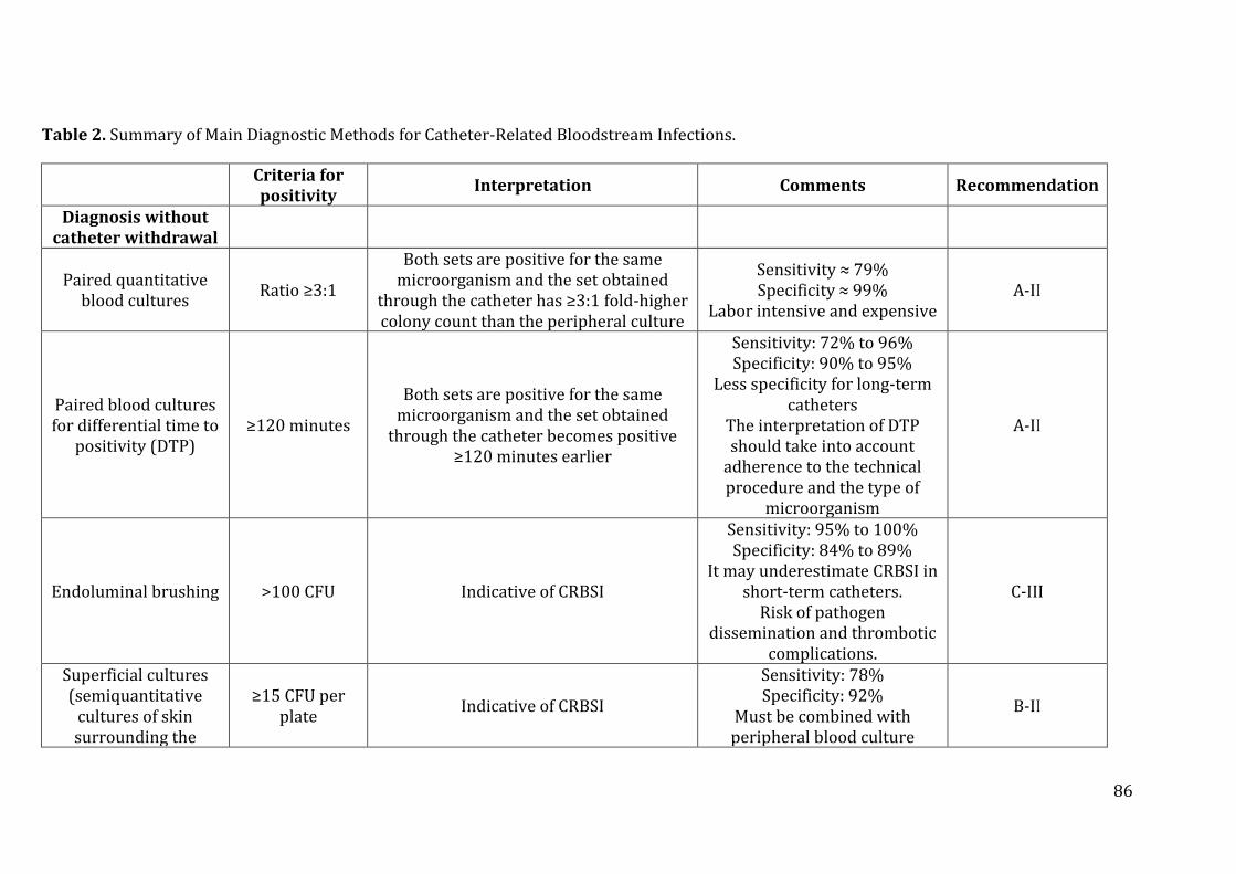
86
Table 2. Summary of Main Diagnostic Methods for Catheter-Related Bloodstream Infections.
Criteria for positivity
Interpretation Comments Recommendation
Diagnosis without catheter withdrawal
Paired quantitative blood cultures
Ratio ≥3:1
Both sets are positive for the same microorganism and the set obtained
through the catheter has ≥3:1 fold-higher colony count than the peripheral culture
Sensitivity ≈ 79% Specificity ≈ 99%
Labor intensive and expensive A-II
Paired blood cultures for differential time to
positivity (DTP) ≥120 minutes
Both sets are positive for the same microorganism and the set obtained
through the catheter becomes positive ≥120 minutes earlier
Sensitivity: 72% to 96% Specificity: 90% to 95%
Less specificity for long-term catheters
The interpretation of DTP should take into account
adherence to the technical procedure and the type of
microorganism
A-II
Endoluminal brushing >100 CFU Indicative of CRBSI
Sensitivity: 95% to 100% Specificity: 84% to 89%
It may underestimate CRBSI in short-term catheters.
Risk of pathogen dissemination and thrombotic
complications.
C-III
Superficial cultures (semiquantitative
cultures of skin surrounding the
≥15 CFU per plate
Indicative of CRBSI
Sensitivity: 78% Specificity: 92%
Must be combined with peripheral blood culture
B-II
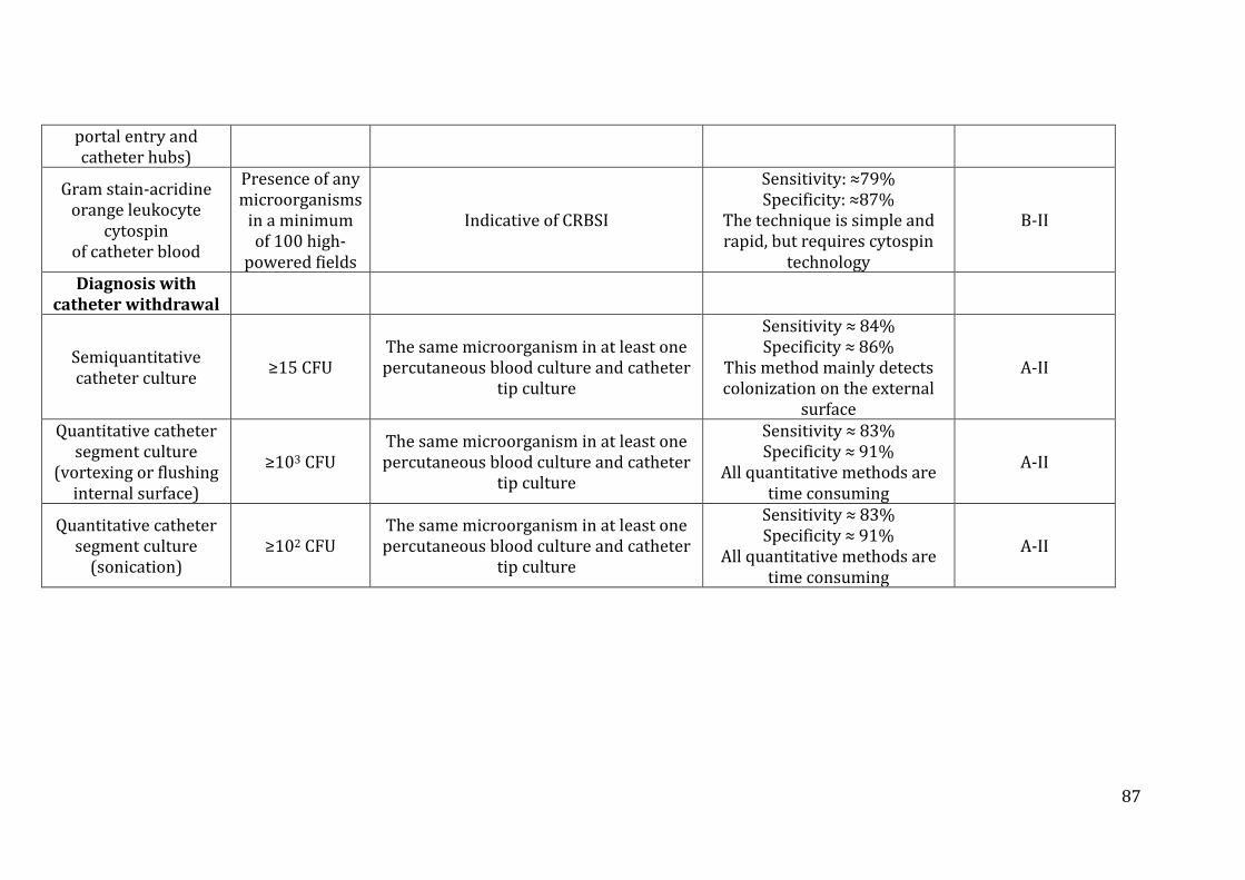
87
portal entry and catheter hubs)
Gram stain-acridine orange leukocyte
cytospin of catheter blood
Presence of any microorganisms
in a minimum of 100 high-
powered fields
Indicative of CRBSI
Sensitivity: ≈79% Specificity: ≈87%
The technique is simple and rapid, but requires cytospin
technology
B-II
Diagnosis with catheter withdrawal
Semiquantitative catheter culture
≥15 CFU The same microorganism in at least one percutaneous blood culture and catheter
tip culture
Sensitivity ≈ 84% Specificity ≈ 86%
This method mainly detects colonization on the external
surface
A-II
Quantitative catheter segment culture
(vortexing or flushing internal surface)
≥103 CFU The same microorganism in at least one percutaneous blood culture and catheter
tip culture
Sensitivity ≈ 83% Specificity ≈ 91%
All quantitative methods are time consuming
A-II
Quantitative catheter segment culture
(sonication) ≥102 CFU
The same microorganism in at least one percutaneous blood culture and catheter
tip culture
Sensitivity ≈ 83% Specificity ≈ 91%
All quantitative methods are time consuming
A-II
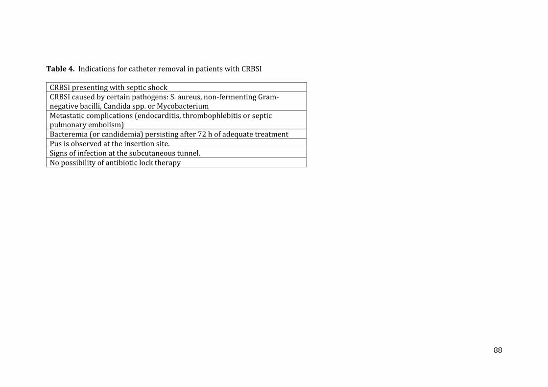
88
Table 4. Indications for catheter removal in patients with CRBSI
CRBSI presenting with septic shock CRBSI caused by certain pathogens: S. aureus, non-fermenting Gram-negative bacilli, Candida spp. or Mycobacterium Metastatic complications (endocarditis, thrombophlebitis or septic pulmonary embolism) Bacteremia (or candidemia) persisting after 72 h of adequate treatment Pus is observed at the insertion site. Signs of infection at the subcutaneous tunnel. No possibility of antibiotic lock therapy
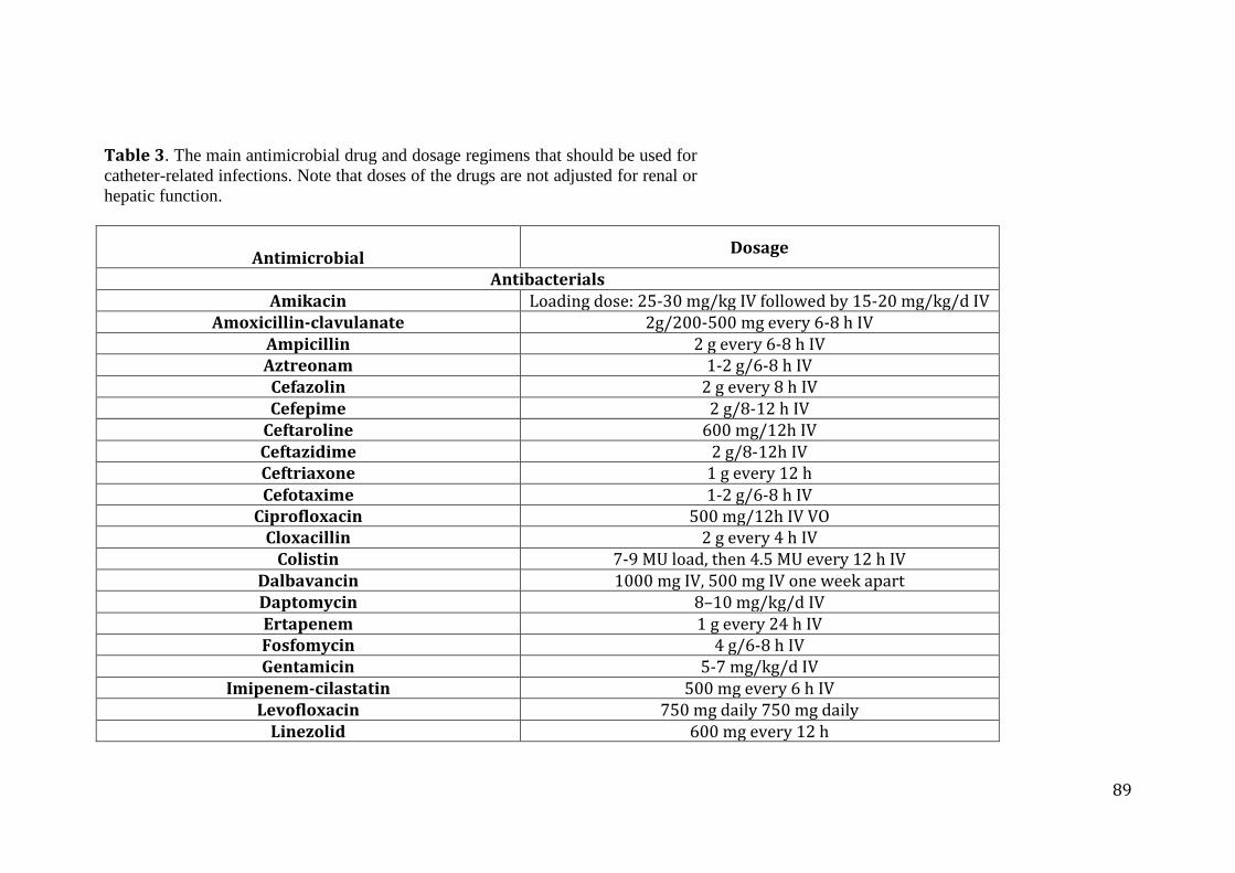
89
Table 3. The main antimicrobial drug and dosage regimens that should be used for
catheter-related infections. Note that doses of the drugs are not adjusted for renal or
hepatic function.
Antimicrobial
Dosage
Antibacterials Amikacin Loading dose: 25-30 mg/kg IV followed by 15-20 mg/kg/d IV
Amoxicillin-clavulanate 2g/200-500 mg every 6-8 h IV Ampicillin 2 g every 6-8 h IV Aztreonam 1-2 g/6-8 h IV Cefazolin 2 g every 8 h IV Cefepime 2 g/8-12 h IV
Ceftaroline 600 mg/12h IV Ceftazidime 2 g/8-12h IV Ceftriaxone 1 g every 12 h Cefotaxime 1-2 g/6-8 h IV
Ciprofloxacin 500 mg/12h IV VO Cloxacillin 2 g every 4 h IV
Colistin 7-9 MU load, then 4.5 MU every 12 h IV Dalbavancin 1000 mg IV, 500 mg IV one week apart Daptomycin 8–10 mg/kg/d IV Ertapenem 1 g every 24 h IV Fosfomycin 4 g/6-8 h IV Gentamicin 5-7 mg/kg/d IV
Imipenem-cilastatin 500 mg every 6 h IV Levofloxacin 750 mg daily 750 mg daily
Linezolid 600 mg every 12 h
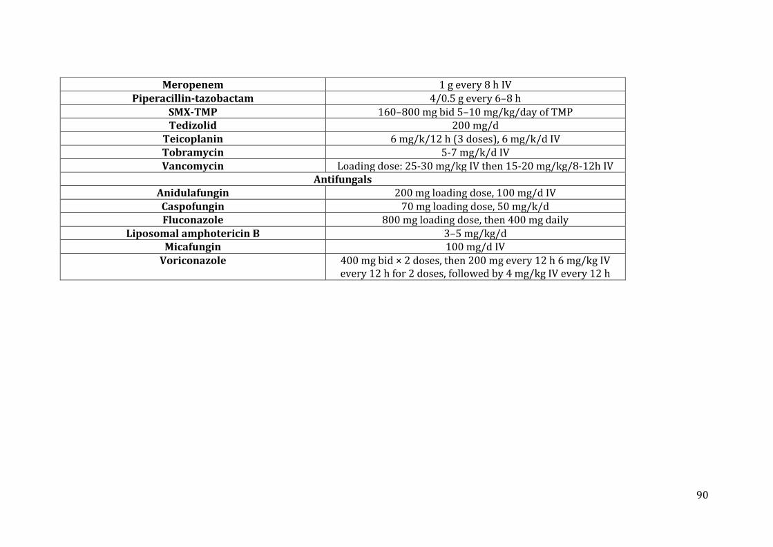
90
Meropenem 1 g every 8 h IV Piperacillin-tazobactam 4/0.5 g every 6–8 h
SMX-TMP 160–800 mg bid 5–10 mg/kg/day of TMP Tedizolid 200 mg/d
Teicoplanin 6 mg/k/12 h (3 doses), 6 mg/k/d IV Tobramycin 5-7 mg/k/d IV Vancomycin Loading dose: 25-30 mg/kg IV then 15-20 mg/kg/8-12h IV
Antifungals Anidulafungin 200 mg loading dose, 100 mg/d IV
Caspofungin 70 mg loading dose, 50 mg/k/d Fluconazole 800 mg loading dose, then 400 mg daily
Liposomal amphotericin B 3–5 mg/kg/d Micafungin 100 mg/d IV
Voriconazole 400 mg bid × 2 doses, then 200 mg every 12 h 6 mg/kg IV every 12 h for 2 doses, followed by 4 mg/kg IV every 12 h
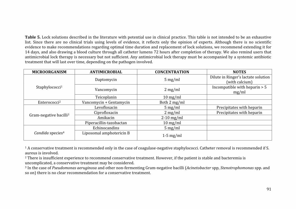
91
Table 5. Lock solutions described in the literature with potential use in clinical practice. This table is not intended to be an exhaustive list. Since there are no clinical trials using levels of evidence, it reflects only the opinion of experts. Although there is no scientific evidence to make recommendations regarding optimal time duration and replacement of lock solutions, we recommend extending it for 14 days, and also drawing a blood culture through all catheter lumens 72 hours after completion of therapy. We also remind users that antimicrobial lock therapy is necessary but not sufficient. Any antimicrobial lock therapy must be accompanied by a systemic antibiotic treatment that will last over time, depending on the pathogen involved.
MICROORGANISM ANTIMICROBIAL CONCENTRATION NOTES
Staphylococci1
Daptomycin 5 mg/ml Dilute in Ringer’s lactate solution
(with calcium)
Vancomycin 2 mg/ml Incompatible with heparin > 5
mg/ml Teicoplanin 10 mg/ml
Enterococci2 Vancomycin + Gentamycin Both 2 mg/ml
Gram-negative bacilli3
Levofloxacin 5 mg/ml Precipitates with heparin Ciprofloxacin 2 mg/ml Precipitates with heparin
Amikacin 2-10 mg/ml Piperacillin-tazobactan 10 mg/ml
Candida species4 Echinocandins 5 mg/ml
Liposomal amphotericin B
1-5 mg/ml
1 A conservative treatment is recommended only in the case of coagulase-negative staphylococci. Catheter removal is recommended if S. aureus is involved. 2 There is insufficient experience to recommend conservative treatment. However, if the patient is stable and bacteremia is uncomplicated, a conservative treatment may be considered. 3 In the case of Pseudomonas aeruginosa and other non-fermenting Gram-negative bacilli (Acinetobacter spp, Stenotrophomonas spp. and so on) there is no clear recommendation for a conservative treatment.
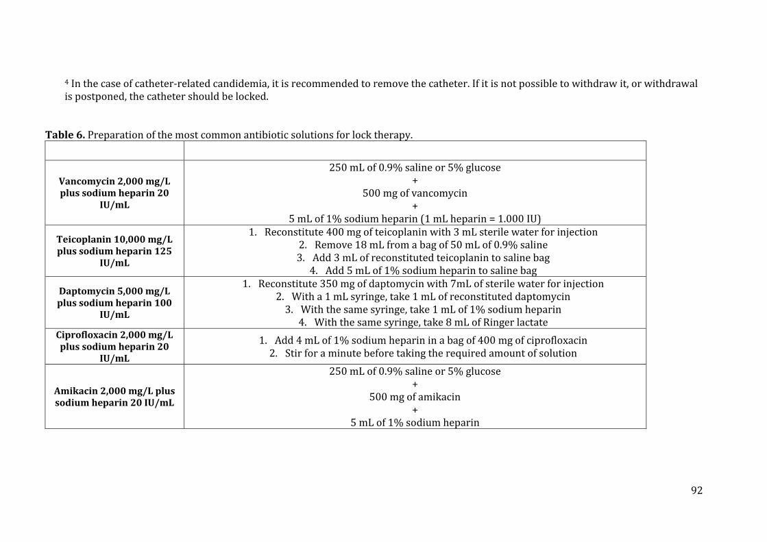
92
4 In the case of catheter-related candidemia, it is recommended to remove the catheter. If it is not possible to withdraw it, or withdrawal is postponed, the catheter should be locked.
Table 6. Preparation of the most common antibiotic solutions for lock therapy.
Vancomycin 2,000 mg/L plus sodium heparin 20
IU/mL
250 mL of 0.9% saline or 5% glucose +
500 mg of vancomycin +
5 mL of 1% sodium heparin (1 mL heparin = 1.000 IU)
Teicoplanin 10,000 mg/L plus sodium heparin 125
IU/mL
1. Reconstitute 400 mg of teicoplanin with 3 mL sterile water for injection 2. Remove 18 mL from a bag of 50 mL of 0.9% saline 3. Add 3 mL of reconstituted teicoplanin to saline bag
4. Add 5 mL of 1% sodium heparin to saline bag
Daptomycin 5,000 mg/L plus sodium heparin 100
IU/mL
1. Reconstitute 350 mg of daptomycin with 7mL of sterile water for injection 2. With a 1 mL syringe, take 1 mL of reconstituted daptomycin
3. With the same syringe, take 1 mL of 1% sodium heparin 4. With the same syringe, take 8 mL of Ringer lactate
Ciprofloxacin 2,000 mg/L plus sodium heparin 20
IU/mL
1. Add 4 mL of 1% sodium heparin in a bag of 400 mg of ciprofloxacin 2. Stir for a minute before taking the required amount of solution
Amikacin 2,000 mg/L plus sodium heparin 20 IU/mL
250 mL of 0.9% saline or 5% glucose +
500 mg of amikacin +
5 mL of 1% sodium heparin
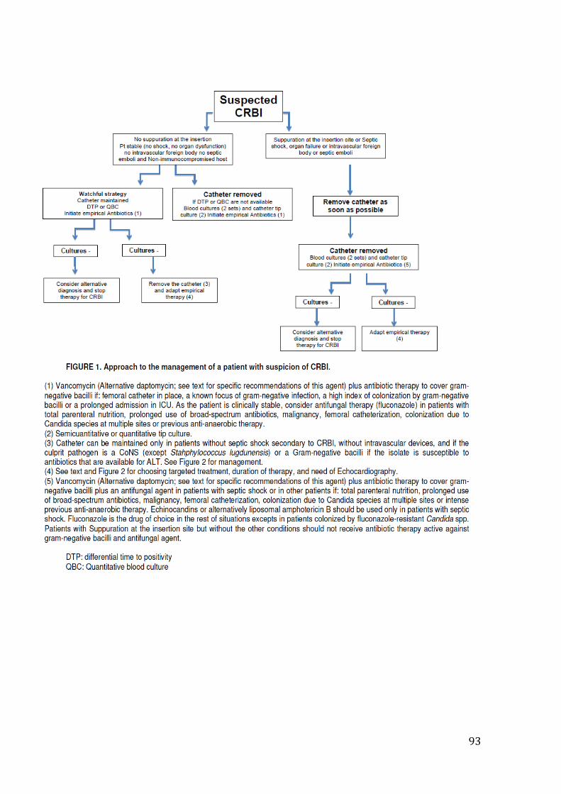
93
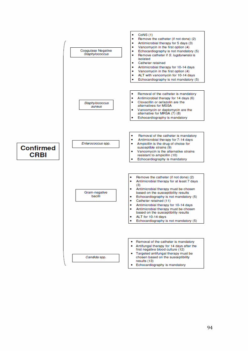
94
95