Embed Size (px)
Citation preview
Susan Liu HokiGillian Lieberman, MD
Role of Imaging in the Diagnosis and Mangagement of Acute Cerebral Infarction
Susan Liu Hoki, Harvard Medical School Year IIIGillian Lieberman, MD
July 2002
2
Susan Liu HokiGillian Lieberman, MD
Goals
• Understand how imaging studies help in stroke diagnosis and management through examining findings in patients at different stages of acute ischemic stroke
• Know the advantages of different tests in stroke imaging
3
Susan Liu HokiGillian Lieberman, MD
Background: Stroke
• Stroke is a lay term meaning a condition due to vascular lesions of the brain caused by hemorrahage, embolism, thrombosis, or rupturing aneurysm
• Primarily diagnosed clinically and confirmed and followed through imaging tests
4
Susan Liu HokiGillian Lieberman, MD
Background: Stroke TypesMost common stroke etiologies:
1) Cerebral Infarction 80%2) Primary Intracranial Hemorrhage 15%3) Nontraumatic subarachnoid hemorrhage 5%
* FOCUS: Acute Cerebral Infarction
5
Susan Liu HokiGillian Lieberman, MD
Menu of Radiological Tests• Cerebral Angiogram• CT: w/ or w/o contrast
CT angiogram (CTA)• MR: w/ or w/o contrast
T1 or T2 weighted (T1WI, T2WI)FLAIRDiffusion weighted image (DWI) SusceptibilityMR angiogram

6
Susan Liu HokiGillian Lieberman, MD
Cerebral Angiogram• Gold standard in the
past• Outdated and
replaced by MRI/MRA
• High risk of producing further thrombus formation in brain and causing renal failure
Courtesy of Dr. Steve Reddy, BIDMC
7
Susan Liu HokiGillian Lieberman, MD
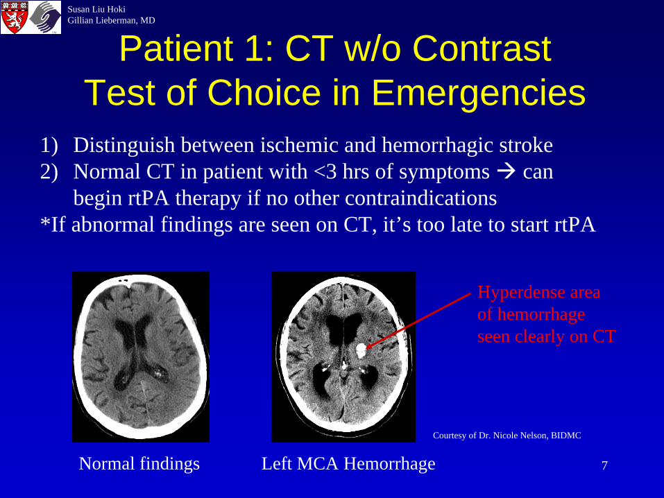
Patient 1: CT w/o Contrast Test of Choice in Emergencies
Normal findings Left MCA Hemorrhage
Courtesy of Dr. Nicole Nelson, BIDMC
Hyperdense area of hemorrhage seen clearly on CT
1) Distinguish between ischemic and hemorrhagic stroke2) Normal CT in patient with <3 hrs of symptoms can
begin rtPA therapy if no other contraindications*If abnormal findings are seen on CT, it’s too late to start rtPA
8
Susan Liu HokiGillian Lieberman, MD
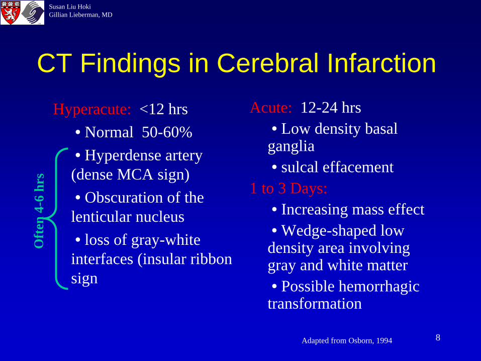
CT Findings in Cerebral InfarctionHyperacute: <12 hrs
• Normal 50-60%• Hyperdense artery (dense MCA sign)• Obscuration of the lenticular nucleus• loss of gray-white interfaces (insular ribbon sign
Acute: 12-24 hrs• Low density basal ganglia• sulcal effacement
1 to 3 Days:• Increasing mass effect• Wedge-shaped low density area involving gray and white matter• Possible hemorrhagic transformation
Adapted from Osborn, 1994
Of t
en 4
- 6 h
rs
9
Susan Liu HokiGillian Lieberman, MD
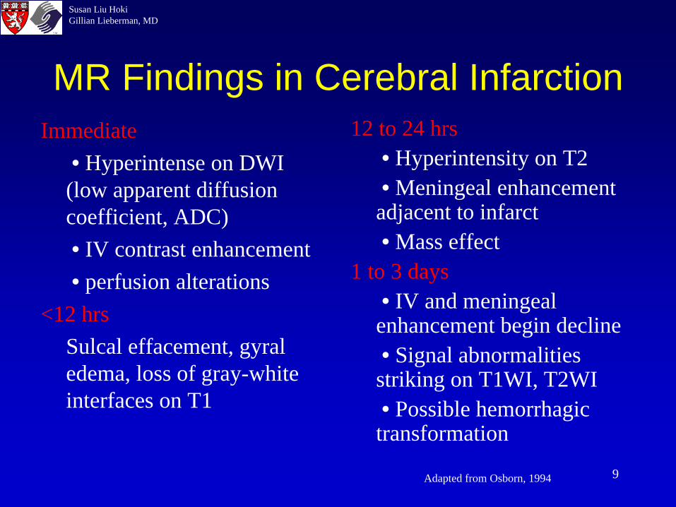
MR Findings in Cerebral InfarctionImmediate
• Hyperintense on DWI (low apparent diffusion coefficient, ADC)• IV contrast enhancement• perfusion alterations
<12 hrsSulcal effacement, gyral edema, loss of gray-white interfaces on T1
12 to 24 hrs• Hyperintensity on T2• Meningeal enhancement adjacent to infarct• Mass effect
1 to 3 days• IV and meningeal enhancement begin decline• Signal abnormalities striking on T1WI, T2WI• Possible hemorrhagic transformation
Adapted from Osborn, 1994
10
Susan Liu HokiGillian Lieberman, MD
Treatment and Further Imaging• After the initial CT scan, patient is treated with
1) tPA2) anti-coagulants3) antiplatelet aggreating agents
• Further studies help in evaluating response to treatment and extent of brain damage not detected on the initial CT
• The following images presented will be of CT, CTA, MR, and MRA studies, which are most commonly used at large hospitals
11
Susan Liu HokiGillian Lieberman, MD
The Ideal Early Patient
• Comes into the ER presenting with a recent onset of stroke symptoms
• CT scan shows no hemorrhage and no changes seen in ischemia
• Patient has no other contraindications for rtPA therapy rtPA is administered
• Follow progress with sensitive MR studies
12
Susan Liu HokiGillian Lieberman, MD
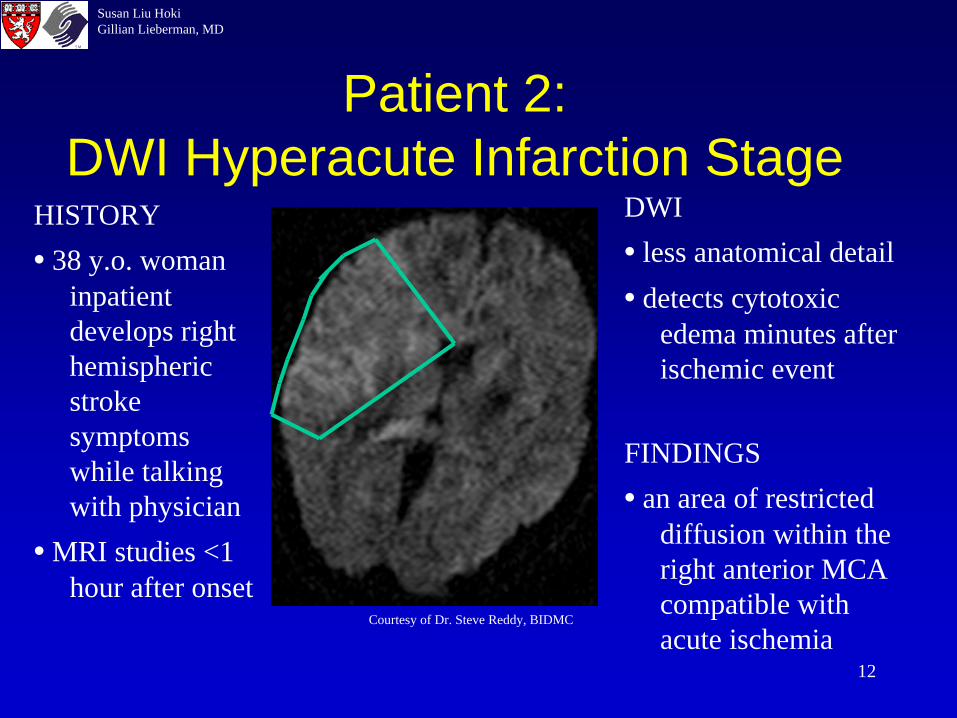
Patient 2: DWI Hyperacute Infarction Stage
HISTORY• 38 y.o. woman
inpatient develops right hemispheric stroke symptoms while talking with physician
• MRI studies <1 hour after onset
DWI• less anatomical detail• detects cytotoxic
edema minutes after ischemic event
FINDINGS• an area of restricted
diffusion within the right anterior MCA compatible with acute ischemia
Courtesy of Dr. Steve Reddy, BIDMC
13
Susan Liu HokiGillian Lieberman, MD
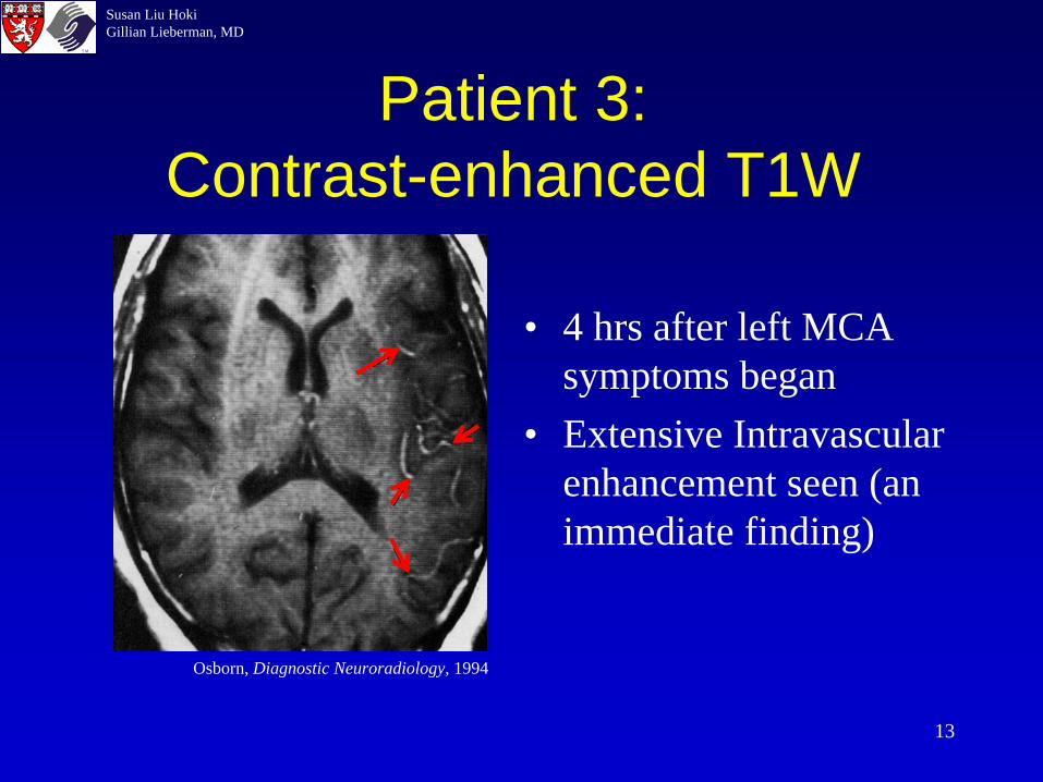
Patient 3: Contrast-enhanced T1W
• 4 hrs after left MCA symptoms began
• Extensive Intravascular enhancement seen (an immediate finding)
Osborn, Diagnostic Neuroradiology, 1994
14
Susan Liu HokiGillian Lieberman, MD
Our Patient: Ms. JB
79 y.o. brought by her son who found her sitting on a chair in her bedroom, unresponsive.
• CC: unable to move left arm/leg• no known cardiovascular disease• PMH: TB exposure s/p Rx, sinusitis, arthritis• DX: Acute Stroke
15
Susan Liu HokiGillian Lieberman, MD
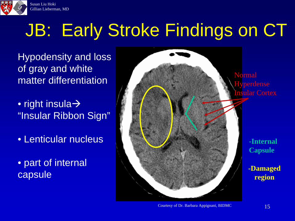
JB: Early Stroke Findings on CT
Normal Hyperdense Insular Cortex
Courtesy of Dr. Barbara Appignani, BIDMC
Hypodensity and loss of gray and white matter differentiation
• right insula“Insular Ribbon Sign”
• Lenticular nucleus
• part of internal capsule
-Internal Capsule
-Damaged region
16
Susan Liu HokiGillian Lieberman, MD
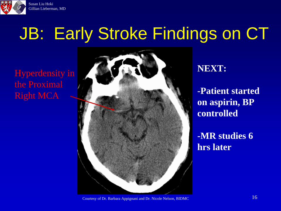
Hyperdensity in the Proximal Right MCA
Courtesy of Dr. Barbara Appignani and Dr. Nicole Nelson, BIDMC
JB: Early Stroke Findings on CT
NEXT:
-Patient started on aspirin, BP controlled
-MR studies 6 hrs later
17
Susan Liu HokiGillian Lieberman, MD
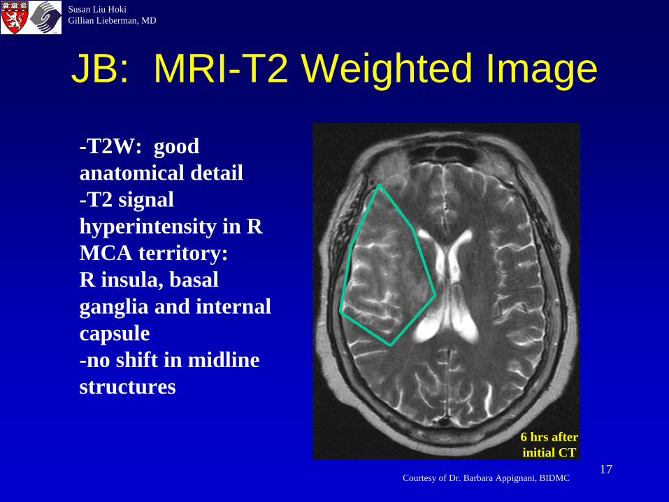
JB: MRI-T2 Weighted Image
Courtesy of Dr. Barbara Appignani, BIDMC
-T2W: good anatomical detail-T2 signal hyperintensity in R MCA territory:R insula, basal ganglia and internal capsule-no shift in midline structures
6 hrs after initial CT
18
Susan Liu HokiGillian Lieberman, MD
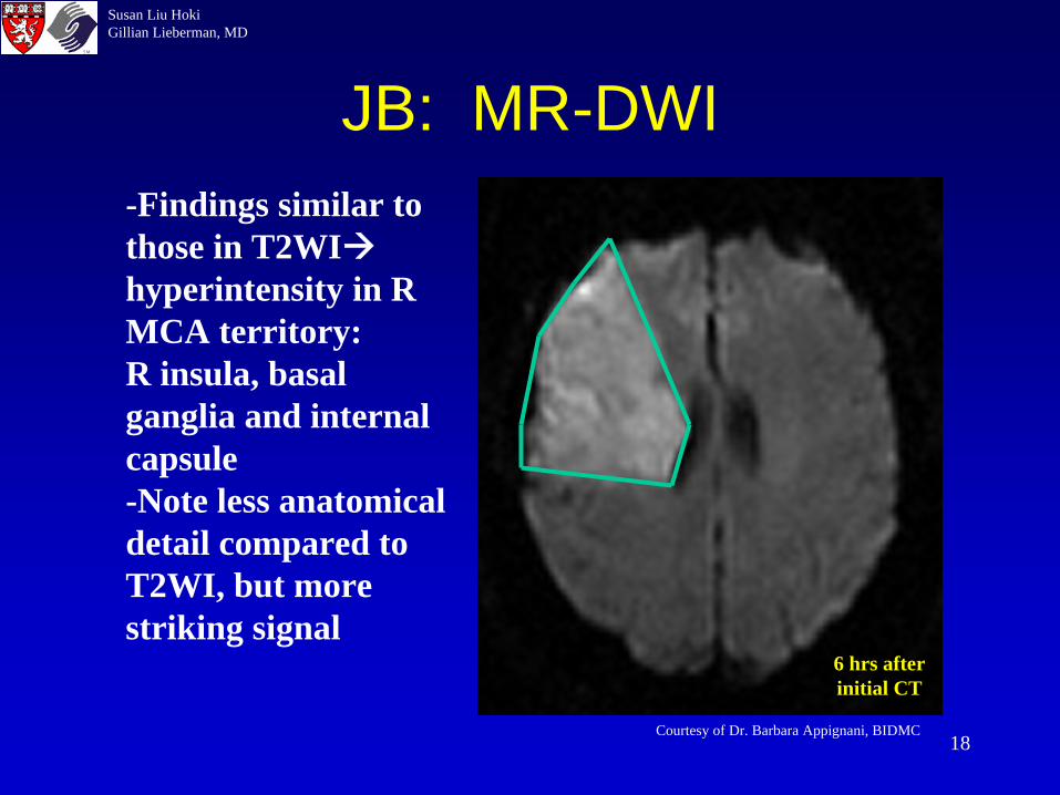
JB: MR-DWI
Courtesy of Dr. Barbara Appignani, BIDMC
-Findings similar to those in T2WIhyperintensity in R MCA territory:R insula, basal ganglia and internal capsule-Note less anatomical detail compared to T2WI, but more striking signal
6 hrs after initial CT

19
Susan Liu HokiGillian Lieberman, MD
JB: MRA 3-D Reconstruction
Courtesy of Dr. Barbara Appignani, BIDMC
-KEY FINDING:Absence of flow in R MCA branches
-Study pinpoints artery location with problem in flow through in R MCA territory
Circle of WillisInternal Carotid ArteryAnterior Cerebral ArteryMiddle Cerebral Artery (MCA)
Absence of flow
C
20
Susan Liu HokiGillian Lieberman, MD
Patient 3: Ms. FL• 86 y.o. with a history of atrial fibrillation
who was at home walking towards her husband when she collapsed and became somulent
• Unable to speak and had difficulty moving her right side clinical dx of acute stroke
• CT scan w/o contrast 2 hrs after onset was normal Timing appropriate for rtPA
• rtPA contraindicated since INR>1.5
21
Susan Liu HokiGillian Lieberman, MD
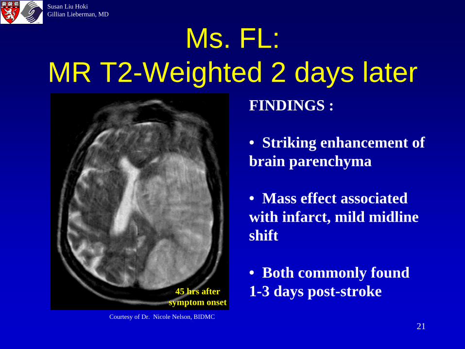
Ms. FL: MR T2-Weighted 2 days later
Courtesy of Dr. Nicole Nelson, BIDMC
FINDINGS :
• Striking enhancement of brain parenchyma
• Mass effect associated with infarct, mild midline shift
• Both commonly found1-3 days post-stroke45 hrs after
symptom onset
22
Susan Liu HokiGillian Lieberman, MD
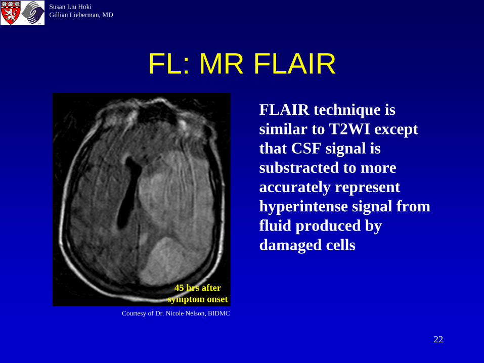
FL: MR FLAIR
Courtesy of Dr. Nicole Nelson, BIDMC
FLAIR technique is similar to T2WI except that CSF signal is substracted to more accurately represent hyperintense signal from fluid produced by damaged cells
45 hrs after symptom onset

23
Susan Liu HokiGillian Lieberman, MD
CT Findings in Cerebral InfarctionHyperacute: <12 hrs
• Normal 50-60%• Hyperdense artery (dense MCA sign)• Obscuration of the lenticular nucleus• loss of gray-white interfaces (insular ribbon sign
Acute: 12-24 hrs• Low density basal ganglia• sulcal effacement
1 to 3 Days:• Increasing mass effect• Wedge-shaped low density area involving gray and white matter• Possible hemorrhagic transformation
Adapted from Osborn, Diagnostic Neuroradiology, 1994
Of t
en 4
- 6 h
rs
24
Susan Liu HokiGillian Lieberman, MD
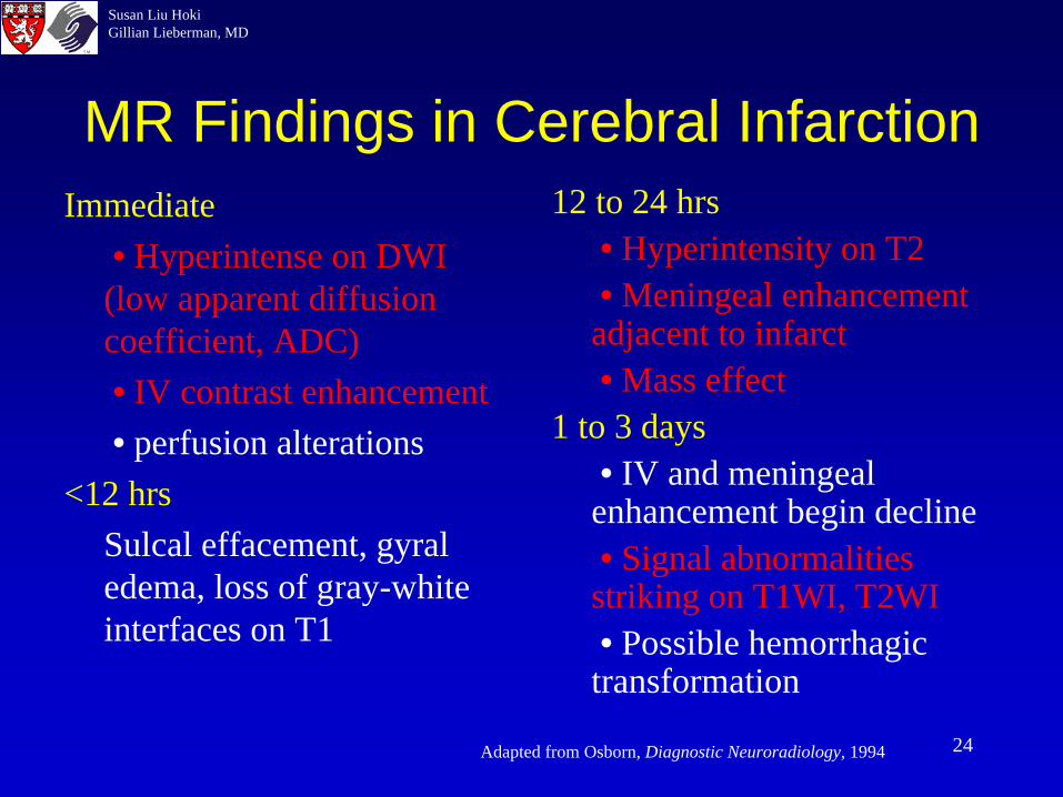
MR Findings in Cerebral InfarctionImmediate
• Hyperintense on DWI (low apparent diffusion coefficient, ADC)• IV contrast enhancement• perfusion alterations
<12 hrsSulcal effacement, gyral edema, loss of gray-white interfaces on T1
12 to 24 hrs• Hyperintensity on T2• Meningeal enhancement adjacent to infarct• Mass effect
1 to 3 days• IV and meningeal enhancement begin decline• Signal abnormalities striking on T1WI, T2WI• Possible hemorrhagic transformation
Adapted from Osborn, Diagnostic Neuroradiology, 1994
25
Susan Liu HokiGillian Lieberman, MD
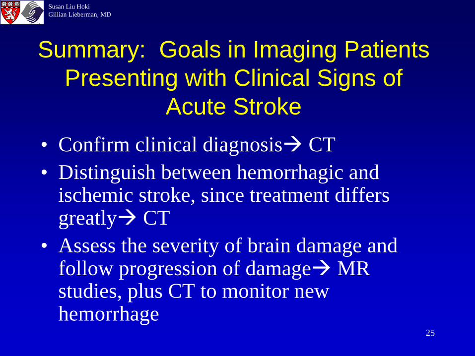
Summary: Goals in Imaging Patients Presenting with Clinical Signs of
Acute Stroke• Confirm clinical diagnosis CT• Distinguish between hemorrhagic and
ischemic stroke, since treatment differs greatly CT
• Assess the severity of brain damage and follow progression of damage MR studies, plus CT to monitor new hemorrhage
26
Susan Liu HokiGillian Lieberman, MD
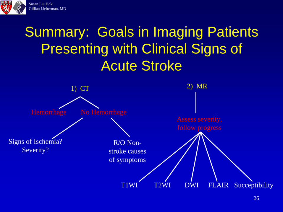
Summary: Goals in Imaging Patients Presenting with Clinical Signs of
Acute Stroke2) MR1) CT
Hemorrhage No Hemorrhage
Signs of Ischemia?Severity?
R/O Non- stroke causes of symptoms
Assess severity, follow progress
T1WI T2WI DWI FLAIR Succeptibility
27
Susan Liu HokiGillian Lieberman, MD
Summary• Neuroradiology is important in confirming
the diagnosis of acute cerebral infarction and monitoring progression
• Advances in MR techniques make early diagnosis and assessment possible, which are necessary in thrombolytic therapy to prevent irreversible brain damage
28
Susan Liu HokiGillian Lieberman, MD
References• Adams Jr., Harold P., Gregory J. del Zoppo, and Rüdiger von
Kummer. Management of Stroke: A Practical Guide for the Prevention, Evaluation, and Treatment of Acute Stroke, 2nd ed., West Islip, NY: Professional Communications, Inc., 2002.
• Castillo, Mauricio. Neuroradiology Companion, 2nd ed., Philadelphia: Lippincott-Raven Publishers, 1999.
• Nolte, John. The Human Brain, 4th ed., St. Louis: Mosby—Year Book, Inc., 1999./SLIDE #11
• Osborn, Anne G. Diagnostic Neuroradiology. St. Louis: Mosby— Year Book, Inc., 1994./SLIDE # 8, 9, 13

29
Susan Liu HokiGillian Lieberman, MD
Acknowledgements
• Barbara Appignani, MD• Nicole Nelson, MD• Steve Reddy, MD
• Gillian Lieberman, MD• Pamela Lepkowski• Larry Barbaras and
Cara Lyn D’amour

Susan Liu HokiGillian Lieberman, MD
The End