Embed Size (px)
Citation preview
THAI AGRICULTURAL STANDARD
TAS 10351-2007
DIAGNOSIS OF AMERICAN FOULBROOD IN BEE
National Bureau of Agricultural Commodity and Food Standards
Ministry of Agriculture and Cooperatives
ICS 11.220 ISBN 978-974-403-566-0
UNOFFICAL TRANSLATION
THAI AGRICULTURAL STANDARD
TAS 10351-2007
DIAGNOSIS OF AMERICAN FOULBROOD IN BEE
National Bureau of Agricultural Commodity and Food Standards
Ministry of Agriculture and Cooperatives
50 Phaholyothin Road, Ladyao, Chatuchak, Bangkok 10900 Telephone (662) 561 2277 www.acfs.go.th
Published in the Royal Gazette Vol.125 Special Section 3 D,
dated 4 January B.E.2551 (2008)
Ad hoc Sub-Committee on the Elaboration of Standards for Diagnosis of Bee Diseases 1. Director General of the Department of Livestock Development Chairperson Mrs.Laddawalaya Ratananakorn, Representative of Director General 2. Representative of the Department of Livestock Development Mrs.Laddawalaya Ratananakorn
3. Representative of the Department of Agricultural Extension Mr.Udom Jeeraswetkul 4. Representative of the National Institute of Animal Health, Department of Livestock Development Mrs. Wantanee Neramitmansook Mrs. Montakan Vongpakorn (alternate) 5. Representative of the Faculty of Agriculture, Kasetsart University Assoc. Prof. Savitee Malaipan 6. Representative of the Faculty of Science, Chulalongkorn University Prof. Siriwat Wongsiri 7. Representative of the Faculty of Science, Chiang Mai University Asst. Prof. Panuwan Chantawannakul 8. Representative of the Faculty of Science,
Naresuan University Mr.Somluck Vongsamanode 9. Representative of the Faculty of Science, Ramkhamhaeng University Asst. Prof. Somnuk Bunkoet 10. Representative of the Northern beekeeper Association of Thailand Mr. Sirisak Toewtrakul 11. Representative of the National Bureau of Agricultural Commodity
and Food Standards Mrs. Oratai Silapanapaporn Mrs. Nantana Posanacharoen (alternate) Ms.Yupa Laojindapun (alternate) 12. Experts Mr. Yongyoot Waikakul Asst. Prof. Anchalee Sawatthum
(2)
13. Representatives of the Office of Commodity and System Standards, Secretary National Bureau of Agricultural Commodity Ms.Darunee Tuntasuvan Mr. Songkhla Chulakasian 14. Representatives of the Office of Commodity and System Standards, Assistant Secretary National Bureau of Agricultural Commodity Ms. Mintra Lukkana Ms. Sakranmanee Krajangwong
(3)
Thai Agricultural Standard (TAS) on Diagnosis of American Foulbrood in Bee was established for certifying the bee commodity and products that are free from American foulbrood disease and to be used as standard manual in diagnosis of American foulbrood disease in laboratory. The laboratory diagnostic techniques are including pathological method, microscopic examination, spore detection, Holst milk test, bacteriological method, immunological method and polymerase chain reaction. By complying with the measure, this will promote production of safe and quality bees and bee products for human consumption both nationally and internationally. It also will notify the status of American foulbrood disease of the country to the World Organization for Animal Health (OIE). The establishment of this standard is based on the information of the following document: OIE. 2004. American foulbrood. In Manual of Diagnostic Tests and Vaccines for Terrestrial Animals. Chapter 2.9.2. World Organization for Animal Health., Paris, France.
Remark:
The standard title has been revised from “Thai Agricultural Commodity and Food Standard (TACFS)” to “Thai Agricultural Standard (TAS)” in accordance with the enforcement of The Agricultural Standards Act B.E. 2551 (2008).
NOTIFICATION OF THE NATIONAL COMMITTEE ON AGRICULTURAL COMMODITY AND FOOD STANDARDS
SUBJECT: THAI AGRICULTURAL COMMODITY AND FOOD STANDARD: DIAGNOSIS OF AMERICAN FOULBROOD IN BEE
B.E. 2550 (2007) ___________________________________________________________________________
The resolution of the 2/2550 session of the National Committee on Agricultural Commodity and Food Standards dated 28 August B.E.2550 (2007) endorsed the Thai Agricultural Commodity and Food Standard entitled Diagnosis of American Foulbrood in Bee. This standard would be of benefits for quality improvement, facilitating trade and protecting consumers.
By virtue of the Cabinet Resolution on Appointment and Authorization of the National Committee on Agricultural Commodity and Food Standards dated 3 April B.E.2550 (2007), the Notification on Thai Agricultural Commodity and Food Standard entitled Diagnosis of American Foulbrood in Bee is hereby issued as voluntary standard, details of which are attached herewith.
Notified on 26 September B.E.2550 (2007)
Prof. Thira Sutabutra
Minister of Agriculture and Cooperatives Chairperson of the National Committee on Agricultural Commodity and Food Standards
TAS 10351-2007
THAI AGRICULTURAL STANDARD
DIAGNOSIS OF AMERICAN FOULBROOD IN BEE
1. SCOPE
Thai Agricultural Standard (TAS) on the Diagnosis of American Foulbrood in Bee determines the diagnostic techniques for American foulbrood disease (AFB) in laboratory by using pathological method, microscopic examination, spore detection, Holst milk test, bacteriological method, immunological method, and polymerase chain reaction (PCR).
2. DEFINITIONS
For the purpose of this standard:
2.1 Honey bee means farm bee cultivated for their honey production. In Thailand there are two species, Asian honey bee (Apis cerana) and European or Western honey bee (Apis mellifera). However, under this standard it covers only the European or Western honey bee.
2.2 American foulbrood means the brood disease which is caused by the bacteria Paenibacillus larvae larvae (P. l. larvae).
2.3 Bee brood means eggs, larvae and pupae in bee comb cells.
2.4 Bee wax means a substance that worker bees produce from wax glands to construct bee comb.
2.5 Cell means the hexagons that are built up to bee hive.
2.6 Bee colony means a honey bee colony consisting of a queen bee, worker bees, drone bees, brood comb and food comb.
2.7 Honey means sweet liquid that honey bees produce from nectar or any part of plants or honey dew from insect of Hemoptera Family and stored in bee comb.
2.8 Bee pollen means mixture of flower pollen which honey bee collected for larval food.
2.9 Royal jelly or bee milk means a whitish creamy liquid substance secreted by worker bees and serves as food for all young larvae and as the only food for larvae that will develop into queen bees. This does not include the dehydrated royal jelly.
2.10 Bee farm means the place that keep bees in order to harvest honey and other bee products.
2.11 Screening test or rapid test means the primary test which is rapid and cheap.
2.12 Confirmation test means the laboratory test designed to confirm the positives from screening test. This test has acceptably high specificity and sensitivity.
TAS 10351-2007 2
2.13 Sensitivity means the capacity of the test that could correctly identify the positive results from the infected population.
2.14 Specificity means the capacity of the test that gives negative results from the uninfected population.
3. DIAGNOSTIC TECHNIQUES
Diagnosis of AFB is done by laboratory diagnostic techniques such as pathological method, bacteriological method, polymerase chain reaction (PCR). The sensitivity and specificity among the mentioned approaches are different.
When the results of chosen laboratory tests are given, the Veterinary authority shall interpret the results with epizootology, pathology and clinical signs as described in ANNEX A in order to treat and control the disease effectively.
3.1 SAMPLE COLLECTION
3.1.1 The sample of comb shall be collected for approximately 20 cm2 in size containing as much of the dead or discoloured brood as possible. Little or no honey shall be present in the sample. The sample shall be placed in closed container and sent to the laboratory as soon as possible.
3.1.2 If a portion of comb cannot be sent, the probe or wooden stick used to examine cell contents may have enough material on it for any test. This sample can be sent to the laboratory for the diagnosis.
3.1.3 If samples are bee products such as honey, royal jelly, bee pollen, and bee wax, they shall be collected about 50 g in cleaned container and sent to the laboratory as soon as possible.
3.1.4 The bacterial culture shall be kept in slants at 4 ºC which may be kept up to 6 months or in lyophilized form which the cultured medium is composed of sucrose (10%) yeast extract (5%) in 0.1 M KH2PO4, pH 6.6. The dried culture shall be kept at 4 ºC which may be kept for several years.
3.1.5 The sample shall be legibly labelled and the information such as sample type, bee farm’s name, hive No., location, date and collector shall be recorded.
3.2 SCREENING TEST OR RAPID TEST
3.2.1 Pathological method (Clinical test)
The principle of this test is to observe any clinical symptoms from bee cell. If bee larvae or pupae are infected with AFB, the larval or pupal color changes to creamy brown and then to a dark brown. The diseased brood eventually dries out to form characteristic brittle scales that adhere tightly to the lower sides of the cell. Sometimes, the larval remains are difficult to locate. The infected dead larvae could be conveniently located by using ultra-violet or near ultra-violet wavelength. Exposure between 310 to 400 nm will cause any infected dead larvae or AFB scale material to fluoresce. Careful attention shall be made when using this technique as both honey and pollen will also fluoresce.
TAS 10351-2007 3
3.2.2 Microscopic examination
The microscopic technique is to observe the morphology of P. l. larvae to distinguish the AFB from other brood diseases. The method has low specificity so that it is commonly used only as a rapid test for preliminary check.
This technique is outlined as follows:
(1) About two drops of water is mixed with the sample.
(2) The suspension is transferred by loop and smeared on a glass slide as thin film.
(3) The slide is air-dried and fixed by heat.
(4) The slide is stained by carbol fuchsin or a suitable spore stain for 30 s.
(5) Any excess stain is washed off with water and the slide is gently dried.
(6) Immersion oil is dropped on the slide and then the bacteria are examined by light microscope at magnification of 1 000 X.
(7) If the sample is AFB, the spore of P. l. larvae is ellipsoidal and about 1.3 m x 0.6 m. The vegetative form of the bacteria could be also observed during first ten days of infection. It is violet and long rod after Gram staining.
3.2.3 Detection of spores in honey
(1) Honey sample (25 ml) is diluted with 25 ml of sterile water, heated at 45-50 º C, and shaken to distribute any spores that may be present.
(2) The diluted honey is transferred into 44 mm width dialysis tube that has been tied at one end. The open end is tied after filling. The tubes are submerged in running water for 18 h or in a water bath with three water changes over the same time period. After dialysis, the contents are centrifuged at 2 000 g for 20 min.
(3) Honey samples can be prepared without the dialysis step (2). The honey sample is centrifuged at 3 000 g for 30 min.
(4) The supernatant liquid is discarded and the deposit is resuspended in sterile water to make up a final volume of 10 ml. The suspension is heated at 80°C for 10 min to kill nonspore-forming bacteria and centrifuged at 2 000 g for 20 min
(5) The sediment is plated on to solid media such as Brain heart infusion agar or direct streaking on the a suitable agar plate which supplemented with 3 µg/ml nalidixic acid to inhibit the growth of P. alvei.
(6) If the culture is P. l. larvae, the bacterial colony is opaque white to cream, about 1 mm to 3 mm after 2 to 4 days cultivation at 34-37 ºC. If the cells of P. l. larvae are stained and inspected under light microscope, the spore and vegetative cells of P. l. larvae could be observed as violet and long rod.
3.2.4. Holst milk test
The Holst milk test use is on the fact that a high level of protease is produced by sporulating P. l. larvae. The test is performed by suspending a disease larvae or dried dead larvae (scale) in a test tube containing 1-4 ml of 1% skim milk in water. The tube is incubated at 37 ºC. If P. l. larvae is present, the suspension is clear in 10-20 min.
TAS 10351-2007 4
3.3 CONFIRMATION TEST
3.3.1. Bacteriological method
This method is based on the detection of pathogenic bacteria in bee hive which shall be done by bacterial cultivation and isolation steps in order to obtain pure culture. The media used are blood agar, J-medium or any suitable media. The bacteria identification is further confirmed by a set of biochemical tests to confirm the presence of P. l. larvae.
3.3.1.1. Culture techniques
Sample preparation
(1) To culture P. l. larvae, spore suspensions are prepared by mixing diseased material sample in 9 ml of sterile water or phosphate buffer saline (PBS) or suitable liquid media in test tubes. The suspension is heated at 80°C for 10 min to kill non-sporulating bacteria.
(2) If the samples are honey and royal jelly, the sample shall be centrifuged to remove access water and to enhance the bacterial spore concentration. In case of bee wax, the sample shall be extracted in chloroform before cultivation.
(3) If the sample is pollen, the sample shall be mixed with sterile water and removed the pollen by filtration before cultivation step.
(4) A sterile cotton swab is used to transfer a portion of the suspension onto the surface of Petri dishes containing solid medium, which are then incubated for 2-4 days at 34-37°C.
(5) Individual colonies are small and opaque. The bacteria are further identified by biochemical tests.
3.3.1.2 Biochemical tests
P. l. larvae can be identified by its biochemical characteristics. The bacteria could reduce nitrate to nitrite, produce acid from glucose and trehalose, and could digest gelatin and casein. They are unable to digest starch and utilize citrate. The bacteria also are facultative anaerobe and grow in liquid media. About two small colonies from suspected sample (3.3.1.1) shall be tested as follows:
3.3.1.2.1 Nitrate reduction test
(1) Brain Heart Infusion with Thiamine (BHIT) (Appendix C), containing potassium nitrate (1-2 mg/l of medium) is prepared.
(2) The suspected culture is inoculated in media (1) and incubated at 37 ºC until the bacterial growth is observed.
(3) A few drops of sulphanilic acid-alpha-naphthyl reagent are added to the culture media.
(4) The resulting liquid medium produces a red colour if any nitrate has been reduced to nitrite. This test shall be used for confirmation and shall not be employed alone.
3.3.1.2.2 Catalase test
(1) A drop of 3% hydrogen peroxide is placed on an actively growing culture on solid medium.
TAS 10351-2007 5
(2) Most aerobic bacteria break down the peroxide to water and oxygen, producing bubbly foam, but P. l. larvae is almost always negative for this reaction.
3.3.1.2.3 Production of acid from carbohydrates
(1) J-broth (ANNEX C), which glucose is omitted, is prepared. The additional 0.5 % of the test substrate is separately sterilized in aqueous solution. The carbohydrates used are L(+) –arabinose, l D(+)-glucose, D(+) xylose, and D(+)-trehalose.
(2) The suspect bacteria are inoculated in the media and grown for 14 days at 37 ºC.
(3) The cultures are tested by removing 1 ml to a spot plate, mixing sample with a drop of 0.04% alcoholic bromocresol purple.
(4) Result interpretation is done by observing the colour of the indicator. P. l. larvae produces acid aerobically from glucose and trehalose. No acid is produced from arabinose and xylose.
3.3.1.2.4 Hydrolysis of starch
(1) Starch hydrolysis is assayed by using J-medium which glucose is omitted. Additional starch (1%) is substituted for the glucose in J-medium. The medium is sterilized at 121 ºC for 15 min and poured into Petri dish when it is cooled down to 45 ºC.
(2) The suspected culture is inoculated on plates and incubated at 37ºC for 5-10 days.
(3) The plates are flooded with iodine solution at room temperature for 15-30 min.
(4) If the culture is P. l. larvae, no clearing zone will be observed.
3.3.1.2.5 Hydrolysis of casein
(1) The medium for the test is added with 10% skim milk and 3 % agar. The medium is sterilized at 121 ºC for 15 min and poured into Petri dish when it is cooled down to 45 ºC.
(2) The suspected culture aged 24 h is inoculated on the medium (1) and incubated at 37 ºC for 7 days.
(3) If the suspected culture is P. l. larvae, the clear zone around the growing culture will be observed due to the casein hydrolysis.
3.3.1.2.6 Citrate utilization
(1) J-media which is glucose omitted is added with 2 g of sodium citrate. The medium is sterilized.
(2) The suspected culture aged 3-4 days is inoculated on the medium (1) and incubated at 37 ºC for 14-21 days.
(3) The growing culture is then mixed with phenol red on a spot plate.
(4) If the phenol red does not change its color, the suspected culture is P. l. larvae due to the inability to use citrate.
3.3.1.2.7 Growth in nutrient broth
(1) Nutrient broth, which is composed of beef extract 3 g, peptone 5 g, and water 1 000 ml, is prepared and sterilized.
(2) The suspected culture is inoculated at 37 ºC for 14 days.
TAS 10351-2007 6
(3) After the bacterial growth, the bacteria are sub-cultured for ten times.
(4) If the suspected culture is P. l. larvae, the bacteria growth is not observed in nutrient broth as the inability to sustain their growth in nutrient broth.
3.3.1.2.8 Hydrolysis of gelatin
(1) Gelatin (12%) medium in test tube is prepared and its pH is adjusted to 7.0.
(2) The suspected culture is inoculated and incubated at 28 ºC and the hydrolysis of gelatin is observed every three days for four weeks. Prior to observe the hydrolysis of gelatin, the tested medium is incubated at 20 ºC for 4 h.
(3) If the suspected culture is P. l. larvae, the gelatin is liquified because P. l. larvae could hydrolyse gelatin.
Note: The use of commercial kits for biochemical characterization shall be taken into consideration to use in biochemical characterization of P. l. larvae.
3.3.2 Immunological method
These methods rely on specific reaction between antigen and antibody. Most of them use polyclonal rabbit serum developed against pure culture of P. l. larvae. For example, there are immunodiffusion test1, immunoflorescence antibody test; IFA. However, these techniques might show cross reactivity with the other closely related and non-pathogenic bacteria such as P. alvei.
Apart from this, enzyme linked immunosorbent assay2 (ELISA) is developed by using monoclonal antibody specific to P. l. larvae or Lateral flow device is also used as a rapid confirmatory on-site diagnosis of AFB with accurate result.
3.3.3 Polymerase chain reaction (PCR)
The detection of P. l. larvae DNA shall be done by purifying the bacterial DNA and amplifying its 16 S rRNA gene as follows:
(1) Genomic DNA of samples is purified as ANNEX D. PCR reactions are set up as 50 l mixtures containing 1-3 l of template DNA, 2 mM MgCl2, 50 pmol/l forward and reverse primers which are specific to 16 S rRNA gene of P. l. larvae (Table 1), 25 M of each deoxyribonucleotide triphosphate3 (dNTP) and 1-1.25 U/l of Taq polymerase.
(2) The amplification of a specific DNA fragment occurs in a thermocycler under the following PCR conditions:
Step 1: denaturation occur at 95 ºC for 1-15 min
Step 2: amplification occurs at 30 cycles of 93 ºC for 1 min, 55 ºC for 30 s and 72 ºC for 1 min.
Step 3: final extension is completed at 72 ºC for 5 min 1 Peng .Y.S. & Peng K.Y. (1979). A study on the possible utilization of immunodiffusion and immunofluorescence techniques as diagnostic methods for American foulbrood of honeybees (Apis mellifera). J. Invertebr. Pathol., 33, 284-289. 2 Olsen P.E., Grant G.A., Nelson D.L. & Rice W.A. (1990). Detection of American foulbrood disease of the honeybee, using a monoclonal antibody specific to Bacillus larvae in an enzyme-linked immunosorbent assay. Can. J. Microbiol., 36, 732-735. 3 dNTP (deoxyribonucleotide triphosphate) consists of dATP, dCTP, dGTP and dTTP
TAS 10351-2007 7
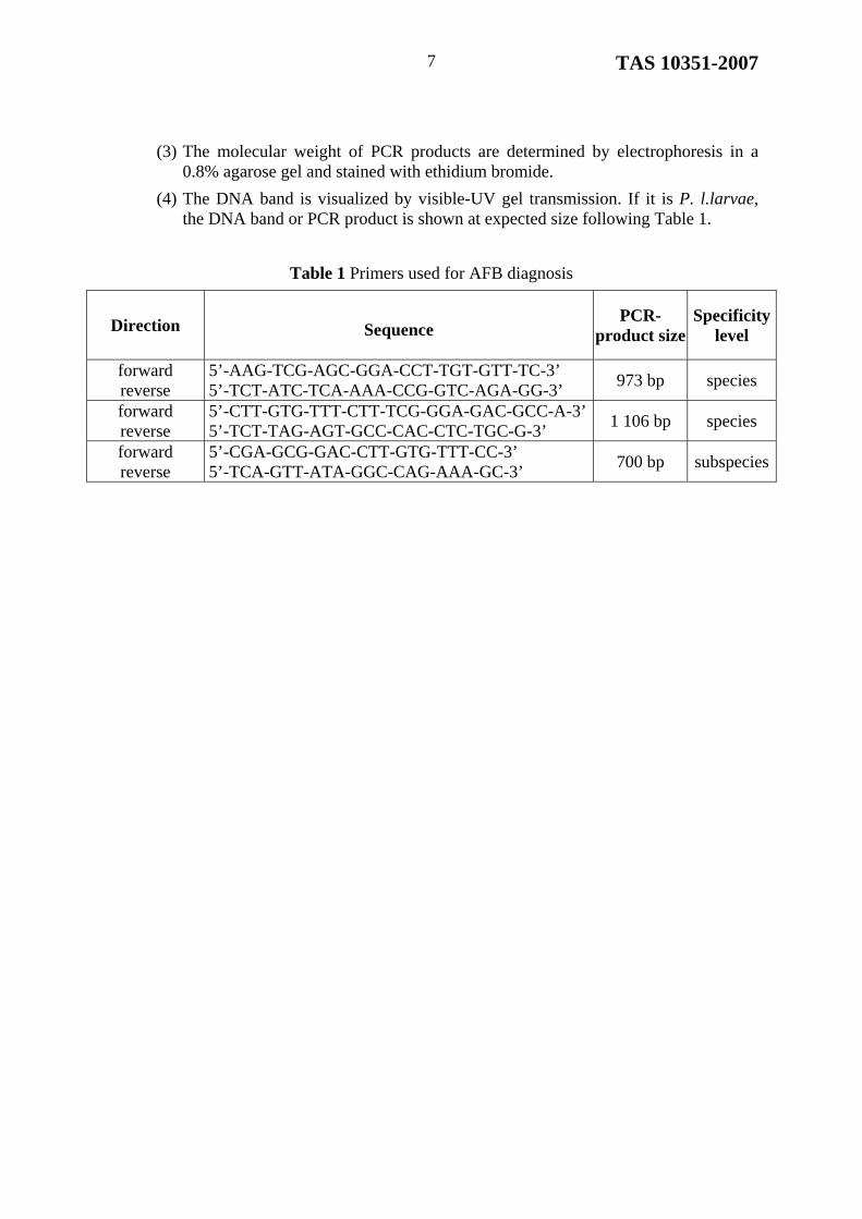
(3) The molecular weight of PCR products are determined by electrophoresis in a 0.8% agarose gel and stained with ethidium bromide.
(4) The DNA band is visualized by visible-UV gel transmission. If it is P. l.larvae, the DNA band or PCR product is shown at expected size following Table 1.
Table 1 Primers used for AFB diagnosis
Direction
Sequence
PCR-product size
Specificity level
forward reverse
5’-AAG-TCG-AGC-GGA-CCT-TGT-GTT-TC-3’ 5’-TCT-ATC-TCA-AAA-CCG-GTC-AGA-GG-3’
973 bp species
forward reverse
5’-CTT-GTG-TTT-CTT-TCG-GGA-GAC-GCC-A-3’ 5’-TCT-TAG-AGT-GCC-CAC-CTC-TGC-G-3’
1 106 bp species
forward reverse
5’-CGA-GCG-GAC-CTT-GTG-TTT-CC-3’ 5’-TCA-GTT-ATA-GGC-CAG-AAA-GC-3’
700 bp subspecies
TAS 10351-2007 8
ANNEX A
EPIZOOTOLOGY, PATHOLOGY AND CLINICAL SIGNS
A.1 EPIZOOTOLOGY
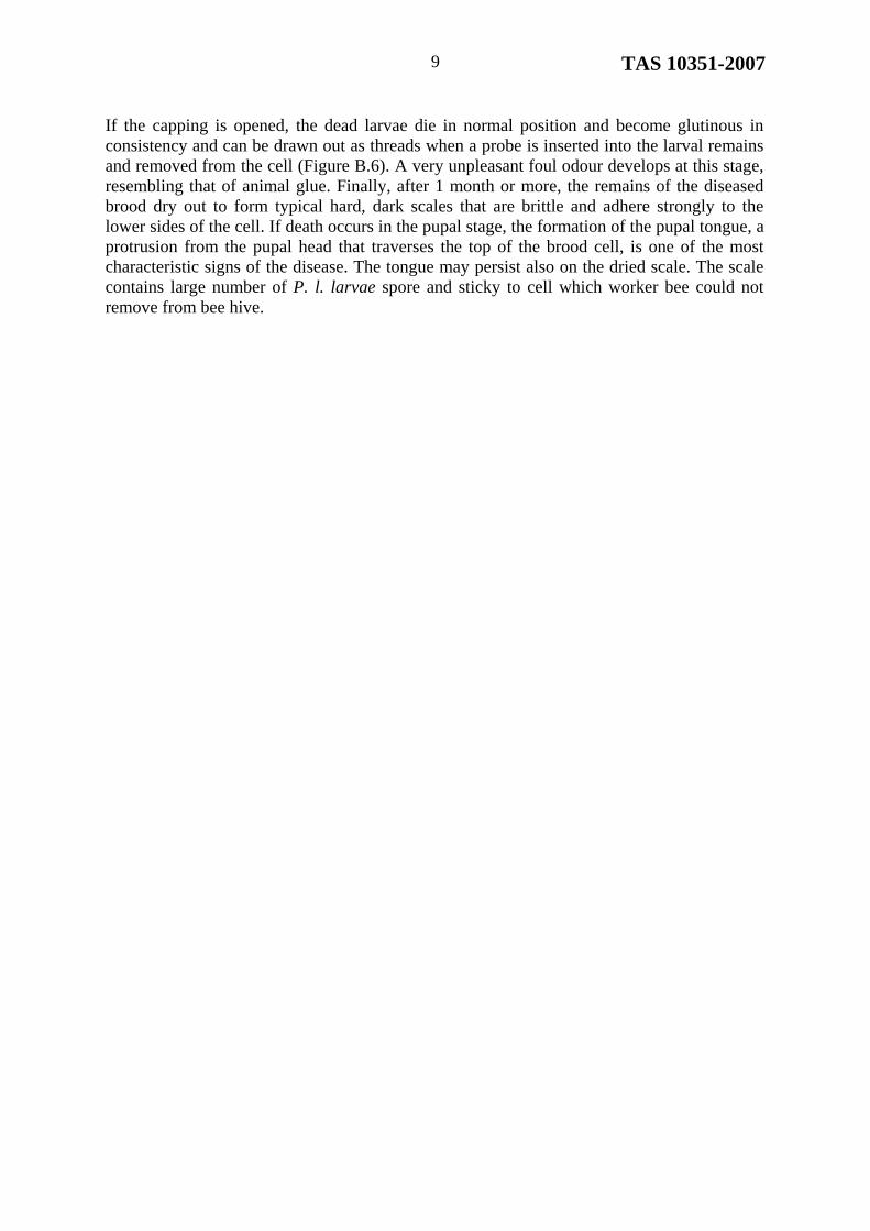
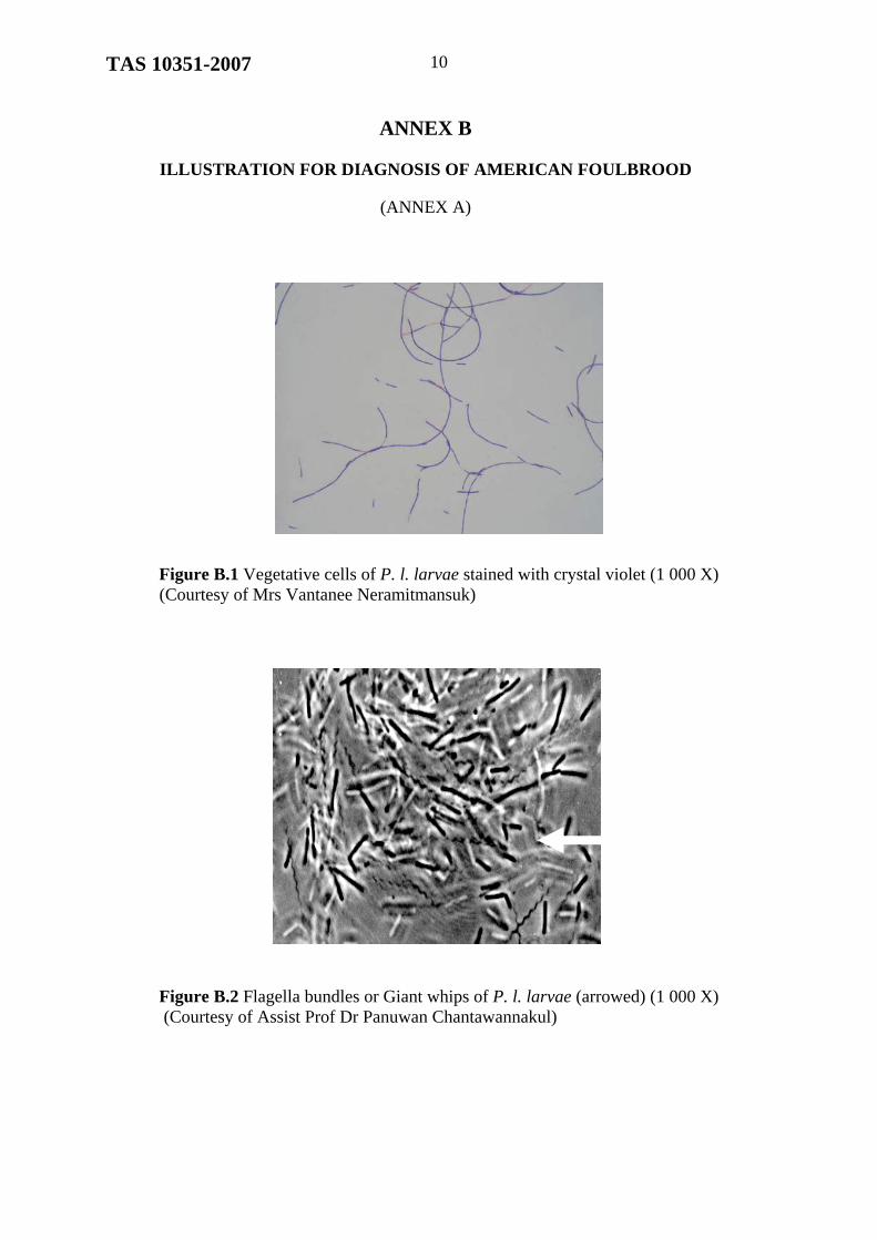
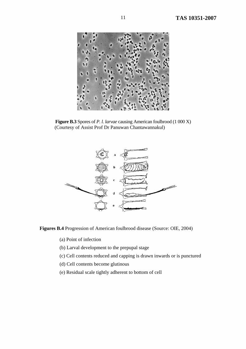
Ameircan foulbrood (AFB) is the fatal and widespread disease of European honey bee (Apis mellifera). It is caused by Paenibacillus larvae larvae (P.l. larvae). The bacterium is a slender rod with slightly rounded ends and occasionally grows in chains. It varies greatly in size from 1.5 to 6.0 µm long and 0.5 µm wide (Figure B.1). It is sometimes found as giant whips under specific conditions (Figure B.2). The causative organism can produce over one billion spores in each infected larva. The spores are oval, approximately twice as long as they are wide, and about 0.6 x 1.3 µm (Figure B.3). They are extremely heat stable and resistant to chemical agents for long period of time therefore, they are difficult to eradicate. The most effective treatment of AFB is to destroy the infected bee hive including bee brood and adults by burning and cleaning all the beekeeping equipment following Veterinary authority’s recommendation.
AFB could be found in beekeeping throughout the world and it affects beekeeping, bee products and international trade. It has been included in the list of infectious animal disease of World Organization for Animal Health or OIE. In Thailand, it is listed under the Ministerial Regulation on the Additional Animal Epidemic B.E. 2538 (1995) empowered by the Animal Epidemics Act B.E. 2499 (1956). The survey of AFB was done jointly by Department of Livestock Development and Department of Agricultural Extension in Year 2003, which supported by the National Bureau of Agricultural Commodity and Food Standards (ACFS). Thailand was found to be free from AFB.
A.2 PATHOLOGY
The infection can be transmitted to a larva from nurse bees or by spores remaining at the base of a brood cell. Although the larval stages of worker bees, drones and queen are susceptible to infection, under natural conditions infected queen and drone larvae are rarely seen. The susceptibility of larvae to AFB decreases with increasing age. Exchanging combs containing the remains of infected larvae is the most common means of spreading the disease from colony to colony. In addition, feeding or robbing of spore-laden honey, artificial swarms, the contaminated food and water sources, and the introduction of queens from infected colonies can also spread the disease.
A.3 CLINICAL SIGNS
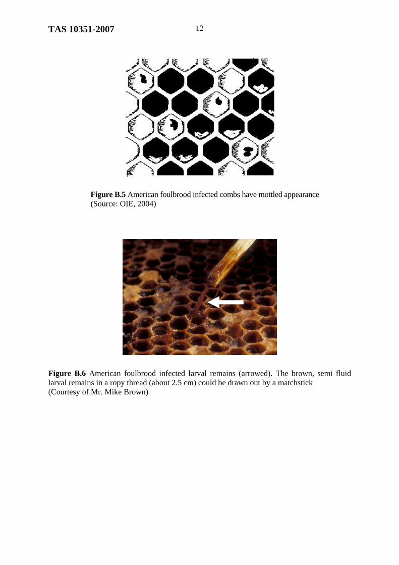
Symptoms of AFB are easily observed from the characteristics of dead bee brood. Since the causative agent, P. l. larvae, could produce proteolytic enzymes to digest the bee brood, and the brood then die within 10-15 days of infection. The diseased larva or pupa appears darkened and eventually dries out to form characteristic brittle scales that adhere tightly to the lower sides of the cell (Figure B.4). The larva or pupa changes colour, first to a creamy and eventually to a dark brown. AFB usually shows clinical signs after cell capping. The capping of cell appears moist and darkened and becomes concave and punctured as the infection progress. In severely infected colonies, the combs appear to be mottled due to a pattern of healthy capped brood, uncapped cells containing the remains of infected larvae, and empty cells. (Figure B.5).
TAS 10351-2007 9
If the capping is opened, the dead larvae die in normal position and become glutinous in consistency and can be drawn out as threads when a probe is inserted into the larval remains and removed from the cell (Figure B.6). A very unpleasant foul odour develops at this stage, resembling that of animal glue. Finally, after 1 month or more, the remains of the diseased brood dry out to form typical hard, dark scales that are brittle and adhere strongly to the lower sides of the cell. If death occurs in the pupal stage, the formation of the pupal tongue, a protrusion from the pupal head that traverses the top of the brood cell, is one of the most characteristic signs of the disease. The tongue may persist also on the dried scale. The scale contains large number of P. l. larvae spore and sticky to cell which worker bee could not remove from bee hive.
TAS 10351-2007 10
ANNEX B
ILLUSTRATION FOR DIAGNOSIS OF AMERICAN FOULBROOD
(ANNEX A)
Figure B.1 Vegetative cells of P. l. larvae stained with crystal violet (1 000 X)
(Courtesy of Mrs Vantanee Neramitmansuk)
Figure B.2 Flagella bundles or Giant whips of P. l. larvae (arrowed) (1 000 X) (Courtesy of Assist Prof Dr Panuwan Chantawannakul)
TAS 10351-2007 11
Figure B.3 Spores of P. l. larvae causing American foulbrood (1 000 X)
(Courtesy of Assist Prof Dr Panuwan Chantawannakul)
Figures B.4 Progression of American foulbrood disease (Source: OIE, 2004)
(a) Point of infection
(b) Larval development to the prepupal stage
(c) Cell contents reduced and capping is drawn inwards or is punctured
(d) Cell contents become glutinous
(e) Residual scale tightly adherent to bottom of cell
TAS 10351-2007 12
Figure B.5 American foulbrood infected combs have mottled appearance (Source: OIE, 2004)
Figure B.6 American foulbrood infected larval remains (arrowed). The brown, semi fluid larval remains in a ropy thread (about 2.5 cm) could be drawn out by a matchstick (Courtesy of Mr. Mike Brown)
TAS 10351-2007 13
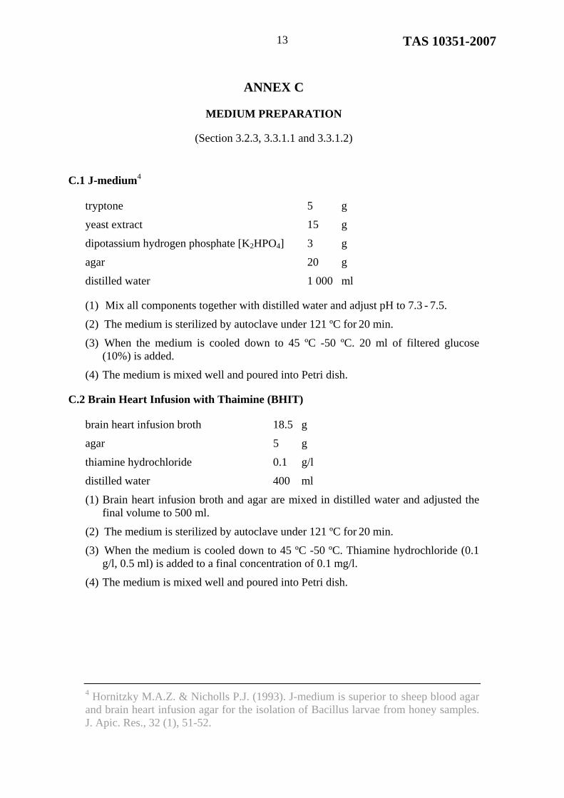
ANNEX C
MEDIUM PREPARATION
(Section 3.2.3, 3.3.1.1 and 3.3.1.2)
C.1 J-medium4
tryptone 5 g
yeast extract 15 g
dipotassium hydrogen phosphate [K2HPO4] 3 g
agar 20 g
distilled water 1 000 ml
(1) Mix all components together with distilled water and adjust pH to 7.3 - 7.5.
(2) The medium is sterilized by autoclave under 121 ºC for 20 min.
(3) When the medium is cooled down to 45 ºC -50 ºC. 20 ml of filtered glucose (10%) is added.
(4) The medium is mixed well and poured into Petri dish.
C.2 Brain Heart Infusion with Thaimine (BHIT)
brain heart infusion broth 18.5 g
agar 5 g
thiamine hydrochloride 0.1 g/l
distilled water 400 ml
(1) Brain heart infusion broth and agar are mixed in distilled water and adjusted the final volume to 500 ml.
(2) The medium is sterilized by autoclave under 121 ºC for 20 min.
(3) When the medium is cooled down to 45 ºC -50 ºC. Thiamine hydrochloride (0.1 g/l, 0.5 ml) is added to a final concentration of 0.1 mg/l.
(4) The medium is mixed well and poured into Petri dish.
4 Hornitzky M.A.Z. & Nicholls P.J. (1993). J-medium is superior to sheep blood agar and brain heart infusion agar for the isolation of Bacillus larvae from honey samples. J. Apic. Res., 32 (1), 51-52.

TAS 10351-2007 14
ANNEX D
DNA EXTRACTION
(Section 3.3.3.1(1))
D.1 CHEMICALS
50 mM Tris, 5 mM EDTA pH 8.0
lysozyme
10% SDS
saturated phenol solution
phenol/chloroform/isoamylalcohol 25:24:1
chloroform/isoamylalcohol 24:1
3M sodium acetate pH 5.2
ethanol
RNase A 10 mg/ml
TE buffer
D.2 DNA EXTRACTION PROTOCAL
(1) A bacterial colony from cultured plate is inoculated in suitable medium under anaerobic condition and 5%-10% CO2 at 35 oC to 37 oC for 4 days.
(2) If samples for extraction are from infected or dead larvae, the samples are grounded in one ml of phosphate buffer saline. The debris is centrifuged at 800 g for 10 min. The supernatant (200 l) is then heated to 95 oC for 15 min. The supernatant is again centrifuged at 14 500 g for 2 min. The supernatant (10 l) is used for PCR template.
(3) The bacterial cells from (1) are harvested by centrifuged at 2 000 g for 10 min at 4 oC and washed three times in suitable buffer.
(4) The cells are suspended in 450 l of 50 mM Tris containing 5 mM EDTA, pH 8.0 and lysozyme 2 mg/ml. The mixture is then incubated at 37 oC for 60 min.
(5) SDS (10% w/v, 22.5 l) is added to a final concentration of 0.5% (w/v). The cells are then lysed and the clear viscous solution could be observed in the tube.
(6) An equal volume of saturated phenol solution is added and mixed several times and then micro-centrifuged for 3 min to 5 min to obtain aqueous phase where DNA is present.
(7) The aqueous solution from (6) is extracted repeatedly by saturated phenol until the white precipitate between phenol and aqueous phase is absent.
(8) The clear lysate (about 400 l) is added with 3 M sodium acetate (40 l) and the DNA is precipitated by adding a twice volume of ethanol (880 l).
TAS 10351-2007 15
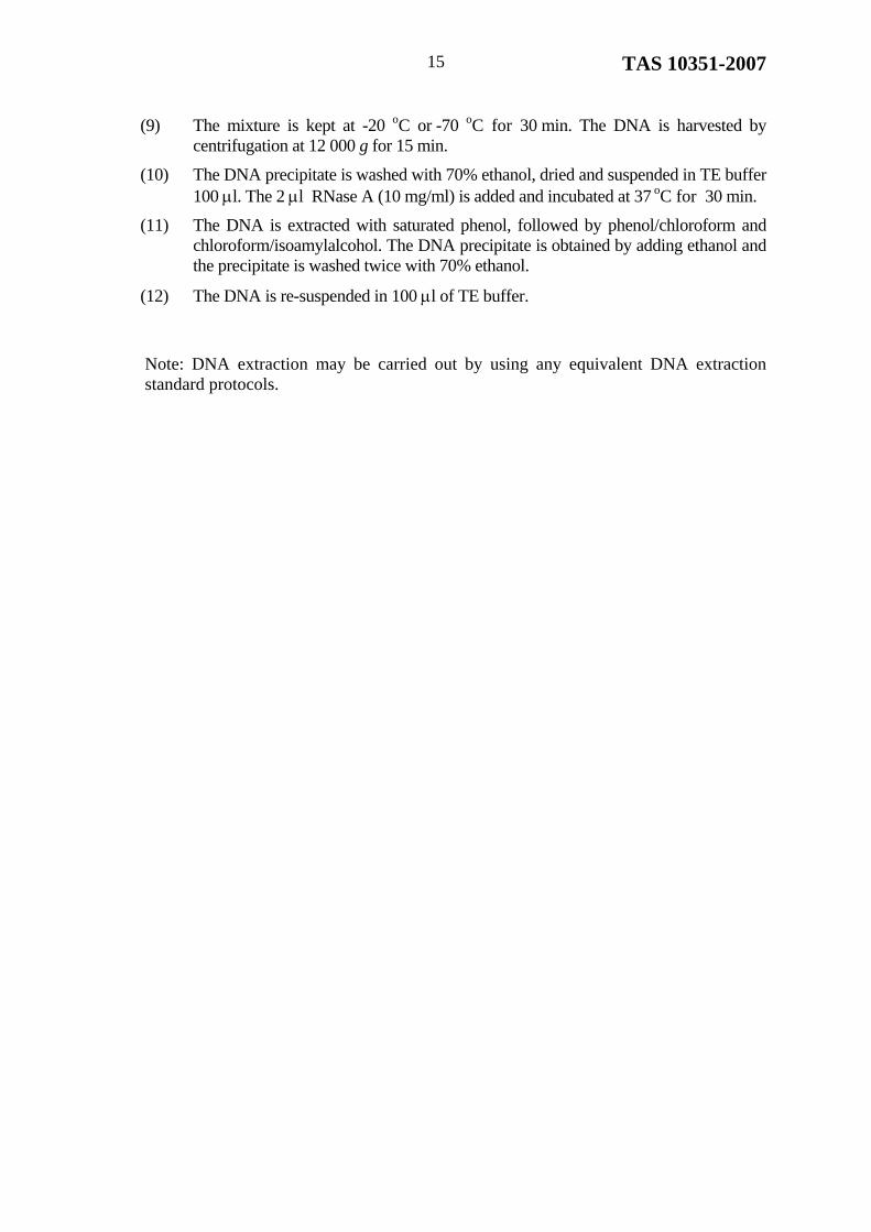
(9) The mixture is kept at -20 oC or -70 oC for 30 min. The DNA is harvested by centrifugation at 12 000 g for 15 min.
(10) The DNA precipitate is washed with 70% ethanol, dried and suspended in TE buffer 100 l. The 2 l RNase A (10 mg/ml) is added and incubated at 37 oC for 30 min.
(11) The DNA is extracted with saturated phenol, followed by phenol/chloroform and chloroform/isoamylalcohol. The DNA precipitate is obtained by adding ethanol and the precipitate is washed twice with 70% ethanol.
(12) The DNA is re-suspended in 100 l of TE buffer.
Note: DNA extraction may be carried out by using any equivalent DNA extraction standard protocols.
TAS 10351-2007 16
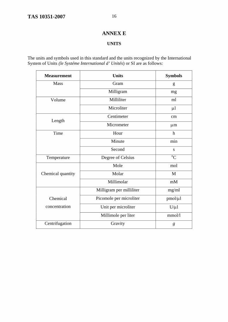
ANNEX E
UNITS
The units and symbols used in this standard and the units recognized by the International System of Units (le Systéme International d’ Unités) or SI are as follows:
Measurement Units Symbols
Gram g Mass
Milligram mg
Milliliter ml Volume
Microliter l
Centimeter cm Length
Micrometer m
Hour h
Minute min
Time
Second s
Temperature Degree of Celsius oC
Mole mol
Molar M Chemical quantity
Millimolar mM
Milligram per milliliter mg/ml
Picomole per microliter pmol/l
Unit per microliter U/l
Chemical
concentration
Millimole per liter mmol/l
Centrifugation Gravity g