Embed Size (px)
Citation preview
PROCEEDINGS Open Access
Diagnostic reproducibility on a digital evaluationsystem slide in cytology and histology inoncologic screeningStefania Lega1, Paola Crucitti1, Paola Pierotti1, Roberta Rapezzi1, Priscilla Sassoli de’ Bianchi2, Carlo Naldoni2,Arrigo Bondi1*
From 11th European Congress on Telepathology and 5th International Congress on Virtual MicroscopyVenice, Italy. 6-9 June 2012
BackgroundDiagnostic reproducibility and accuracy in cytology andhistology are the main issues in Oncologic Screening ofcervix, breast and colorectal cancer: it can be achieved byprograms for quality assurance (QA). The slides set stan-dard represents the most used method to compare diag-nostic proficiency, the chance of interpreting microscopicdigital photographs provided an interesting alternative toread conventional microscope slides.The whole digital slide observed in a computer screen
is a third, interesting, option to reach the goal. In fact allthe information on conventional samples are transferredinto a file, easily archived, catalogued, duplicated oradvice for quality control, but is especially available atdistance and from multiple locations simultaneously withdrastic reduction of time needed to achieve proficiencytest reproducibility [1].The production of digital slides with modern scanners
is relatively simple and quick. All suppliers offer servicesinto private or public networks server in the literature [2]and software able to track scanned cases stored in com-prehensive database to build large casistic archives online[3] Tools are already available for a teleconference dis-cussion of cases with vision of cytological preparationson line [4,5], educational programs with integrated digitalslides are poorly developed or proficiency tests for conti-nuing education and professional updating are easilyaccessible.A project on Virtual Microscopy and Digital Pathology
has been conducted in Emilia-Romagna Region (Italy)with the goal to promote quality in diagnostic cytology
and histology in Screening programs by testing a differentsystem involving pathologists and cytologists using digitalslides, with a faster and reproducible program easier tomanage than standard diagnostic sets and by distance forretraining of patrhologists with a final consensus meeting.The aim has been reached with the realization of a man-
agement system for cytological and histological whole-slides digital images and related clinical data and thebuilding of a picture archive and communication system(PACS) among pathologists of our (and probably other)region. This must be backed by software for the realizationof network slide seminars to perform periodic diagnosticreproducibility and proficiency test. The cases, collectedand properly catalogued in an online, easily accessible andsystemic digital archive of slides, with diagnoses discussedin clinical and pathologic audit meetings and validated byexperts, can be used as diagnostic reference tools (caseregistry online ). The cataloguing and indexing is per-formed with NAP codes derived from SNOMED [6],which contains terms and definitions in Italian and Englishand encompasses extensive synonyms and complexresearches.
Material and methodsThe cancer screening group of the Emilia-RomagnaRegion (Italy) set up a picture, archive and communica-tion system (PACS) devoted to pathologists for coopera-tive diagnosis, teaching and training, teleconsulting,documentation of rare cases and pilot experiences;furthermore selected cases are catalogued in the PACSwith the aim to check the diagnostic concordance inregional oncologic screening (cervix, breast and colon).The PACS system is made by two Aperio scanner and an
1Anatomia Patologica Ospedale Maggiore, Azienda USL di Bologna, ItalyFull list of author information is available at the end of the article
Lega et al. Diagnostic Pathology 2013, 8(Suppl 1):S18http://www.diagnosticpathology.org/content/8/S1/S18
© 2013 Lega et al; licensee BioMed Central Ltd. This is an Open Access article distributed under the terms of the Creative CommonsAttribution License (http://creativecommons.org/licenses/by/2.0), which permits unrestricted use, distribution, and reproduction inany medium, provided the original work is properly cited.
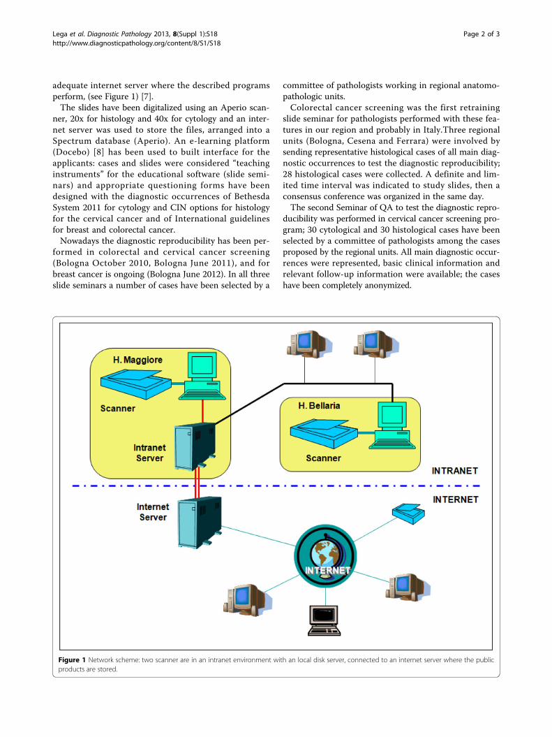
adequate internet server where the described programsperform, (see Figure 1) [7].The slides have been digitalized using an Aperio scan-
ner, 20x for histology and 40x for cytology and an inter-net server was used to store the files, arranged into aSpectrum database (Aperio). An e-learning platform(Docebo) [8] has been used to built interface for theapplicants: cases and slides were considered “teachinginstruments” for the educational software (slide semi-nars) and appropriate questioning forms have beendesigned with the diagnostic occurrences of BethesdaSystem 2011 for cytology and CIN options for histologyfor the cervical cancer and of International guidelinesfor breast and colorectal cancer.Nowadays the diagnostic reproducibility has been per-
formed in colorectal and cervical cancer screening(Bologna October 2010, Bologna June 2011), and forbreast cancer is ongoing (Bologna June 2012). In all threeslide seminars a number of cases have been selected by a
committee of pathologists working in regional anatomo-pathologic units.Colorectal cancer screening was the first retraining
slide seminar for pathologists performed with these fea-tures in our region and probably in Italy.Three regionalunits (Bologna, Cesena and Ferrara) were involved bysending representative histological cases of all main diag-nostic occurrences to test the diagnostic reproducibility;28 histological cases were collected. A definite and lim-ited time interval was indicated to study slides, then aconsensus conference was organized in the same day.The second Seminar of QA to test the diagnostic repro-
ducibility was performed in cervical cancer screening pro-gram; 30 cytological and 30 histological cases have beenselected by a committee of pathologists among the casesproposed by the regional units. All main diagnostic occur-rences were represented, basic clinical information andrelevant follow-up information were available; the caseshave been completely anonymized.
Figure 1 Network scheme: two scanner are in an intranet environment with an local disk server, connected to an internet server where the publicproducts are stored.
Lega et al. Diagnostic Pathology 2013, 8(Suppl 1):S18http://www.diagnosticpathology.org/content/8/S1/S18
Page 2 of 3
A 30 days interval was indicated to study the slides, thena consensus conference has been programmed at the endof the evaluation to present the results and discuss cases.Before the meeting each participant received a report withthe gold standard diagnosis performed buy the committeeand her/his diagnosis and concordance result.
Results and discussion15 pathologists of regional units attended the colon-rec-tal QA and the diagnostic reproducibility have beenevaluated matching their results with the final gold stan-dard diagnosis reached during the consensus conference.The observed agreement was 69% and the overall per-formance of the participating pathologists was assessedwith a statistical analysis using Cohen’s kappa: the aver-age value was 0.64 (substantial).95 cytologists and 32 histopathologists have been
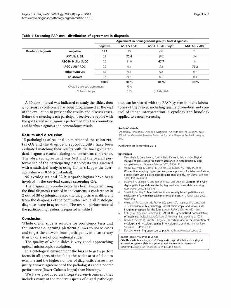
involved in the cervical cancer screening QA.The diagnostic reproducibility has been evaluated using
the final diagnosis reached in the consensus conference: in2 out of 30 cytological cases the diagnosis was differentfrom the diagnosis of the committee, while all histologicdiagnoses were in agreement. The overall performance ofthe participating readers is reported in table 1.
ConclusionWhole digital slide is suitable for proficiency tests andthe internet e-learning platform allows to share casesand to get the answers from participants, in a easier waythan by of a set of conventional slides.The quality of whole slides is very good, approaching
optical microscopic resolution.In a cytological environment the bias is to get a perfect
focus in all parts of the slide; the wider area of slide toexamine and the higher number of diagnostic classes mayjustify a worse agreement of the pathologists and a poorerperformance (lower Cohen’s kappa) than histology.We have produced an integrated environment that
includes many of the modern aspects of digital pathology
that can be shared with the PACS system in many labora-tories of the region, including quality promotion and con-trol of image interpretation in cytology and histologyapplied to cancer screening.
Authors’ details1Anatomia Patologica Ospedale Maggiore, Azienda USL di Bologna, Italy.2Direzione Generale Sanità e Politiche Sociali – Regione Emilia-Romagna,Italy.
Published: 30 September 2013
References1. Demichelis F, Della Mea V, Forti S, Dalla Palma P, Beltrami CA: Digital
storage of glass slides for quality assurance in histopathology andcytopathology. J Telemed Telecare 2002, 8:138-142.
2. Wilbur DC, Madi K, Colvin RB, Duncan LM, Faquin WC, Ferry JA, et al:Whole-slide imaging digital pathology as a platform for teleconsultation:a pilot study using paired subspecialist correlations. Arch Pathol Lab Med2009, 133:1949-1953.
3. Huisman A, Looijen A, van den Brink SM, van Diest PJ: Creation of a fullydigital pathology slide archive by high-volume tissue slide scanning.Hum Pathol 2010, 41:751-757.
4. Saysell E, Routley C: Telemedicine in community-based palliative care:evaluation of a videolink teleconference project. Int J Palliat Nurs 2003,9:489-495.
5. Weinstein RS, Graham AR, Richter LC, Barker GP, Krupinski EA, Lopez AM,et al: Overview of telepathology, virtual microscopy, and whole slideimaging: prospects for the future. Hum Pathol 2009, 40:1057-1069.
6. College of American Pathologists: SNOMED - Systematized nomenclatureof medicine. Skokie,Ill.,USA, College of American Pathologists;, 2 1979.
7. Bondi A, Pierotti P, Crucitti P, Lega S: The virtual slide in the promotion ofcytologic and hystologic quality in oncologic screenings. Ann Ist SuperSanita 2010, 46:144-150.
8. Docebo: e-learning open source platform. [http://www.docebo.org].
doi:10.1186/1746-1596-8-S1-S18Cite this article as: Lega et al.: Diagnostic reproducibility on a digitalevaluation system slide in cytology and histology in oncologicscreening. Diagnostic Pathology 2013 8(Suppl 1):S18.
Table 1 Screening PAP test - distribution of agreement in diagnosis
Agreement in homogeneous groups: final diagnoses
negative ASCUS L SIL ASC-H H SIL / SqCC AGC AIS / ADC
Reader’s diagnosis negative 85.1 15 6.6 2.1
ASCUS/ L SIL 5.1 72.4 20.2 3.6
ASC-H/ H SIL/ SqCC 2.9 11.9 67.7 14
AGC / AIS/ ADC 2.9 0.3 5.2 79.2
other tumours 3.5 0.2 0.2 0.7
no answer 0.5 0.2 0.1 0.4
100% 100% 100% 100%
Overall observed agreement 73%
Cohen’s Kappa 0.63 (substantial)
Lega et al. Diagnostic Pathology 2013, 8(Suppl 1):S18http://www.diagnosticpathology.org/content/8/S1/S18
Page 3 of 3