Embed Size (px)
Citation preview
Hindawi Publishing CorporationJournal of Respiratory MedicineVolume 2013, Article ID 756483, 6 pageshttp://dx.doi.org/10.1155/2013/756483
Research ArticleDiagnostic Utility of Transbronchial NeedleAspiration in Malignant Endobronchial Lesions:Relevance to Lesions’ Characteristics
Sherif A. A. Mohamed,1 Yousef Ahmed,1 Khaled Hussein,1
Nashwa M. A. Abd El-Aziz,2 and Yasser Gamal3
1 Department of Chest Diseases, Faculty of Medicine, Assiut University, Assiut 71516, Egypt2 Department of Medical Oncology, South Egypt Cancer Institute, Assiut University, Assiut 71516, Egypt3 Department of Pathology, Faculty of Medicine, Assiut University, Assiut 71516, Egypt
Correspondence should be addressed to Sherif A. A. Mohamed; [email protected]
Received 27 June 2013; Revised 9 August 2013; Accepted 22 August 2013
Academic Editor: Akira Mogi
Copyright © 2013 Sherif A. A. Mohamed et al. This is an open access article distributed under the Creative Commons AttributionLicense, which permits unrestricted use, distribution, and reproduction in any medium, provided the original work is properlycited.
In this prospective study, we aimed to report our experience with the diagnostic utility of transbronchial needle aspiration (TBNA)in patients with malignant endobronchial lesions detected during routine bronchoscopy. Ninety-four patients were enrolled.TBNA and conventional diagnostic techniques (CDTs: forceps biopsy, brushing, and washing) were performed in all patients.Endobronchial lesions were classified into exophytic mass lesions (EMLs), submucosal disease (SD), and peribronchial disease(PD). The diagnostic yields of TBNA and CDT alone and together were compared according to the lesions’ types, histopathology,and locations. During 3-year period, the addition of TBNA to CDT improved bronchoscopic sensitivity from 70.2% to 94.7% inall lesion types. Addition of TBNA to CDT increased the diagnostic success from 74% to 95% and from 50% to 94% in NSCLCand SCLC, respectively. The diagnostic success was increased in all localizations by the addition of TBNA to CDT, particularlyfor lesions located at the trachea, main bronchi, and upper lobes. We conclude that the addition of TBNA to CDT increases thediagnostic yield in patients with visible malignant endobronchial lesions, particularly in peribronchial disease, and improves thediagnostic yield of bronchoscopy, in both NSCLC and SCLC and in all bronchoscopic locations, particularly in central and upperlobar lesions.
1. Introduction
Transbronchial needle aspiration (TBNA) is a modality thatallows the bronchoscopist to sample tissue from the deepersubmucosa as well as from the close extraluminal areasof the endobronchial tree. TBNA is a beneficial, safe, andminimally invasive technique that was proved to be usefulin the diagnosis and staging of lung cancer [1]. Previously,the utility of TBNA was restricted to mediastinal lymphnode and extrabronchial lesion sampling. Its use has beenexpanded to complement conventional diagnostic techniques(CDTs) such as bronchial washing (BW), bronchial brushing(BB), and forceps biopsy (FB) in the diagnosis of endo-bronchial lung cancer [2]. However, only few studies [2–7] had addressed the diagnostic utility of TBNA in visible
endobronchial lung cancer. Moreover, despite that TBNAproved cost-effective diagnostic utility in visible malignantendobronchial lesions [8], studies evaluating that utility indeveloping countries are still lacking. Lung cancer maypresent either as a parenchymal lesion or as endobronchialdisease. The latter may manifest as an exophytic mass lesion(EML), submucosal infiltration (submucosal disease, SD),or extrinsic compression from peribronchial disease (PD)[1, 2].The impact of different characteristics of endobronchiallesions (e.g., type, histopathologic subtype, location) on thediagnostic utility of TBNA needs to be elucidated [2, 7].The aims of this study were; first, to report our experiencewith the diagnostic utility of TBNA and its contributions toCDT in visible endobronchial lesions of the patients whounderwent TBNAduring fiberoptic bronchoscopy (FOB) and
2 Journal of Respiratory Medicine
had the final diagnosis of primary lung cancer and Second,to evaluate this utility in relation to different endobronchiallesions’ characteristics (i.e., type, histopathologic subtype,and location).
2. Patients and Methods
Assiut University Hospital is a large tertiary hospital in UpperEgypt, to which many patients with suspected or knownlung cancer are referred. Patients who had the preliminarydiagnosis of lung cancer in whom EML, SD, or PD wasdetected by routine FOB underwent TBNA in addition to thebronchoscopic CDT and were prospectively enrolled fromJanuary, 2010 to December, 2012 into this study. Patients withpreexisting known malignancy were excluded. Submucosaldisease was defined as erythema, vascular flares, enhancedrugal pattern, thickening or loss of mucosal markings,and bronchial narrowing [1, 2, 7]. Peribronchial diseasewas defined as luminal narrowing secondary to extrinsiccompression [1, 2]. Lesions located in trachea and in mainand lobar bronchi were classified as “central,” and thosewith segmental and subsegmental localization were classifiedas “peripheral” [6]. All patients underwent all procedures(CDT and TBNA).The sequence was always washing, TBNA,forceps biopsy, and brushing to minimize specimen contam-ination and avoid false-positive results.
Prior to bronchoscopy, all patients underwent chestradiography and computed tomography (CT) of the chest.Complete blood count and coagulation tests (bleeding-aggregation and prothrombin time) were performed. Patientsfasted overnight and were given 10mg of diazepam and0.5mg of atropine intramuscularly as premedication 30minprior to the examination. Local anesthesia was performedwith a lidocaine solution. A flexible fiberoptic bronchoscopy(Pentax SB 15; Pentax, Japan) under local anaesthesia wasinserted via nasal or oral route with the patient lying insupine position. A 13mm 21-gauge cytology needle (NA-2C-1; Olympus Corporation) was used for TBNA. The trans-bronchial needle was introduced into the bronchoscope withthe needle inside the metal hub while visualizing the tracheathrough the scope. When the metal hub was visible, theneedle was advanced. In the presence of PD, the needlewas inserted into the lesion according to the “pushing”technique [9]. In EML, the needle was penetrated into themass directly. In patients with SD, the needle was insertedat a 45∘ angle into the puncture site. One cytologic sampleconsisted of at least three subsequent aspirations from thesame region, penetrating the bronchial wall in an area<2 cm2.A 50mL syringe was used to apply suction [6, 7], and itwas detached before retraction of the needle in order toavoid contamination. The aspirated material was blown intofour or five slides, smeared, immediately fixed with 95%alcohol, and sent for cytologic examination. After the TBNAprocedure, FB and BB were performed in a conventionalmanner. Four adequate FB specimens were performed andthen fixed in formaldehyde solution. Histologic specimenswere stained with hematoxylin-eosin, and cytologic speci-mens were stained by the Papanicolaou technique. On-sitecytopathologic assessment was not performed. All specimens
Table 1: Diagnostic yield of TBNA, CDT, and CDT plus TBNAwithrespect to the type of lesions∗.
Condition TBNAno. (%)
CDTno. (%)
CDT + TBNAno. (%) 𝑃 value
EML (𝑛 = 34) 27 (79.4) 28 (82.3) 33 (97) 0.063SD (𝑛 = 23) 21 (91) 17 (74) 22 (95.6) 0.063PD (𝑛 = 37) 34 (92) 21 (57) 34 (92) <0.001SPD (𝑛 = 60) 55 (91.7) 38 (63.3) 56 (93.3) <0.001Total (𝑛 = 94) 82 (87.2) 66 (70.2) 89 (94.7) <0.001∗CDT versus CDT + TBNA; EML: exophytic mass lesions; SD: submucosaldisease; PD: peribronchial disease; SPD: submucosal-peribronchial disease.
were interpreted by one cytopathologist. Results of cytologicanalysis were considered positive only when a sufficientnumber of definitely malignant cells were observed. Allspecimens having cellular atypia and abnormal cells highlysuggestive of malignancy were considered negative. The pri-mary outcome tomeasurewas to evaluate the diagnostic yieldfrom TBNA, CDT, and TBNA plus CDT; yields of CDT werecompared to those of CDT plus TBNA. We also studied andcompared the diagnostic yields from individual proceduresand questioned for the best yield of their combination(s).The secondary outcome was to address the diagnostic yieldof TBNA and its contribution to CDT in relation to the typeof the endobronchial lesion (i.e., EML, SD, or PD), histologicsubtypes, and location. Any procedure-related complicationsor damage to the bronchoscope were documented.The studywas approved by the local ethical committee, and a writtenconsent was obtained from every patient for enrollment intothis study.
Data were analysed using the statistical package for theSocial Sciences (Windows version 16.0; SPSS Inc., Chicago,IL, USA). Statistical analysis of the difference in diagnosticyield between the two groups was performed using McNe-mar’s test. Fisher’s exact test was used to detect any differencebetween locations. A 𝑃 value <0.05 was considered to besignificant.
3. Results
During a 3-year period, 94 patients had bronchoscopicallyvisible endobronchial lesions. Samples of TBNA, forcepsbiopsy, brushing, and washing were collected from allpatients. With these diagnostic methods, 89 patients (94.7%)had a diagnosis of primary lung cancer, but in 5 patients,no diagnosis was made. These five patients were diagnosedby other techniques (pleural biopsy, CT-guided biopsy, andtransthoracic needle biopsy). The patients included 81 (86%)men and 13 (14%) women, with a mean age of 58.39 ± 6.14years and range of 42–78 years. Histopathologic diagnosiswas nonsmall cell lung cancer (NSCLC) in 78 (83%) patientsand small cell lung cancer (SCLC) in 16 (17%) patients.Bronchoscopically, EML, SD, and PD were encounteredin 34/94, 36%, 23/94, 24%, and 37/94, 40% of patients,respectively. Diagnostic yield of TBNA, CDT, and CDT plusTBNA is shown in Table 1. Diagnosis was made by CDT in 66of 94 patients (70.2%).The addition of TBNA toCDT enabled
Journal of Respiratory Medicine 3
histopathologic diagnosis in another 23 patients (24.5%) andimproved the sensitivity of bronchoscopy from 70.2 to 94.7%(𝑃 < 0.001). The sensitivities of CDT, TBNA, and CDTplus TBNAwere compared for different endobronchial lesiontypes. The addition of TBNA to CDT increased sensitivity inall lesion types, and this increase was statistically significantin the peribronchial disease group (𝑃 < 0.001). In this group,diagnosis was made solely by TBNA in 13 patients (35% ofthe PD group). When the 60 submucosal and peribronchialdiseases (SPDs) were considered together, the addition ofTBNA to CDT increased the diagnostic yield from 63 to93%, and the difference was highly significant (𝑃 < 0.001).Despite that no statistical difference was observed in theEML group, TBNA identified five additional patients withlung cancer. Table 2 demonstrates the different diagnosticyields from individual procedures and their combination.Rate of diagnostic yield was the highest for TBNA (82/94patients, 87.2%), followed by forceps biopsy (52/94 patients,55.3%), brushing (49/94 patients, 52%), and washing (24/94patients, 25.5%). The rate of diagnostic yield was also thehighest for TBNA in both EML and SPD groups, 79.4% and91.7%, respectively. Interestingly, diagnostic yield fromTBNAwas far superior to the yield from combination of forcepsbiopsy and brushing in total lesions and SPD (87.2% versus70.2% and 91.7% versus 63.3%, resp.). Combination of TBNAwith forceps and brushing provided the best diagnostic yield,97%, 93.3%, and 94.7%, in EML, SPD, and total lesions,respectively.
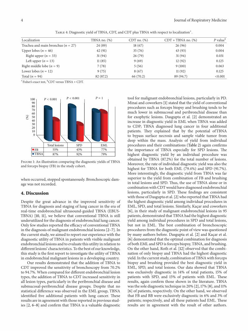
TBNA was exclusively diagnostic in 13 patients (14%of all patients): 9 patients with SPD (15% of patients withSPD) and 4 patients with EML (15% of patients with EML).Among those 4 patients with EML, there were two patientswith small cell carcinoma who had crush artifacts on theFB and were solely diagnosed by TBNA. Forceps biopsyand brushing were exclusively diagnostic in six (6%) andthree (3%) of patients; respectively, and all these patientshad EML. Bronchial washing was not exclusively diagnosticin situations in which all other procedures were negative.Comparing the diagnostic yield of TBNA and forceps biopsyfor all types of endobronchial lesions revealed a highlysignificant statistical difference (82/94, 87.2% versus 52/94,55.3%; 𝑃 < 0.001). (Figure 1) This difference was primarilydue to difference in the SPD group (55/60, 91.7% versus26/60, 43%; 𝑃 < 0.001). The diagnostic yield of TBNA withrespect to histologic subtypes is demonstrated in Table 3. Anincrement from 74.4 to 95% was observed in diagnostic yieldinNSCLCwhenTBNAwas combinedwithCDT (𝑃 < 0.001).The diagnostic efficacy improved from 58.8 to 91.2% by theaddition of TBNA to CDT in the peribronchial disease groupfor NSCLC (𝑃 < 0.001). However, the addition of TBNAto CDT did not result in a statistically significant differencein EML and SD groups for NSCLC. In the SCLC group, thediagnosis was made by CDT in 8 of 16 patients (50%), andthe diagnosis was made by CDT plus TBNA in 15 patients(94%) (𝑃 = 0.016). However, when the differences betweenCDT and CDT plus TBNA were analysed in different typesof lesions within the SCLC group, there was no significantstatistical difference for any of these lesions (60% versus100%, 50% versus 87.5%, and 33.3% versus 100% for EML,
Table 2: Diagnostic yield of individual procedures and theircombinations∗.
Procedure EML(𝑛 = 34)
SPD(𝑛 = 60)
Total(𝑛 = 94)
Solediagnostictechnique
BW 10 (29.4) 14 (23.3) 24 (25.5) 0 (0)BB 21 (61.8) 28 (50) 49 (52) 3 (3)FB 26 (76.5) 26 (43) 52 (55.3) 6 (6)TBNA 27 (79.4) 55 (91.7) 82 (87.2) 13 (14)FB + BB 28 (82.3) 38 (63.3) 66 (70.2)FB + TBNA 27 (79.4) 55 (91.7) 82 (87.2)FB + BB + TBNA 33 (97) 56 (93.3) 89 (94.7)∗Values given as no. (%) BW: bronchial washing; BB: bronchial brushing; FB:forceps biopsy; TBNA: transbronchial needle aspiration.
Table 3: Diagnostic yield of TBNA, CDT, and CDT plus TBNAwithrespect to the histopathologic subtype∗.
Histopathologicsubtype/lesion
TBNAno. (%)
CDTno. (%)
CDT + TBNAno. (%) 𝑃 value
NSCLC (𝑛 = 78)EML (𝑛 = 29) 24 (82.8) 25 (86.2) 28 (96.6) 0.25SD (𝑛 = 15) 15 (100) 13 (86.7) 15 (100) 0.5PD (𝑛 = 34) 31 (91.2) 20 (58.8) 31 (91.2) 0.001Total (𝑛 = 78) 70 (90) 58 (74.4) 74 (95) <0.001
SCLC (𝑛 = 16)EML (𝑛 = 5) 3 (60) 3 (60) 5 (100) 0.25SD (𝑛 = 8) 6 (75) 4 (50) 7 (87.5) 0.125PD (𝑛 = 3) 3 (100) 1 (33.3) 3 (100) 0.125Total (𝑛 = 16) 12 (75) 8 (50) 15 (94) 0.016
∗CDT versus TBNA+CDT; NSCLC: nonsmall cell lung cancer; SCLC: smallcell lung cancer.
SD, and PD, resp.). The diagnostic utility of CDT and CDTplus TBNAaccording to the location of endobronchial lesionswas addressed (Table 4). The lesions were in the tracheaor main bronchi in 27 patients (29%), in upper lobes in46 patients (49%), in the right middle lobe in 9 patients(9.3%), and in the lower lobes in 12 patients (12.7%). Theaddition of TBNA to CDT increased the diagnostic yieldirrespective of the location of the lesion, and this increasewas significant in trachea and main bronchi and bronchiof the upper lobes. When the right and left upper lobeswere compared, this significance was attributed to rightupper lobe lesions rather than left upper lobe ones. Right-or left-sided malignant lesions demonstrated no differencedue to diagnostic efficacy. Also, no statistically significantdifference was observed among the diagnostic yield of eachmethod with regard to the lesion being central or peripheral.Results from distribution of malignant cell types obtainedby cytologic assessment from TBNA were in complete con-cordance with those obtained by histologic assessment byforceps biopsy. Severe complications such as major bleedingand pneumothorax were not observed. Minor bleeding,
4 Journal of Respiratory Medicine
Table 4: Diagnostic yield of TBNA, CDT, and CDT plus TBNA with respect to localization∗.
Localization TBNA no. (%) CDT no. (%) CDT + TBNA no. (%) 𝑃 value§
Trachea and main bronchus (𝑛 = 27) 24 (89) 18 (67) 26 (96) 0.004Upper lobes (𝑛 = 46) 42 (91) 35 (76) 43 (93) 0.004
Right upper (𝑛 = 33) 31 (94) 26 (79) 31 (94) 0.031Left upper (𝑛 = 13) 11 (85) 9 (69) 12 (92) 0.125
Right middle lobe (𝑛 = 9) 7 (78) 5 (56) 9 (100) 0.063Lower lobes (𝑛 = 12) 9 (75) 8 (67) 11 (92) 0.125Total (𝑛 = 94) 82 (87.2) 66 (70.2) 89 (94.7) <0.001∗Fisher’s exact test, §CDT versus TBNA + CDT.
0102030405060708090
100
(%)
P < 0.001 P < 0.001 P = 1
Total lesions SPD EMLFBTBNA
55% 43% 77%87% 92% 79%
Figure 1: An illustration comparing the diagnostic yields of TBNAand forceps biopsy (FB) in the study cohort.
when occurred, stopped spontaneously. Bronchoscopic dam-age was not recorded.
4. Discussion
Despite the great advance in the improved sensitivity ofTBNA for diagnosis and staging of lung cancer in the era ofreal-time endobronchial ultrasound-guided TBNA (EBUS-TBNA) [10, 11], we believe that conventional TBNA is stillunderutilized for the diagnosis of endobronchial lung cancer.Only few studies reported the efficacy of conventional TBNAin the diagnosis of malignant endobronchial lesions [2–7]. Inthe current study, we aimed to report our experience with thediagnostic utility of TBNA in patients with visible malignantendobronchial lesions and to evaluate this utility in relation todifferent lesions’ characteristics. To the best of our knowledge,this study is the first report to investigate the utility of TBNAin endobronchial malignant lesions in a developing country.
Our results demonstrated that the addition of TBNA toCDT improved the sensitivity of bronchoscopy from 70.2%to 94.7%. When compared for different endobronchial lesiontypes, the addition of TBNA to CDT increased sensitivity inall lesion types, particularly in the peribronchial disease andsubmucosal-peribronchial disease groups. Despite that nostatistical difference was observed in the EML group, TBNAidentified five additional patients with lung cancer. Theseresults are in agreement with those reported in previous stud-ies [2, 6–8] and confirm that TBNA is a valuable diagnostic
tool for malignant endobronchial lesions, particularly in PD.Minai and coworkers [1] stated that the yield of conventionalprocedures such as forceps biopsy and brushing tends to bemuch lower in submucosal and peribronchial disease thanfor exophytic lesions. Dasgupta et al. [2] demonstrated anincrease in diagnostic yield in EML when TBNA was addedto CDP; TBNA diagnosed lung cancer in four additionalpatients. They explained that by the potential ofTBNAto bypass surface necrosis and sample viable tumor fromdeep within the mass. Analysis of yield from individualprocedures and their combinations (Table 2) again confirmsthe importance of TBNA especially for SPD lesions. Thehighest diagnostic yield by an individual procedure wasobtained by TBNA (87.2%) for the total number of lesions.Moreover, the rate of individual diagnostic yield was also thehighest for TBNA for both EML (79.4%) and SPD (91.7%).More interestingly, the diagnostic yield from TBNA was farsuperior to the yield from combination of FB and brushingin total lesions and SPD. Thus, the use of TBNA alone or incombinationwithCDTwould have diagnosed endobronchiallesions, particularly in SPD. These findings are consistentwith those of Dasgupta et al. [2] who reported that TBNAhadthe highest diagnostic yield among individual procedures inEML, SPD, and total lesions. Similarly, Kacar and coworkers[6], in their study of malignant endobronchial lesions in 95patients, demonstrated that TBNAhad the highest diagnosticyield among individual procedures in SPD and total lesions,but not in EML. The best combination of bronchoscopicprocedures from the diagnostic point of view was questionedby many authors before. Dasgupta et al. [2] and Kacar et al.[6] demonstrated that the optimal combination for diagnosisof both EML and SPD is forceps biopsy, TBNA, and brushing.On the other hand, Roth et al. [12] observed that the combi-nation of only biopsy and TBNA had the highest diagnosticyield. In the current study, combination of TBNAwith forcepsbiopsy and brushing provided the best diagnostic yield inEML, SPD, and total lesions. Our data showed that TBNAwas exclusively diagnostic in 14% of total patients, 15% ofpatients with SPD, and 15% of patients with EML. Theseresults, again confirm those shown in the literature. TBNAwas the sole diagnostic technique in 20% [2], 17% [8], and 11%[6] of patients, respectively. On the other hand, we observedthat FB and BB were exclusively diagnostic in 6% and 3% ofpatients; respectively, and all these patients had EML. Theseresults are in agreement with the result of other authors.
Journal of Respiratory Medicine 5
Forceps biopsy and BB were the sole diagnostic techniquesin 3% and 6% [2], 13.8% and 1.5% [8], and 5% and 4% [6]of patients, respectively. Recent advances in bronchoscopictechniques had shown safety and higher diagnostic yield forendobronchial lesions using hot biopsy forceps or cryobiopsyin comparison to conventional biopsy [13–15]. Our datashowed that bronchial washingwas not exclusively diagnosticin situations in which all other procedures were negative.Several authors found that bronchial washing in relation toother sampling techniques for diagnosing bronchial tumoursdoes not influence the diagnostic yield [2, 6, 15]. Notably,previous studies comparing the diagnostic yields of TBNAand forceps biopsy for malignant endobronchial lesionsshowed contradicting results. Roth et al. [12] and Lundgrenet al. [16] reported a greater yield with forceps biopsy thanTBNA (60% versus 40% and 69% versus 46%), respectively.However, reversed trends were shown by other authors [2–4].Shure and Fedullo [4] showed an increased yield with TBNA(71%) compared with forceps biopsy (55%), similar to Buirskiet al. [3] and Dasgupta et al. [2] who reported yields of 100%versus 43% and 85% versus 43%, respectively. Our results(Figure 1) demonstrated a greater diagnostic yield of TBNA incomparison to forceps biopsy for all types of endobronchiallesions (87.2% versus 55.3%; 𝑃 < 0.001). This difference wasprimarily resulting from differences in the SPD group.
When histologic subtypes are analyzed, the diagnosticsensitivity increased by addition of TBNA to CDT from74.4% to 95% (𝑃 < 0.001) and from 50% to 94% (𝑃 =0.016) in NSCLC and SCLC, respectively. In NSCLC group,increased sensitivity was attributed to peribronchial disease.In SCLC group, there was no significant difference for anysubtype of lesions within the group. The later observationcould be due to small numbers of patients within eachof the lesions’ subtypes. This small number did not giveenough power for statistical comparison. Our results are inaccordance to those reported by Caglayan and coworkers[7]. They reported an increased sensitivity by the additionof TBNA to CDT in both NSCLC and SCLC from 61% to87% and from 74% to 100%, respectively. Similarly, Uskulet al. [17] in their study of 81 patients demonstrated that,by the addition of TBNA to CDT, diagnostic success wasincreased from 52% to 76% in the NSCLC group and from81% to 95% in the SCLC group. Crush artifacts producedduring biopsy sampling of small cell cancer may result in anegative result with the use of forceps biopsy. This situationwas seen in two of our cases with EML in which TBNAwas exclusively diagnostic. Jones et al. [18] have reported onthe critical diagnostic value of TBNA in five patients withsmall cell cancer who demonstrated crush artifacts on nondiagnostic forceps biopsy. Schenk and colleagues [19] hadthe best diagnostic rate with TBNA in SCLC (55%), and thelowest rate was in adenocarcinoma (35%). They attributedthis difference to the increased cell count per volume inSCLC its more central location and that individual cells ofsmall cell carcinomas are less adherent to one another thanthose of adenocarcinoma. The diagnostic utility of CDT andCDT plus TBNA according to the location of endobronchiallesions was addressed. Our results demonstrated that theaddition of TBNA to CDT had increased the diagnostic yield
irrespective of the location of the lesion, and this increasewas significant in trachea and main bronchi and bronchiof the upper lobes. Interestingly, our results are consistentwith those in the literature. Caglayan et al. [7] demonstratedincreased sensitivity of CDT plus TBNA versus CDT in thetrachea and main bronchi, upper lobes, and right middlelobe lesions. In another study, although the diagnostic successwas increased in all localizations by the addition of TBNA toCDT, significant increase was observed only for the lesionslocated at trachea and the main bronchi [17]. Anatomically,because of the acute angulation of the bronchoscope neededto reach them, FB may be difficult to perform in upper lobelesions, particularly if they are distally located. No majorcomplication due to TBNA of endobronchial lesions wasreported in the literature [2–4, 6–8] which is consistentwith our findings. Finally, despite that cost effectiveness wasnot carried out, the current study might have importantimplications. Being a safe and cost-effective, TBNA is auseful diagnostic tool in the hands of bronchoscopists indeveloping countries. This will avoid unnecessary, repeat,and/or expensive procedures, thus improving the outcomesof bronchoscopy for lung cancer.
5. Conclusion
Our results confirm previous data that TBNA is a safe andvaluable diagnostic technique for malignant endobronchiallesions. Utility of TBNA is evident as a complementary toolfor CDT in the diagnosis of malignant endobronchial lesions,particularly in peribronchial disease. Addition of TBNA toCDT improves the diagnostic yield of bronchoscopy, inboth NSCLC and SCLC and in all bronchoscopic locations;particularly in central and upper lobar lesions.
Conflict of Interests
All authors do not have potential conflict of interests.
Disclosure
No portion of this work has been published previously oris under consideration for publication elsewhere and is anoriginal paper. The authors declare no financial support forthis work.
References
[1] O. A. Minai, A. Dasgupta, and A. C. Mehta, “Transbronchialneedle aspiration of central and peripheral lesions,” in Interven-tional Bronchoscopy, C. T. Bolliger and P. N. Mathur, Eds., vol.30, pp. 66–79, Karger, Basel, Switzerland, 2000.
[2] A. Dasgupta, P. Jain, O. A.Minai et al., “Utility of transbronchialneedle aspiration in the diagnosis of endobronchial lesions,”Chest, vol. 115, pp. 1237–1241, 1999.
[3] G. Buirski, P. M. A. Calverley, and N. J. Douglas, “Bronchialneedle aspiration in the diagnosis of bronchial carcinoma,”Thorax, vol. 36, no. 7, pp. 508–511, 1981.
[4] D. Shure and P. F. Fedullo, “Transbronchial needle aspirationin the diagnosis of submucosal and peribronchial bronchogeniccarcinoma,” Chest, vol. 88, no. 1, pp. 49–51, 1985.
6 Journal of Respiratory Medicine
[5] S. Bilaceroglu, O. Gunel, U. Cagirici, and K. Perim, “Com-parison of endobronchial needle aspiration with forceps andbrush biopsies in the diagnosis of endobronchial lung cancer,”Monaldi Archives for Chest Disease, vol. 52, no. 1, pp. 13–17, 1997.
[6] N. Kacar, F. Tuksavul, O. Edipoglu, S. Ermete, and S. Z.Guclu, “Effectiveness of transbronchial needle aspiration inthe diagnosis of exophytic endobronchial lesions and submu-cosal/peribronchial diseases of the lung,” Lung Cancer, vol. 50,no. 2, pp. 221–226, 2005.
[7] B. Caglayan, U. A. Akturk, A. Fidan et al., “Transbronchialneedle aspiration in the diagnosis of endobronchial malignantlesions: a 3-year experience,” Chest, vol. 128, no. 2, pp. 704–708,2005.
[8] J. A. Gullon, R. Fernandez, A. Medina et al., “Transbronchialneedle aspiration in bronchogenic carcinoma with visiblelesions: diagnostic yield and cost,” Archivos de Bronconeumolo-gia, vol. 39, no. 11, pp. 496–500, 2003.
[9] K.-P. Wang, “Transbronchial needle aspiration and percuta-neous needle aspiration for staging and diagnosis of lungcancer,” Clinics in Chest Medicine, vol. 16, no. 3, pp. 535–552,1995.
[10] L. Kokkonouzis, A. S. Strimpakos, L. Lampaditis, S. Tsimpoukis,and K. N. Syrigos, “The role of endobronchial ultrasound inlung cancer diagnosis and staging: a comprehensive review,”Clinical Lung Cancer, vol. 13, pp. 408–415, 2012.
[11] S. Mohamed, K. Yasufuku, T. Nakajima et al., “Analysis ofcell cycle-related proteins in mediastinal lymph nodes ofpatients with N2-NSCLC obtained by EBUS-TBNA: relevanceto chemotherapy response,”Thorax, vol. 63, no. 7, pp. 642–647,2008.
[12] K. Roth, J. A. Hardie, A. H. Andreassen, F. Leh, and T. M.L. Eagan, “Predictors of diagnostic yield in bronchoscopy: aretrospective cohort study comparing different combinations ofsampling techniques,” BMC Pulmonary Medicine, vol. 8, article2, 2008.
[13] A. Khan, A. N. Aggarwal, R. Agarwal, A. Bal, and D. Gupta,“A randomized controlled trial of electrocoagulation-enabledbiopsy versus conventional biopsy in the diagnosis of endo-bronchial lesions,” Respiration, vol. 81, no. 2, pp. 129–133, 2011.
[14] C. Schumann, J. Hetzel, A. J. Babiak et al., “Cryoprobe biopsyincreases the diagnostic yield in endobronchial tumor lesions,”Journal of Thoracic and Cardiovascular Surgery, vol. 140, no. 2,pp. 417–421, 2010.
[15] J. Dionısio, “Diagnostic flexible bronchoscopy and accessorytechniques,” Revista Portuguesa de Pneumologia, vol. 18, no. 2,pp. 99–106, 2012.
[16] R. Lundgren, F. Bergman, and T. Angstrom, “Comparisonof transbronchial fine needle aspiration biopsy, aspiration ofbronchial secretion, bronchial washing, brush biopsy and for-ceps biopsy in the diagnosis of lung cancer,” European Journalof Respiratory Diseases, vol. 64, no. 5, pp. 378–385, 1983.
[17] B. T. Uskul, H. Turker, A. Melikoglu, A. Yilmaz, S. Boga, andC. Ulman, “Value of transbronchial needle aspiration in thediagnosis of endobronchial malignant lesions,” Tuberkuloz veToraks, vol. 55, pp. 259–265, 2007.
[18] D. F. Jones, R. Chin Jr., J. O. Cappellari, and E. F. Haponik,“Endobronchial needle aspiration in the diagnosis of small-cellcarcinoma,” Chest, vol. 105, no. 4, pp. 1151–1154, 1994.
[19] D. A. Schenk, C. L. Bryan, J. H. Bower, and D. L. Myers, “Trans-bronchial needle aspiration in the diagnosis of bronchogeniccarcinoma,” Chest, vol. 92, no. 1, pp. 83–85, 1987.

Submit your manuscripts athttp://www.hindawi.com
Stem CellsInternational
Hindawi Publishing Corporationhttp://www.hindawi.com Volume 2014
Hindawi Publishing Corporationhttp://www.hindawi.com Volume 2014
MEDIATORSINFLAMMATION
of
Hindawi Publishing Corporationhttp://www.hindawi.com Volume 2014
Behavioural Neurology
EndocrinologyInternational Journal of
Hindawi Publishing Corporationhttp://www.hindawi.com Volume 2014
Hindawi Publishing Corporationhttp://www.hindawi.com Volume 2014
Disease Markers
Hindawi Publishing Corporationhttp://www.hindawi.com Volume 2014
BioMed Research International
OncologyJournal of
Hindawi Publishing Corporationhttp://www.hindawi.com Volume 2014
Hindawi Publishing Corporationhttp://www.hindawi.com Volume 2014
Oxidative Medicine and Cellular Longevity
Hindawi Publishing Corporationhttp://www.hindawi.com Volume 2014
PPAR Research
The Scientific World JournalHindawi Publishing Corporation http://www.hindawi.com Volume 2014
Immunology ResearchHindawi Publishing Corporationhttp://www.hindawi.com Volume 2014
Journal of
ObesityJournal of
Hindawi Publishing Corporationhttp://www.hindawi.com Volume 2014
Hindawi Publishing Corporationhttp://www.hindawi.com Volume 2014
Computational and Mathematical Methods in Medicine
OphthalmologyJournal of
Hindawi Publishing Corporationhttp://www.hindawi.com Volume 2014
Diabetes ResearchJournal of
Hindawi Publishing Corporationhttp://www.hindawi.com Volume 2014
Hindawi Publishing Corporationhttp://www.hindawi.com Volume 2014
Research and TreatmentAIDS
Hindawi Publishing Corporationhttp://www.hindawi.com Volume 2014
Gastroenterology Research and Practice
Hindawi Publishing Corporationhttp://www.hindawi.com Volume 2014
Parkinson’s Disease
Evidence-Based Complementary and Alternative Medicine
Volume 2014Hindawi Publishing Corporationhttp://www.hindawi.com


![Transbronchial Needle Aspiration Staging of Bronchogenic ...downloads.hindawi.com/journals/dte/1996/237680.pdfChest, 80,48-50. [18] Transbronchialneedle bronchogenic carcinoma, In:](https://img.pdfslide.net/doc/110x75/5fef28f6c0cad34ae7313439/transbronchial-needle-aspiration-staging-of-bronchogenic-chest-8048-50-18.jpg)