Embed Size (px)
Citation preview
Research ArticleRole of Transbronchial Lung Cryobiopsies in DiffuseParenchymal Lung Diseases: Interest of a Sequential Approach
Benjamin Bondue,1 Thierry Pieters,2 Patrick Alexander,3 Paul De Vuyst,1
Maria Ruiz Patino,4 Delphine Hoton,5 Myriam Remmelink,6 and Dimitri Leduc1
1Department of Pneumology, Hopital Erasme, Universite Libre de Bruxelles, Brussels, Belgium2Department of Pneumology, Cliniques Universitaires Saint-Luc, Universite Catholique de Louvain, Brussels, Belgium3Department of Pneumology, AZ Glorieux, Ronse, Belgium4Department of Thoracic Surgery, Hopital Erasme, Universite Libre de Bruxelles, Brussels, Belgium5Department of Pathology, Cliniques Universitaires Saint-Luc, Universite Catholique de Louvain, Brussels, Belgium6Department of Pathology, Hopital Erasme, Universite Libre de Bruxelles, Brussels, Belgium
Correspondence should be addressed to Benjamin Bondue; [email protected]
Received 23 December 2016; Revised 23 March 2017; Accepted 2 April 2017; Published 20 April 2017
Academic Editor: Charlie Strange
Copyright © 2017 Benjamin Bondue et al. This is an open access article distributed under the Creative Commons AttributionLicense, which permits unrestricted use, distribution, and reproduction in any medium, provided the original work is properlycited.
Background. Transbronchial lung cryobiopsies (TBLCs) are a promising diagnostic tool in the setting of diffuse parenchymal lungdiseases (DPLDs). However, no comparisonwith surgical lung biopsy (SLB) in the same patient is available.Methods.Thediagnosticyield and safety data of TBLCs, as well as the result of SLB performed after TBLCs, were analysed in a multicentric Belgian study.A SLB was performed after TBLCs in absence of a definite pathological diagnosis or if a NSIP pattern was observed without relatedcondition identified following multidisciplinary discussion. Results. Between April 2015 and November 2016, 30 patients wereincluded. Frequent complications included pneumothorax (20%) and bleeding (severe 7%, moderate 33%, and mild 53%). Therewas no mortality. The overall diagnostic yield was 80%. A SLB was performed in six patients (three without definite histologicalpattern and three with an NSIP). The surgical biopsy changed the pathological diagnosis into a UIP pattern in five patients andconfirmed a NSIP pattern in one patient. Conclusion. TBLCs are useful in the diagnostic work-up of DPLDs avoiding a SLB in 80%of the patients. However, surgical biopsies, performed as a second step after TBLCs because of an indefinite diagnosis or a NSIPpattern, provide additional information supporting the interest of a sequential approach in these patients.
1. Introduction
Diffuse parenchymal lung diseases (DPLDs) are a heterogen-eous group of diseases with a variable amount of fibrosisand inflammation. For prognostic and therapeutic purposes,a precise diagnosis is required. This is performed by the con-frontation of clinical, radiological, and, when available, path-ological data during a multidisciplinary discussion (MDD)[1, 2]. If the high resolution CT (HRCT) scan is typical fora usual interstitial pneumonia (UIP), no biopsy of any kindis indicated to confirm a UIP pattern [2]. In the other cases,if a lung biopsy is required, a surgical lung biopsy (SLB) isrecommended as conventional transbronchial biopsy usingforceps provides generally too small specimens to bringinformation in the diagnosis of the majority of DPLDs [1, 2].
However, SLB is an invasive procedure with significantcomorbidities requiring hospitalization for a few days anda systematic chest drainage. The rate of postoperative mor-tality of SLB ranges between 0% and 3,6% [3–7]. Therefore,the indication of SLBs has to be carefully taken by themultidisciplinary team and the development of less invasivetechnique has emerged: thoracic surgeons, who originallyperformed an open lung biopsy, developed the VATS surgery,and, for a few years, transbronchial lung cryobiopsy (TBLC)has been described for the diagnosis of DPLDs.
Compared toconventional transbronchial biopsies, TBLCsprovide specimens significantly larger with approximatelythree to six times the amount of alveolar tissue without crushartifacts [8–10]. As a consequence, the diagnostic usefulness
HindawiPulmonary MedicineVolume 2017, Article ID 6794343, 7 pageshttps://doi.org/10.1155/2017/6794343
2 Pulmonary Medicine
(and the possibility of identifying a UIP pattern) of trans-bronchial cryobiopsy clearly exceeds that of conventionaltransbronchial biopsy obtained with forceps [9–14]. In arecent meta-analysis including 994 patients from 15 studies,Ravaglia and colleagues revealed an overall diagnostic yieldof 81% for TBLCs in DPLDs [6]. Bronchial bleeding andpneumothorax are the main complications after TBLCs. Theprobability of developing a pneumothorax was 6% in themeta-analysis of Ravaglia et al. [6] but reached up to 28%in some studies depending of the distance between the chestwall and the cryoprobes, the underlying disease, and theexperience of the endoscopist [14]. Severe bleeding is rare andclinically significant bleeding (severe and moderate) can besafely controlled by the use of a Fogarty balloon [6, 14].
A remaining question is how to position TBLCs com-pared to SLB in the assessment of an undefined DPLD.However, no data compared directly, in the same patient,the diagnostic yield of SLB and TBLC. As performing thesetwo techniques in a patient at the same time point couldpresent some ethical limitations, we rather evaluated the yieldof a sequential approach in which a SLB is performed afterTBLCs when this latter technique is inconclusive or providesan unspecific diagnosis (including a histological pattern ofnonspecific interstitial pneumonia NSIP).
2. Material and Methods
2.1. Subjects. A multicentric prospective observational studywas performed between April 2015 and November 2016 inthree Belgian hospitals and was approved by the ethicalcommittees of the nonleading hospitals and by the leadingethical committee of the Erasme hospital (ref P2015/192).Written informed consent for participation in the studywas obtained from each patient before any study procedure.Patients were included if they present a DPLD requiring alung biopsy as evaluated by the multidisciplinary team. Allindications and biopsy results were discussed within multi-disciplinary teams experienced in management of interstitiallung diseases. These multidisciplinary teams include at leastone chest physician, one pathologist, one thoracic radiologist,one specialist in internal medicine, or one rheumatologist.Patients had to be at least 18 years old, with a forcedvital capacity (FVC) higher than 50% of predicted value, adiffusing capacity for carbon monoxide (DLCO) higher than30% of predicted value, and a pulmonary systolic arterialpressure estimated by echocardiography less than 40mmHgor measured by a right heart catheterization showing a meanpulmonary artery pressure (mPAP) less than 25mmHg atrest. Exclusion criteria included also coagulopathy (plateletcount < 100000/mm3, prothrombin time international nor-malized ratio-INR > 1.5, and activated partial thromboplastintime-APTT > 35), hypoxemia (PaO
2< 55mmHg on room
air), or hypercapnia (PaCO2> 45mmHg), severe under-
lying cardiac disease, and suspicion of pleuroparenchymalfibroelastosis based on the HRCT aspect (presence of adense pleural and subpleural consolidation with a reticularpattern, predominantly in the upper lobes) [15]. Collagenvascular disease-associated interstitial lung disease (CVD-ILD) and drug-induced interstitial lung disease (D-ILD)were
not formally excluded but we try to avoid lung biopsies inthese patients as the diagnosis can be achieved by othermeans.
2.2. Experimental Protocol. Patients could choose betweenSLBs and TBLCs taking into account the advantages anddisadvantages of both techniques. If TBLCs were preferred,patients were included in the study and informed of the pos-sibility of having a surgical biopsy following the endoscopicprocedure in case of unclear pathological diagnosis or ahistopathologic pattern suggestive ofNSIP. Indeed, idiopathicNSIP, which is nowadays considered as a specific entity [1],is defined histologically by varying amounts of interstitialinflammation and fibrosis with a uniform appearance [16].However, the assessment of this pathological uniformity isbased on the analysis of SLBs that are bigger than TBLCs andperformed in different lobes. Therefore, TBLCs could missother specific features such as UIP lesions that could changethe final diagnosis and the prognosis of the patient [17].
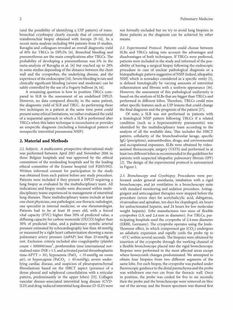
Of note, a SLB was not performed in patients witha histological NSIP pattern following TBLCs if a relatedcondition (such as a hypersensitivity pneumonitis) wasidentified by the multidisciplinary team according to theanalysis of all the available data. This includes the HRCTpattern, cellularity of the bronchoalveolar lavage, specificIgG (precipitins), autoantibodies, drugs, and environmentaland occupational exposures. SLBs were obtained by video-assisted thoracoscopic surgery (VATS) and performed in atleast two different lobes as recommended in the guidelines forpatients with suspected idiopathic pulmonary fibrosis (IPF)[2]. The design of the experimental protocol is summarizedin Figure 1.
2.3. Bronchoscopy and Cryobiopsy. Procedures were per-formed under general anesthesia, intubation with a rigidbronchoscope, and jet ventilation in a bronchoscopy suitewith standard monitoring and sedation procedure. Antiag-gregant and anticoagulant therapies were stopped before theprocedure (seven days for acetylsalicylic acid, dabigatran,rivaroxaban and apixaban, ten days for clopidogrel, six hoursfor unfractionated heparin, and 24 hours for low molecularweight heparin). Erbe manufactures two sizes of flexiblecryoprobes (1,9, and 2,4mm in diameter). For TBLCs, par-ticipating hospitals used the cryoprobe of 2.4mm diameter(ERBE, Germany). The cryoprobe operates using the Joule-Thomson effect, in which compressed gas (CO
2) undergoes
an adiabatic expansion and rapidly cools the probe tip to−45∘C within several seconds. The biopsies were obtained byinsertion of the cryoprobe through the working channel ofa flexible bronchoscope placed into the rigid bronchoscope.Biopsies were performed in the most affected areas exceptwhere honeycomb changes predominated. We attempted toobtain four biopsies from two different segments of thesame lobe. For each biopsy, the cryoprobe was pushed underfluoroscopic guidance to the distal parenchyma and the probewas withdrawn one-two cm from the thoracic wall. Oncein position, the probe was cooled for five to six seconds;then the probe and the bronchoscope were removed en blocout of the airway, and the frozen specimen was thawed first
Pulmonary Medicine 3
unde�ned DPLD
Indication for lung biopsy
TBLC
No diagnosis Speci�c pattern(except NSIP)
NSIP
SLB
Final diagnosis
MDD
MDD
MDD
MDD
Figure 1: Algorithm summarizing the study protocol. DPLD: diffuse parenchymal lung disease; MDD: multidisciplinary discussion; SLB:surgical lung biopsy; NSIP: nonspecific interstitial pneumonia; TBLC: transbronchial lung cryobiopsy.
in saline at room temperature and afterwards transferred toformalin for fixation. To control potential severe bleeding,a noninflated Fogarty balloon, previously placed in thelobar bronchus close to the sampled segment, was inflatedimmediately after biopsy and then deflated in case of absenceof hemorrhage. The bleeding was scored as follows: score 0 ifno bleeding, score 1 if bleeding stopped with aspiration onlyand/or insufflation of the Fogarty balloon less than 5minutes,score 2 if bleeding stopped by cold saline instillation and/orprolonged use of the Fogarty balloon (more than 5 minutes),and score 3 if life-threatening bleeding requiring any ofthe following: embolization, selective bronchial intubation,transfusion, admission in an intensive care unit (ICU), orresulting in death or prolonged hospitalization. Within 3 hafter the procedure, a chest X-ray was obtained to excludepneumothorax. All patients stay in the hospital for onenight after the procedure for monitoring (the aim of thismonitoring was to identify relapse of bleeding and subacutepneumothoraces).
2.4. Biopsy Specimens. Biopsy specimens were fixed in 10%formalin and embedded in paraffin. Hematoxylin and eosinas well as Masson’s Trichrome, Giemsa, staining was per-formed and immunostaining against pancytokeratins wasperformed. Specimens were reviewed by a pathologist expert
in interstitial lung disease. If case of doubt regarding thediagnosis, two other pathologists experts in interstitial lungdiseases could review the specimens to obtain a consensualdiagnosis.
3. Results
30 patientswere included in the study. Patient’s characteristicsare summarized in Table 1. The chest HRCT showed aninconsistent UIP pattern in the majority of the patients(80%) whereas a typical UIP pattern was present in onepatient. In this patient, pleural plaques were also noticedand the lung biopsy was performed to count asbestos bodies(differential diagnosis between idiopathic pulmonary fibrosisand asbestosis). Of note, cryobiopsies were sufficient formin-eralogical analysis. 333 asbestos bodies per gram of dry tissuewere identified and the diagnosis of asbestosis was thereforerejected (for an asbestosis, 5000 asbestos bodies per gram ofdry tissue or more are required) [18].
The mean number of biopsies by patient was 4,2 (range2–5) and the mean specimen area was 17mm2 (range10–40mm2). Cryobiopsies were mainly performed in thelower lobes (23 patients) whereas the upper lobes werebiopsied in six and the middle lobe in one patient. Maincomplications were pneumothorax and bleeding (Table 2).
4 Pulmonary Medicine
Table 1: Clinical characteristics of the patients (𝑁 = 30).
Gender Male𝑁 (%) 14 (47)Age, years Median (range) 62 (26–80)
Smoking historyCurrent𝑁 (%) 3 (10)Former𝑁 (%) 17 (57)Never𝑁 (%) 10 (33)
BMI Median (range) 29 (17–39)FVC, % predicted value Median (range) 73 (54–104)DLCo, % predicted value Median (range) 50 (30–76)
HRCTTypical UIP𝑁 (%) 1 (3)Possible UIP𝑁 (%) 5 (17)
Inconsistent UIP𝑁 (%) 24 (80)Number ofbiopsies/patient Mean (range) 4,2 (2–5)
Size of biopsies (mm2)
By specimen Mean(range)
16,6(9,5–40)
By patient Mean(range)
71,0(42–106)
BMI: body mass index; HRCT: high resolution chest tomography; FVC:forced vital capacity; DLCO: diffusion capacity of the lung for carbonmonoxide.
Table 2: Complications and histological diagnosis of the TBLCs(𝑁 = 30).
Hemorrhage𝑁 (%)
Grade 0 2 (7)Grade 1 16 (53)Grade 2 10 (33)Grade 3 2 (7)
Pneumothorax All 6 (20)𝑁 (% total patients) Requiring chest drainage 3 (10)
Detailed histologicaldiagnosis𝑁 (%)
HP 8 (27)UIP 7 (23)NSIP 6 (20)
Sarcoidosis 2 (7)DIP 1 (3)
Amyloidosis 1 (3)Eosinophilic pn. 1 (3)Adenocarcinoma 1 (3)Undetermined 3 (10)
HP: hypersensitivity pneumonia; UIP: usual interstitial pneumonia; NSIP:nonspecific interstitial pneumonia; DIP: desquamative interstitial pneumo-nia; eosinophilic pn.: eosinophilic pneumonia.
Pneumothorax occurred in six patients (20%) and requiredchest tube in three patients (50%), simple aspiration in twopatients (33%), and only observation in one. Bleeding was inthe majority of the cases mild (53%, grade 1) and moderate(33%, grade 2). Severe bleeding occurred in two patients andrequired a prolonged use of the Fogarty balloon and injectionof cold saline. One of these patients was also admitted inICU for overnight surveillance. With respect to the limitednumber of patient, we cannot definitely conclude but there
was no clear relationship between the number or the sizeof the biopsies and the risk of pneumothorax and bleeding(supplemental data, Figure S1 in Supplementary Materialavailable online at https://doi.org/10.1155/2017/6794343).
Interestingly, the mean hospitalization time after theprocedure was 1,3 days taking into account the mandatoryovernight monitoring. No significant chest pain amongpatients without pneumothorax was reported as well asprolonged air leak, infection, acute exacerbation, or death.
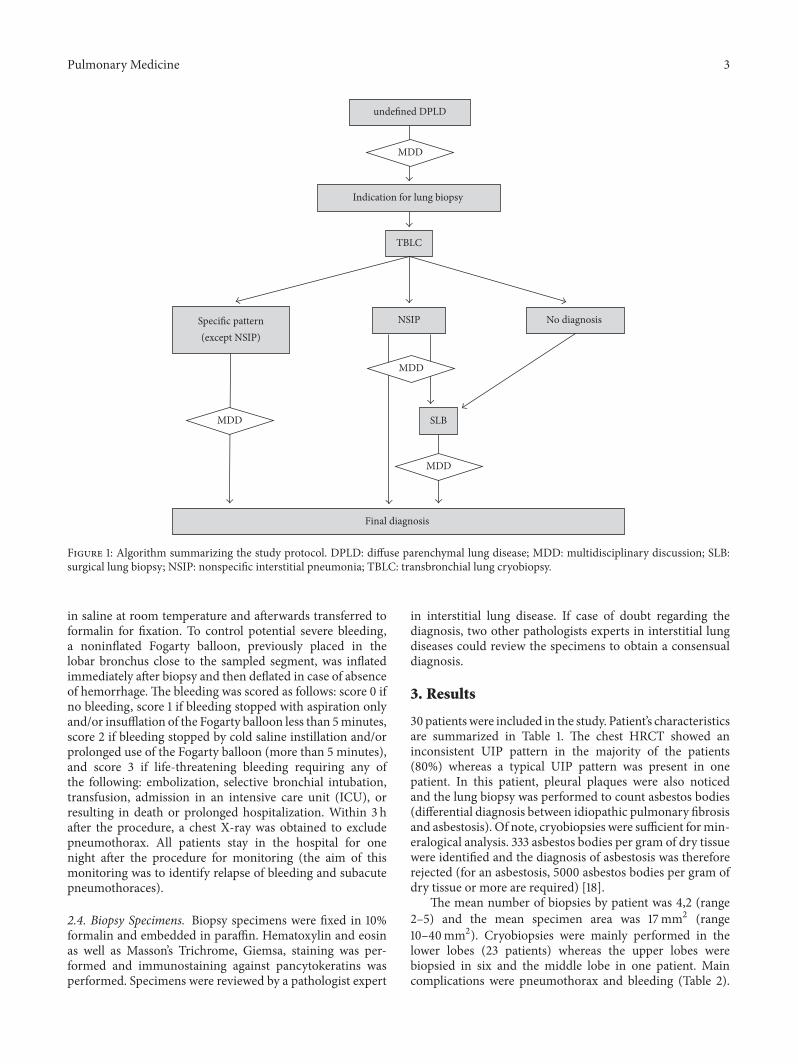
The overall diagnostic yield of TBLCs was 80% (24/30).This corresponds to patients with either a specific histologicalpattern other than an NSIP or patients with an NSIP forwhich this pattern could be related to a specific condition.The detailed diagnoses obtained are summarized in Figure 2and Table 2. A specific histological pattern (other than NSIP)was identified in 21 patients. Among these, the final diagnosis,after discussion within the multidisciplinary team, was IPFin seven patients and hypersensitivity pneumonitis (HP) ineight patients (the other less frequent diagnoses are listed inFigure 2). In three patients, no definite histological diagnosiswas identified, and, in six patients, an NSIP pattern wasobserved (four cellular and two fibrotic NSIP patterns). Inthree out of the six patients with an NSIP pattern, the pres-ence of this histological pattern helped the multidisciplinaryteam to identify a hypersensitivity pneumonitis taking intoaccount the other available clinical data such as exposures,BAL lymphocytosis, presence of specific antibodies (precip-itins), and radiological features. Therefore, in these patients,no surgical biopsy was proposed after TBLCs.
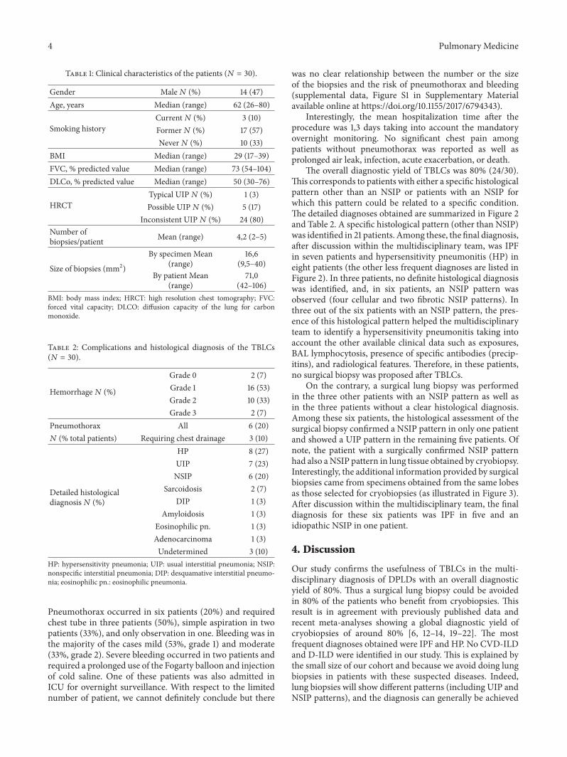
On the contrary, a surgical lung biopsy was performedin the three other patients with an NSIP pattern as well asin the three patients without a clear histological diagnosis.Among these six patients, the histological assessment of thesurgical biopsy confirmed a NSIP pattern in only one patientand showed a UIP pattern in the remaining five patients. Ofnote, the patient with a surgically confirmed NSIP patternhad also aNSIP pattern in lung tissue obtained by cryobiopsy.Interestingly, the additional information provided by surgicalbiopsies came from specimens obtained from the same lobesas those selected for cryobiopsies (as illustrated in Figure 3).After discussion within the multidisciplinary team, the finaldiagnosis for these six patients was IPF in five and anidiopathic NSIP in one patient.
4. Discussion
Our study confirms the usefulness of TBLCs in the multi-disciplinary diagnosis of DPLDs with an overall diagnosticyield of 80%. Thus a surgical lung biopsy could be avoidedin 80% of the patients who benefit from cryobiopsies. Thisresult is in agreement with previously published data andrecent meta-analyses showing a global diagnostic yield ofcryobiopsies of around 80% [6, 12–14, 19–22]. The mostfrequent diagnoses obtained were IPF and HP. No CVD-ILDand D-ILD were identified in our study. This is explained bythe small size of our cohort and because we avoid doing lungbiopsies in patients with these suspected diseases. Indeed,lung biopsies will show different patterns (including UIP andNSIP patterns), and the diagnosis can generally be achieved
Pulmonary Medicine 5
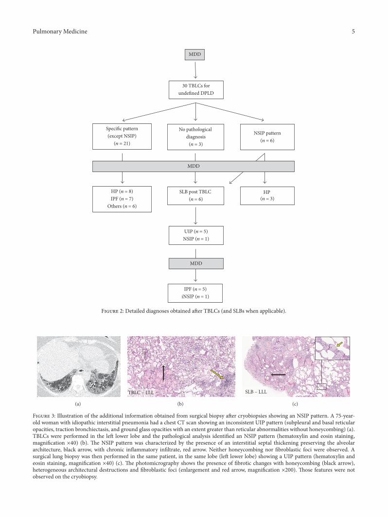
30 TBLCs forunde�ned DPLD
Speci�c pattern(except NSIP)
(n = 21)
No pathological diagnosis
(n = 3)
NSIP pattern(n = 6)
MDD
MDD
HP (n = 3)
SLB post TBLC (n = 6)
UIP (n = 5)NSIP (n = 1)
HP (n = 8)IPF (n = 7)
Others (n = 6)
MDD
IPF (n = 5)iNSIP (n = 1)
Figure 2: Detailed diagnoses obtained after TBLCs (and SLBs when applicable).
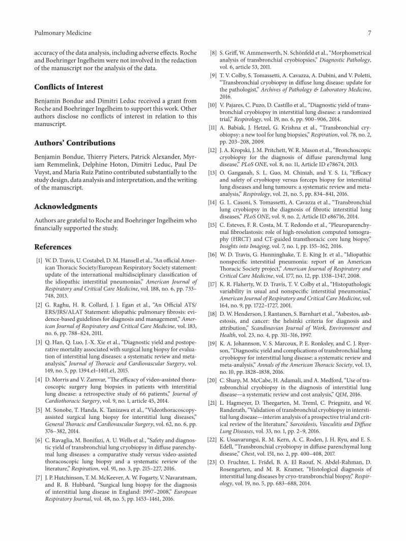
(a)
TBLC – LLL
(b)
SLB – LLL
(c)
Figure 3: Illustration of the additional information obtained from surgical biopsy after cryobiopsies showing an NSIP pattern. A 75-year-old woman with idiopathic interstitial pneumonia had a chest CT scan showing an inconsistent UIP pattern (subpleural and basal reticularopacities, traction bronchiectasis, and ground glass opacities with an extent greater than reticular abnormalities without honeycombing) (a).TBLCs were performed in the left lower lobe and the pathological analysis identified an NSIP pattern (hematoxylin and eosin staining,magnification ×40) (b). The NSIP pattern was characterized by the presence of an interstitial septal thickening preserving the alveolararchitecture, black arrow, with chronic inflammatory infiltrate, red arrow. Neither honeycombing nor fibroblastic foci were observed. Asurgical lung biopsy was then performed in the same patient, in the same lobe (left lower lobe) showing a UIP pattern (hematoxylin andeosin staining, magnification ×40) (c). The photomicrography shows the presence of fibrotic changes with honeycombing (black arrow),heterogeneous architectural destructions and fibroblastic foci (enlargement and red arrow, magnification ×200). Those features were notobserved on the cryobiopsy.

6 Pulmonary Medicine
on the basis of other clinical data (clinical history, presence ofextrapulmonary signs/symptoms, presence of autoantibod-ies, and results of extrapulmonary investigations). Anotherconcern could be the capacity of cryobiopsies to identifyoverlap of histologic patterns and lymphoid nodules oftenproved to be related to CVD-ILD [1].
Compared to SLB, other benefits of TBLCs are lowermorbidity and shorter hospitalization time (slightly morethan one day after the procedure in our study) supporting therecently reported cost-effectiveness of this technique [20].Wealso confirm that bleeding and pneumothorax are the maincomplications following TBLCs. Bleeding is mostly mild tomoderate. Severe bleeding can occur and is controlled bythe use of a Fogarty balloon. Pneumothorax rate is around20%, as expected when biopsies are performed between1 and 2 cm of the thoracic wall, but the placement of achest tube is not always mandatory [6, 14, 23]. There wasno clear relationship between the number, the size, or thelocalisation of the biopsies and the risk of pneumothorax andbleeding. However, the study was not designed to analysethose relationships. Indeed, the cohort was too small withonly one patient having two biopsies whereas the others hadfour or five biopsies,mostly in the lower lobes.This resulted ina significant underrepresentation ofmany subgroups to allowaccurate comparisons and formal conclusions.
In order to definitely compare the ability of TBLCs toidentify accurately a specific histological pattern, both tech-niques (TBLCs and SLBs) should be performed in the samepatients. However, such data have not been published so farand raise ethical limitations. Of interest, our study providesdata from some patients having a sequential approach withboth procedures (TBLCs first, and afterwards SLB) evalu-ating the added value of SLB to TBLC alone in terms ofdiagnostic yield. As we thought it is unethical to performthe two procedures in all patients, we selected two specificsituations in which we hypothesized that SLB could providecomplementary information, that is, when the histologicalpattern was inconclusive or showed a NSIP (except if arelated condition/etiology could be identified supporting theaccuracy of this histological diagnosis). The rationale forperforming SLB after TBLCs in such situations is based onretrospective analysis showing a slightly lower diagnosticyield for TBLC than for SLB (82.8% versus 98.7%, resp.) [6].Moreover, in 26% of IPF patients who benefit from a SLB,a discordant UIP pattern is present with a UIP pattern in alobe and a NSIP in another one [17]. The accuracy of TBLCsto identify such discordant patterns has not been studied sofar and could be lower than for SLB taking into account thenotion that the sizes of the specimens are lower and generallyperformed in only one lobe. Therefore, in the present study,we proposed to perform a SLB after TBLC in patients with anunclear histological pattern following TBLCs or when a NSIPpattern was present without related condition.
This situation occurred in six patients out of 30 (20%).Interestingly, in five out of these six patients (83%) the SLBprovided additional information. Indeed, the histologicaldiagnosis was a typical UIP pattern, changing significantlythe diagnosis, the prognosis, and the treatment of these fivepatients, as the final multidisciplinary diagnosis was IPF. In
the majority of these five patients, the cryobiopsies wereperformed in the lower lobes (4/5) whereas the surgicalbiopsy was performed in two lobes (the upper and the lowerlobes). Interestingly, it is not the surgical specimen obtainedfrom the noncryobiopsied lobewho induces the change in thediagnosis (as illustrated in the Figure 3). In fact, SLB providesmore information mostly because the size of the biopsieswas greater and therefore more pathological features couldbe identified. Finally, idiopathic NSIP was only diagnosed inone patient out of 30 (3%) confirming that this diagnosis isrelatively rare.
In conclusion, our data confirm the role of TBLCs forthemultidisciplinary diagnosis of DPLDs when a lung biopsyis required. Morbidity and hospitalization time are lowerthan after surgical lung biopsy [6]. The diagnostic yieldis around 80%, thus avoiding a SLB in 80% of the cases.Therefore, our data support that TBLCs should be the firstline procedure when the analysis of a lung biopsy is required.However, when an inconclusive result or a NSIP patternwithout related condition (idiopathic NSIP) is obtained, aSLB performed after TBLC could provide complementaryinformation. Altogether, those data, even preliminary andlimited, support the concept of the sequential approach asmade for the mediastinal staging of lung cancer. In thisapproach, endoscopic procedure (TBLCs) is performed firstand SLB is reserved for inconclusive or NSIP results aftercryobiopsies. Other studies are required to confirm thesepreliminary data and to support this sequential approach. Inthe meanwhile, chest physicians should cautiously interpretNSIP results, especially in idiopathic condition.
Abbreviations
APTT: Activated partial thromboplastin timeBMI: Body mass indexCVD-ILD: Collagen vascular disease-associated
interstitial lung diseaseD-ILD: Drug-induced interstitial lung diseaseDLCO: Diffusion capacity for carbon monoxideDPLD: Diffuse parenchymal lung diseaseFVC: Forced vital capacityHP: Hypersensitivity pneumonitisHRCT: High resolution computed tomographyICU: Intensive care unitINR: Prothrombin time international
normalized ratioIPF: Idiopathic pulmonary fibrosisMDD: Multidisciplinary discussionmPAP: Mean pulmonary artery pressureNSIP: Nonspecific interstitial pneumoniaSLB: Surgical lung biopsyTBLC: Transbronchial lung cryobiopsyUIP: Usual interstitial pneumoniaVATS: Video-assisted thoracoscopic surgery.
Disclosure
Benjamin Bondue had full access to all of the data in the studyand takes responsibility for the integrity of the data and the
Pulmonary Medicine 7
accuracy of the data analysis, including adverse effects. Rocheand Boehringer Ingelheimwere not involved in the redactionof the manuscript nor the analysis of the data.
Conflicts of Interest
Benjamin Bondue and Dimitri Leduc received a grant fromRoche and Boehringer Ingelheim to support this work. Otherauthors disclose no conflicts of interest in relation to thismanuscript.
Authors’ Contributions
Benjamin Bondue, Thierry Pieters, Patrick Alexander, Myr-iam Remmelink, Delphine Hoton, Dimitri Leduc, Paul DeVuyst, andMaria Ruiz Patino contributed substantially to thestudy design, data analysis and interpretation, and thewritingof the manuscript.
Acknowledgments
Authors are grateful to Roche and Boehringer Ingelheimwhofinancially supported the study.
References
[1] W.D. Travis, U. Costabel, D.M.Hansell et al., “An official Amer-icanThoracic Society/European Respiratory Society statement:update of the international multidisciplinary classification ofthe idiopathic interstitial pneumonias,” American Journal ofRespiratory and Critical Care Medicine, vol. 188, no. 6, pp. 733–748, 2013.
[2] G. Raghu, H. R. Collard, J. J. Egan et al., “An Official ATS/ERS/JRS/ALAT Statement: idiopathic pulmonary fibrosis: evi-dence-based guidelines for diagnosis and management,” Amer-ican Journal of Respiratory and Critical Care Medicine, vol. 183,no. 6, pp. 788–824, 2011.
[3] Q. Han, Q. Luo, J.-X. Xie et al., “Diagnostic yield and postope-rative mortality associated with surgical lung biopsy for evalua-tion of interstitial lung diseases: a systematic review and meta-analysis,” Journal of Thoracic and Cardiovascular Surgery, vol.149, no. 5, pp. 1394.e1–1401.e1, 2015.
[4] D. Morris and V. Zamvar, “The efficacy of video-assisted thora-coscopic surgery lung biopsies in patients with interstitiallung disease: a retrospective study of 66 patients,” Journal ofCardiothoracic Surgery, vol. 9, no. 1, article 45, 2014.
[5] M. Sonobe, T. Handa, K. Tanizawa et al., “Videothoracoscopy-assisted surgical lung biopsy for interstitial lung diseases,”General Thoracic and Cardiovascular Surgery, vol. 62, no. 6, pp.376–382, 2014.
[6] C. Ravaglia, M. Bonifazi, A. U.Wells et al., “Safety and diagnos-tic yield of transbronchial lung cryobiopsy in diffuse parenchy-mal lung diseases: a comparative study versus video-assistedthoracoscopic lung biopsy and a systematic review of theliterature,” Respiration, vol. 91, no. 3, pp. 215–227, 2016.
[7] J. P. Hutchinson, T.M.McKeever, A.W. Fogarty, V.Navaratnam,and R. B. Hubbard, “Surgical lung biopsy for the diagnosisof interstitial lung disease in England: 1997–2008,” EuropeanRespiratory Journal, vol. 48, no. 5, pp. 1453–1461, 2016.
[8] S. Griff,W. Ammenwerth, N. Schonfeld et al., “Morphometricalanalysis of transbronchial cryobiopsies,” Diagnostic Pathology,vol. 6, article 53, 2011.
[9] T. V. Colby, S. Tomassetti, A. Cavazza, A. Dubini, and V. Poletti,“Transbronchial cryobiopsy in diffuse lung disease: update forthe pathologist,” Archives of Pathology & Laboratory Medicine,2016.
[10] V. Pajares, C. Puzo, D. Castillo et al., “Diagnostic yield of trans-bronchial cryobiopsy in interstitial lung disease: a randomizedtrial,” Respirology, vol. 19, no. 6, pp. 900–906, 2014.
[11] A. Babiak, J. Hetzel, G. Krishna et al., “Transbronchial cry-obiopsy: a new tool for lung biopsies,” Respiration, vol. 78, no. 2,pp. 203–208, 2009.
[12] J. A. Kropski, J. M. Pritchett,W. R.Mason et al., “Bronchoscopiccryobiopsy for the diagnosis of diffuse parenchymal lungdisease,” PLoS ONE, vol. 8, no. 11, Article ID e78674, 2013.
[13] O. Ganganah, S. L. Guo, M. Chiniah, and Y. S. Li, “Efficacyand safety of cryobiopsy versus forceps biopsy for interstitiallung diseases and lung tumours: a systematic review and meta-analysis,” Respirology, vol. 21, no. 5, pp. 834–841, 2016.
[14] G. L. Casoni, S. Tomassetti, A. Cavazza et al., “Transbronchiallung cryobiopsy in the diagnosis of fibrotic interstitial lungdiseases,” PLoS ONE, vol. 9, no. 2, Article ID e86716, 2014.
[15] C. Esteves, F. R. Costa, M. T. Redondo et al., “Pleuroparenchy-mal fibroelastosis: role of high-resolution computed tomogra-phy (HRCT) and CT-guided transthoracic core lung biopsy,”Insights into Imaging, vol. 7, no. 1, pp. 155–162, 2016.
[16] W. D. Travis, G. Hunninghake, T. E. King Jr. et al., “Idiopathicnonspecific interstitial pneumonia: report of an AmericanThoracic Society project,” American Journal of Respiratory andCritical Care Medicine, vol. 177, no. 12, pp. 1338–1347, 2008.
[17] K. R. Flaherty, W. D. Travis, T. V. Colby et al., “Histopathologicvariability in usual and nonspecific interstitial pneumonias,”American Journal of Respiratory and Critical Care Medicine, vol.164, no. 9, pp. 1722–1727, 2001.
[18] D.W. Henderson, J. Rantanen, S. Barnhart et al., “Asbestos, asb-estosis, and cancer: the helsinki criteria for diagnosis andattribution,” Scandinavian Journal of Work, Environment andHealth, vol. 23, no. 4, pp. 311–316, 1997.
[19] K. A. Johannson, V. S. Marcoux, P. E. Ronksley, and C. J. Ryer-son, “Diagnostic yield and complications of transbronchial lungcryobiopsy for interstitial lung disease: a systematic review andmeta-analysis,” Annals of the AmericanThoracic Society, vol. 13,no. 10, pp. 1828–1838, 2016.
[20] C. Sharp,M.McCabe, H. Adamali, and A.Medford, “Use of tra-nsbronchial cryobiopsy in the diagnosis of interstitial lungdisease—a systematic review and cost analysis,” QJM, 2016.
[21] L. Hagmeyer, D. Theegarten, M. Treml, C. Priegnitz, and W.Randerath, “Validation of transbronchial cryobiopsy in intersti-tial lung disease—interim analysis of a prospective trial and crit-ical review of the literature,” Sarcoidosis, Vasculitis and DiffuseLung Diseases, vol. 33, no. 1, pp. 2–9, 2016.
[22] K. Ussavarungsi, R. M. Kern, A. C. Roden, J. H. Ryu, and E. S.Edell, “Transbronchial cryobiopsy in diffuse parenchymal lungdisease,” Chest, vol. 151, no. 2, pp. 400–408, 2017.
[23] O. Fruchter, L. Fridel, B. A. El Raouf, N. Abdel-Rahman, D.Rosengarten, and M. R. Kramer, “Histological diagnosis ofinterstitial lung diseases by cryo-transbronchial biopsy,” Respir-ology, vol. 19, no. 5, pp. 683–688, 2014.

Submit your manuscripts athttps://www.hindawi.com
Stem CellsInternational
Hindawi Publishing Corporationhttp://www.hindawi.com Volume 2014
Hindawi Publishing Corporationhttp://www.hindawi.com Volume 2014
MEDIATORSINFLAMMATION
of
Hindawi Publishing Corporationhttp://www.hindawi.com Volume 2014
Behavioural Neurology
EndocrinologyInternational Journal of
Hindawi Publishing Corporationhttp://www.hindawi.com Volume 2014
Hindawi Publishing Corporationhttp://www.hindawi.com Volume 2014
Disease Markers
Hindawi Publishing Corporationhttp://www.hindawi.com Volume 2014
BioMed Research International
OncologyJournal of
Hindawi Publishing Corporationhttp://www.hindawi.com Volume 2014
Hindawi Publishing Corporationhttp://www.hindawi.com Volume 2014
Oxidative Medicine and Cellular Longevity
Hindawi Publishing Corporationhttp://www.hindawi.com Volume 2014
PPAR Research
The Scientific World JournalHindawi Publishing Corporation http://www.hindawi.com Volume 2014
Immunology ResearchHindawi Publishing Corporationhttp://www.hindawi.com Volume 2014
Journal of
ObesityJournal of
Hindawi Publishing Corporationhttp://www.hindawi.com Volume 2014
Hindawi Publishing Corporationhttp://www.hindawi.com Volume 2014
Computational and Mathematical Methods in Medicine
OphthalmologyJournal of
Hindawi Publishing Corporationhttp://www.hindawi.com Volume 2014
Diabetes ResearchJournal of
Hindawi Publishing Corporationhttp://www.hindawi.com Volume 2014
Hindawi Publishing Corporationhttp://www.hindawi.com Volume 2014
Research and TreatmentAIDS
Hindawi Publishing Corporationhttp://www.hindawi.com Volume 2014
Gastroenterology Research and Practice
Hindawi Publishing Corporationhttp://www.hindawi.com Volume 2014
Parkinson’s Disease
Evidence-Based Complementary and Alternative Medicine
Volume 2014Hindawi Publishing Corporationhttp://www.hindawi.com


![Interstitial lung disease (ILD), or diffuse parenchymal lung disease … · 2018-10-28 · Interstitial lung disease (ILD), or diffuse parenchymal lung disease (DPLD),[[1] is a group](https://img.pdfslide.net/doc/110x75/5e7d31d2ec5074254471c7d0/interstitial-lung-disease-ild-or-diffuse-parenchymal-lung-disease-2018-10-28.jpg)