Embed Size (px)
Citation preview
Diagnostic Work-up of Renal Insufficiency in Solid Organ
Transplant Recipients
Beatrice P. Concepcion MD Assistant Professor of Medicine
Division of Nephrology and Hypertension Vanderbilt University Medical Center
Objectives
• To differentiate Acute Kidney Injury (AKI) from Chronic Kidney Disease (CKD)
• To enumerate common causes of AKI in solid organ transplant (SOT) recipients
• To understand the role of different diagnostic tests in the work-up of causes of AKI in SOT recipients
• To enumerate common causes of CKD in SOT recipients • To understand the utility of different diagnostic tests in SOT recipients
with CKD
CASE 1: In your office
• 65/M with a history of kidney transplant in 2012, HTN and DM, just moved to Nashville, complaining of fatigue, nausea and vomiting.
• Lab check: BUN 75, Creatinine 3.8

THE DIAGNOSTIC APPROACH
• Acute, subacute or chronic ??? • Baseline creatinine • Clinical elements of the case
• Onset of symptoms • Urine output • Rate of rise of creatinine
• Renal ultrasound: size and echogenicity of kidneys * • Anemia * • Hypocalcemia and hyperphosphatemia
CASE 1: In your office
• 65/M with a history of kidney transplant in 2012, HTN and DM, just moved to Nashville, complaining of fatigue, nausea and vomiting.
• Lab check: BUN 75, Creatinine 3.8 • Sudden decrease in urine output • Hgb 13.1, Phos 3.5, Ca 10.2, iPTH 30 • Renal ultrasound: normal sized-kidneys with normal echogenicity • Additional records: creatinine 1 month ago was 1.2
ACUTE KIDNEY INJURY • Abrupt loss of kidney function • Retention of urea and other nitrogenous waste products • Dysregulation of extracellular volume and electrolytes • Easily detected by measurement of the serum creatinine which is
used to estimate the glomerular filtration rate (GFR) *
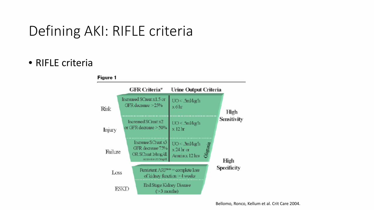
Defining AKI: RIFLE criteria
• RIFLE criteria
Bellomo, Ronco, Kellum et al. Crit Care 2004.
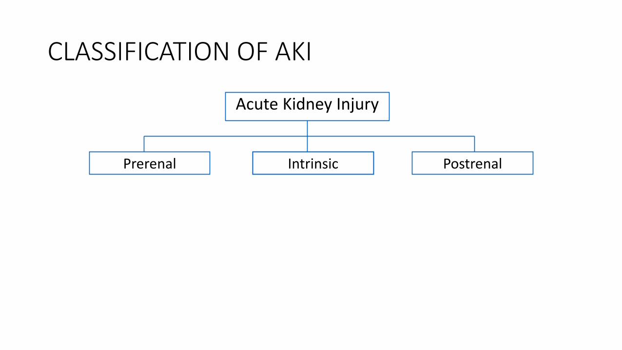
CLASSIFICATION OF AKI
Acute Kidney Injury
Prerenal Intrinsic Postrenal

In formulating AKI differentials, remember, a kidney transplant is…
• A TRANSPLANT. • Rejection: Cellular, Antibody-mediated • Infection: Pyelonephritis, BK nephropathy • Med side effects: CNI toxicity, thrombotic microangiopathy
• SOLITARY. • Obstruction • Renal Artery Stenosis • Renal Vein Thrombosis
• A KIDNEY. • Disease Recurrence • Everything else!

Case 2: In the ER
• 75/M with a liver transplant and h/o prostate CA, complaining of decreased urine output x 3 days.
• Lab check: BUN 65, Creatinine 6.8 (baseline 1.2 a year ago) • UA: pH 5.5, sg 1.010, 1 RBC, no protein

CLASSIFICATION OF AKI
Acute Kidney Injury
Prerenal Intrinsic Postrenal
• Obstruction from renal pelvis to urethra • Bilateral obstruction or unilateral with solitary functioning
kidney • Increased pressure leads nephron destruction • Oliguria/anuria common • UA generally not helpful • Hydronephrosis on imaging

CLASSIFICATION OF AKI
Acute Kidney Injury
Prerenal Intrinsic Postrenal
• Prostate hypertrophy • Neurogenic bladder • Intraureteral obstruction – crystals (uric acid, acyclovir,
indinavir), stones, clots, tumor • Extraureteral obstruction – tumor (cervical, prostate),
retroperitoneal fibrosis*
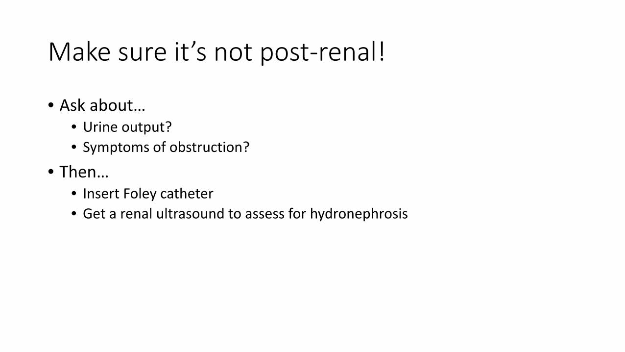
Make sure it’s not post-renal!
• Ask about… • Urine output? • Symptoms of obstruction?
• Then… • Insert Foley catheter • Get a renal ultrasound to assess for hydronephrosis
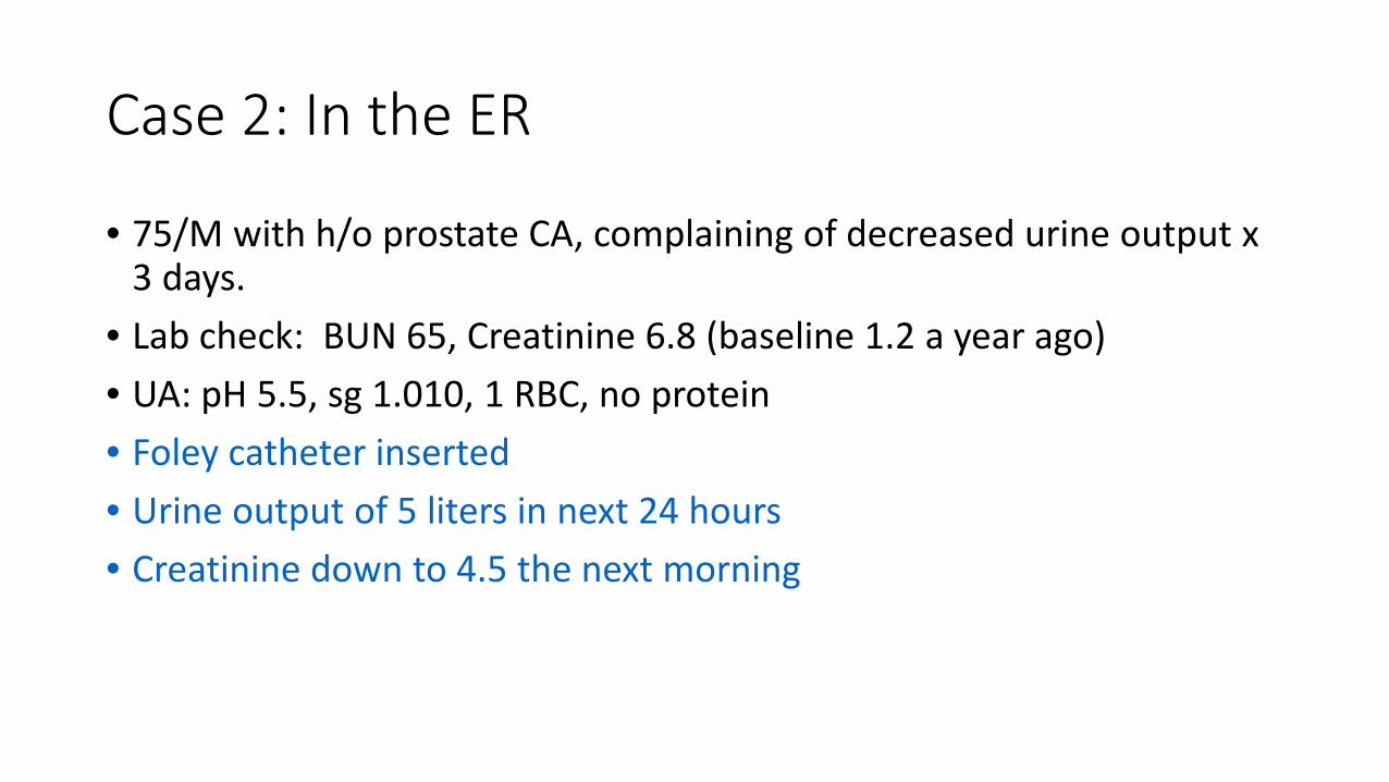
Case 2: In the ER
• 75/M with h/o prostate CA, complaining of decreased urine output x 3 days.
• Lab check: BUN 65, Creatinine 6.8 (baseline 1.2 a year ago) • UA: pH 5.5, sg 1.010, 1 RBC, no protein • Foley catheter inserted • Urine output of 5 liters in next 24 hours • Creatinine down to 4.5 the next morning
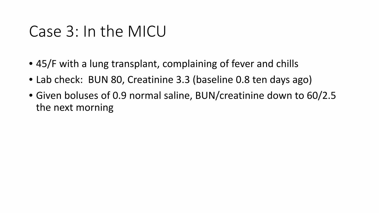
Case 3: In the MICU
• 45/F with a lung transplant, complaining of fever and chills • Lab check: BUN 80, Creatinine 3.3 (baseline 0.8 ten days ago) • Given boluses of 0.9 normal saline, BUN/creatinine down to 60/2.5
the next morning
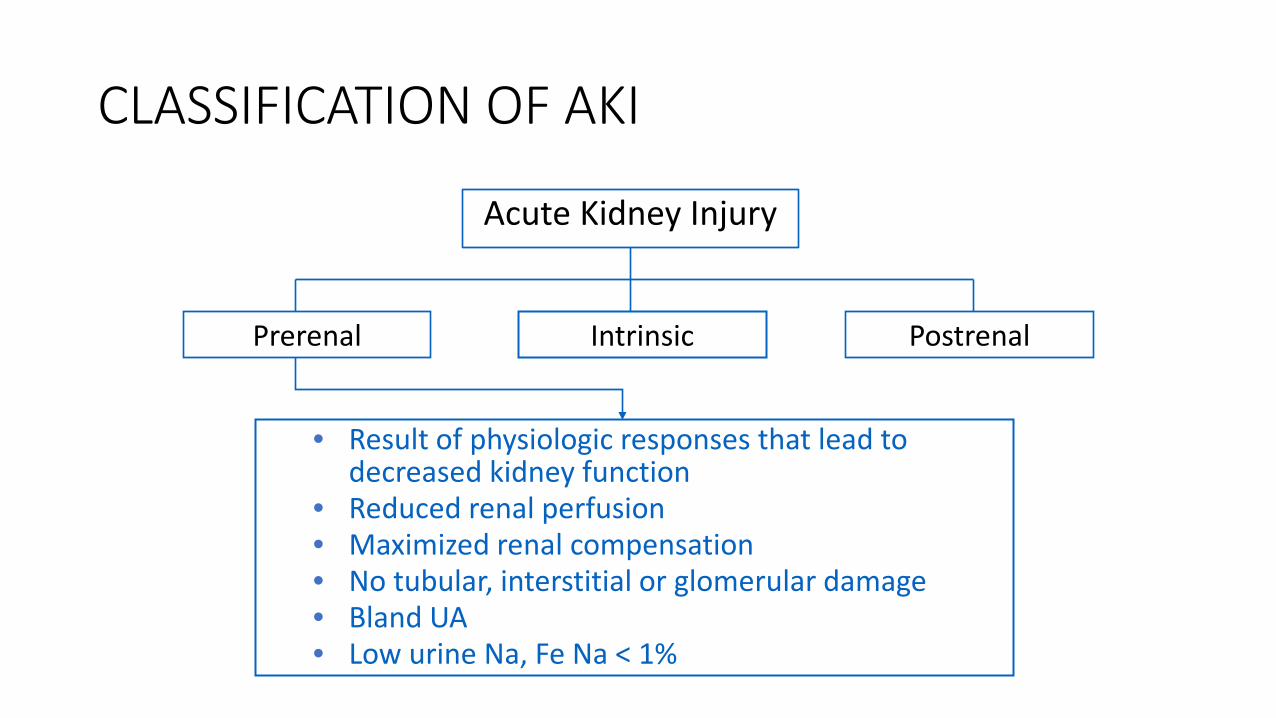
CLASSIFICATION OF AKI
Acute Kidney Injury
Prerenal Intrinsic Postrenal
• Result of physiologic responses that lead to decreased kidney function
• Reduced renal perfusion • Maximized renal compensation • No tubular, interstitial or glomerular damage • Bland UA • Low urine Na, Fe Na < 1%
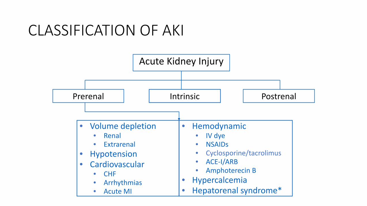
CLASSIFICATION OF AKI
Acute Kidney Injury
Prerenal Intrinsic Postrenal
• Volume depletion • Renal • Extrarenal
• Hypotension • Cardiovascular
• CHF • Arrhythmias • Acute MI
• Hemodynamic • IV dye • NSAIDs • Cyclosporine/tacrolimus • ACE-I/ARB • Amphoterecin B
• Hypercalcemia • Hepatorenal syndrome*

Case 5: In the Transplant Unit
• 75/F s/p Kidney Transplant (2002), HTN and DM, admitted with abdominal pain, diarrhea, vomiting x 3 days
• Meds include: prograf, prednisone, lisinopril • Took Ibuprofen x 3 doses for pain • BP 90/60, HR 65, RR 18 • Lab check: BUN 65, Creat 3.5 (baseline 2.3 two months ago) • Prograf trough level=18
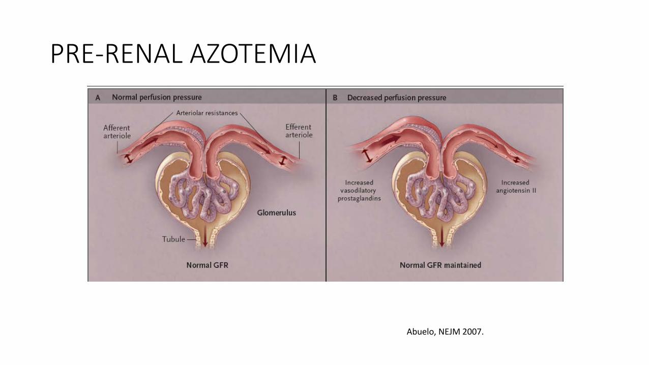
PRE-RENAL AZOTEMIA
Abuelo, NEJM 2007.
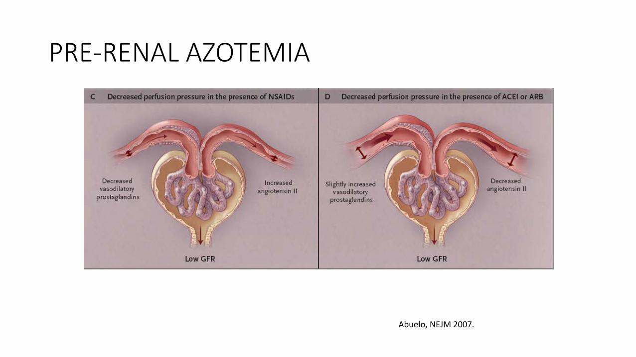
PRE-RENAL AZOTEMIA
Abuelo, NEJM 2007.
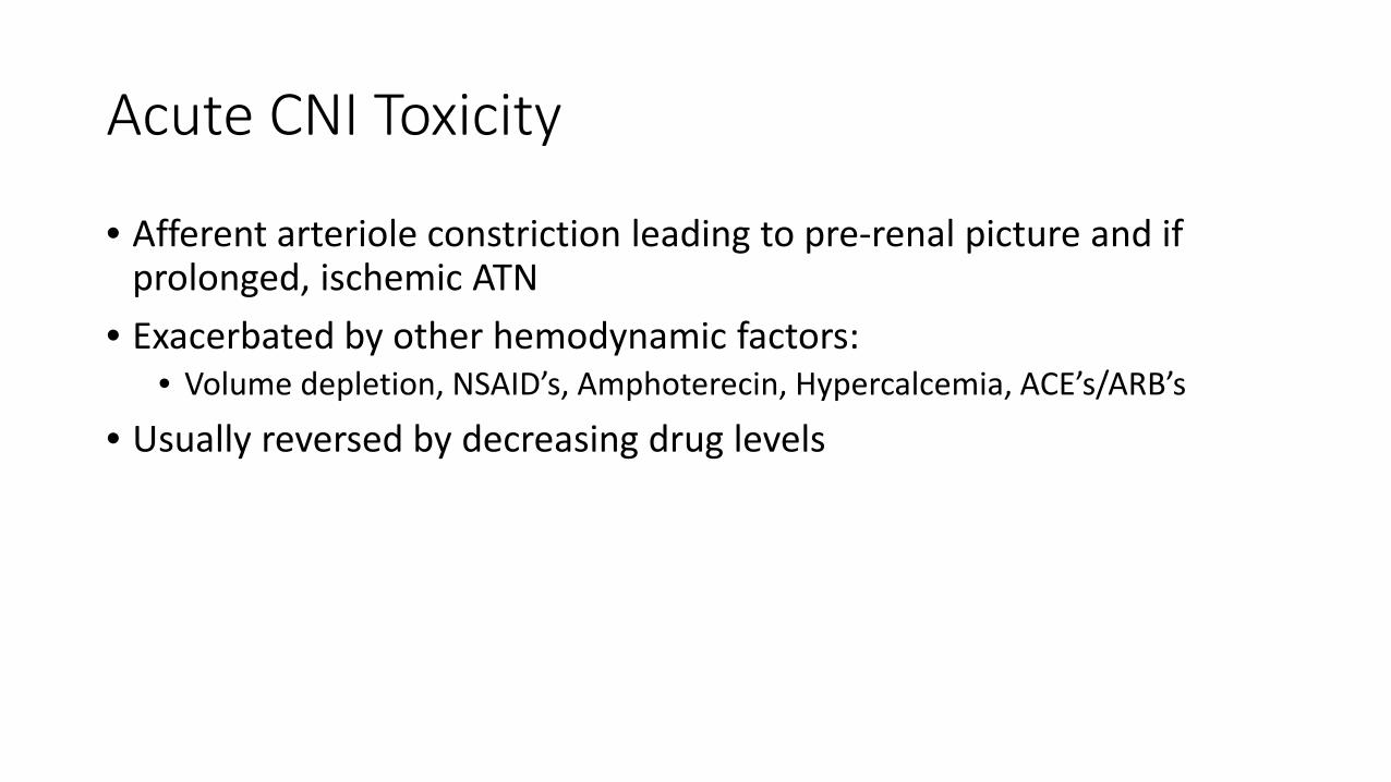
Acute CNI Toxicity
• Afferent arteriole constriction leading to pre-renal picture and if prolonged, ischemic ATN
• Exacerbated by other hemodynamic factors: • Volume depletion, NSAID’s, Amphoterecin, Hypercalcemia, ACE’s/ARB’s
• Usually reversed by decreasing drug levels
Case 5: In the Transplant Unit
• Given 2 liters normal saline for volume depletion • Lisinopril and prograf held • Educated about avoidance of NSAIDS! • Creatinine improved to 2.5 after 2 days
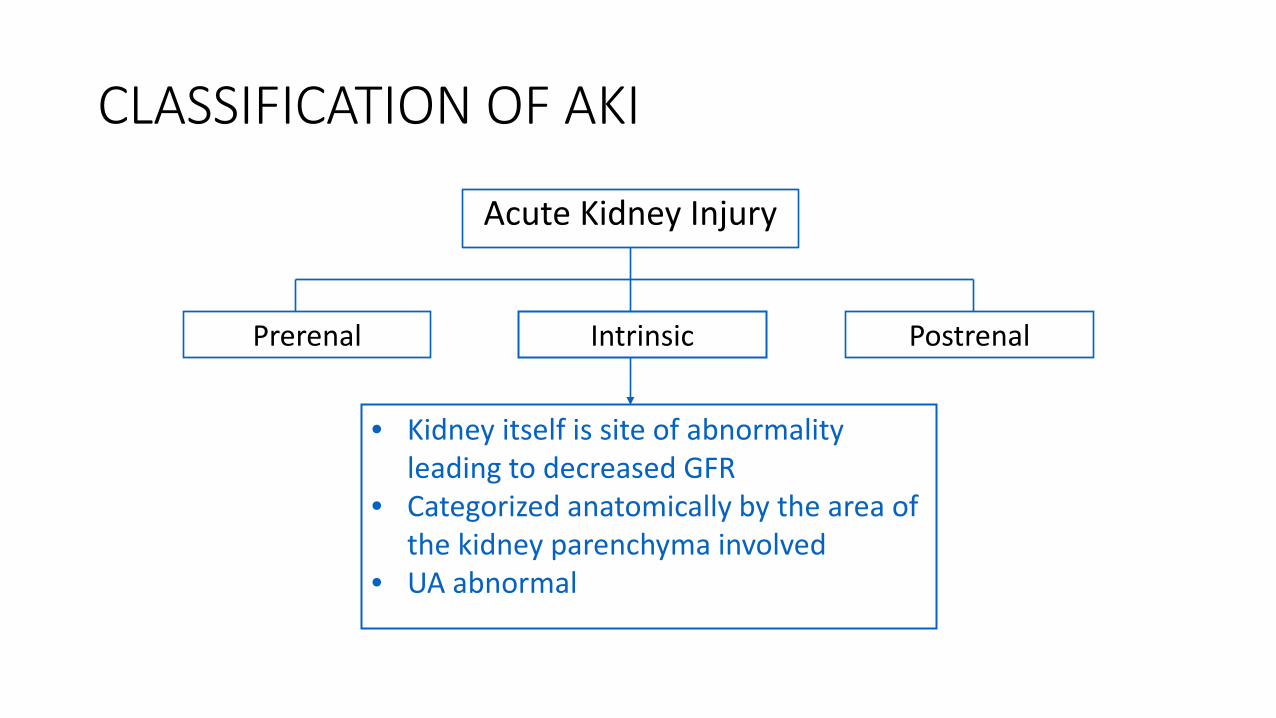
CLASSIFICATION OF AKI
Acute Kidney Injury
Prerenal Intrinsic Postrenal
• Kidney itself is site of abnormality leading to decreased GFR
• Categorized anatomically by the area of the kidney parenchyma involved
• UA abnormal

CLASSIFICATION OF AKI
Acute Kidney Injury
Prerenal Intrinsic Postrenal
• Glomerular • Vascular • Interstitial • Tubular
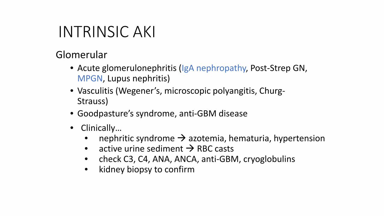
INTRINSIC AKI Glomerular
• Acute glomerulonephritis (IgA nephropathy, Post-Strep GN, MPGN, Lupus nephritis)
• Vasculitis (Wegener’s, microscopic polyangitis, Churg-Strauss)
• Goodpasture’s syndrome, anti-GBM disease • Clinically…
• nephritic syndrome azotemia, hematuria, hypertension • active urine sediment RBC casts • check C3, C4, ANA, ANCA, anti-GBM, cryoglobulins • kidney biopsy to confirm
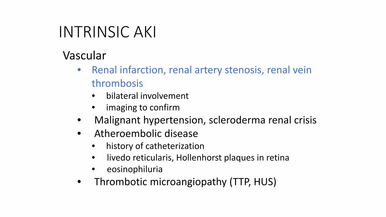
INTRINSIC AKI Vascular
• Renal infarction, renal artery stenosis, renal vein thrombosis • bilateral involvement • imaging to confirm
• Malignant hypertension, scleroderma renal crisis • Atheroembolic disease
• history of catheterization • livedo reticularis, Hollenhorst plaques in retina • eosinophiluria
• Thrombotic microangiopathy (TTP, HUS)
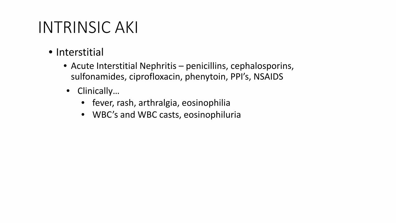
INTRINSIC AKI • Interstitial
• Acute Interstitial Nephritis – penicillins, cephalosporins, sulfonamides, ciprofloxacin, phenytoin, PPI’s, NSAIDS
• Clinically… • fever, rash, arthralgia, eosinophilia • WBC’s and WBC casts, eosinophiluria
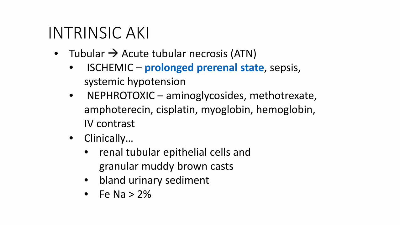
INTRINSIC AKI • Tubular Acute tubular necrosis (ATN)
• ISCHEMIC – prolonged prerenal state, sepsis, systemic hypotension
• NEPHROTOXIC – aminoglycosides, methotrexate, amphoterecin, cisplatin, myoglobin, hemoglobin, IV contrast • Clinically… • renal tubular epithelial cells and
granular muddy brown casts • bland urinary sediment • Fe Na > 2%

IT’S ALL IN THE UA!!!
• pH • Specific gravity • Blood • RBC • WBC • Protein • Granular casts • Eosinophils
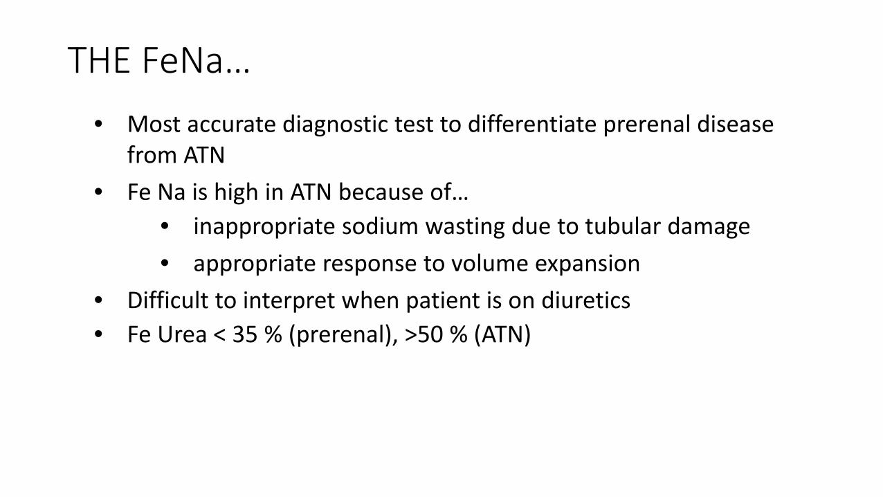
THE FeNa… • Most accurate diagnostic test to differentiate prerenal disease
from ATN • Fe Na is high in ATN because of…
• inappropriate sodium wasting due to tubular damage • appropriate response to volume expansion
• Difficult to interpret when patient is on diuretics • Fe Urea < 35 % (prerenal), >50 % (ATN)
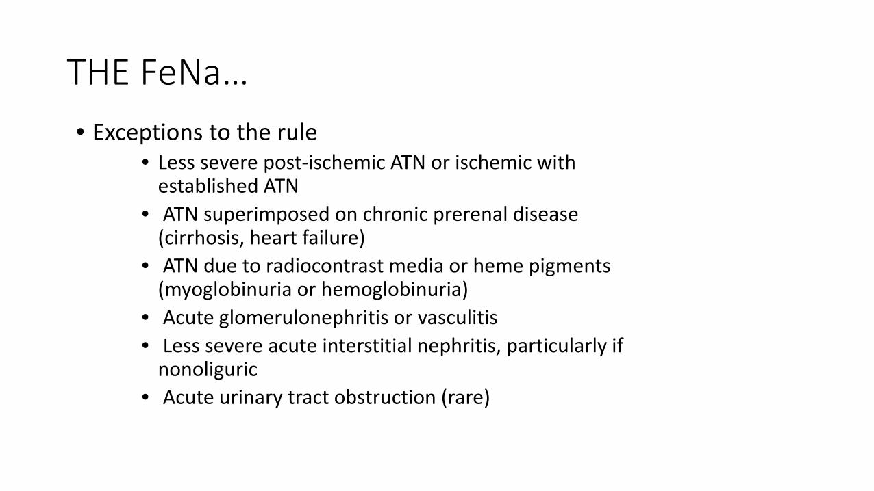
THE FeNa… • Exceptions to the rule
• Less severe post-ischemic ATN or ischemic with established ATN
• ATN superimposed on chronic prerenal disease (cirrhosis, heart failure)
• ATN due to radiocontrast media or heme pigments (myoglobinuria or hemoglobinuria)
• Acute glomerulonephritis or vasculitis • Less severe acute interstitial nephritis, particularly if
nonoliguric • Acute urinary tract obstruction (rare)
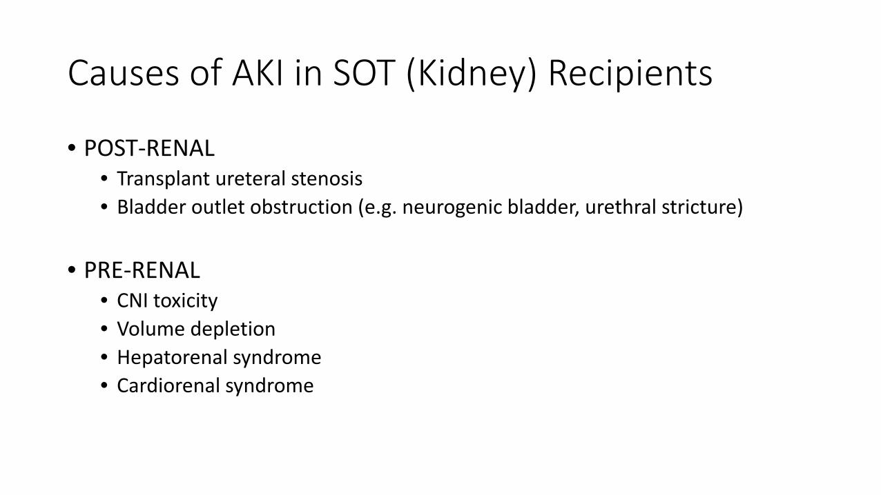
Causes of AKI in SOT (Kidney) Recipients
• POST-RENAL • Transplant ureteral stenosis • Bladder outlet obstruction (e.g. neurogenic bladder, urethral stricture)
• PRE-RENAL • CNI toxicity • Volume depletion • Hepatorenal syndrome • Cardiorenal syndrome
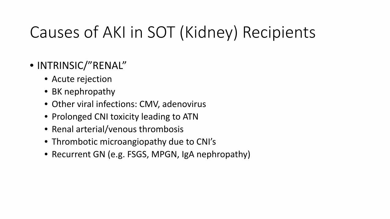
Causes of AKI in SOT (Kidney) Recipients
• INTRINSIC/”RENAL” • Acute rejection • BK nephropathy • Other viral infections: CMV, adenovirus • Prolonged CNI toxicity leading to ATN • Renal arterial/venous thrombosis • Thrombotic microangiopathy due to CNI’s • Recurrent GN (e.g. FSGS, MPGN, IgA nephropathy)
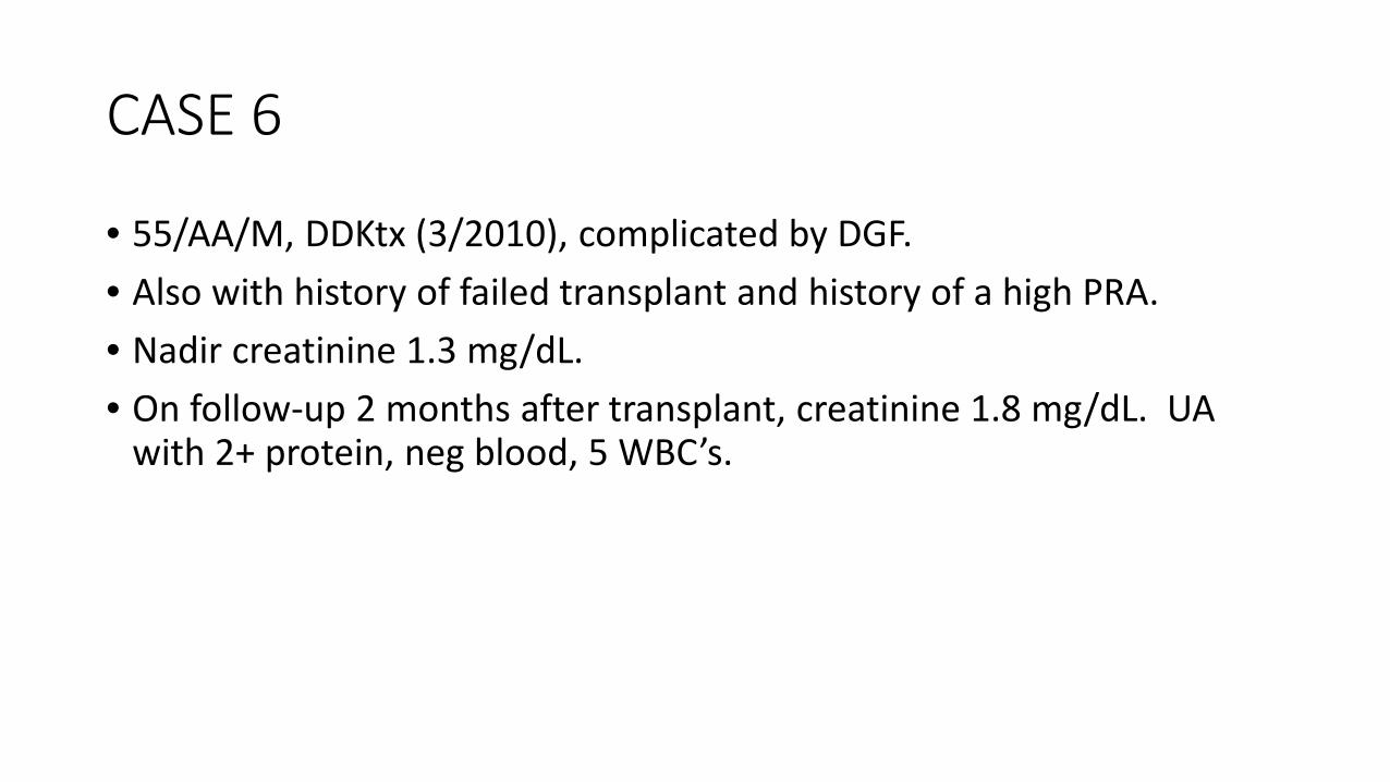
CASE 6
• 55/AA/M, DDKtx (3/2010), complicated by DGF. • Also with history of failed transplant and history of a high PRA. • Nadir creatinine 1.3 mg/dL. • On follow-up 2 months after transplant, creatinine 1.8 mg/dL. UA
with 2+ protein, neg blood, 5 WBC’s.
Case 6: Acute Rejection of a Kidney Transplant
• Acute Cellular Rejection • Acute Humoral Rejection • Both
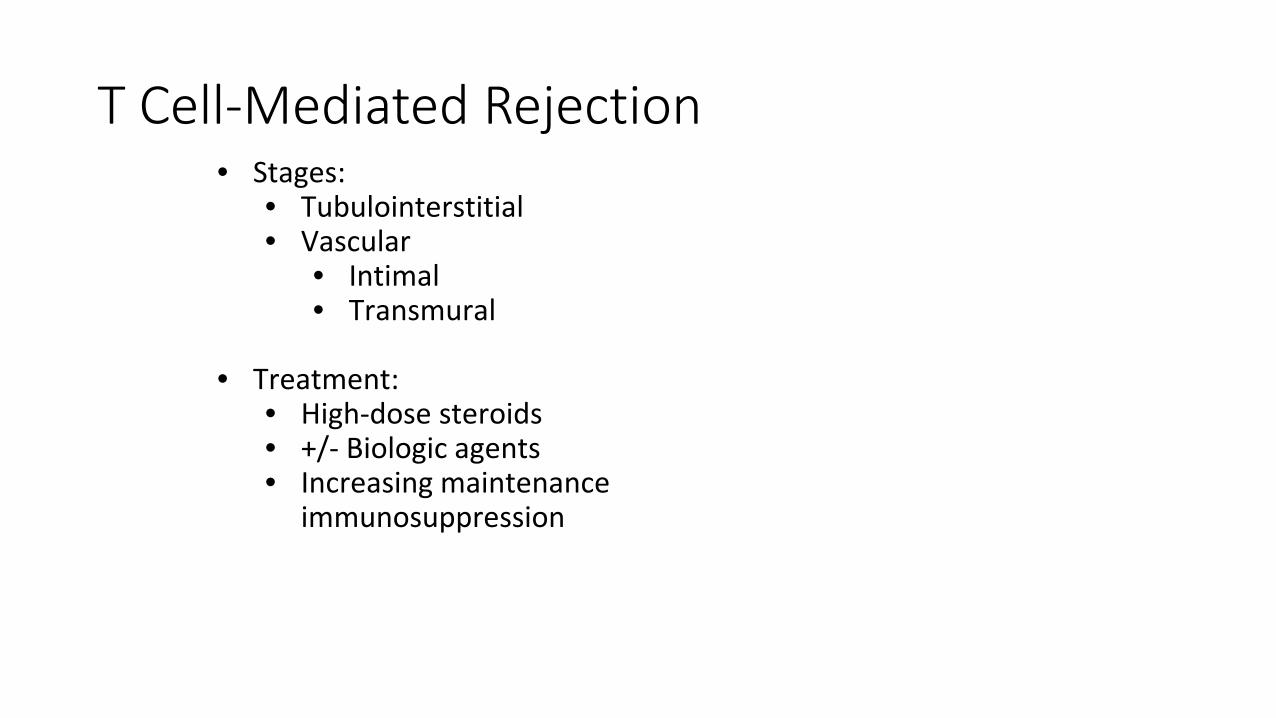
T Cell-Mediated Rejection • Stages:
• Tubulointerstitial • Vascular
• Intimal • Transmural
• Treatment: • High-dose steroids • +/- Biologic agents • Increasing maintenance
immunosuppression

Antibody-Mediated Rejection • Diagnosis:
• Allograft Dysfunction • C4d positivity • Donor-specific Ab’s
• Treatment: • IVIG • Plasmapheresis • Rituximab • Thymoglobulin • Splenectomy
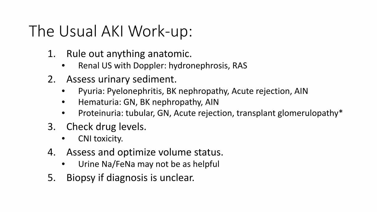
The Usual AKI Work-up: 1. Rule out anything anatomic.
• Renal US with Doppler: hydronephrosis, RAS 2. Assess urinary sediment.
• Pyuria: Pyelonephritis, BK nephropathy, Acute rejection, AIN • Hematuria: GN, BK nephropathy, AIN • Proteinuria: tubular, GN, Acute rejection, transplant glomerulopathy*
3. Check drug levels. • CNI toxicity.
4. Assess and optimize volume status. • Urine Na/FeNa may not be as helpful
5. Biopsy if diagnosis is unclear.
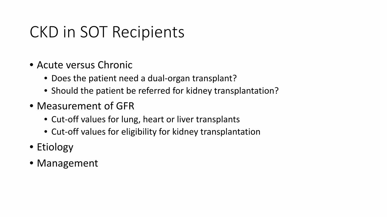
CKD in SOT Recipients
• Acute versus Chronic • Does the patient need a dual-organ transplant? • Should the patient be referred for kidney transplantation?
• Measurement of GFR • Cut-off values for lung, heart or liver transplants • Cut-off values for eligibility for kidney transplantation
• Etiology • Management

How do you know it’s CKD? • There is a good reason for CKD (e.g. longstanding DM/HTN). • Sustained reduction in GFR (3 months) • Urine sodium NOT low • Proteinuria • Ultrasound findings
• Small kidneys • Echogenic kidneys • Cortical thinning
• Biopsy findings • Glomerulosclerosis • Tubular atrophy and interstitial fibrosis

Measurements of GFR • Estimated GFR
• Cockroft-Gault • MDRD • CKD-EPI
• Measured GFR • Creatinine clearance
• 24-hour urine collection (24-hour creatinine to ensure adequate collection) • DTPA
Etiology of CKD in SOT recipients
• Diabetes • Hypertension • CNI toxicity • Prolonged ATN • Others:
• Glomerular disease • ADPKD
Chronic Allograft Nephropathy
• Chronic rejection • Transplant glomerulopathy
• CNI toxicity • BK nephropathy • Diabetes • Hypertension • Others:
• Recurrent Disease • De novo glomerulonephritis • Nephrocalcinosis
Management of CKD
• Treat underlying cause if possible. • Blood pressure control, goal of at least <130/80 • Reduction in proteinuria with RAAS blockade • Avoidance of nephrotoxic agents • Avoidance of volume depletion and hypotension