Embed Size (px)
Citation preview
7/28/2019 Differences Between EMGs of Forearm Skeletal Muscles for Flick Strokes
http://slidepdf.com/reader/full/differences-between-emgs-of-forearm-skeletal-muscles-for-flick-strokes 1/4151
International Journal of Table Tennis Sciences, No.6(2010)
Differences between EMGs of Forearm Skeletal Muscles for Flick Strokes
against Backspin and No-spin Services in Table Tennis
Kazuto YOSHIDA1, Koji SUGIYAMA
2, Shin Murakoshi
3
Faculty of Education, Shizuoka University, Shizuoka, Japan
(1Tel: +81-54-238-4692; E-mail: [email protected])
(
2
Tel: +81-54-238-4997; E-mail: [email protected])(3Tel: +81-54-238-4665; E-mail: [email protected])
Abstract: We have conducted an experimental study to clarify the differences between the EMGs for the forearm
skeletal muscles when receiving backspin and no-spin services with a forehand flick stroke. An elite Japanese table
tennis player participated as a subject in this study. A Chinese coach, acting as a server, sent a service ball and the
subject returned it with a forehand flick stroke. The service ball speed was approximately 4 m/s. The receiver was
informed whether the service ball had spin or not. A significant difference (p<0.05) between the two kinds of services
was shown for the electrical discharge amount of M. extensor carpi ulnarise (Backspin: 122.06±42.10 and No-spin:
87.12±36.39microV). There were no significant differences for other muscles. It is assumed that the electrical
discharge of M. extensor carpi ulnarise of the subject for 15ms just before the impact is concerned with controlling
the racket surface.
Keywords: table tennis, forehand flick stroke, EMG, forearm skeletal muscle, racket control
1.Purpose
There are several techniques in table tennis for
returning balls that land near the net. These techniques
are considered extremely important, and closely related
to competition results.
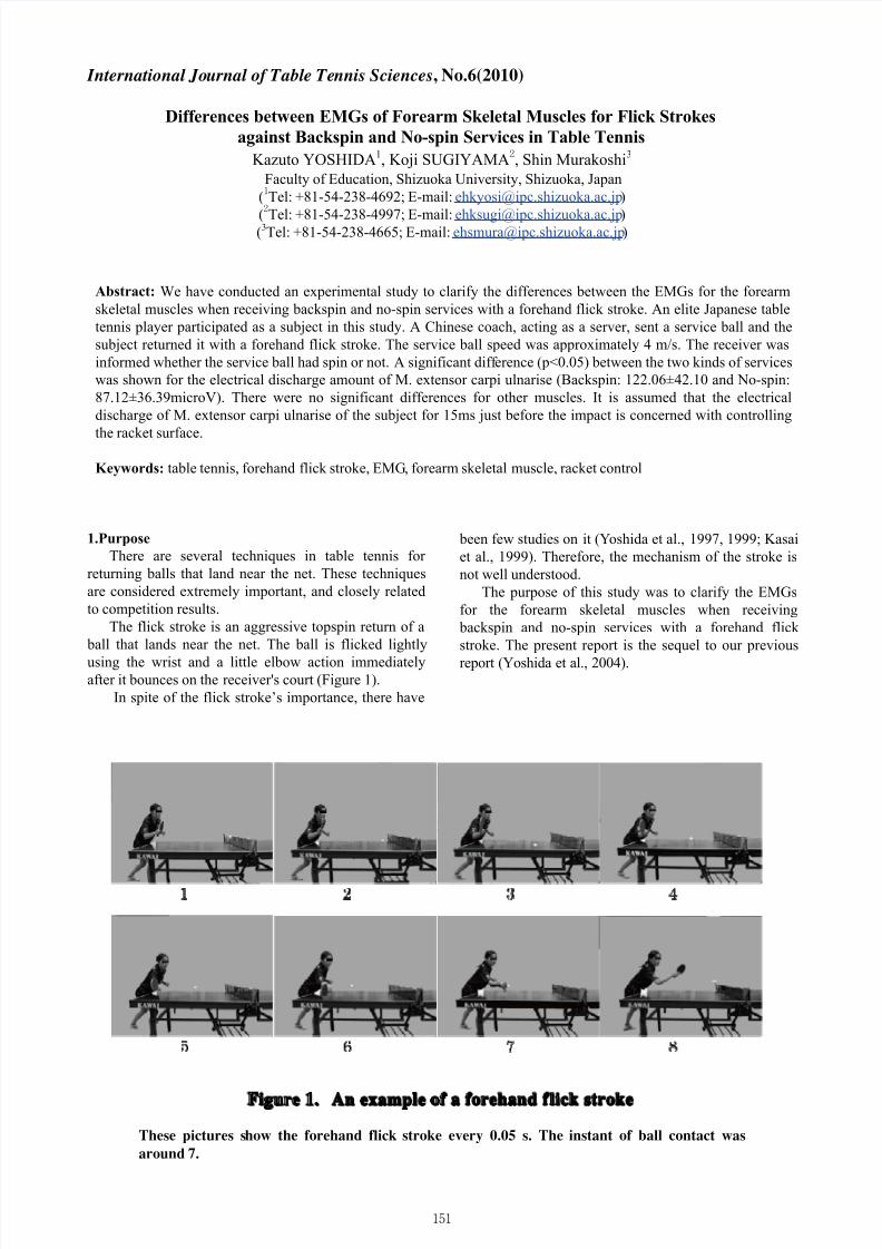
The flick stroke is an aggressive topspin return of a
ball that lands near the net. The ball is flicked lightly
using the wrist and a little elbow action immediatelyafter it bounces on the receiver's court (Figure 1).
In spite of the flick stroke’s importance, there have
been few studies on it (Yoshida et al., 1997, 1999; Kasai
et al., 1999). Therefore, the mechanism of the stroke is
not well understood.
The purpose of this study was to clarify the EMGs
for the forearm skeletal muscles when receiving
backspin and no-spin services with a forehand flick
stroke. The present report is the sequel to our previous
report (Yoshida et al., 2004).
Figure 1. An example of a forehand f lick stroke
These pictures show the forehand flick stroke every 0.05 s. The instant of ball contact was
around 7.
7/28/2019 Differences Between EMGs of Forearm Skeletal Muscles for Flick Strokes
http://slidepdf.com/reader/full/differences-between-emgs-of-forearm-skeletal-muscles-for-flick-strokes 2/4152
Kazuto Yoshida, et al.
2.Methods
2.1 Subject
An elite table tennis player participated as a subject
in the present study. He was a finalist in the All
American Open U-22 (1999) and a member of the
Japanese national team of the 2001 Universiade. Table
1 shows the characteristics of the subject. We obtainedhis agreement to attend the experiment after explaining
its purpose and safety aspects.
2.2 Experimental Procedure
A Chinese coach acting as a server sent a service
ball and the subject returned it with a forehand flick
stroke. The service balls were controlled to land in a
circle of 20cm radius on the right half court of the
receiver. The center of this circle was 50cm from the end
line and 40cm from the sideline. The service ball speed
was approximately 4m/s. The receiver was required to
return the ball into a 25cm radius circle on the right half court of the server. The center of this circle was 95 cm
from the net and 30 cm from the sideline. Two cases,
backspin (B) and no-spin (N), were examined. There
was little variability in service ball speed and spin. The
receiver was informed whether or not the service ball
would have spin before the server served. The tests were
repeated for each case until the receiver succeeded in
returning the ball precisely more than 5 times as required.
In practice, about 10 trials were necessary for each case.
2.3 Measurement Items
Measurements of muscular activities were made for
the following muscles: extensor carpi ulnaris, extensor
digitorum communis, extensor carpi radialis lognus and
brevis, flexor carpi radialis, and pronator teres. Muscular
electrical discharge was measured by a surface dipole
dielectric method. After treatment to reduce skin
resistance, miniature bio-electrodes 12mm in diameter
(NT-611U: Nihonkoden, Tokyo, Japan) were set at
20mm centers along the line of muscles following the
Zipp method (1982). The angles of the elbow joint and
wrist joint were measured by goniometers (M110,
M180: Penny and Giles, Gwent, UK). The angle of the
elbow joint showed flexion and extension, and the
angles of the wrist joint showed flexion and extension,abduction and adduction. Acceleration sensors
(AS-100HA: Kyowa Electronic Instruments, Tokyo,
Japan) were installed on the table and racket to record
the moment of bouncing. Using a data analyzing system
(MP100WS: Biopac Systems, California, USA) and PC
(iMac: Apple Computer, California, USA), all the analog
signals were sampled at a sampling frequency of 1kHz
and converted to digital data for further processing.
Furthermore, the motion of the subject in the test wasrecorded by a digital video camera (DCR-TRV10: Sony,
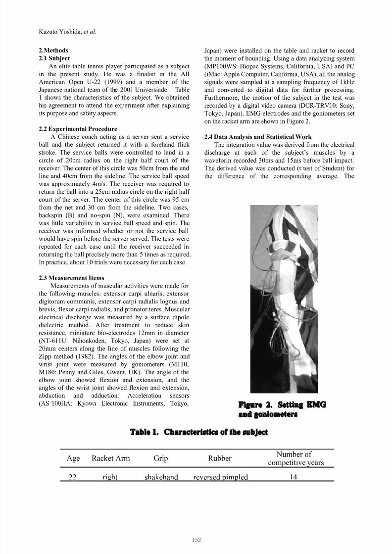
Tokyo, Japan). EMG electrodes and the goniometers set
on the racket arm are shown in Figure 2.
2.4 Data Analysis and Statistical Work
The integration value was derived from the electrical
discharge at each of the subject’s muscles by a
waveform recorded 30ms and 15ms before ball impact.
The derived value was conducted (t test of Student) for
the difference of the corresponding average. The
Number of competitive years
22 right shakehand reversed pimpled 14
Age Racket Arm Rubber Grip
Table 1. Characteristics of the subject
Figure 2. Setting EMG
and goniometers
7/28/2019 Differences Between EMGs of Forearm Skeletal Muscles for Flick Strokes
http://slidepdf.com/reader/full/differences-between-emgs-of-forearm-skeletal-muscles-for-flick-strokes 3/4153
Differences between EMGs of Forearm Skeletal Muscles for Flick Strokes
significant level was p<0.05.
3. Results
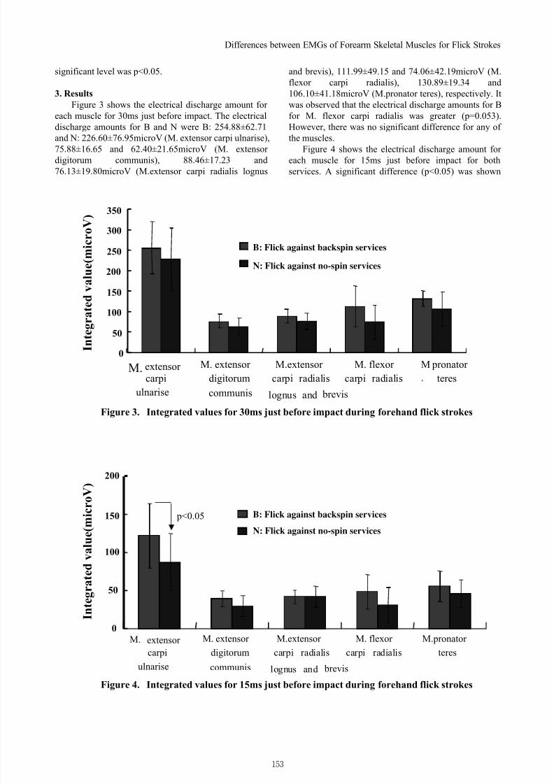
Figure 3 shows the electrical discharge amount for
each muscle for 30ms just before impact. The electrical
discharge amounts for B and N were B: 254.88±62.71
and N: 226.60±76.95microV (M. extensor carpi ulnarise),75.88±16.65 and 62.40±21.65microV (M. extensor
digitorum communis), 88.46±17.23 and
76.13±19.80microV (M.extensor carpi radialis lognus
and brevis), 111.99±49.15 and 74.06±42.19microV (M.
flexor carpi radialis), 130.89±19.34 and
106.10±41.18microV (M.pronator teres), respectively. It
was observed that the electrical discharge amounts for B
for M. flexor carpi radialis was greater (p=0.053).
However, there was no significant difference for any of
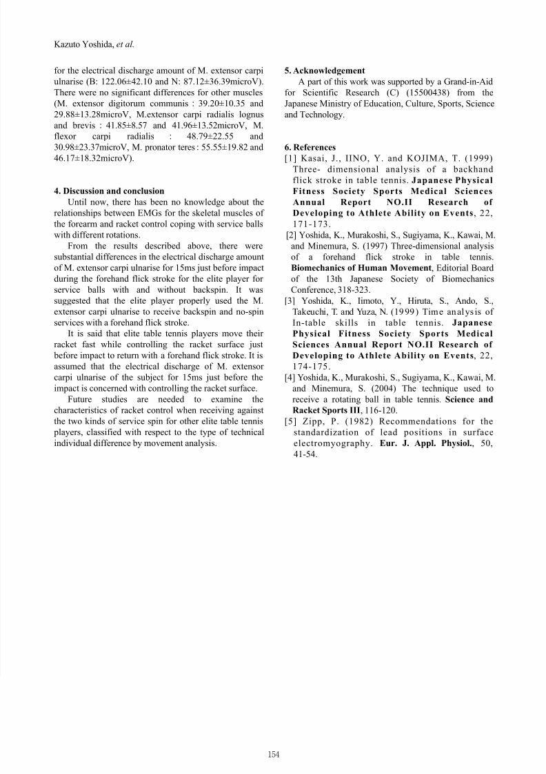
the muscles.Figure 4 shows the electrical discharge amount for
each muscle for 15ms just before impact for both
services. A significant difference (p<0.05) was shown
0
50
100
150
200
250
300
350
B: Flick against back-spin services
N: Flick against no services
M. extensor carpi
ulnarise
M. extensor
digitorum
M.extensor
carpi radialis
lognus and brevis
M. flexor
carpi radialis
M
.
pronator
teres
communis
I n t e g r a t e d v a l u
e ( m i c r o V )
0
50
100
150
200
M. extensor
carpi
ulnarise
M. extensor
digitorum
M.extensor
carpi radialis
lognus and brevis
M. flexor
carpi radialis
M.pronator
teres
communis
p<0.05 B: Flick against back-spin services
N: Flick against no services
I n t e g r a t e d v a l u e ( m i c r o V )
Figure 4. Integrated values for 15ms just before impact during forehand flick strokes
Figure 3. Integrated values for 30ms just before impact during forehand flick strokes
B: Flick against backspin services
N: Flick against no-spin services
B: Flick against backspin services
N: Flick against no-spin services
7/28/2019 Differences Between EMGs of Forearm Skeletal Muscles for Flick Strokes
http://slidepdf.com/reader/full/differences-between-emgs-of-forearm-skeletal-muscles-for-flick-strokes 4/4154
Kazuto Yoshida, et al. for the electrical discharge amount of M. extensor carpi
ulnarise (B: 122.06±42.10 and N: 87.12±36.39microV).
There were no significant differences for other muscles
(M. extensor digitorum communis:39.20±10.35 and
29.88±13.28microV, M.extensor carpi radialis lognus
and brevis:41.85±8.57 and 41.96±13.52microV, M.
flexor carpi radialis : 48.79±22.55 and30.98±23.37microV, M. pronator teres:55.55±19.82 and
46.17±18.32microV).
4. Discussion and conclusion
Until now, there has been no knowledge about the
relationships between EMGs for the skeletal muscles of
the forearm and racket control coping with service balls
with different rotations.
From the results described above, there were
substantial differences in the electrical discharge amount
of M. extensor carpi ulnarise for 15ms just before impactduring the forehand flick stroke for the elite player for
service balls with and without backspin. It was
suggested that the elite player properly used the M.
extensor carpi ulnarise to receive backspin and no-spin
services with a forehand flick stroke.
It is said that elite table tennis players move their
racket fast while controlling the racket surface just
before impact to return with a forehand flick stroke. It is
assumed that the electrical discharge of M. extensor
carpi ulnarise of the subject for 15ms just before the
impact is concerned with controlling the racket surface.
Future studies are needed to examine the
characteristics of racket control when receiving against
the two kinds of service spin for other elite table tennis
players, classified with respect to the type of technical
individual difference by movement analysis.
5. Acknowledgement
A part of this work was supported by a Grand-in-Aid
for Scientific Research (C) (15500438) from the
Japanese Ministry of Education, Culture, Sports, Science
and Technology.
6. References
[1] Kasai, J., IINO, Y. and KOJIMA, T. (1999)
Three- dimensional analysis of a backhand
flick stroke in table tennis. Japanese Physical
Fitness Society Sports Medical Sciences
Annual Report NO.II Research of
Developing to Athlete Ability on Events , 22,
171-173.
[2] Yoshida, K., Murakoshi, S., Sugiyama, K., Kawai, M.
and Minemura, S. (1997) Three-dimensional analysis
of a forehand flick stroke in table tennis.
Biomechanics of Human Movement, Editorial Boardof the 13th Japanese Society of Biomechanics
Conference, 318-323.
[3] Yoshida, K., Iimoto, Y., Hiruta, S., Ando, S.,
Takeuchi, T. and Yuza, N. (1999) Time analys is of
In-table skills in table tennis. Japanese
Physical Fitness Society Sports Medical
Sciences Annual Report NO.II Research of
Developing to Athlete Ability on Events, 22,
174-175.
[4] Yoshida, K., Murakoshi, S., Sugiyama, K., Kawai, M.
and Minemura, S. (2004) The technique used to
receive a rotating ball in table tennis. Science and
Racket Sports III, 116-120.
[5] Zipp, P. (1982) Recommendations for the
standardization of lead positions in surface
electromyography. Eur. J. Appl. Physiol., 50,
41-54.