Embed Size (px)
Citation preview
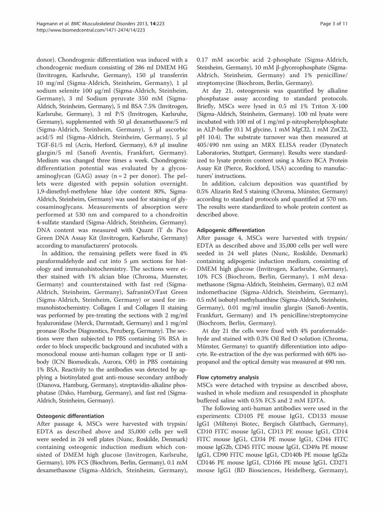
Hagmann et al. BMC Musculoskeletal Disorders 2013, 14:223http://www.biomedcentral.com/1471-2474/14/223
RESEARCH ARTICLE Open Access
Different culture media affect growthcharacteristics, surface marker distribution andchondrogenic differentiation of human bonemarrow-derived mesenchymal stromal cellsSebastien Hagmann1, Babak Moradi1, Sebastian Frank1, Thomas Dreher1, Peer Wolfgang Kämmerer2,Wiltrud Richter3 and Tobias Gotterbarm1*
Abstract
Background: Bone marrow-derived mesenchymal stromal cells (BM-MSCs) play an important role in modern tissueengineering, while distinct variations of culture media compositions and supplements have been reported. BecauseMSCs are heterogeneous regarding their regenerative potential and their surface markers, these parameters werecompared in four widely used culture media compositions.
Methods: MSCs were isolated from bone marrow and expanded in four established cell culture media. MSC yield/1000MNCs, passage time and growth index were observed. In P4, typical MSC surface markers were analysed by fluorescencecytometry. Additionally, chondrogenic, adipogenic and osteogenic differentiation potential were evaluated.
Results: Growth index and P0 cell yield varied importantly between the media. The different expansion media had asignificant influence on the expression of CD10, CD90, CD105, CD140b CD146 and STRO-1. While no significantdifferences were observed regarding osteogenic and adipogenic differentiation, chondrogenic differentiation wassuperior in medium A as reflected by GAG/DNA content.
Conclusions: The choice of expansion medium can have a significant influence on growth, differentiationpotential and surface marker expression of mesenchymal stromal cells, which is of fundamental importancefor tissue engineering procedures.
Keywords: Mesenchymal stromal cells, Expansion media, Surface markers, Osteogenic differentiation,Chondrogenic differentiation, Adipogenic differentiation
BackgroundSince their recognition in the 1960s and 70s [1,2], mes-enchymal stem or stromal cells (MSC) are consideredone of the most promising targets for regenerative medi-cine. MSC were first isolated from animal bone marrow(BM) [3], but a variety of tissues in humans, such as adi-pose tissue [4], cord blood [5], peripheral blood [6] andconnective tissues [7] have also proven to be a yieldingsource for these cells.
* Correspondence: [email protected] of Orthopedics, Trauma Surgery and Spinal Cord Injury,University Hospital Heidelberg, Germany Schlierbacher Landstrasse 200a,69118 Heidelberg, GermanyFull list of author information is available at the end of the article
© 2013 Hagmann et al.; licensee BioMed CentCommons Attribution License (http://creativecreproduction in any medium, provided the or
The potency of human MSC (hMSC) to differentiate intovarious cell lines, such as fibroblasts, myofibroblasts, osteo-blasts, chondroblasts and adipocytes [8,9] put these cellsinto the current focus of tissue engineering, particularly inthe fields of bone [10,11] and cartilage [12,13] regener-ation, but also in myocardial infarction [14,15].Minimal criteria for MSC have been postulated by the
International Society for Cellular Therapy [16]: theymust express CD105, CD73 and CD90 and lack expres-sion of hematopoietic markers such as CD45, CD34,CD14 and CD11b. In addition, MSC must be capable ofdifferentiating into fibroblasts, osteoblasts, adipocytesand chondroblasts under specific in vitro conditions.
ral Ltd. This is an Open Access article distributed under the terms of the Creativeommons.org/licenses/by/2.0), which permits unrestricted use, distribution, andiginal work is properly cited.

Hagmann et al. BMC Musculoskeletal Disorders 2013, 14:223 Page 2 of 11http://www.biomedcentral.com/1471-2474/14/223
However, there is an enormous variance in cell isola-tion techniques and expansion conditions for MSCs[12,17-19]. Basal culture media and supplements havebecome a considerable market with the growth of mes-enchymal stromal cell research. Up to this day, only littleattention was paid to the distinct variations of culturemedia, growth factors and other supplements used inthis field of research.Yet there is increasing evidence that the choice of cul-
ture media has an important influence on the biologicalproperties of MSCs [9,19-21]. Besides, recent studies re-port that bone marrow derived MSC may consist of sev-eral distinct subpopulations with diverse regenerativepotential [22,23]. However, the properties of these sub-populations and their usability in tissue engineering areonly marginally explored.The aim of this study therefore was to analyse the influ-
ence of four different widely used culture media on growthparameters and surface marker distribution of human BM-MSCs. Rather than determining the influence of single cul-ture additives, actual media compositions that were in usein our laboratory were compared to each other with themain focus on the impact on surface marker distribution.
MethodsBone marrow donorsHuman MSCs were isolated from bone marrow of 12donors (mean age 33 ± 21.6 years, 6 female, 6 male).Bone marrow aspiration was performed in the tibia (n = 2),femur (n = 2) or the iliac crest (n = 8) within the frameworkof total joint arthroplasty or osteotomy. The study protocolhad been approved by the ethics committee of the Uni-versity of Heidelberg, Germany. All patients providedinformed consent according to the latest version of theHelsinki Declaration.
Mesenchymal stromal cells (MSCs)Bone marrow was intraoperatively diluted to 1:1 isotonicsaline solution (B. Braun Melsungen, Germany) with5000 I.E. of heparine (ratiopharm, Ulm, Germany).Mononuclear cells (MNCs) were isolated from these bonemarrow samples by Ficoll Paque plus (GE Healthcare,Uppsala, Sweden) gradient centrifugation. After Ficoll sep-aration, the cells of all donors were distributed into fourequal samples and resuspended in different cell culturemedia as described below at a density of 5 × 10⁵ cells/cm2
(equals 2.5 × 10⁶ cells/ml) in T75 cm2 cell culture flasks(Nunc, Roskilde, Denmark). MSCs were then culturedunder the exact same conditions, at 37°C with 6% CO2 ina humidified thermostat, except for the use of four differ-ent culture media:
1. Dulbecco’s modified Eagle’s medium low glucose(DMEM-LG, Invitrogen, Karlsruhe, Germany) with 10%
fetal calf serum (FCS, Biochrom, Berlin, Germany) and 1%penicilline/streptomycine (Invitrogen, Karlsruhe, Germany)(referred to as medium A).2. Alpha minimum essential medium (αMEM) with
L-glutamine with 10% fetal calf serum (FCS, Biochrom,Berlin, Germany) and 1% penicilline/streptomycine(Invitrogen, Karlsruhe, Germany) (medium B).3. A variation of “Verfaillie” medium (medium C) [18],
consisting of 547.5 ml Dulbecco’s modified Eagle’s mediumhigh glucose (DMEM-HG, Invitrogen, Karlsruhe, Germany)and L-glutamine (Invitrogen, Karlsruhe, Germany), 40 mlMCDB (Sigma-Aldrich, Steinheim, Germany), 20 mlFCS (Biochrom, Berlin, Germany), 10 ml penicilline/streptomycine (Invitrogen, Karlsruhe, Germany), 20 mlIST (Sigma-Aldrich, Steinheim, Germany), 2 ml dexa-methasone (Sigma-Aldrich, Steinheim, Germany), 500 μlascorbic acid (Sigma-Aldrich, Steinheim, Germany),10 ng/ml PDGF-BB (Miltenyi Biotec, Bergisch Gladbach,Germany), 10 ng/ml EGF (Miltenyi Biotec, BergischGladbach, Germany).4. A variation of “Bernese chondrocyte medium”
(medium D) [24,25], consisting of Dulbecco’s modi-fied Eagle’s medium F12 + L-glutamine (Invitrogen,Karlsruhe, Germany), 1% penicilline/streptomycine (Invit-rogen, Karlsruhe, Germany), 10% FCS (Biochrom, Berlin,Germany), 10 μl/50 ml TGF-ß1 (Acris, Herford, Germany),2.5 μl/50 ml FGF-2 (Acris, Herford, Germany).After 24 hours, non-adherent cells were discarded;
afterwards, medium replacement was carried out every72 hours.Cells were inspected daily for confluence by polarisa-
tion microscopy. When reaching 80% confluence, cellswere detached by incubation with trypsine (Biochrom,Berlin, Germany), harvested and washed with the corre-sponding complete medium. Cells were counted in tripli-cates after staining with trypan blue 0.4% (Sigma-Aldrich,Steinheim, Germany). MSCs were then resuspended andfurther cultured according to the protocol detailed above.The end of this first passage was defined as P1. The proced-ure was repeated until the end of the fourth passage (P4).
Growth parametersCells were counted before and after Ficoll separation,and after each passage in triplicates as detailed above,and passage time was analyzed. The following parame-ters were then calculated: MSC yield in P0, P1, P2, P3and P4 per 1000 BM-MNCs and MSC growth index(MSCs before passage x/MSCs after passage x) and com-pared between the media.
Chondrogenic differentiationAfter P4, MSCs were harvested with trypsine/EDTA asdescribed above and centrifuged into pellets containing5 × 105 cells (n = 6 donors per group, n = 3-5 pellets per
Hagmann et al. BMC Musculoskeletal Disorders 2013, 14:223 Page 3 of 11http://www.biomedcentral.com/1471-2474/14/223
donor). Chondrogenic differentiation was induced with achondrogenic medium consisting of 286 ml DMEM HG(Invitrogen, Karlsruhe, Germany), 150 μl transferrin10 mg/ml (Sigma-Aldrich, Steinheim, Germany), 1 μlsodium selenite 100 μg/ml (Sigma-Aldrich, Steinheim,Germany), 3 ml Sodium pyruvate 350 mM (Sigma-Aldrich, Steinheim, Germany), 5 ml BSA 7.5% (Invitrogen,Karlsruhe, Germany), 3 ml P/S (Invitrogen, Karlsruhe,Germany), supplemented with 50 μl dexamethasone/5 ml(Sigma-Aldrich, Steinheim, Germany), 5 μl ascorbicacid/5 ml (Sigma-Aldrich, Steinheim, Germany), 5 μlTGF-ß1/5 ml (Acris, Herford, Germany), 6.9 μl insulineglargin/5 ml (Sanofi Aventis, Frankfurt, Germany).Medium was changed three times a week. Chondrogenicdifferentiation potential was evaluated by a glycos-aminoglycan (GAG) assay (n = 2 per donor). The pel-lets were digested with pepsin solution overnight.1,9-dimethyl-methylene blue (dye content 80%, Sigma-Aldrich, Steinheim, Germany) was used for staining of gly-cosaminoglycans. Measurements of absorption wereperformed at 530 nm and compared to a chondroitin4-sulfate standard (Sigma-Aldrich, Steinheim, Germany).DNA content was measured with Quant iT ds PicoGreen DNA Assay Kit (Invitrogen, Karlsruhe, Germany)according to manufacturers’ protocols.In addition, the remaining pellets were fixed in 4%
paraformaldehyde and cut into 5 μm sections for hist-ology and immunohistochemistry. The sections were ei-ther stained with 1% alcian blue (Chroma, Muenster,Germany) and counterstained with fast red (Sigma-Aldrich, Steinheim, Germany), SafraninO/Fast Green(Sigma-Aldrich, Steinheim, Germany) or used for im-munohistochemistry. Collagen I and Collagen II stainingwas performed by pre-treating the sections with 2 mg/mlhyaluronidase (Merck, Darmstadt, Germany) and 1 mg/mlpronase (Roche Diagnostics, Penzberg, Germany). The sec-tions were then subjected to PBS containing 5% BSA inorder to block unspecific background and incubated with amonoclonal mouse anti-human collagen type or II anti-body (ICN Biomedicals, Aurora, OH) in PBS containing1% BSA. Reactivity to the antibodies was detected by ap-plying a biotinylated goat anti-mouse secondary antibody(Dianova, Hamburg, Germany), streptavidin-alkaline phos-phatase (Dako, Hamburg, Germany), and fast red (Sigma-Aldrich, Steinheim, Germany).
Osteogenic differentiationAfter passage 4, MSCs were harvested with trypsin/EDTA as described above and 35,000 cells per wellwere seeded in 24 well plates (Nunc, Roskilde, Denmark)containing osteogenic induction medium which con-sisted of DMEM high glucose (Invitrogen, Karlsruhe,Germany), 10% FCS (Biochrom, Berlin, Germany), 0.1 mMdexamethasone (Sigma-Aldrich, Steinheim, Germany),
0.17 mM ascorbic acid 2-phosphate (Sigma-Aldrich,Steinheim, Germany), 10 mM β-glycerophosphate (Sigma-Aldrich, Steinheim, Germany) and 1% penicilline/streptomycine (Biochrom, Berlin, Germany).At day 21, osteogenesis was quantified by alkaline
phosphatase assay according to standard protocols.Briefly, MSCs were lysed in 0.5 ml 1% Triton X-100(Sigma-Aldrich, Steinheim, Germany). 100 ml lysate wereincubated with 100 ml of 1 mg/ml p-nitrophenylphosphatein ALP-buffer (0.1 M glycine, 1 mM MgCl2, 1 mM ZnCl2,pH 10.4). The substrate turnover was then measured at405/490 nm using an MRX ELISA reader (DynatechLaboratories, Stuttgart, Germany). Results were standard-ized to lysate protein content using a Micro BCA ProteinAssay Kit (Pierce, Rockford, USA) according to manufac-turers’ instructions.In addition, calcium deposition was quantified by
0.5% Alizarin Red S staining (Chroma, Münster, Germany)according to standard protocols and quantified at 570 nm.The results were standardized to whole protein content asdescribed above.
Adipogenic differentiationAfter passage 4, MSCs were harvested with trypsin/EDTA as described above and 35,000 cells per well wereseeded in 24 well plates (Nunc, Roskilde, Denmark)containing adipogenic induction medium, consisting ofDMEM high glucose (Invitrogen, Karlsruhe, Germany),10% FCS (Biochrom, Berlin, Germany), 1 mM dexa-methasone (Sigma-Aldrich, Steinheim, Germany), 0.2 mMindomethacine (Sigma-Aldrich, Steinheim, Germany),0.5 mM isobutyl methylxanthine (Sigma-Aldrich, Steinheim,Germany), 0.01 mg/ml insulin glargin (Sanofi-Aventis,Frankfurt, Germany) and 1% penicilline/streptomycine(Biochrom, Berlin, Germany).At day 21 the cells were fixed with 4% paraformalde-
hyde and stained with 0.3% Oil Red O solution (Chroma,Münster, Germany) to quantify differentiation into adipo-cyte. Re-extraction of the dye was performed with 60% iso-propanol and the optical density was measured at 490 nm.
Flow cytometry analysisMSCs were detached with trypsine as described above,washed in whole medium and resuspended in phosphatebuffered saline with 0.5% FCS and 2 mM EDTA.The following anti-human antibodies were used in the
experiments: CD105 PE mouse IgG1, CD133 mouseIgG1 (Miltenyi Biotec, Bergisch Glattbach, Germany),CD10 FITC mouse IgG1, CD13 PE mouse IgG1, CD14FITC mouse IgG1, CD34 PE mouse IgG1, CD44 FITCmouse IgG2b, CD45 FITC mouse IgG1, CD49a PE mouseIgG1, CD90 FITC mouse IgG1, CD140b PE mouse IgG2aCD146 PE mouse IgG1, CD166 PE mouse IgG1, CD271mouse IgG1 (BD Biosciences, Heidelberg, Germany),
Hagmann et al. BMC Musculoskeletal Disorders 2013, 14:223 Page 4 of 11http://www.biomedcentral.com/1471-2474/14/223
CD340 mouse IgG1 (Genway, San Diego, USA), HLA-ABCPE (Dako, Glostrup, Denmark), for MSC analysis. STRO-1mouse IgM (R +D Systems, Wiesbaden, Germany) was la-beled with a secondary goat anti-mouse FITC antibody(Dako, Glostrup, Denmark). Isotype matched control anti-bodies (IgG1 FITC and PE, IgG2a FITC and PE, IG2bFITC, all Dako, Glostrup, Denmark) were used for assess-ment of background fluorescence.One-color and two color cytometry was performed
using a FACS ScanW analyser (BD Biosciences, San Jose,USA) and the Cellquest ProW Software (BD Biosciences,San Jose, USA). Positive fluorescence was defined as anyevent above the background fluorescence, which was de-fined by a histogram cut-off where 99.5% of the eventsin isotype antibody labeled cells were considered nega-tive. All results regarding flow cytometry are displayedas % positive cells.
Statistical analysisStatistical analysis was performed using the SPSS com-puter software (SPSS Inc. Released 2009. PASW Statis-tics for Windows, Version 18.0. Chicago: SPSS Inc.). Athree steps testing of normal distribution was performedfor each data set: 1. Graphic display (QQ-plot, histogramand box plot), 2. Ratio analysis, 3. Kolmorogov-Smirnov(with Lilliefors significance correction) and Shapiro-Wilks testing. Analyses of variance (ANOVA) followedby Bonferroni correction were performed to comparethe results for parametric data (osteogenic differentiationresults). For non-parametric data, Friedman tests wereperformed to compare the four groups, followed byWilcoxon tests for the comparison of two groups forpaired data (chondrogenic differentiation results). AKruskal-Wallis test for the comparison of four groups,
0
1
2
3
4
5
6
7
8
9
10
A B
Pro
life
rati
on
in
dex
P1
0
100
200
300
400
500
600
A B C D
nu
mb
er o
f P
0 ce
lls/
1000
BM
-MN
Cs
a
*
b
**
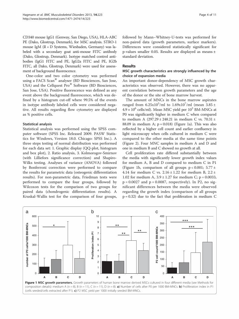
Figure 1 MSC growth parameters. Growth parameters of human bone mcomposition details): medium A (n = 8), B (n = 11), C (n = 11), D (n = 8). a) N(cells seeded/cells extracted after P1). c) P2 MSC yield per 1000 initially see
followed by Mann–Whitney-U-tests was performed fornon-paired data (growth parameters, surface markers).Differences were considered statistically significant forp-values smaller 0.05. Results are displayed as means ±standard deviation.
ResultsMSC growth characteristics are strongly influenced by thechoice of expansion mediaAn important donor-dependency of MSC growth char-acteristics was observed. However, there was no appar-ent correlation between growth parameters and the ageof the donor or the site of bone marrow harvest.The amount of MNCs in the bone marrow aspirates
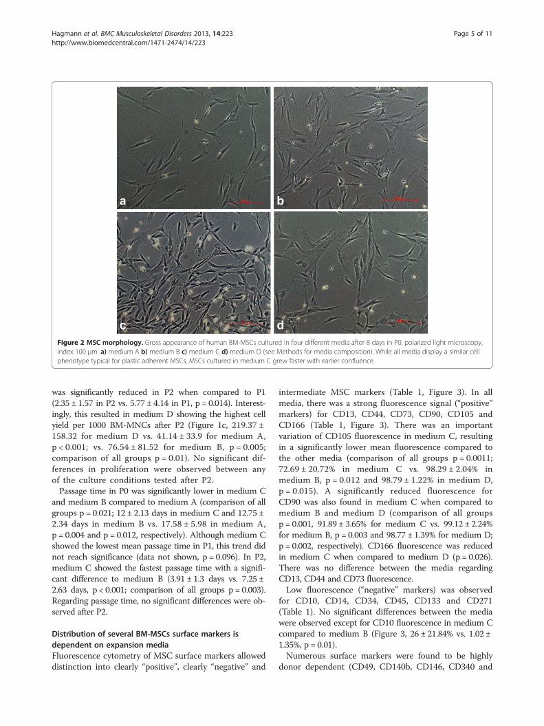
ranged from 6.25x10⁵/ml to 1.69x10⁷/ml (mean 3.85 ±4.9 × 10⁶ cells/ml). Mean MSC yield per 103 BM-MNCs atP0 was significantly higher in medium C when comparedto medium A (297.29 ± 248.21 in medium C vs. 70.31 ±88.09 in medium A; p = 0.018) (Figure 1a). This was alsoreflected by a higher cell count and earlier confluency inlight microscopy when cells cultured in medium C werecompared to the other media at the same time points(Figure 2). Four MNC samples in medium A and D andone in medium B and C showed no growth at all.Cell proliferation rate differed substantially between
the media with significantly lower growth index valuesfor medium A, B and D compared to medium C in P1(Figure 1b, comparison of all groups p < 0.001; 5.77 ±4.14 for medium C vs. 2.16 ± 1.22 for medium B, 2.2 ±1.02 for medium A, 3.9 ± 1.27 for medium C; p = 0.0033,p = 0.0027 and p = 0.0087, respectively). In P2, no sig-nificant differences between the media were observedregarding the growth index (comparison of all groupsp = 0.32) due to the fact that proliferation in medium C
0
50
100
150
200
250
300
350
400
A B C D
P2
cell
co
un
t/10
00 B
M-M
NC
s
C D
**
c
*****
***
arrow derived MSCs cultured in four different media (see Methods forumber of cells after P0 per 1000 BM-MNCs. b) Proliferation index in P1ded BM-MNCs.
ba
dcFigure 2 MSC morphology. Gross appearance of human BM-MSCs cultured in four different media after 8 days in P0, polarized light microscopy,index 100 μm. a) medium A b) medium B c) medium C d) medium D (see Methods for media composition). While all media display a similar cellphenotype typical for plastic adherent MSCs, MSCs cultured in medium C grew faster with earlier confluence.
Hagmann et al. BMC Musculoskeletal Disorders 2013, 14:223 Page 5 of 11http://www.biomedcentral.com/1471-2474/14/223
was significantly reduced in P2 when compared to P1(2.35 ± 1.57 in P2 vs. 5.77 ± 4.14 in P1, p = 0.014). Interest-ingly, this resulted in medium D showing the highest cellyield per 1000 BM-MNCs after P2 (Figure 1c, 219.37 ±158.32 for medium D vs. 41.14 ± 33.9 for medium A,p < 0.001; vs. 76.54 ± 81.52 for medium B, p = 0.005;comparison of all groups p = 0.01). No significant dif-ferences in proliferation were observed between anyof the culture conditions tested after P2.Passage time in P0 was significantly lower in medium C
and medium B compared to medium A (comparison of allgroups p = 0.021; 12 ± 2.13 days in medium C and 12.75 ±2.34 days in medium B vs. 17.58 ± 5.98 in medium A,p = 0.004 and p = 0.012, respectively). Although medium Cshowed the lowest mean passage time in P1, this trend didnot reach significance (data not shown, p = 0.096). In P2,medium C showed the fastest passage time with a signifi-cant difference to medium B (3.91 ± 1.3 days vs. 7.25 ±2.63 days, p < 0.001; comparison of all groups p = 0.003).Regarding passage time, no significant differences were ob-served after P2.
Distribution of several BM-MSCs surface markers isdependent on expansion mediaFluorescence cytometry of MSC surface markers alloweddistinction into clearly “positive”, clearly “negative” and
intermediate MSC markers (Table 1, Figure 3). In allmedia, there was a strong fluorescence signal (“positive”markers) for CD13, CD44, CD73, CD90, CD105 andCD166 (Table 1, Figure 3). There was an importantvariation of CD105 fluorescence in medium C, resultingin a significantly lower mean fluorescence compared tothe other media (comparison of all groups p = 0.0011;72.69 ± 20.72% in medium C vs. 98.29 ± 2.04% inmedium B, p = 0.012 and 98.79 ± 1.22% in medium D,p = 0.015). A significantly reduced fluorescence forCD90 was also found in medium C when compared tomedium B and medium D (comparison of all groupsp = 0.001, 91.89 ± 3.65% for medium C vs. 99.12 ± 2.24%for medium B, p = 0.003 and 98.77 ± 1.39% for medium D;p = 0.002, respectively). CD166 fluorescence was reducedin medium C when compared to medium D (p = 0.026).There was no difference between the media regardingCD13, CD44 and CD73 fluorescence.Low fluorescence (“negative” markers) was observed
for CD10, CD14, CD34, CD45, CD133 and CD271(Table 1). No significant differences between the mediawere observed except for CD10 fluorescence in medium Ccompared to medium B (Figure 3, 26 ± 21.84% vs. 1.02 ±1.35%, p = 0.01).Numerous surface markers were found to be highly
donor dependent (CD49, CD140b, CD146, CD340 and
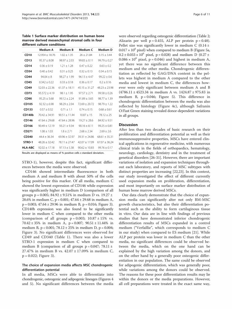
Table 1 Surface marker distribution on human bonemarrow derived mesenchymal stromal cells in fourdifferent culture conditions
Medium A Medium B Medium C Medium D
CD10 12.954 ± 18.76 1.02 ± 1.35 26 ± 21.84 3.15 ± 3.44
CD13 95.37 ± 8.08 98.97 ± 2.03 99.83 ± 0.11 99.79 ± 0.27
CD14 0.36 ± 0.19 1.21 ± 1.28 0.47 ± 0.22 0.63 ± 0.2
CD34 0.40 ± 0.42 0.31 ± 0.25 0.32 ± 0.13 0.34 ± 0.15
CD44 94.04 ± 8 98.27 ± 1.99 96.13 ± 4.47 99.22 ± 0.6
CD45 0.342 ± 0.22 0.33 ± 0.18 0.36 ± 0.17 0.2 ± 0.16
CD49 52.03 ± 22.36 61.37 ± 18.11 45.15 ± 31.27 48.23 ± 23.98
CD73 95.572 ± 4.19 98.1 ± 1.95 97.57 ± 2.71 99.58 ± 0.26
CD90 95.25 ± 5.68 99.12 ± 2.24 91.89 ± 3.65 98.77 ± 1.39
CD105 92.32 ± 6.98 98.29 ± 2.04 72.69 ± 20.72 98.79 ± 1.22
CD133 0.57 ± 0.32 0.71 ± 1.1 0.74 ± 0.15 0.68 ± 0.61
CD140b 70.42 ± 34.91 90.51 ± 11.44 10.87 ± 15 78.12 ± 25
CD146 47.64 ± 29.68 47.64 ± 28.96 19.27 ± 28.6 84.92 ± 9.51
CD166 90.49 ± 12.19 93.21 ± 9.34 90.16 ± 8.11 99.25 ± 0.81
CD271 1.08 ± 1.03 1.8 ± 2.71 2.68 ± 2.34 2.69 ± 2.6
CD340 44.4 ± 36.34 69.96 ± 32.97 39.31 ± 24.86 68.61 ± 35.31
STRO-1 48.26 ± 32.42 78.11 ± 27.47 42.07 ± 17.09 67.07 ± 36.24
HLA-ABC 92.02 ± 17.18 97.13 ± 5.38 90.62 ± 10.83 99.76 ± 0.17
Results are displayed as means of % positive cells ± standard deviation.
Hagmann et al. BMC Musculoskeletal Disorders 2013, 14:223 Page 6 of 11http://www.biomedcentral.com/1471-2474/14/223
STRO-1), however, despite this fact, significant differ-ences between the media were observed.CD146 showed intermediate fluorescence in both
medium A and medium B with about 50% of the cellsbeing positive for this marker. Of all media, medium Cshowed the lowest expression of CD146 while expressionwas significantly higher in medium D (comparison of allgroups p = 0.005; 84.92 ± 9.51% in medium D vs. 19.27 ±28.6% in medium C, p = 0.001; 47.64 ± 29.68 in medium A,p = 0.003; 47.64 ± 29.96 in medium B, p = 0.016; Figure 3).CD140b expression was also found to be significantlylower in medium C when compared to the other media(comparison of all groups p = 0.003; 10.87 ± 15% vs.70.42 ± 35% in medium A, p = 0.007; 90.51 ± 11.44% inmedium B, p < 0.001; 78.12 ± 25% in medium D, p = 0.004;Figure 3). No significant differences were observed forCD49 and CD340 (Table 1). There was also a lowerSTRO-1 expression in medium C when compared tomedium B (comparison of all groups p = 0.047; 78.11 ±27.47% in medium B vs. 42.07 ± 17.09% in medium C,p = 0.022; Figure 3).
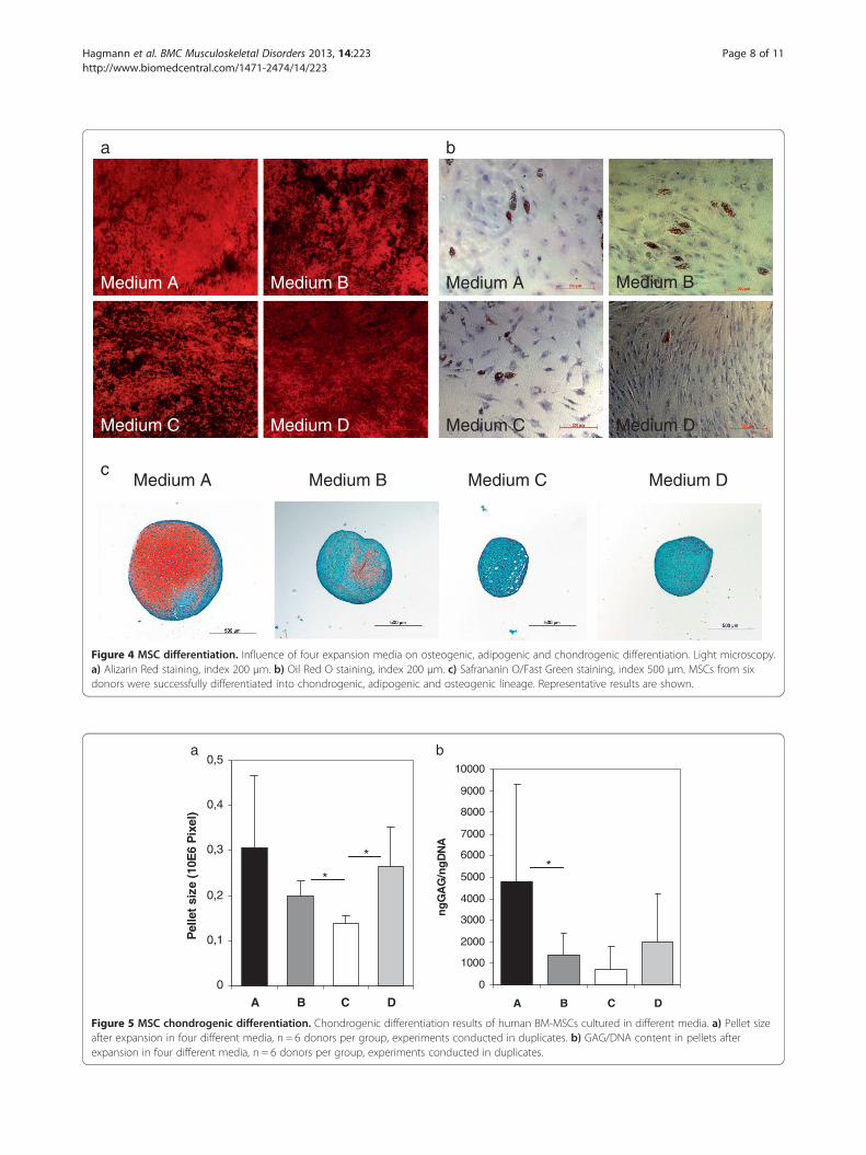
The choice of expansion media affects MSC chondrogenicdifferentiation potentialIn all media, MSCs were able to differentiate intochondrogenic, osteogenic and adipogenic lineages (Figures 4and 5). No significant differences between the media
were observed regarding osteogenic differentiation (Table 2;Alizarin per well p = 0.455, ALP per protein p = 0.48).Pellet size was significantly lower in medium C (0.14 ±0.017 × 106 pixel) when compared to medium B (Figure 5a,0.2 ± 0.033 × 106 pixel, p = 0.028) and medium D (0.27 ±0.086 × 106 pixel, p = 0.046) and highest in medium A,yet there was no significant difference between thismedium and the other media. Chondrogenic differen-tiation as reflected by GAG/DNA content in the pel-lets was highest in medium A compared to the othermedia and lowest in medium C, the differences how-ever were only significant between medium A and B(4786.11 ± 4523.34 in medium A vs. 1424.87 ± 975.83 inmedium B, p = 0.046; Figure 5). This difference inchondrogenic differentiation between the media was alsoreflected by histology (Figure 4c), although SafraninO/Fast Green staining revealed donor-dependent variationsin all groups.
DiscussionAfter less than two decades of basic research on theirproliferation and differentiation potential as well as theirimmunosuppressive properties, MSCs have entered clin-ical applications in regenerative medicine, with numerousclinical trials in the fields of orthopaedics, hematology,neurology, cardiology, dentistry and in the treatment ofgenetical disorders [26-31]. However, there are importantvariations of isolation and expansion techniques through-out each laboratory, and reports of MSC subtypes withdistinct properties are increasing [22,23]. In this context,our study investigated the effect of different currentlyused expansion media on proliferation, differentiation,and most importantly on surface marker distribution ofhuman bone marrow derived MSCs.Our data clearly demonstrate that the choice of expan-
sion media can significantly alter not only BM-MSCgrowth characteristics, but also their differentiation po-tential such as the ability to form cartilaginous tissuein vitro. Our data are in line with findings of previousstudies that have demonstrated inferior chondrogenicdifferentiation results of MSCs expanded in a controlmedium (“Verfaillie”, which corresponds to medium Cin our study) when compared to ES medium [21]. WhileALP per protein was lower in medium C than the othermedia, no significant differences could be observed be-tween the media, which on the one hand can beexplained by the high variation among the donors, andon the other hand by a generally poor osteogenic differ-entiation in our population. The same could be observedfor adipogenic differentiation, which was generally poor,while variations among the donors could be observed.The reasons for these poor differentiation results may liewithin the donors or the media preparations. However,all cell preparations were treated in the exact same way,
0
10
20
30
40
50
60
70
80
90
100
A B C D
% S
TR
O-1
+ c
ells
0
10
20
30
40
50
60
70
80
90
100
A B C D
% C
D14
6+ c
ells
0
10
20
30
40
50
60
70
80
90
100
A B C D
% C
D14
0b+
cel
ls
0
10
20
30
40
50
60
70
80
90
100
A B C D
% C
D10
5+ c
ells
0
10
20
30
40
50
60
70
80
90
100
A B C D
% C
D90
+ c
ells
0
10
20
30
40
50
60
70
80
90
100
A B C D
% C
D10
+ c
ells
* *******cba
ed f
**
*
**
*****
****
**
Figure 3 MSC surface marker distribution. Differences in surface marker distribution between of human BM-MSCs cultured in four differentmedia (see Table 1 for data). All results are displayed as % positive cells, means of n = 10 for medium B and C and n = 6 for medium A and D.a) CD10, b) CD90, c) CD105, d) CD140b, e) CD146, f) STRO-1.
Hagmann et al. BMC Musculoskeletal Disorders 2013, 14:223 Page 7 of 11http://www.biomedcentral.com/1471-2474/14/223
thus indicating that in this context, chondrogenic differen-tiation potential is more affected by the choice of expan-sion media than adipogenic and osteogenic differentiation.Our study adds to findings that quantify differences
in surface marker expression depending on the expan-sion media applied [32]. An important study in thisfield was published in 2006, revealing the impact notonly of the choice of expansion media and growth fac-tors, but also of basic factors such as plating densityand flask manufacturer on MSC characteristics [19].The experiments conducted in this study also revealedthat certain MSC surface markers (CD44, MAB1470,STRO-1 and HLA-DR) showed intermediate fluores-cence when MSCs were expanded in DMEM-HG, how-ever without quantifying these results. Our study revealscharacteristic changes in the distribution of CD10, CD140band CD146 that to our best knowledge have not been pub-lished yet, with consistent results of some more stable
markers such as CD14, CD34 and CD45 as “negativemarkers” and CD13, CD44, CD73 and CD166 as “positive”markers [33].Another recent study has compared four different ex-
pansion media (among these DMEM and Alpha-MEM)and analysed adipose-tissue derived MSCs regardingtheir surface markers, concluding that the expression ofCD49d, CD54 (being lowest in DMEM-KO) and CD117was inconsistent at all passages and in all four mediawhile the other surface markers did not significantlyalter between the media [34]. This is not in accordancewith other studies [32] and our own findings that clearlyindicate significant differences in the expression ofCD10 (not tested in the above study), CD90, CD105,CD140b (not tested in the above study) and CD146 (nottested in the above study). This discrepancy could be ei-ther due to differences in the study population or by as-suming different characteristics of adipose-tissue derived
Medium BMedium A
Medium C Medium D
Medium A Medium B
Medium C Medium D
a b
cMedium A Medium B Medium C Medium D
Figure 4 MSC differentiation. Influence of four expansion media on osteogenic, adipogenic and chondrogenic differentiation. Light microscopy.a) Alizarin Red staining, index 200 μm. b) Oil Red O staining, index 200 μm. c) Safrananin O/Fast Green staining, index 500 μm. MSCs from sixdonors were successfully differentiated into chondrogenic, adipogenic and osteogenic lineage. Representative results are shown.
0
1000
2000
3000
4000
5000
6000
7000
8000
9000
10000
A B C D
ng
GA
G/n
gD
NA
0
0,1
0,2
0,3
0,4
0,5
A B C D
Pel
let
size
(10
E6
Pix
el)
a b
*
**
Figure 5 MSC chondrogenic differentiation. Chondrogenic differentiation results of human BM-MSCs cultured in different media. a) Pellet sizeafter expansion in four different media, n = 6 donors per group, experiments conducted in duplicates. b) GAG/DNA content in pellets afterexpansion in four different media, n = 6 donors per group, experiments conducted in duplicates.
Hagmann et al. BMC Musculoskeletal Disorders 2013, 14:223 Page 8 of 11http://www.biomedcentral.com/1471-2474/14/223
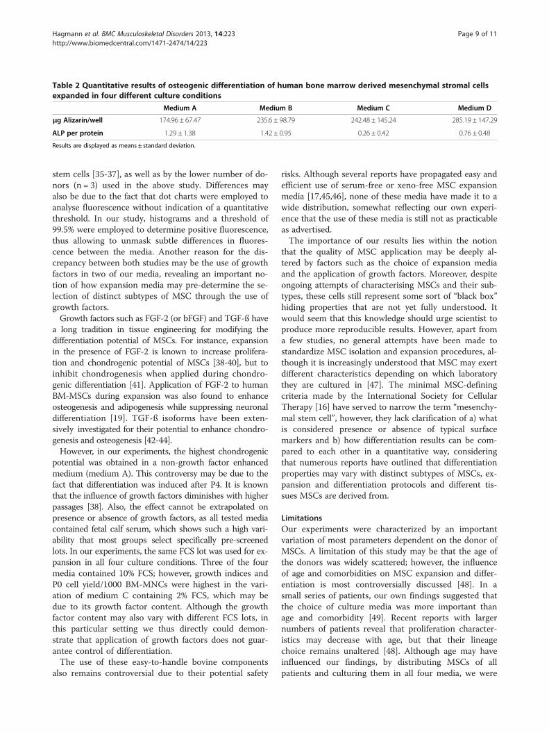
Table 2 Quantitative results of osteogenic differentiation of human bone marrow derived mesenchymal stromal cellsexpanded in four different culture conditions
Medium A Medium B Medium C Medium D
μg Alizarin/well 174.96 ± 67.47 235.6 ± 98.79 242.48 ± 145.24 285.19 ± 147.29
ALP per protein 1.29 ± 1.38 1.42 ± 0.95 0.26 ± 0.42 0.76 ± 0.48
Results are displayed as means ± standard deviation.
Hagmann et al. BMC Musculoskeletal Disorders 2013, 14:223 Page 9 of 11http://www.biomedcentral.com/1471-2474/14/223
stem cells [35-37], as well as by the lower number of do-nors (n = 3) used in the above study. Differences mayalso be due to the fact that dot charts were employed toanalyse fluorescence without indication of a quantitativethreshold. In our study, histograms and a threshold of99.5% were employed to determine positive fluorescence,thus allowing to unmask subtle differences in fluores-cence between the media. Another reason for the dis-crepancy between both studies may be the use of growthfactors in two of our media, revealing an important no-tion of how expansion media may pre-determine the se-lection of distinct subtypes of MSC through the use ofgrowth factors.Growth factors such as FGF-2 (or bFGF) and TGF-ß have
a long tradition in tissue engineering for modifying thedifferentiation potential of MSCs. For instance, expansionin the presence of FGF-2 is known to increase prolifera-tion and chondrogenic potential of MSCs [38-40], but toinhibit chondrogenesis when applied during chondro-genic differentiation [41]. Application of FGF-2 to humanBM-MSCs during expansion was also found to enhanceosteogenesis and adipogenesis while suppressing neuronaldifferentiation [19]. TGF-ß isoforms have been exten-sively investigated for their potential to enhance chondro-genesis and osteogenesis [42-44].However, in our experiments, the highest chondrogenic
potential was obtained in a non-growth factor enhancedmedium (medium A). This controversy may be due to thefact that differentiation was induced after P4. It is knownthat the influence of growth factors diminishes with higherpassages [38]. Also, the effect cannot be extrapolated onpresence or absence of growth factors, as all tested mediacontained fetal calf serum, which shows such a high vari-ability that most groups select specifically pre-screenedlots. In our experiments, the same FCS lot was used for ex-pansion in all four culture conditions. Three of the fourmedia contained 10% FCS; however, growth indices andP0 cell yield/1000 BM-MNCs were highest in the vari-ation of medium C containing 2% FCS, which may bedue to its growth factor content. Although the growthfactor content may also vary with different FCS lots, inthis particular setting we thus directly could demon-strate that application of growth factors does not guar-antee control of differentiation.The use of these easy-to-handle bovine components
also remains controversial due to their potential safety
risks. Although several reports have propagated easy andefficient use of serum-free or xeno-free MSC expansionmedia [17,45,46], none of these media have made it to awide distribution, somewhat reflecting our own experi-ence that the use of these media is still not as practicableas advertised.The importance of our results lies within the notion
that the quality of MSC application may be deeply al-tered by factors such as the choice of expansion mediaand the application of growth factors. Moreover, despiteongoing attempts of characterising MSCs and their sub-types, these cells still represent some sort of “black box”hiding properties that are not yet fully understood. Itwould seem that this knowledge should urge scientist toproduce more reproducible results. However, apart froma few studies, no general attempts have been made tostandardize MSC isolation and expansion procedures, al-though it is increasingly understood that MSC may exertdifferent characteristics depending on which laboratorythey are cultured in [47]. The minimal MSC-definingcriteria made by the International Society for CellularTherapy [16] have served to narrow the term “mesenchy-mal stem cell”, however, they lack clarification of a) whatis considered presence or absence of typical surfacemarkers and b) how differentiation results can be com-pared to each other in a quantitative way, consideringthat numerous reports have outlined that differentiationproperties may vary with distinct subtypes of MSCs, ex-pansion and differentiation protocols and different tis-sues MSCs are derived from.
LimitationsOur experiments were characterized by an importantvariation of most parameters dependent on the donor ofMSCs. A limitation of this study may be that the age ofthe donors was widely scattered; however, the influenceof age and comorbidities on MSC expansion and differ-entiation is most controversially discussed [48]. In asmall series of patients, our own findings suggested thatthe choice of culture media was more important thanage and comorbidity [49]. Recent reports with largernumbers of patients reveal that proliferation character-istics may decrease with age, but that their lineagechoice remains unaltered [48]. Although age may haveinfluenced our findings, by distributing MSCs of allpatients and culturing them in all four media, we were
Hagmann et al. BMC Musculoskeletal Disorders 2013, 14:223 Page 10 of 11http://www.biomedcentral.com/1471-2474/14/223
able to detect differences that could be solely assignedto the choice of culture media. However, we were ableto observe an important variation among the donorsthat could not be attributed to age. The heterogeneityof MSC preparations of different donors is a knownproblem [50] that may have had an important influ-ence on growth parameters and differentiation, butsurface markers as well.It could be criticized that two media containing differ-
ent growth factors were examined along with two mediawithout growth factors and that three media contained10% FCS while one contained 2% FCS. However, in thisstudy, our aim was to compare four media that are actu-ally widely used in different laboratories rather than todetermine the influence of each component. In ouropinion, this approach creates a clearer image of how al-legedly trivial decisions may have an influence on theoutcome of an experiment. It is clear that in the future,experiments attributing the observed differences to sin-gle media components will have to be conducted.
ConclusionsThis study adds to several reports that emphasize onhow the choice of expansion media can influence notonly growth characteristics, but also differentiation po-tential of MSCs. This study is in accordance with previ-ous findings reporting differences in the expression oftypical MSC markers depending on the media applied.We were able to detect additional MSC markers affectedby the expansion conditions that to our best knowledgehave not been reported yet. Our findings suggest that at-tempts to further investigate how different media com-ponents affect MSC characteristics are necessary. Moreimportantly, our findings are in favour of pursuing stan-dardized protocols in MSC isolation and expansion.
AbbreviationsAlpha-MEM: Alpha minimum essential medium; BM: Bone marrow;CD: Cluster of differentiation; DMEM-LG: Dulbecco’s modified Eagle’smedium low glucose; FCS: Fetal calf serum; FGF-2: Fibroblast growth factor 2;GAG: Glycosaminoglycans; hMSCs: Human mesenchymal stromal cells;MNCs: Mononuclear cells.
Competing interestsThe authors declare that they have no competing interests.
Authors’ contributionsSH, BM, WR and TG conceived of the study. SH drafted the manuscript. SH, TD,TG, BM and SF provided the bone marrow. SH and SF carried out theexperiments. SF, WR, PWK and SH performed the statistical analysis. SH, WR, BM,TD, PWK and TG participated in study design and coordination and helped todraft the manuscript. All authors read and approved the final manuscript.
AcknowledgementsWe would like to acknowledge Rene Wetzel, Patrick Göthlich, Marc Hoffmannand Elena Tripel for their support. The study was carried out with funding by thestate of Baden-Württemberg, Germany. None of the authors received externalfunding in connection with the study presented in this publication.
Author details1Department of Orthopedics, Trauma Surgery and Spinal Cord Injury,University Hospital Heidelberg, Germany Schlierbacher Landstrasse 200a,69118 Heidelberg, Germany. 2Maxillofacial and Plastic Surgery, UniversityMedical Center, Mainz, Germany. 3Research Center for ExperimentalOrthopedics, University Hospital Heidelberg, Heidelberg, Germany.
Received: 22 April 2013 Accepted: 19 July 2013Published: 30 July 2013
References1. Becker AJ, Mc CE, Till JE: Cytological demonstration of the clonal nature
of spleen colonies derived from transplanted mouse marrow cells.Nature 1963, 197:452–454.
2. Friedenstein AJ, Deriglasova UF, Kulagina NN, Panasuk AF, Rudakowa SF,Luria EA, Ruadkow IA: Precursors for fibroblasts in different populations ofhematopoietic cells as detected by the in vitro colony assay method.Exp Hematol 1974, 2:83–92.
3. Friedenstein AJ, Piatetzky S II, Petrakova KV: Osteogenesis in transplants ofbone marrow cells. J Embryol Exp Morphol 1966, 16:381–390.
4. Zuk PA, Zhu M, Mizuno H, Huang J, Futrell JW, Katz AJ, Benhaim P, Lorenz HP,Hedrick MH: Multilineage cells from human adipose tissue: implications forcell-based therapies. Tissue Eng 2001, 7:211–228.
5. Bieback K, Kern S, Kluter H, Eichler H: Critical parameters for the isolationof mesenchymal stem cells from umbilical cord blood. Stem Cells 2004,22:625–634.
6. Kuznetsov SA, Mankani MH, Gronthos S, Satomura K, Bianco P, Robey PG:Circulating skeletal stem cells. J Cell Biol 2001, 153:1133–1140.
7. da Silva ML, Chagastelles PC, Nardi NB: Mesenchymal stem cells reside invirtually all post-natal organs and tissues. J Cell Sci 2006, 119:2204–2213.
8. Jiang Y, Jahagirdar BN, Reinhardt RL, Schwartz RE, Keene CD, Ortiz-Gonzalez XR,Reyes M, Lenvik T, Lund T, Blackstad M, et al: Pluripotency of mesenchymalstem cells derived from adult marrow. Nature 2002, 418:41–49.
9. Wagner W, Wein F, Seckinger A, Frankhauser M, Wirkner U, Krause U, Blake J,Schwager C, Eckstein V, Ansorge W, Ho AD: Comparative characteristics ofmesenchymal stem cells from human bone marrow, adipose tissue, andumbilical cord blood. Exp Hematol 2005, 33:1402–1416.
10. Kallai I, van Lenthe GH, Ruffoni D, Zilberman Y, Muller R, Pelled G, Gazit D:Quantitative, structural, and image-based mechanical analysis ofnonunion fracture repaired by genetically engineered mesenchymalstem cells. J Biomech 2010, 43:2315–2320.
11. Lyons FG, Al-Munajjed AA, Kieran SM, Toner ME, Murphy CM, Duffy GP,O’Brien FJ: The healing of bony defects by cell-free collagen-basedscaffolds compared to stem cell-seeded tissue engineered constructs.Biomaterials 2010, 31:9232–9243.
12. Goepfert C, Slobodianski A, Schilling AF, Adamietz P, Portner R: Cartilageengineering from mesenchymal stem cells. Adv Biochem Eng Biotechnol2010, 123:163–200.
13. Guo J, Lin GS, Bao CY, Hu ZM, Hu MY: Anti-inflammation role formesenchymal stem cells transplantation in myocardial infarction.Inflammation 2007, 30:97–104.
14. Hatzistergos KE, Quevedo H, Oskouei BN, Hu Q, Feigenbaum GS, Margitich IS,Mazhari R, Boyle AJ, Zambrano JP, Rodriguez JE, et al: Bone marrowmesenchymal stem cells stimulate cardiac stem cell proliferation anddifferentiation. Circ Res 2010, 107:913–922.
15. Mazhari R, Hare JM: Mechanisms of action of mesenchymal stem cells incardiac repair: potential influences on the cardiac stem cell niche.Nat Clin Pract Cardiovasc Med 2007, 4(Suppl 1):S21–26.
16. Dominici M, Le Blanc K, Mueller I, Slaper-Cortenbach I, Marini F, Krause D,Deans R, Keating A, Prockop D, Horwitz E: Minimal criteria for definingmultipotent mesenchymal stromal cells. The International Society forCellular Therapy position statement. Cytotherapy 2006, 8:315–317.
17. Miwa H, Hashimoto Y, Tensho K, Wakitani S, Takagi M: Xeno-freeproliferation of human bone marrow mesenchymal stem cells.Cytotechnology 2012, 64:301–308.
18. Reyes M, Lund T, Lenvik T, Aguiar D, Koodie L, Verfaillie CM: Purificationand ex vivo expansion of postnatal human marrow mesodermalprogenitor cells. Blood 2001, 98:2615–2625.
19. Sotiropoulou PA, Perez SA, Salagianni M, Baxevanis CN, Papamichail M:Characterization of the optimal culture conditions for clinical scaleproduction of human mesenchymal stem cells. Stem Cells 2006, 24:462–471.
Hagmann et al. BMC Musculoskeletal Disorders 2013, 14:223 Page 11 of 11http://www.biomedcentral.com/1471-2474/14/223
20. Apel A, Groth A, Schlesinger S, Bruns H, Schemmer P, Buchler MW, Herr I:Suitability of human mesenchymal stem cells for gene therapy dependson the expansion medium. Exp Cell Res 2009, 315:498–507.
21. Dexheimer V, Frank S, Richter W: Proliferation as a requirement for in vitrochondrogenesis of human mesenchymal stem cells. Stem Cells Dev 2012,21:2160–2169.
22. Buhring HJ, Battula VL, Treml S, Schewe B, Kanz L, Vogel W: Novel markersfor the prospective isolation of human MSC. Ann N Y Acad Sci 2007,1106:262–271.
23. Buhring HJ, Treml S, Cerabona F, de Zwart P, Kanz L, Sobiesiak M:Phenotypic characterization of distinct human bone marrow-derivedMSC subsets. Ann N Y Acad Sci 2009, 1176:124–134.
24. Barbero A, Grogan S, Schafer D, Heberer M, Mainil-Varlet P, Martin I: Agerelated changes in human articular chondrocyte yield, proliferation andpost-expansion chondrogenic capacity. Osteoarthritis Cartilage 2004,12:476–484.
25. Grogan SP, Barbero A, Diaz-Romero J, Cleton-Jansen AM, Soeder S,Whiteside R, Hogendoorn PC, Farhadi J, Aigner T, Martin I, Mainil-Varlet P:Identification of markers to characterize and sort human articularchondrocytes with enhanced in vitro chondrogenic capacity.Arthritis Rheum 2007, 56:586–595.
26. Chen SL, Fang WW, Ye F, Liu YH, Qian J, Shan SJ, Zhang JJ, Chunhua RZ,Liao LM, Lin S, Sun JP: Effect on left ventricular function of intracoronarytransplantation of autologous bone marrow mesenchymal stem cell inpatients with acute myocardial infarction. Am J Cardiol 2004, 94:92–95.
27. ClinicalTrials.gov by the National Library of Medicine (NLM) at the NationalInstitutes of Health (NIH). http:www.clinicaltrials.gov.
28. Horwitz EM, Gordon PL, Koo WK, Marx JC, Neel MD, McNall RY, Muul L,Hofmann T: Isolated allogeneic bone marrow-derived mesenchymal cellsengraft and stimulate growth in children with osteogenesis imperfecta:Implications for cell therapy of bone. Proc Natl Acad Sci U S A 2002,99:8932–8937.
29. Koc ON, Day J, Nieder M, Gerson SL, Lazarus HM, Krivit W: Allogeneicmesenchymal stem cell infusion for treatment of metachromaticleukodystrophy (MLD) and Hurler syndrome (MPS-IH). Bone MarrowTransplant 2002, 30:215–222.
30. Le Blanc K, Rasmusson I, Sundberg B, Gotherstrom C, Hassan M, Uzunel M,Ringden O: Treatment of severe acute graft-versus-host disease with thirdparty haploidentical mesenchymal stem cells. Lancet 2004, 363:1439–1441.
31. Mazzini L, Fagioli F, Boccaletti R, Mareschi K, Oliveri G, Olivieri C, Pastore I,Marasso R, Madon E: Stem cell therapy in amyotrophic lateral sclerosis: amethodological approach in humans. Amyotroph Lateral Scler Other MotorNeuron Disord 2003, 4:158–161.
32. Haack-Sorensen M, Friis T, Bindslev L, Mortensen S, Johnsen HE, Kastrup J:Comparison of different culture conditions for human mesenchymalstromal cells for clinical stem cell therapy. Scand J Clin Lab Invest 2008,68:192–203.
33. Fekete N, Gadelorge M, Furst D, Maurer C, Dausend J, Fleury-Cappellesso S,Mailander V, Lotfi R, Ignatius A, Sensebe L, et al: Platelet lysate from wholeblood-derived pooled platelet concentrates and apheresis-derivedplatelet concentrates for the isolation and expansion of human bonemarrow mesenchymal stromal cells: production process, content andidentification of active components. Cytotherapy 2012, 14:540–554.
34. Dhanasekaran M, Indumathi S, Rashmi M, Rajkumar JS, Sudarsanam D:Unravelling the retention of proliferation and differentiation potency inextensive culture of human subcutaneous fat-derived mesenchymalstem cells in different media. Cell Prolif 2012, 45:516–526.
35. Strioga M, Viswanathan S, Darinskas A, Slaby O, Michalek J: Same or not thesame? Comparison of adipose tissue-derived versus bone marrow-derivedmesenchymal stem and stromal cells. Stem Cells Dev 2012, 21:2724–2752.
36. Vishnubalaji R, Al-Nbaheen M, Kadalmani B, Aldahmash A, Ramesh T:Comparative investigation of the differentiation capability of bone-marrow- and adipose-derived mesenchymal stem cells by qualitativeand quantitative analysis. Cell Tissue Res 2012, 347:419–427.
37. Xie X, Wang Y, Zhao C, Guo S, Liu S, Jia W, Tuan RS, Zhang C: Comparativeevaluation of MSCs from bone marrow and adipose tissue seeded inPRP-derived scaffold for cartilage regeneration. Biomaterials 2012,33:7008–7018.
38. Cheng T, Yang C, Weber N, Kim HT, Kuo AC: Fibroblast growth factor 2enhances the kinetics of mesenchymal stem cell chondrogenesis.Biochem Biophys Res Commun 2012, 426:544–550.
39. Stewart AA, Byron CR, Pondenis H, Stewart MC: Effect of fibroblast growthfactor-2 on equine mesenchymal stem cell monolayer expansion andchondrogenesis. Am J Vet Res 2007, 68:941–945.
40. Buckley CT, Kelly DJ: Expansion in the presence of FGF-2 enhances thefunctional development of cartilaginous tissues engineered usinginfrapatellar fat pad derived MSCs. J Mech Behav Biomed Mater 2012,11:102–111.
41. Weiss S, Hennig T, Bock R, Steck E, Richter W: Impact of growth factors andPTHrP on early and late chondrogenic differentiation of humanmesenchymal stem cells. J Cell Physiol 2010, 223:84–93.
42. Dickhut A, Dexheimer V, Martin K, Lauinger R, Heisel C, Richter W:Chondrogenesis of human mesenchymal stem cells by local transforminggrowth factor-beta delivery in a biphasic resorbable carrier. Tissue Eng Part A2010, 16:453–464.
43. Park H, Temenoff JS, Tabata Y, Caplan AI, Raphael RM, Jansen JA, Mikos AG:Effect of dual growth factor delivery on chondrogenic differentiation ofrabbit marrow mesenchymal stem cells encapsulated in injectablehydrogel composites. J Biomed Mater Res A 2009, 88:889–897.
44. Zhao L, Hantash BM: TGF-beta1 regulates differentiation of bone marrowmesenchymal stem cells. Vitam Horm 2011, 87:127–141.
45. Chase LG, Yang S, Zachar V, Yang Z, Lakshmipathy U, Bradford J, Boucher SE,Vemuri MC: Development and characterization of a clinically compliant xeno-free culture medium in good manufacturing practice for human multipotentmesenchymal stem cells. Stem Cells Transl Med 2012, 1:750–758.
46. Lindroos B, Boucher S, Chase L, Kuokkanen H, Huhtala H, Haataja R, VemuriM, Suuronen R, Miettinen S: Serum-free, xeno-free culture media maintainthe proliferation rate and multipotentiality of adipose stem cells in vitro.Cytotherapy 2009, 11:958–972.
47. Wagey R, Short B: Isolation, enumeration, and expansion of humanmesenchymal stem cells in culture. Methods Mol Biol 2013, 946:315–334.
48. Dexheimer V, Mueller S, Braatz F, Richter W: Reduced reactivation fromdormancy but maintained lineage choice of human mesenchymal stemcells with donor age. PLoS One 2011, 6:e22980.
49. Hagmann S, Moradi B, Frank S, Bäsig A-M, Dreher T, Richter W, Gotterbarm T:Chondrogenic and immunophenotypic properties of mesenchymal stemcells from osteoarthritis patients (abstract). Osteoarthritis Cartilage 2011,20(Suppl. 1):S277.
50. Phinney DG, Kopen G, Righter W, Webster S, Tremain N, Prockop DJ: Donorvariation in the growth properties and osteogenic potential of humanmarrow stromal cells. J Cell Biochem 1999, 75:424–436.
doi:10.1186/1471-2474-14-223Cite this article as: Hagmann et al.: Different culture media affectgrowth characteristics, surface marker distribution and chondrogenicdifferentiation of human bone marrow-derived mesenchymal stromalcells. BMC Musculoskeletal Disorders 2013 14:223.
Submit your next manuscript to BioMed Centraland take full advantage of:
• Convenient online submission
• Thorough peer review
• No space constraints or color figure charges
• Immediate publication on acceptance
• Inclusion in PubMed, CAS, Scopus and Google Scholar
• Research which is freely available for redistribution
Submit your manuscript at www.biomedcentral.com/submit